UNIVERSITY OF BEIRA INTERIOR
Engineering
Computer-Aided Detection and Diagnosis of Breast
Cancer in 2D and 3D Medical Imaging Through
Multifractal Analysis
Filipe Cruz Gomes Soares
Thesis for obtaining the degree of Doctor of Philosophy in
Computer Science and Engineering
(3rd Cycle Studies)
Advisor: Prof. Doctor Mário Marques Freire (University of Beira Interior)
Co-advisor: Eng. João Seabra Ferreira Pinto (Siemens S.A. Healthcare Sector)
Thesis prepared at Siemens S.A. Healthcare Sector, at Instituto de Telecomunicações, within Multimedia Signal Processing Covilhã Group and at University of Beira Interior, and submitted to University of Beira Interior for defense in a public examination session.
Work nanced by the Portuguese Fundação para a Ciência e a Tecnologia through the grant identi ed by SFRH/BDE/15624/2006 under the program QREN POPH Type 4.1 Advanced Training, co-funded by the European Social Fund and by national funds from the Portuguese Ministério da Educação e Ciência.
Dedicatory
Acknowledgments
I would like to acknowledge Siemens S.A. Healthcare Sector for giving me the opportunity to develop this challenging research project in industry, where I could understand how to excel at innovation.
I am thankful to Fundação para a Ciência e Tecnologia for partially funding together with Siemens these research years under the PhD studentship in industry SFRH/BDE/15624/2006. I also thank Instituto de Telecomunicações for providing me extra resources and supporting my publications in international conferences and specialized training.
I am also grateful to Clínica João Carlos Costa from Viana do Castelo for providing me all the breast magnetic resonance data used in this Thesis.
I have to acknowledge the following people:
My main advisor Professor Mário Marques Freire for the strong motivation and enthusiasm, al-ways pushing me to go further in pursuing the PhD, and for following my work even on distance. Prof. Manuela Pereira for all the valuable teaching and research work shared.
Eng. João Seabra and Eng. Filipe Janela from Siemens S.A. Healthcare Sector for co-supervising my work. I expressly thank Filipe for all the guidance, trust and support, for letting me take responsibility on the project, and also for the professional lessons that he gave me.
My Siemens colleagues Inês Sousa, Celina Lourenço, Pawel Andruszkiewicz, Liliana Caldeira, José Ferrão, Linda Gomes, Catarina Duarte, David Afonso, Catarina Runa, Teresa Mendes, Pedro Almeida, Paulo Cruz and António Martins. Especially, I would like to thank you Inês for the most mind blowing ideas and permanent intellectual stimulus. You were one of the greatest assets from Siemens.
Pawel, Alessia Pagotto and Manuel Stadlbauer for being the coolest roommates in Porto. João, Rui, Marta, Ricardo, Nery, David, Liliana, Guida and Celina Lourenço for their faithful friendship. In particular, I must express my deep gratitude to Celina for her patience, generosity, presence, care, and also for the happiness moments and experiences shared during these years.
Finally, I have to thank in Portuguese:
À minha família por todo o amor, ajuda e atenção, durante estes largos anos de grande desa o. À minha irmã por toda a força e alegria. Em especial à minha Mãe pelo inesgotável carinho, preocupação e admiração incondicional que me fez acreditar sempre num futuro brilhante. Inês, obrigado por seres a minha maior fonte de inspiração e energia, por me fazeres sonhar e ser cada vez melhor. Obrigado pelo teu amor desmedido, pela tua ajuda constante e por estares sempre a meu lado em todos os momentos. Obrigado por me mostrares o lado bom e feliz da vida.
Foreword
This Thesis describes the research work performed in the scope of a doctoral research program and presents its conclusions and contributions. The research activities were carried on in the industry with Siemens S.A. Healthcare Sector, in integration with a research team.
Siemens S.A. Healthcare Sector is one of the world biggest suppliers of products, services and complete solutions in the medical sector. The company offers a wide selection of diagnostic and therapeutic equipment and information systems. Siemens products for medical imaging and in vivo diagnostics include: ultrasound, computer tomography, mammography, digital breast to-mosynthesis, magnetic resonance, equipment to angiography and coronary angiography, nuclear imaging, and many others.
Siemens has a vast experience in Healthcare and at the beginning of this project it was strategi-cally interested in solutions to improve the detection of Breast Cancer, to increase its competi-tiveness in the sector.
The company owns several patents related with self-similarity analysis, which formed the back-ground of this Thesis. Furthermore, Siemens intended to explore commercially the comput-er-aided automatic detection and diagnosis eld for portfolio integration. Therefore, with the high knowledge acquired by University of Beira Interior in this area together with this Thesis, will allow Siemens to apply the most recent scienti c progress in the detection of the breast cancer, and it is foreseeable that together we can develop a new technology with high potential. The project resulted in the submission of two invention disclosures for evaluation in Siemens A.G., two articles published in peer-reviewed journals indexed in ISI Science Citation Index, two other articles submitted in peer-reviewed journals, and several international conference papers. This work on computer-aided-diagnosis in breast led to innovative software and novel processes of research and development, for which the project received the Siemens Innovation Award in 2012.
It was very rewarding to carry on such technological and innovative project in a sociallysensitive area as Breast Cancer.
List of Publications
Articles included in the thesis resulting from the doctoral research
program
1. 3D Lacunarity in Multifractal Analysis of Breast Tumor Lesions in Dynamic
Contrast-En-hanced Magnetic Resonance Imaging
Filipe Soares, Filipe Janela, Manuela Pereira, João Seabra and Mário M. Freire IEEE Transactions on Image Processing, Volume 22, Issue 11, pp. 4422-4435, 2013. DOI: 10.1109/TIP.2013.2273669
2. Classi cation of Breast Masses on Contrast-Enhanced Magnetic Resonance Images Through
Log Detrended Fluctuation Cumulant-Based Multifractal Analysis
Filipe Soares, Filipe Janela, Manuela Pereira, João Seabra and Mário M. Freire IEEE Systems Journal, accepted for publication, 2013.
DOI: 10.1109/JSYST.2013.2284101
3. Review and Performance Evaluation of Multifractal Approaches for Computer-aided
Detection of Microcalci cation Clusters in Mammograms
Filipe Soares, Filipe Janela, Manuela Pereira, João Seabra and Mário M. Freire Submitted for publication in an international journal, 2013.
4. Computer-Aided Detection and Diagnosis of Breast Cancer: Overview on Typical
Sys-tems and Methods in Mammography and Breast Magnetic Resonance Imaging
Filipe Soares, Filipe Janela, Manuela Pereira, João Seabra and Mário M. Freire Submitted for publication in an international journal, 2013.
Other publications resulting from the doctoral research program
not included in the thesis
1. The Role of Self-Similarity for Computer Aided Detection Based on Mammogram
Analy-sis
Filipe Soares, Mário M. Freire, Manuela Pereira, Filipe Janela, João Seabra
Chapter 6: Biomedical Diagnostics and Clinical Technologies: Applying High-Performance Cluster and Grid Computing
IGI Global, 2011, ISBN13: 9781605662800, pp. 181-199. DOI: 10.4018/978-1-60566-280-0.ch006
2. Self-similarity classi cation of breast tumour lesions on dynamic contrast-enhanced
magnetic resonance images - Special Session on Breast CAD
Filipe Soares, Filipe Janela, João Seabra, Manuela Pereira, and Mário Marques Freire International Journal of Computer Assisted Radiology and Surgery
Springer-Verlag, Volume 5, Supplement 1, pp. S203-S205, 2010. DOI: 10.1007/s11548-010-0459-y
3. Multifractal Analysis of Arterial Spin Labeling Functional Magnetic Resonance Imaging
of the Brain
Filipe Soares, Inês Sousa, Filipe Janela, João Seabra, Manuela Pereira, and Mário Marques Freire
Proceedings of the IEEE International Workshop on Medical Measurements and Applica-tions
IEEE Press, 2010, pp. 161-164. DOI: 10.1109/MEMEA.2010.5480209
4. A New Computer-Aided Approach for Breast Cancer Diagnosis Filipe Soares
Proceedings of the 3rd World Cancer Congress - Breast Cancer Conference BIT Life Sciences, 2010, pp. 273
5. Towards the detection of microcalci cations on mammograms through Multifractal
De-trended Fluctuation Analysis
Filipe Soares, Mário Marques Freire, Manuela Pereira, Filipe Janela, and João Seabra Proceedings of the IEEE Paci c Rim Conference on Communications, Computers and Signal Processing
IEEE Computer Society Press, 2009, pp. 677-681. DOI: 10.1109/PACRIM.2009.5291288
6. Self-Similarity Analysis Applied to 2D Breast Cancer Imaging
Filipe Soares, Pawel Andruszkiewicz, Mário Marques Freire, Paulo Cruz, Manuela Pereira Proceedings of the International Conference on Systems and Networks Communications, on the First International Workshop on High Performance Computing Applied to Medical Data and Bioinformatics
IEEE Computer Society Press, 2007, pp. 77-83. DOI: 10.1109/ICSNC.2007.76
Resumo
No cancro da mama a deteção precoce e o diagnóstico correto são de extrema importância na prescrição terapêutica e caz e e ciente, que potencie o aumento da taxa de sobrevivência à doença. A teoria multifractal foi inicialmente introduzida no contexto da análise de sinal e a sua utilidade foi demonstrada na descrição de comportamentos siológicos de bio-sinais e até na deteção e predição de patologias. Nesta Tese, três métodos multifractais foram estendidos para imagens bi-dimensionais (2D) e comparados na deteção de microcalci cações em mamo-gramas. Um destes métodos foi também adaptado para a classi cação de massas da mama, em cortes transversais 2D obtidos por ressonância magnética (RM) de mama, em grupos de massas provavelmente benignas e com suspeição de malignidade. Um novo método de análise multi-fractal usando a lacunaridade tri-dimensional (3D) foi proposto para classi cação de massas da mama em imagens volumétricas 3D de RM de mama. A análise multifractal revelou diferenças na complexidade subjacente às localizações das microcalci cações em relação aos tecidos nor-mais, permitindo uma boa exatidão da sua deteção em mamogramas. Adicionalmente, foram extraídas por análise multifractal características dos tecidos que permitiram identi car os casos tipicamente recomendados para biópsia em imagens 2D de RM de mama. A análise multifractal 3D foi e caz na classi cação de lesões mamárias benignas e malignas em imagens 3D de RM de mama. Este método foi mais exato para esta classi cação do que o método 2D ou o método padrãode análise decontraste cinéticotumoral. Em conclusão, a análise multifractal fornece informação útil para deteção auxiliada por computador em mamogra a e diagnóstico auxiliado por computador em imagens 2D e 3D de RM de mama, tendo o potencial de complementar a interpretação dos radiologistas.
Palavras-chave
Deteção auxiliada por computador (CADe), Diagnóstico auxiliado por computador (CADx), Ma-mogra a, Ressonância magnética de mama,Extração de características, Classi cação, Análise multifractal, Multi-escala, Wavelets, Cancro da mama.
Resumo Alargado
Introdução
Neste capítulo é apresentado um resumo alargado do trabalho de investigação conducente à Tese de Doutoramento intitulada Computer-Aided Detection and Diagnosis of Breast Cancer in 2D and 3D Medical Imaging Through Multifractal Analysis. O enquadramento da Tese é des-crito numa fase inicial, de nindo-se depois o problema abordado, os objetivos do trabalho de investigação e o argumento da Tese. De seguida, são abordados os principais temas objeto de investigação nesta Tese: a deteção de microcalci cações em mamogramas e a classi cação de lesões em imagens de ressonância magnética de mama. As metodologias são brevemente discutidas bem como as contribuições resultantes do trabalho desenvolvido. Por último, apre-sentam-se as principais conclusões.
Enquadramento da Tese
O cancro da mama é curável se detetado precocemente e mediante um tratamento apropri-ado. Além de salvar vidas, espera-se dos médicos que encontrem a forma menos invasiva e dolorosa para veri car o estado em que se encontra a doença. Com respeito ao desconforto que os exames de Mamogra a e Biopsia Mamária podem causar, reduzir o número de deteções falso-positivas torna-se um problema igualmente importante como a redução de falso-negati-vas. A anatomia complexa da mama é uma inevitável fonte da estrutura altamente irregular dos mamogramas, que constitui uma informação delicada de analisar pelos radiologistas, a quem se espera que distingam anomalias muito subtis de uma massa de ambiguidade global. Além disso, a variabilidade entre dois casos acresce a di culdade na decisão humana, que enfatiza a necessidade de ferramentas de processamento de imagem con áveis que possam assistir o processo de deteção de anomalias e diagnóstico em imagens da mama. A nalidade do trabalho enquadra-se no desenvolvimento de novos métodos não lineares de estimação de auto-semelhança, aplicável à imagiologia, que possam auxiliar a deteção da patologia do can-cro da mama, segmentando regiões mamárias de risco para otimizar o processo de diagnóstico. Pretende-se apurar histologicamente o estado e evolução do cancro da mama, descrevendo a natureza fractal e multifractal dos objetos presentes nas imagens recolhidas determinando o grau de auto-semelhança. A metodologia a desenvolver de sistemas de apoio à decisão au-xiliada por computador deverá permitir não só a deteção ou diagnóstico automático a partir de imagens de Mamogra a como de Ressonância Magnética (RM). Trata-se de um projeto de investigação inovador, com uma iminente aplicação prática, conseguindo conjugar num único trabalho de Doutoramento os aspetos do desenvolvimento cientí co e a sua implementação em ambiente industrial, numa área onde a empresa Siemens S.A. tem vindo a apostar fortemente. Com prestadores de cuidados de saúde e outros parceiros de negócio interessados nos resul-tados do projeto, perspetiva-se a oportunidade de concretização de um protótipo e respetivo produto. Contudo, o projeto de investigação envolve ainda restrições de con dencialidade dos
casos clínicos utilizados para validação, e tem como principal risco a concorrência industrial neste mercado e o forte crescimento da investigação e desenvolvimento nesta área.
Descrição do Problema e Objetivos de Investigação
O objetivo do trabalho descrito nesta Tese é a melhoria da deteção e diagnóstico precoce do cancro da mama, através do desenvolvimento de sistemas de apoio à decisão auxiliada por computador, baseados nas propriedades de auto-semelhança dos tecidos mamários. Sistemas de deteção auxiliada por computador (CADe) são desenvolvidos para extração de sinais preco-ces de anormalidade, em particular microcalci cações, das imagens mamográ cas. Sistemas de diagnóstico auxiliado por computador (CADx) são implementados para classi cação da ma-lignidade de lesões mamárias em imagens 2D e 3D obtidas por RM de mama. O trabalho de investigação desenvolvido pode ser dividido em três objetivos principais, correspondentes aos três principais capítulos da Tese, conforme se descreve a seguir.
Aplicação de métodos de análise de imagem multifractal a mamogramas para
extração automática de microcalci cações, que constituem sinais precoces de
anormalidade no tecido mamário
a) Generalização para 2D dos três principais métodos multifractais: Multifractal Detrended Fluctuation Analysis (MF-DFA), Modulus Maxima Wavelet Transform (MMWT) e Wavelet Le-aders Multifractal Formalism (WLMF).
b) Desenvolvimento de uma estrutura comum que inclua os três métodos, MF-DFA, MMWT and WLMF, para análise de imagens mamográ cas.
c) Comparação dos três métodos, MF-DFA, MMWT and WLMF, em termos da sua capacidade de extração de microcalci cações e e ciência computacional.
d) Redução da deteção de falsos positivos usando a auto-semalhança para criar um mapa de potenciais estruturas a remover, por exemplo: estruturas lineares como os vasos sanguí-neos.
Extração de características das lesões mamárias relacionadas com a sua
morfo-logia e textura, por análise multifractal de imagens 2D de RM de mama
a) Aplicação do método MF-DFA a imagens 2D de RM de mama correspondentes a cortes de tumores ou lesões mamárias.
b) Identi cação dos descritores matemáticos dos espectros multifractais relevantes para a discriminação de lesões mamárias em imagens de RM de mama.
Computer-Aided Detection and Diagnosis of Breast Cancer in 2D and 3D Medical Imaging Through Multifractal Analysis
c) Extração de propriedades de auto-semelhança por análise multifractal baseada nos cu-mulantes logarítmicos da utuação destendenciada dos cortes de lesões mamárias em imagens RM de mama.
d) Avaliação dos descritores e propriedades multifractais num esquema de classi cação su-pervisionada para distinção de lesões suspeitas de malignidade das potencialmente benig-nas em imagens RM de mama.
Desenvolvimento de um novo método de análise multifractal usando a
lacuna-ridade 3D como uma medida para obter propriedades multifractais de imagens
volumétricas de RM de mama
a) Estimação do expoente de escala multifractal usando a lacunaridade como a medida mul-tifractal.
b) Investigação do uso da teoria multifractal condicionada pela medida lacunaridade 3D para classi cação de lesões mamárias em imagens volumétricas de RM de mama.
c) Extração de características dos novos espectros multifractais para classi cação automá-tica de lesões benignas e malignas em imagens volumétricas de RM de mama.
d) Comparação da capacidade de discriminação entre lesões benignas e malignas com os métodos MF-DFA 2D e 3D e 3TP (standard clínico para análise da cinética do tumor) num esquema de classi cação supervisionada.
Argumento da Tese
Esta tese propõe uma nova abordagem para a deteção e classi cação de características do can-cro da mama. Especi camente, o argumento de tese é o seguinte:
O tecido mamário apresenta alto grau de complexidade, revelando propriedades de auto-se-melhança passíveis de serem descritas matematicamente por análise multifractal. O tecido mamário normal e regiões com potencial tumoral mostram comportamento multifractal dis-tinto, o que pode ser usado para a deteção precoce de cancro da mama assistida por computa-dor em mamogra as. Características multifractais são bem correlacionadas com o estado de evolução de um tumor e fornecem uma indicação da probabilidade de malignidade através de diagnóstico assistido por computador em imagens 2D e 3D de RM de mama.
Principais Contribuições
Abordagens multifractais para deteção auxiliada por computador de clusters de
microcalci cações em mamogramas
Os métodos multifractais generalizados para 2D e aplicados a um conjunto de mamogramas de duas bases de dados públicas foram e cazes na deteção de microcalci cações. O método 2D MF-DFA resultou numa melhor performance de deteção do que os outros dois métodos baseados em wavelets (MMWT and WLMF), independentemente da resolução espacial das imagens na base de dados. O método WLMF demonstrou a melhor e ciência computacional, no entanto a perfor-mance de deteção é apenas mediana. A análise multifractal permite obter características dos tecidos mamários que estão correlacionadas com a caracterização da complexidade subjacente às lesões mamárias, que constituem sinais precoces de cancro da mama. Estas características mostraram-se úteis na identi cação de microcalci cações e na eliminação de falsos positivos, como estruturas lineares que evidenciam características distintas. A análise multifractal de ma-mogramas permite, assim, obter informação útil para sistemas de deteção precoce de cancro da mama auxiliados por computador.
Classi cação de massas mamárias em imagens de ressonância magnética de
mama com contraste dinâmico através de cumulantes logarítmicos obtidos da
análise baseada em utuações destendenciadas
Foi desenvolvido um sistema de apoio à decisão que permite identi car casos de massas mamá-rias tipicamente recomendadas para biópsia a partir de imagens RM de mama 2D. Este sistema utiliza descritores matemáticos dos espectros multifractais e cumulantes logarítmicos num es-quema de classi cação supervisionada que proporciona uma recomendação de biópsia. A e cá-cia do sistema de apoio à decisão é elevada na distinção de lesões com suspeita de malignidade, principalmente com uma das oito características estudadas.
Análise multifractal com lacunaridade 3D de lesões tumorais da mama em
ima-gens volumétricas de ressonância magnética com contraste dinâmico
A presença de características multifractais nas imagens volumétricas de RM de mama foi con r-mada através da observação de prevalência de múltiplos graus de auto-semelhança a múltiplas escalas. Uma combinação de características multifractais foi obtida da análise multifractal usando a lacunaridade 3D como medida e demonstrou-se e caz na classi cação de lesões benig-nas e maligbenig-nas. Este método foi mais exato na determinação da probabilidade de malignidade do que o 2D MF-DFA ou o standard clínico para análise da cinética tumoral, 3TP. Desta forma, o método proposto para extração de características multifractais e classi cação tem o potencial de complementar a interpretação dos radiologistas e vir a ser usado num sistema de diagnóstico
Computer-Aided Detection and Diagnosis of Breast Cancer in 2D and 3D Medical Imaging Through Multifractal Analysis
assistido por computador (CADx).
Discussão da Metodologia
Abordagens multifractais para deteção auxiliada por computador de clusters de
microcalci cações em mamogramas
A deteção auxiliada por computador de padrões mamográ cos é frequentemente baseada na caracterização de texturas. A análise multifractal pode ser usada para caracterizar texturas de imagens, no entanto, esta abordagem é raramente aplicada no contexto da deteção de cancro da mama em imagens de mamogra a. Este capítulo revê e investiga a generalização dos três principais métodos multifractais recentemente propostos: Multifractal Detrended Fluctuation Analysis (MF-DFA), Modulus Maxima Wavelet Transform (MMWT) and Wavelet Leaders Multifrac-tal Formalism (WLMF). Pretende-se avaliar se as generalizações 2D destes métodos podem ser usadas na extração de elementos de importância clínica para a deteção do cancro da mama. Os métodos foram implementados numa plataforma comum e aplicados à deteção de microcalci -cações em mamogramas. A avaliação foi feita sobre duas bases de dados públicas com diferente resolução espacial de imagem, relacionando a sensibilidade com o número de falsos positivos da deteção, através de curvas FROC (Free-Response Receiver Operating Characteristic). A per-formance dos métodos na deteção de microcalci cações e os seus custos computacionais foram comparados. No conjunto de 290 imagens médicas, o método MF-DFA obteve um desempenho superior independentemente da resolução das imagens nas bases de dados. No entanto, em ambos os algoritmos foi veri cado o impacto de uma maior resolução de imagem nos resulta-dos superiores da deteção. É de salientar que o método baseado em wavelets MMWT foi mais sensível à alteração da base de dados. O método WLMF apresenta uma performance de dete-ção mediana mas melhor e ciência computacional. A inspedete-ção de singularidades e respetivas
utuações a múltiplas escalas revelou que o estudo multifractal é muito importante para a caracterização da complexidade subjacente às potenciais localizações de microcalci cações. A análise multifractal de mamogramas permite, assim, obter informação útil para sistemas de deteção precoce de cancro da mama auxiliados por computador.
Classi cação de massas em imagens de ressonância magnética de mama com
contraste dinâmico através de cumulantes logarítmicos obtidos da análise
mul-tifractal baseada em utuações destendenciadas
Foi desenvolvido um sistema de apoio à decisão que permite identi car casos de massas mamá-rias tipicamente recomendadas para biópsia a partir de imagens RM de mama 2D com contraste dinâmico. Este sistema utiliza descritores matemáticos dos espectros multifractais e cumulan-tes logarítmicos num esquema de classi cação supervisionada que proporciona uma recomenda-ção de biópsia. Os outputs da classi carecomenda-ção foram comparados com o diagnóstico do radiologista baseado no breast imaging-reporting and data system (BIRADS). Os resultados mostram que o
cumulante logarítmico c2 é o mais e caz na identi cação dos casos tipicamente recomendados para biópsia. A e cácia do sistema de apoio à decisão é cerca de 94% na distinção de lesões com suspeição de malignidade, com uma das oito características estudadas, o cumulante c2. O método proposto de análise multifractal pode contribuir para novas técnicas de classi cação que auxiliem os radiologistas na identi cação mais exata de casos que necessitem biópsia.
Análise multifractal com lacunaridade 3D de lesões tumorais da mama em
ima-gens volumétricas de ressonância magnética com contraste dinâmico
A RM de mama com contraste dinâmico é especialmente robusta para diagnóstico de cancro em casos de alto risco, devido à sua elevada sensibilidade. No entanto, a especi cidade pode ser comprometida uma vez que as diferenças entre as cinéticas do contraste dinâmico são subtis en-tre massas benignas e malignas. Nesta Tese é proposto um método multifractal 3D que permite caracterizar a complexidade (arranjo espacial de texturas) dos tumores mamários a múltiplas escalas. Propriedades de auto-semelhança são extraídas da estimação do expoente de escala multifractal de cada caso clínico, usando a lacunaridade 3D como medida multifractal. Estas propriedades incluem diversos descritores dos espectros multifractais que re etem a morfologia e estrutura espacial interna das lesões relativamente ao tecido normal. Os resultados sugerem que a combinação de várias características multifractais é e caz na distinção entre lesões be-nignas e malignas, como avaliado pela performance de um método de classi cação baseado em support vector machine com área da curva de receiver operating characteritics (ROC) de 0.96. Adicionalmente, a presença de multifractalidade nas imagens volumétricas de RM de mama com contraste dinâmico foi con rmada, já que múltiplos graus de auto-semelhança existem a múltiplas escalas. O método proposto de extração de características multifractais e classi -cação tem o potencial de complementar a interpretação do radiologista e futuros sistemas de diagnóstico assistido por computador (CADx).
Conclusão
Em conclusão, a análise multifractal fornece informação útil para deteção auxiliada por com-putador em mamogra a e diagnóstico auxiliado por comcom-putador em imagens 2D e 3D de RM de mama, tendo o potencial de complementar a interpretação dos radiologistas.
Computer-Aided Detection and Diagnosis of Breast Cancer in 2D and 3D Medical Imaging Through Multifractal Analysis
Abstract
The early detection and accurate diagnosis of breast cancer is of utmost importance in pro-viding effective and ef cient treatment in order to increase survival rates. The multifractal theory was rst introduced for signal analysis and has shown its utility in describing physio-logic behaviors of bio-signals and even in detecting and predicting pathology. In this Thesis, three multifractal analysis methods have been extended to two-dimensional (2D) images and compared in the detection of microcalci cations in mammograms. One of these methods was adapted for classi cation of breast masses in 2D cross-sectional breast magnetic resonance (MR) images in suspicious malignant and probably benign groups. A novel multifractal analysis method using three-dimensional (3D) lacunarity is proposed for classi cation of breast masses in 3D volumetric MR images. The multifractal analysis revealed differences in the underlying complexity of the microcalci cations relatively to the normal tissue allowing a good accuracy of their detection in mammograms. Moreover, it provided meaningful features that allowed identifying the typically biopsy-recommended cases from 2D breast MR images. The 3D multi-fractal analysis method was also effective in the classi cation of malignant and benign lesions in 3D breast MR images. This method was more accurate in estimation of the likelihood of malignancy than the 2D method and the standard analysis of tumor enhancement kinetics. In conclusion, multifractal analysis provides useful information for computer-aided detection in mammography and for computer-aided diagnosis in 2D and 3D breast MR images and have the potential to complement the interpretation of the radiologists.
Keywords
Computer-Aided Detection (CADe), Computer-Aided Diagnosis (CADx), Mammography, Breast Magnetic Resonance Imaging (MRI), Feature Extraction, Classi cation, Multifractal Analysis, Mul-tiscale, Wavelets, Breast Cancer.
Contents
Dedicatory v Acknowledgments vii Foreword ix List of Publications xi Resumo xiii Resumo Alargado xv Abstract xxi Contents xxiiiList of Figures xxvii
List of Tables xxxi
Acronyms xxxiii
Chapter 1
Introduction 1
I Thesis Focus and Scope . . . 1 II Research Objectives . . . 3 III Thesis Statement . . . 4 IV Main Contributions . . . 5 V Thesis Organization . . . 6 References . . . 7
Chapter 2
Computer-Aided Detection and Diagnosis of Breast Cancer: Overview on Typical Systems and Methods in Mammography and Breast Magnetic Resonance Imaging 9
Abstract . . . 11 I Breast Cancer Imaging . . . 11 II CAD in Mammography and Breast MRI . . . 14 III Commercial CAD for Mammography and Breast MRI . . . 17 IV CADe and CADx Typical Shemes . . . 20 V Survey on CADe and CADx Methods . . . 22
V.A Feature extraction for CADe detection of clustered microcalci cations in Mam-mography . . . 22
V.B Feature extraction for CADe detection of masses in Mammography . . . 26 V.C Classi cation for CADx diagnosis of microcalci cations and masses in
Mammog-raphy . . . 30 V.D CADx diagnosis of masses in DCE-MRI of the breast . . . 33 VI Conclusion . . . 39 References . . . 41
Chapter 3
Review and Performance Evaluation of Multifractal Approaches for Computer-aided De-tection of Microcalci cation Clusters in Mammograms 53
Abstract . . . 55 I Introduction . . . 55 I.A Review of Prior Work on Detection of Mammographic Abnormalities . . . 56 I.B Multifractal Image Analysis . . . 57 I.C Overview of the Article . . . 57 II Detection Model and Multifractal Approaches . . . 57 II.A Pre-Processing and Breast Region Detection . . . 57 II.B Multifractal-based Feature Extraction . . . 58 II.C Selection of Findings and False Positive Reduction . . . 61 III Results . . . 62 III.A Experiments . . . 62 III.B Detection Performance . . . 62 III.C Computational Ef ciency . . . 63 IV Discussion . . . 64 V Conclusion . . . 65 References . . . 65
Chapter 4
Classi cation of Breast Masses on Contrast-Enhanced Magnetic Resonance Images Through Log Detrended Fluctuation Cumulant-Based Multifractal Analysis 69
Abstract . . . 71 I Introduction . . . 71 II Materials and Methods . . . 72 II.A Image Acquisition . . . 73 II.B Dataset Characterization and Tumor Selection . . . 73 II.C Multifractal Analysis . . . 74 II.D Self-similarity Extraction . . . 75 III Results . . . 76 IV Discussion . . . 76 V Conclusion . . . 79 References . . . 79
Computer-Aided Detection and Diagnosis of Breast Cancer in 2D and 3D Medical Imaging Through Multifractal Analysis
Chapter 5
3D Lacunarity in Multifractal Analysis of Breast Tumor Lesions in Dynamic
Contrast-En-hanced Magnetic Resonance Imaging 81
Abstract . . . 83 I Introduction . . . 83 II Background and Theory . . . 85 II.A Multifractal Analysis . . . 85 II.B Lacunarity Estimation . . . 86 III 3D Multifractal Scaling Exponent Lacunarity Analysis (MF-SELA) . . . 86 III.A Pre-Processing and VOI Selection . . . 87 III.B 3D lacunarity Estimation With Gliding Cube . . . 87 III.C Multifractal Analysis With 3D Lacunarity . . . 87 III.D Self-Similarity and Scaling Dynamics as Descriptors . . . 88 IV Experimental Setup and Performance Assessment . . . 88 IV.A Image Acquisition . . . 88 IV.B Tumor Collection and Diagnosis . . . 89 IV.C SVM-Based Classi cation . . . 90 IV.D ROC Analysis . . . 90 V Results . . . 90 VI Discussion . . . 92 VII Conclusion . . . 94 References . . . 94
Chapter 6
List of Figures
Chapter 2
Computer-Aided Detection and Diagnosis of Breast Cancer: Overview on Typical Systems and Methods in Mammography and Breast Magnetic Resonance Imaging
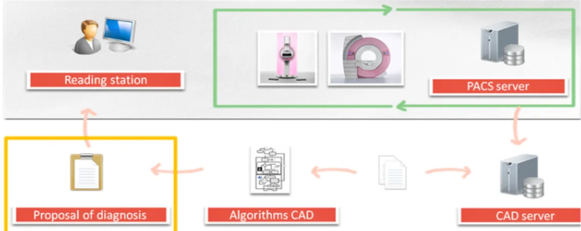
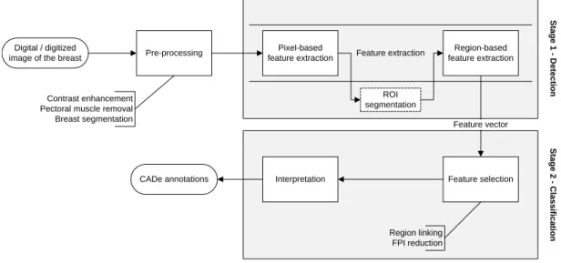
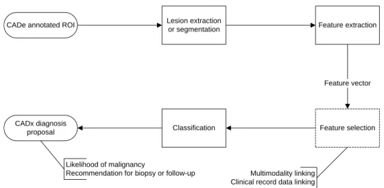
Figure 1. CAD embedded in the clinical cycle of breast imaging. . . 15 Figure 2. Flowchart of a typical CADe system in breast imaging. . . 20 Figure 3. Flowchart of a typical CADx system in breast imaging. . . 21
Chapter 3
Review and Performance Evaluation of Multifractal Approaches for Computer-aided De-tection of Microcalci cation Clusters in Mammograms
Figure 1. Proposed microcalci cation detection model. The rst three blocks of the ow are pre-processing steps before the core feature extraction, where the method for multifractal image analysis is employed. The clustering and self-similarity analysis aim for false positive reduction. . . 58 Figure 2. Fragment of a mammogram and extracted microcalci cations. . . 61 Figure 3. Radius vectors with centroid in C to create the boundary pro le of a
micro-calci cation with closed contour L. . . 62 Figure 4. Two cropped ROI sizes from the same MiniMIAS mammogram, 128 x 128
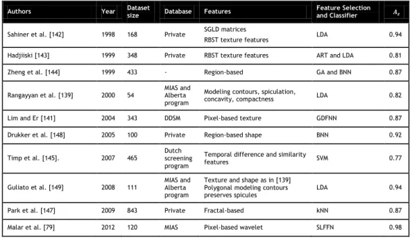
(blue squares) and 256 x 256 (black circles), and their multifractal spec-trums f(α) via Legendre transform with 2D MF-DFA. . . 62 Figure 5. (a) Detection of microcalci cations in mammogram ROIs. From top to
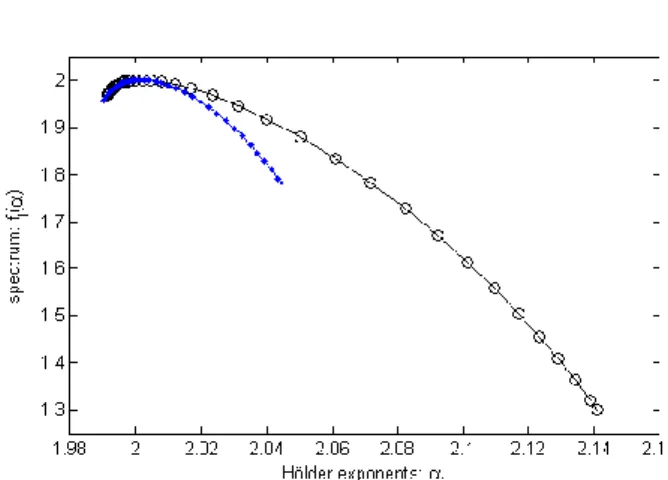
bot-tom: mdb209, mdb219, mdb223 and mdb249. (b) Legendre spectrums with 2D MF-DFA. . . 63 Figure 6. FROC curves showing detection performance using the three multifractal
methods on different datasets. 2D MF-DFA with DDSM has the best perfor-mance with bigger area under the curve. It points 85% of sensitivity for the detection of microcalci cations at 0.5 False Positives per Image. . . 63 Figure 7. Comparison of computational ef ciency by CPU time in seconds (s)
aver-aged per case in DDSM dataset, using the three multifractal methods. . . . 64
Chapter 4
Classi cation of Breast Masses on Contrast-Enhanced Magnetic Resonance Images Through Log Detrended Fluctuation Cumulant-Based Multifractal Analysis
Figure 1. Flowchart of the model for Log Detrended Fluctuation Cumulant- Based Multifractal Analysis. . . 73
Figure 2. Morphology features: typical benign case on the left, with oval shaped mass smooth, margin and homogeneous enhancement; typical malignant case on the right with irregular shaped mass, spiculated margin and heterogeneous enhancement. . . 73 Figure 3. Histogram of the longest diameter of the lesions in the dataset. The longest
diameter was measured where the lesion was best visualized as determined by radiologist. . . 74 Figure 4. Scheme of the descriptors used for the multifractal spectrum
characteriza-tion. . . 75 Figure 5. Detrended uctuation function Fq(s) at different scales for q = -2 (left)
and q = 2 (right). PM cases: in black. PB cases: in green. It is shown the presence of scaling range in particular for negative moment q, with the extreme scales showing more deviation from the power law scaling (smaller scales in q = -2 and larger scales in q = 2). Bars from the group of cases represent 95% con dence interval for mean. . . 77 Figure 6. Estimated scaling exponent τ(q) (left) and multifractal spectrum D(h) (right)
for the lesions in the dataset. PM cases: in black. PB cases: in green. . . . 77 Figure 7. Comparison of the three log-cumulants estimated from τ(q) before SVM
analysis for PB (left bar) and PM (right bar) cases. The box-plots show the lower and upper quartile and median. . . 77 Figure 8. Comparison of the ROC curves using SVM with the self-similarity extracted
features, RFE-3 feature set and the 3TP. . . 78 Figure 9. Comparison of computational ef ciency by CPU time in seconds (s) with
achieved area under the ROC curve Az with log-cumulant c2, for multiple expansions of moment q range. The CPU time presented is an average of the total time for running the complete dataset of 70 cases. . . 78
Chapter 5
3D Lacunarity in Multifractal Analysis of Breast Tumor Lesions in Dynamic Contrast-En-hanced Magnetic Resonance Imaging
Figure 1. Flowchart of the model for Multifractal Scaling Exponent Lacunarity Analy-sis (MF-SELA). . . 86 Figure 2. Morphology features of lesions in the dataset. Representation of tumor VOIs
(top). A sliced region of interest of a typical: benign case (bottom left), with oval shaped mass smooth, margin and homogeneous enhancement; ma-lignant case (bottom right), with irregular shaped mass, spiculated margin and heterogeneous enhancement. . . 89 Figure 3. BI-RADS grade of the lesions in the dataset plotted against the kinetic curve
Computer-Aided Detection and Diagnosis of Breast Cancer in 2D and 3D Medical Imaging Through Multifractal Analysis
Figure 4. Histogram of the longest diameter of the lesions in the dataset. The longest diameter was measured where the lesion was best visualized as determined by radiologist. . . 89 Figure 5. Multifractal spectra D(h) of the VOIs of the cases in the dataset. Benign
cases in gray. Malignant cases in black. . . 91 Figure 6. Multifractal scaling exponent τ(q) of the VOIs of the cases in the dataset.
Benign cases in gray. Malignant cases in black. . . 91 Figure 7. Comparison of multifractal descriptors and log-cumulants as features. Top:
For each feature normalized by its mean value, benign cases in gray and malignant cases in black. Bottom: Pooled features values tested for statis-tically signi cant differences with One-way ANOVA resulting in F-statistic = 588.32 and p-value < 0.05. Statistically signi cant differences among descriptors are identi ed by letters according to Post-Hoc Tukey test. . . . 91 Figure 8. ROC curves comparing the classi cation performance of the multifractal
features and the combined parameter (CP ) using SVM with a leave-one-out testing. . . 92
List of Tables
Chapter 2
Computer-Aided Detection and Diagnosis of Breast Cancer: Overview on Typical Systems and Methods in Mammography and Breast Magnetic Resonance Imaging
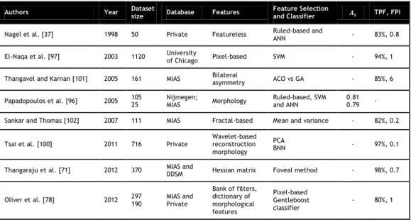
Table I. Representative selection of microcalci cations CADe in Mammography . . . 26 Table II. Representative selection of mass CADe in Mammography . . . 31 Table III. Area under the ROC curve Az of a representative selection of microcalci
-cations CADx in Mammography . . . 34 Table IV. Area under the ROC curve Azof a representative selection of mass CADx in
Mammography . . . 34 Table V. Area under the ROC curve Azof a representative selection of mass CADx in
DCE-MRI . . . 39
Chapter 3
Review and Performance Evaluation of Multifractal Approaches for Computer-aided De-tection of Microcalci cation Clusters in Mammograms
Table I. Assessment of Sensitivity . . . 63
Chapter 4
Classi cation of Breast Masses on Contrast-Enhanced Magnetic Resonance Images Through Log Detrended Fluctuation Cumulant-Based Multifractal Analysis
Table I. Comparison of the area under the ROC curve Az and corresponding standard deviation (STD) using SVM . . . 78
Chapter 5
3D Lacunarity in Multifractal Analysis of Breast Tumor Lesions in Dynamic Contrast-En-hanced Magnetic Resonance Imaging
Table I. Clinical cases in the dataset . . . 90 Table II. Area under the ROC curve Az in discriminating malignant from benign
le-sions with multifractal-based features. Az of the SVM classi er using each feature (leave-one-out cross-validation) . . . 92 Table III. ROC Az of 3TP and two multifractal methods on our dataset of 35 cases . . 93 Table IV. ROC Az among state-of-art studies on their datasets . . . 93
Acronyms
2D Two-Dimensional
3D Three-Dimensional
3TP Three-Time-Points
ACO Ant Colony Optimization
AFUM Average Fraction Under the Minimum
ANN Arti cial Neural Network
ANOVA Analysis of Variance
ART Adaptive Resonance Theory
BI-RADS Breast Imaging - Reporting and Data System
BNN Backpropagation Neural Network
BBN Bayesian Belief Network
BSSA Boundary Self-Similarity Analysis
CAD Computer-aided Medical Imaging Analysis
CADe Computer-Aided Detection
CADx Computer-Aided Diagnosis
CALMA Computer Assisted Library in Mammography CBIR Content-Based Image Retrieval
CC Craniocaudal
CP Combined Spectral Parameter
CPU Central Processing Unit
DBT Digital Breast Tomosynthesis
DCE-MRI Dynamic Contrast-Enhanced Magnetic Resonance Imaging DDSM Digital Database for Screening Mammography
DFA Detrended Fluctuation Analysis
Dh Hausdorff dimension
DoG Difference-of-Gaussian
DWT Discrete Wavelet Transform
EM Expectation Maximization
FD Fractal Dimension
FDA Food and Drug Administration
FFDM Full Field Digital Mammography
FL3D T1-weighted FLASH 3D
FNA Fine Needle Aspiration
FPI False Positives per Image
FROC Free-Response Receiver Operating Characteristic
GA Genetic Algorithm
Gd-DTPA Gadopentetate dimeglumine
GDFNN Generalized Dynamic Fuzzy Neural Networks
H Hurst Parameter
h Hölder Exponent
ICA Independent Component Analysis
kNN k-Nearest-Neighbor
LDA Linear Discriminant Analysis LRA Linear Regression Analysis
LOO Leave-One-Out
LS Left Slope of the curve
MCC Microcalci cation Clusters
MCS Multiple Classi er System
MF-DFA Multifractal Detrended Fluctuation Analysis MF-SELA Multifractal Scaling Exponent Lacunarity Analysis
MI Mutual Information
MIL Multiple-Instance Learning
MiniMIAS Database of Mammograms from Mammographic Image Analysis Society
MIP Maximum Intensity Projection
MLO Mediolateral Oblique
MMMWT Local Maxima along the MMWT Chains
MMWT Modulus Maxima Wavelet Transform
MR Magnetic Resonance
MRF Markov random eld
MRI Magnetic Resonance imaging
PACS Picture Archiving and Communication System
PB Probably Benign and Non-Biopsied
PBT Probabilistic Boosting Tree
PM Probably Malignant and Biopsied
PPV Positive Predictive Value
RBF Radial Basis Function
RBFNN Radial-Based Function Neural Network RBST Rubber Band Straightening Transformation
RFE Recursive Feature Elimination
ROC Receiver Operating Characteristic
ROI Region of Interest
RR Round-robin
RS Right Slope of the Curve
Computer-Aided Detection and Diagnosis of Breast Cancer in 2D and 3D Medical Imaging Through Multifractal Analysis
SLFFN Single Layer Feed Forward Network
SMF Standard Mammogram Form
SVM Support Vector Machine
SVM-RFE Recursive Feature Elimination-based Support Vector Machine
TE Testing Error
UBI University of Beira Interior
VOI Volume of Interest
W Curve Width
WLMF Wavelet Leaders Multifractal Formalism
Chapter 1
Introduction
This Thesis addresses the problem of detection and classi cation of breast cancer by the appli-cation of computer assisted tools for augmenting human functions, namely radiologists on their demanding job of chasing microcalci cations and tumors using data from two medical imaging modalities: Mammography and Magnetic Resonance imaging (MRI). In the form of software and applied mathematics, it is proposed to study self-similarity features found in 2D and 3D images of the breast. The focus, scope and research objectives of the Thesis are described in this chapter, followed by the Thesis statement, the main contributions and the Thesis organization.
I Thesis Focus and Scope
Breast cancer is a malignant tumor originated in the cells of the breast. A malignant tumor is a group of cancer cells that can grow into (invade) surrounding tissues or spread (metastasize) to distant areas of the body. The disease occurs almost exclusively in women, but it can also occur in men. Breast cancer is the most common cancer among women in the western world, aside from non-melanoma skin cancer. Breast cancer is the second leading cause of cancer death in women, exceeded only by lung cancer. Death rates from breast cancer have been declining since early 90's, with larger decreases in women younger than 50. These decreases are believed to be the result of earlier detection through screening and increased awareness, as well as improved treatment [1]. This is a big argument in favor of screening programs that has been focused on traditional imaging modalities of the breast as x-ray mammography, which has been the standard imaging modality for decades [2] [7]. Incidence rates of breast cancer have been increasing in the industrialized world, but this is expected given the higher life expectancy in those countries and the fact that many people are being screened by methods that did not exist a few decades ago. The combination of the characteristics of breast cancer: high incidence, deadly disease, asymptomaticin earlier stages, and high survival rate if detected inthese stages, makes the ght against the disease, through research and development of high-end technology in breast imaging devices, worthy.
Mammographic rst signs of breast cancer usually appear in the form of clusters of microcal-ci cations. These tiny deposits of calmicrocal-cium can be visible long before any palpable lesion has developed and their early detection contributes to the success of the treatment. For diagnosis,
radiologists generally rely on their shape, size, number and distribution. Malignant calci ca-tions are typically very numerous, clustered, small, dot-like or elongated, variable in size, shape and density. Benign calci cations are generally larger, more rounded, smaller in number, more diffusely distributed, and more homogeneous in shape [8]. However, because of the small size of microcalci cations, the comparison and characterization of benign and malignant lesions represents a very complex problem even for an experienced radiologist [9].
The microcalci cations can arise in isolation or together with other areas of high density breast tissue, called masses. The term mass arises from the characteristic well-de ned mammographic appearance, and they tend to be brighter than their surroundings due to the high density within their boundaries. In order to be able to characterize a mass, radiologists generally rely on its contour and different kinds can be observed in mammograms (circumscribed, spiculated, microlobulated, with dense kernel). Usually circumscribed masses are related to benign lesions while spiculated masses are related to malignant lesions.
The fractal geometry has been introduced a long time ago in image analysis through fractal dimension. Fractal compression and fractal encoding exploit the property of self-similarity of fractal objects [10]. Images of breast tissue are characterized by a high degree of self-sim-ilarity, i.e., several parts look as the whole image. In self-similar objects, irregularities are structural deviations from the global regularity. In the case of breast images these irregular-ities may correspond to locations of potential breast lesions and can be characterized under the light of fractal or multifractal analysis. These analyses allow a multiscale mathematical description of changes in the textural information, re ecting self-similarity. Moreover, it is possible to derive a set of mathematical quantities or features, which can be related to the type of breast lesion and malignancy level, constituting the basis of machine aid systems in the detection and diagnosis of breast cancer. The multifractal analysis provides a spectrum of fractal dimensions, characterizing multiple irregularities. This can potentially give more in-formation about the image than the fractal analysis, which is unable to uniquely characterize a texture pattern, as different fractal sets may share the same fractal dimension values and yet have different appearances. Therefore the methods developed in the scope of this Thesis are based on multifractal analysis. Mammography and breast MRI are the gold-standard imag-ing techniques in the detection and diagnosis of breast cancer, respectively. Mammography is the established technique for screening tests and breast MRI is mostly used for tumor staging and treatment planning and follow-up. The usual proceeding for breast tumor detection in screening mammography is visual inspection by a radiologist. As the volumetric anatomical information is projected into a two-dimensional (2D) image plane, mammographic ndings are generally hard to identify because of their superimposition on the breast parenchymal textures. In particular microcalci cations are often overlooked by the radiologist due to their small size, despite being usually an early sign of abnormality. Therefore, machine-aid based on reliable
Computer-Aided Detection and Diagnosis of Breast Cancer in 2D and 3D Medical Imaging Through Multifractal Analysis
image processing tools is valuable and it has been shown to help nding more cancers. On the other hand, in breast MRI decision-support systems are essential, since many benign and malig-nant tumors have similar appearances. Clinical interpretation of the images is based on visual examination of morphology features and contrast-enhancement kinetics and despite following a scoring system it still remains largely subjective. Computer assisted diagnosis may have an important impact on the accuracy, consistency and reproducibility of the diagnosis, preventing unnecessary therapies or invasive procedures, such as biopsies.
II Research Objectives
The aim of the work described in this Thesis is the improvement of breast cancer early detection and diagnosis by developing computer-aided systems based on the self-similarity properties of breast tissues. Computer-aided detection (CADe) systems are developed for extraction of early signs of abnormality, speci cally microcalci cations, from mammographic images. Comput-er-aided diagnosis (CADx) systems are implemented for malignancy classi cation of 2D and 3D images obtained with breast MRI. The work can be divided in three main objectives, correspond-ing to the three main chapters of the Thesis:
1) Application of multifractal image analysis methods to mammograms for automatic extrac-tion of microcalci caextrac-tions, which are early signs of abnormality in breast tissue
a) Generalization for 2D of the main three multifractal methods: Multifractal Detrended Fluctuation Analysis (MF-DFA), Modulus Maxima Wavelet Transform (MMWT) and Wavelet Leaders Multifractal Formalism (WLMF).
b) Development of a common framework including the three methods, MF-DFA, MMWT and WLMF, for mammographic image analysis.
c) Comparison of the three methods, MF-DFA, MMWT and WLMF, in terms of ability for microcalci cation extraction and computacional ef ciency.
d) Reduction of false positive detection by using self-similarity analysis to identify and create a likelihood map of potential structures to remove, for example: normal linear structures as blood vessels.
2) Extraction of multifractal image analysis derived features to characterize the morphology and texture of breast tumor MR images
a) Application of the MF-DFA method to 2D breast MR images corresponding to tumor slices.
b) Identi cation of meaningful mathematical descriptors of the multifractal spectra for discrimination of breast lesions in MRI.
c) Extraction of self-similarity features by log detrended uctuation cumulant-based multifractal analysis of the tumor images.
d) Evaluation of the multifractal descriptors and features in a supervised classi cation scheme for distinguishing suspicious malignant masses in breast MR images.
3) Development of a novel multifractal analysis method using 3D lacunarity as a measure to derive self-similar properties from volumetric breast MR images
a) Estimation of the multifractal scaling exponent using lacunarity as the multifractal measure.
b) Investigation of the use of multifractal theory conditioned by the 3D lacunarity mea-sure, for classi cation of breast lesions in volumetric breast MR images.
c) Extraction of features from the novel multifractal spectra for automated classi ca-tion of malignant and benign lesions.
d) Comparison of the likelihood of malignancy discrimination ability with 2D MF-DFA and Three-Time-Points (3TP) (clinical standard technique for analysis of tumor kinetics) in a supervised classi cation scheme.
III Thesis Statement
This Thesis proposes a new approach for the detection and classi cation of breast cancer fea-tures. Speci cally, the thesis statement is:
Breast tissue presents high degree of complexity showing self-similarity properties mathemat-ically described by multifractal analysis. Healthy breast tissue and potential breast tumor locations show differential multifractal behavior, which can be used for early computer-aided breast cancer detection in mammograms. Multifractal features are well correlated with tu-mor staging and provide an indication of the likelihood of malignancy through computer-aided diagnosis in both 2D and 3D breast MRI.
To support this thesis statement, the following research approach was conducted. The liter-ature on detection and diagnosis of breast cancer is reviewed in order to de ne the problem and research eld. The various modalities for breast imaging are studied and the suitability of its application in each phase of the disease management is analyzed. The several methods of breast lesion detection in mammography and diagnosis in breast MR were reviewed and their performance was evaluated. The multifractal theory was identi ed as a promising area of re-search in computer-aided medical imaging analysis. The few works of multifractal analysis in pattern identi cation were studied in terms of applied mathematics and computerized perfor-mance. In the area of multifractal analysis of breast cancer images even fewer studies were
Computer-Aided Detection and Diagnosis of Breast Cancer in 2D and 3D Medical Imaging Through Multifractal Analysis
found and therefore this was an opportunity to contribute with novelty in the eld. Three multifractal methods are generalized for 2D and applied in detection of microcalci cations in mammography. With the motivation of improving the distinction of benign and malignant le-sions in breast MR images a new multiscale and multifractal 3D characterization of tumors is proposed. This method is compared with the most equivalent 2D method. The output of the methods developed is evaluated by free-response receiver operating characteristic (FROC) and receiver operating characteristic (ROC) curves. Additionally, their computational performance is assessed. The methods developed have the potential of being included in future CADe and CADx systems.
IV Main Contributions
This section brie y describes the main scienti c contributions resulting from the research work presented in this Thesis.
1) Review and Performance Evaluation of Multifractal Approaches for Computer-aided Detec-tion of Microcalci caDetec-tion Clusters in Mammograms
• The multifractal methods generalized for 2D and applied to a set of mammograms from two public databases were able to successfully detect microcalci cations, and their computational performance were also assessed.
• The 2D MF-DFA method has shown to outperform the other two wavelet-based vari-ants of multifractal analysis (MMWT and WLMF), independently from the spatial res-olution of the images in the database. Nevertheless, 2D WLMF is computationally more ef cient having average detection performance.
• The inspection of singularities and their uctuations at multiple resolutions revealed that the multifractal study is very important for the characterization of the under-lying complexity of microcalci cations. Multifractal mammogram analysis provides, therefore, useful information for computer-aided detection.
This work was initially presented in the First International Workshop on High Performance Computing Applied to Medical Data and Bioinformatics and published in the proceedings of the conference [11]. After further developments a presentation was made in the IEEE Paci c Rim Conference on Communications, Computers and Signal Processing with a paper published in the respective proceedings [12]. Finally a journal article was prepared and has been submitted to a IEEE journal [13].
2) Classi cation of Breast Masses on Contrast-Enhanced Magnetic Resonance Images Through Log Detrended Fluctuation Cumulant-Based Multifractal Analysis
• A decision-support system was developed to identify the typically biopsy-recom-mended cases from 2D breast MR images.
• This system makes use mathematical descriptors of the multifractal spectra and log-cumulantfeatures in a supervised classi er scheme to effectively provide a biopsy recommendation.
• The decision-support system presents high accuracy (94%) distinguishing suspicious malignant lesions from probably benign lesions, with one of the eight features stud-ied.
The rst evidence to these ndings was presented in the Special Session on Breast CAD of the conference Computer Assisted Radiology and Surgery and published in the respective proceedings. It was also published in a supplement of the International Journal of Com-puter Assisted Radiology and Surgery from Springer-Verlag [14]. A more complete version of the work was accepted for publication in the IEEE Systems Journal [15].
3) 3D Lacunarity in Multifractal Analysis of Breast Tumor Lesions in Dynamic Contrast-En-hanced Magnetic Resonance Imaging
• The presence of multifractality in breast MR volumetric images was con rmed by prevalence of multiple degrees of self-similarity at multiple scales. A combination of self-similarity characteristics retrieved from the multifractal analysis using 3D lacu-narity asthe measure, was effective for the classi cation of malignant and benign lesions.
• This method was more accurate in estimation of the likelihood of malignancy than 2D MF-DFA and the clinical standard for analysis of tumor kinetics, 3TP. Therefore, the proposed feature extraction and classi cation method have the potential to com-plement the interpretation of the radiologists and supply a computer-aided diagnosis (CADx) system.
The novel multifractal 3D method and application to breast MR images was published in IEEE Transactions on Image Processing [16].
V Thesis Organization
The Thesis is organized as follows:Chapter 1: Introduction
A brief introduction to the Thesis is presented including the focus and scope, Thesis objectives, Thesis statement, and major contributions of the work carried out.
Computer-Aided Detection and Diagnosis of Breast Cancer in 2D and 3D Medical Imaging Through Multifractal Analysis
Chapter 2: Computer-Aided Detection and Diagnosis of Breast Cancer: Overview on Typical Systems and Methods in Mammography and Breast Magnetic Resonance Imaging
The background concepts behind the work developed are presented and discussed including both, an overview of breast cancer imaging modalities as well as a description of typical CAD systems. Finally, a survey on methods constituting CADe and CADx is presented.
Chapter 3: Review and Performance Evaluation of Multifractal Approaches for Comput-er-aided Detection of Microcalci cation Clusters in Mammograms
This chapter presents a comparative of three multifractal methods applied in the detection of microcalci cations in mammograms.
Chapter 4: Classi cation of Breast Masses on Contrast-Enhanced Magnetic Resonance Images Through Log Detrended Fluctuation Cumulant-Based Multifractal Analysis
MF-DFA multifractal method is applied in the classi cation of suspicious malignant images in 2D breast MR images.
Chapter 5: 3D Lacunarity in Multifractal Analysis of Breast Tumor Lesions in Dynamic Con-trast-Enhanced Magnetic Resonance Imaging
A novel multifractal method is proposed using 3D lacunarity for classi cation of benign and malignant breast lesions in volumetric breast MR images. This method was compared with the method of Chapter 4 in the same dataset.
Chapter 6: Conclusion and Future Work
The results presented throughout the Thesis are discussed and the main achievements are sum-marized pointing directions for the future.
References
[1] L. Tabar, M. Yen, B. Vitak, H. Chen, R. Smith, and S. Duffy, Mammography service screening and mortality in breast cancer patients: 20-year follow-up before and after introduction of screening, The Lancet, vol. 361, no. 9367, pp. 1405 1410, 2003.
[2] M. Reddy and R. Given-Wilson, Screening for breast cancer, Womens Heal. Med., vol. 3, no. 1, pp. 22 27, Jan. 2006.
[3] L. Wyld and C. E. Ingram, Screening of the population for breast cancer, Surg. Oxf., vol. 25, no. 6, pp. 254 256, Jun. 2007.
[4] D. Schopper and C. de Wolf, How effective are breast cancer screening programmes by mammography? Review of the current evidence, Eur. J. Cancer, vol. 45, no. 11, pp. 1916 1923, Jul. 2009.
[5] P. Skaane, Studies comparing screen- lm mammography and full- eld digital mammography in breast cancer screening: updated review, Acta Radiol. Stockh. Swed. 1987, vol. 50, no. 1, pp. 3 14, Jan. 2009.
[6] A. M. J. Bluekens, R. Holland, N. Karssemeijer, M. J. M. Broeders, and G. J. den Heeten, Comparison of Digital Screening Mammography and Screen-Film Mammography in the Early Detection of Clinically Relevant Cancers: A Multicenter Study, Radiology, vol. 265, no. 3, pp. 707 714, Jan. 2012.
[7] P. Skaane, A. I. Bandos, R. Gullien, E. B. Eben, U. Ekseth, U. Haakenaasen, M. Izadi, I. N. Jebsen, G. Jahr, M. Krager, L. T. Niklason, S. Hofvind, and D. Gur, Comparison of Digital Mammography Alone and Digital Mammography Plus Tomosynthesis in a Population-based Screening Program, Radiology, vol. 267, no. 1, pp. 47 56, Jan. 2013.
[8] F. van Gelderen, Understanding X-Rays: A Synopsis of Radiology. Springer, 2004.
[9] E. Pisano, M. Yaffe, B. Hemminger, R. Hendrick, L. Niklason, A. Maidment, C. Kimme-Smith, S. Feig, E. Sickles, and M. Braeuning, Current status of full eld digital mammography, Acad. Radiol., vol. 7, no. 4, pp. 266 280, 2000.
[10] F. Davoine, M. Antonini, J.M. Chassery, and M. Barlaud, Fractal Image Compression Based on Delaunay Triangulation and Vector Quantization, IEEE Transactions on Image Processing, special issue on vector quantization, vol. 5, no. 2, pp. 338 346, 1996.
[11] F. Soares, P. Andruszkiewicz, M. M. Freire, P. Cruz, M. Pereira, Self-Similarity Analysis Applied to 2D Breast Cancer Imaging, Proceedings of the International Conference on Systems and Networks Communi-cations, IEEE Computer Society Press, pp. 77 83, 2007.
[12] F. Soares, M. M. Freire, M. Pereira, F. Janela, J. Seabra, Towards the detection of microcalci cations on mammograms through Multifractal Detrended Fluctuation Analysis, Proceedings of the IEEE Paci c Rim Conference on Communications, Computers and Signal Processing, IEEE Computer Society Press, 2009, pp. 677 681, 2009.
[13] F. Soares, F. Janela, M. Pereira, J. Seabra, M. M. Freire, Review and Performance Evaluation of Multi-fractal Approaches for Computer-aided Detection of Microcalci cation Clusters in Mammograms, Submitted for publication in an international peer-reviewed IEEE journal, 2013.
[14] F. Soares, F. Janela, J. Seabra, M. Pereira, M. M. Freire, Self-similarity classi cation of breast tu-mour lesions on dynamic contrast-enhanced magnetic resonance images, - Special Session on Breast CAD,International Journal of Computer Assisted Radiology and Surgery, Springer-Verlag, Volume 5, pp. S203-S205, 2010.
[15] F. Soares, F. Janela, J. Seabra, M. Pereira, M. M. Freire, Classi cation of Breast Masses on Contrast-En-hanced Magnetic Resonance Images Through Log Detrended Fluctuation Cumulant-Based Multifractal Anal-ysis, IEEE Systems Journal, accepted for publication, 2013.
DOI: 10.1109/JSYST.2013.2284101
[16] F. Soares, F. Janela, M. Pereira, J. Seabra, M. M. Freire, 3D Lacunarity in Multifractal Analysis of Breast Tumor Lesions in Dynamic Contrast-Enhanced Magnetic Resonance Imaging, IEEE Transactions on Image Processing, Volume 22, Issue 11, pp. 4422 4435, 2013.
Chapter 2
Computer-Aided Detection and Diagnosis of Breast
Cancer: Overview on Typical Systems and Methods
in Mammography and Breast Magnetic Resonance
Imaging
This chapter consists of the following article:
Computer-Aided Detection and Diagnosis of Breast Cancer: Overview on Typical Systems and Methods in Mammography and Breast Magnetic Resonance Imaging
Filipe Soares, Filipe Janela, Manuela Pereira, João Seabra and Mário M. Freire Submitted for publication in an international peer-reviewed Elsevier journal, 2013.
Computer-Aided Detection and Diagnosis of Breast Cancer in 2D and 3D Medical Imaging Through Multifractal Analysis
Computer-Aided Detection and Diagnosis of Breast
Cancer: Overview on Typical Systems and Methods in
Mammography and Breast Magnetic Resonance Imaging
Filipe Soares
1,2,∗, Filipe Janela
1, Manuela Pereira
2, João Seabra
1and Mário M. Freire
21Siemens S.A. Healthcare Sector, 4456-901 Pera ta, Portugal.
2Instituto de Telecomunicações, Department of Computer Science, University of Beira Interior, 6201-001
Covilhã, Portugal.
Corresponding author. E-mail address: [email protected]
Abstract This paper reviews computer-aided medical imaging analysis (CAD) systems in breast
cancer detection and diagnosis, focused on the two complementary modalities that provide the most detailed images of the breast: Mammography and breast Magnetic Resonance Imaging (MRI). The paper presents an overview of digital image processing and pattern analysis tech-niques to address several areas in CAD of breast cancer, including: contrast enhancement, detection and classi cation of microcalci cations, detection and classi cation of masses. This work is organized as follows. First, the background on breast cancer imaging modalities is intro-duced followed by how CAD can be embedded in the clinical cycle of breast imaging. Then we proceed to the particular case of CAD in mammography and breast MRI. Finally, a survey on this research area is presented, organized by the state-of-art in detection and diagnosis, through feature extraction and classi cation, in mammography and breast MRI.
Key words: Computer-Aided Detection (CADe), Computer-Aided Diagnosis (CADx), Mammogra-phy, Magnetic Resonance Imaging (MRI), Feature Extraction, Classi cation, Multifractal Analysis, Multiscale, Wavelets.
I Breast Cancer Imaging
Breast cancer is the second leading cause of cancer death in women, exceeded only by lung cancer [1]. The declining death rates in the last twenty years in developed countries are