RESUMO.- [Caracterização molecular de rotavírus bo-vino do Grupo A nas regiões Sudeste e Centro-Oeste do Brasil.] Rotavírus é uma importante causa de diarreia neonatal em humanos e várias espécies animais, incluindo bezerros. Foi realizado um estudo a partir de 792 amostras fecais colhidas de bezerros, provenientes de 65 rebanhos
Molecular characterization of group A bovine rotavirus in
southeastern and central-western Brazil, 2009-2010
1Fernanda D.F.Silva2*, Fábio Gregori3, Ana C.S. Gonçalves2, Samir Issa Sa mara2
and Maria G. Buzinaro2
ABSTRACT.-Silva F.D.F., Gregori F., Gonçalves A.C.S., Samara S.I. & Buzinaro M.G. 2012. Molecular characterization of group A bovine rotavirus in sou theastern and central-western Brazil. Pesquisa Veterinária Brasileira 32(3):237-242. Departamento de Medicina Veterinária Preventiva e Reprodução Animal, Faculdade de Ciências Agrárias e Veteriná-rias, Universidade Estadual Paulista “Júlio de Mesquita Filho”, Jaboticabal, SP 14884-900, Brazil. E-mail: [email protected]
Rotavirus is an important cause of neonatal diarrhea in humans and several animal spe-cies, including calves. A study was conducted to examine 792 fecal samples collected from cal-ves among 65 dairy and beef herds distributed in two of Brazil’s major lical-vestock p roducing re-gions, aiming to detect the occurrence of rotavirus and perform a molecular characterization of the rotavirus according to G and P genotypes in these regions. A total of 40 (5.05%) samples tested positive for rotavirus by the polyacrylamide gel electrophoresis (PAGE) technique. The molecular characterization was performed by multiplex s emi-nested RT-PCR reactions, which indicated that the association s of genotypes circulating in herds in Brazil’s southeastern re-gion were G6P[11], G10P[11], G[-]P[5] + [11], G[-]P[6] in the state of São Paulo and G6P[11], G8P[5], G11P[11], G10P[11] in the state of Minas Gerais. In the central-western region, the genotypes G6P[5] + [11], G6P[5], G8P[-], G6P[11], G [-] P[1], G[-] P[11], and G[-] P[5] were detected in the state of Goiás, while the genotypes G6P[5], G8[P11], G6[P11], G8[P1], G8[P5], G6[P1] were circulating in herds in the state of Mato Grosso do Sul. The genotypic diversity of bovine rotavirus found in each region under study underlines the importance of characteri-zing the circulating samples in order to devise the most effective prophylactic measures. INDEX TERMS: Rotavirus, diarrhea, genotypes, cattle, Brazil.
1 Received on September 5, 2011.
Accepted for publication on January 16, 2012. Part of the ϐirst author’s MSc Dissertation.
2 Departamento de Medicina Veterinária Preventiva e Reprodução
Ani-mal, Faculdade de Ciências Agrárias e Veterinárias, Universidade Estadual Paulista “Júlio de Mesquita Filho, Via de Acesso Prof. Paulo Donato Cas-tellane Km 5, Jaboticabal, SP 14884-900, Brazil. *Corresponding author: [email protected]
3 Departamento de Medicina Veterinária Preventiva e Saúde Animal,
Fa-culdade de Medicina Veterinária e Zootecnia, Universidade de São Paulo (USP), Av. Prof. Dr. Orlando Marques de Paiva 87, Cidade Universitária, São Paulo, SP 05508-270, Brazil.
região estudada justiϐica a importância da caracterização das amostras circulantes para medidas proϐiláticas mais efetivas.
TERMOS DE INDEXAÇÃO: Rotavírus, diarreia, genotipos, bovinos, Brasil.
INTRODUCTION
Neonatal diarrhea is an important cause of morbidity and mortality in newborn calves (Lanz 2008), causing econo-mic losses due to the costs of treatment, diagnostics, and poor growth performance (Constable 2004).
Although numerous etiological agents may be impli-cated in this clinical symptom (De La Fuente et al. 1998), group A rotavirus is one of the major causes of acute gas-troenteritis in infants and in many animal species (Estes & Kapikian 2007).
The genus rotavirus belongs to the family Reoviridae
and its gen ome is composed of 11 segments of double--stranded RNA (dsRNA) that encode six structural proteins (VP1 to VP4, VP6 and VP7) and six non-structural proteins (NSP1 to NSP6) (Greenberg & Estes 2009). These segments are classiϐied serologically into seven different groups or species (A-G) (Estes & Kapikian 2007).
Group A rotavirus is classiϐied as G and P genotypes or serotypes according to the genetic or antigenic characteris-tics presented by the proteins VP7 and VP4, both located in the virus outer capsid (Dennehy 2008).
Genotype-speciϐic classiϐica tion is based on the results of revers e transcription reactions followed by the polymerase chain reaction (PCR) or by nucleotide sequence analysis. At least 25 different G genotypes and 33 P genotypes have been described based on molec ular differences (Collins et al. 2010, Esona et al. 2010, Abe et al. 2011). Among these genotypes, G6, G8, and G10 are the most commonly found in cattle, associated with P[1], P[5], or P[11] (Fukai et al. 1999, Ghosh et al. 2008), although the occurrence of human rotavi-rus serotypes G1, G2, and G3, and porcine rotavirotavi-rus s erotype G11 has also been reported (Hussein et al. 1993).
According to Martella et al. (2010), research on genetic variability provides useful data for a better understanding of virus epidemiology, particularly with respect to eviden-ce of inte rspecies transmission of rotaviruses (Steyer et al. 2008, Mat thijnssens et al. 2011), which increases the possi-bility of the emergence of new strains and unusual genoty-pic combinations. This type of research also underpins the development and monitoring of effective vaccines.
The purpose of this study was to ascertain the occur-rence of rotaviruses in calves of dairy and beef herds in the Brazilian states of São Paulo, Minas Gerais (southeastern region), Mato Grosso do Sul, and Goiás (central-western re-gion), which are two of the country’s major beef producing regions, and to characterize the samples testing positive for rotavirus G and P genotypes by RT-PCR (reverse transcrip-tion-polyme rase chain reaction).
MATERIALS AND METHODS
An analysis was made of 792 fecal samples from 10 to 60-day-old calves with and without symptoms of diarrhea, from 65 dairy and
beef herds . The samples were collected in municipalities located in the states of São Paulo, Mato Grosso do Sul, Goiás, and Minas Gerais from November 2009 to December 2010, and were stored at -20°C until analysis.
To detect rotaviruses, the fecal samples were screened by the polyacrylamide gel electrophoresis (PAGE) technique (Herring et al. 1982) modiϐied by Pereira et al. (1985). Fecal suspensions (v/v; 50%) were prepared with ultra-pure water treated with 0.1% diethyl pyrocarbonate (DEPC water) and were clariϐied at 10,000 x g/15 min at 4°C, using only the supernatants in the as-says.
The polymerase chain reaction (PCR) preceded by reverse--transcription of viral RNA was employed to characterize the bo-vi ne rotabo-virus genotypes G and P, using primers and conditions describ ed by Gouvea et al. (1994a,b). The rotavirus strain NCDV was used as the positive control and ultrapure DPE C-treated wa-ter as negative.
Using Trizol reagent (Invitrogen®), the bovine rotavirus RNA was extracted fro m fecal suspens ions clariϐied according to the manufacturer’s instructions.
The cDNA synthesis and the ϐirst-round PCR ampliϐication of VP4 and VP7 genes were performed using a SuperScript® One--Step RT-PCR kit (Invitrogen, Life Technologies). Brieϐly, 5 mL of extracted RNA, previously denatured at 95°C for 5 min, was ad-ded to a mixture containing 0.5 mM of each primer Beg9, End9, End9CRW8, End9UK (for G genotypes) or con3 and con2 (for P genotypes). The ϐinal volume of 25mL was completed with ul-trapure water pretreated with diethyl pyrocarbonate. The cDNA synthesis was performed by incubating the mixture for 30 min at 45°C, followed by a 2-min cycle at 94°C. The resulting cDNA was then ampliϐied in 30 1-min cycles at 94 °C, followed by a 2-min cycle at 45°C and a 1-min cycle at 72°C. The ϐinal extension was performed at 72°C for 10 min.
The second round of ampliϐication (multiplex semi-nested PCR) consisted of adding 0.5 mL of DNA ampliϐied in the previous step to a solution of 1x PCR buffer II, MgCl2 (1.5 mM), dNTPs (0.2 mM each); 0.5 mM of each primer (SBeg9, DT6, ET10, HT8, FT5, BT11 for G genotypes or con2, pB223, pGOTT, pNCDV, pOSU, PUK, for P genotypes), and 0.65U of Platinum Taq polymerase (Invitro-gen®), added with DPEC water to a ϐinal reaction volume of 25 mL. This solution was then heated to 94°C for 2 min, followed by 25 1-min cycles at 94°C, 2-min cycle at 55°C, 1-min cycle at 72°C, and a ϐinal extension of 72°C for 10 min.
A total of 8mL of PCR products were analyzed electrophore-tically in agarose gel 1.5% stained with 0.5 mg/mL of ethidium bromide and e xamined under ultraviol et light.
RESULTS
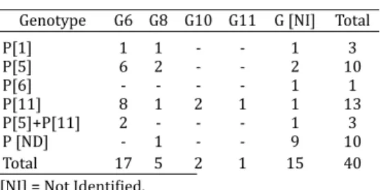
Forty of the samples (5.5%) tested positive for rotaviruses by the PAGE technique. Table 1 lists the G and P genotypes of bovine group A rotavirus identiϐied by RT-PCR. The ro-tavirus genotypes G and P were fully characterized in 25 (62.5%) of the fecal samples. The VP7 gene could not be identiϐied in 15 (37.5%) samples, while the VP4 gene was identiϐie d in 30 (75%) of samples.
G8, G10 and G11, which were detected in ϐive (12.5%), two (5%) and one (2.5%) sample (s), respectively.
Genotypic associations of P[5]+P[11] and of G6P[5]+ P[11] were observed in one and two sam ples, respectively. Associations of genotypes circulating in herds in São Paulo were G6P[11], G10[P11], G[-] P[5] + [11] and G[-] P[6]. The genotypes found circulating in herds in Minas Gerais were G6P[11], G8P[5], G11P[11], G10P[11], while those identi-ϐied in Goiás were G6P[5] + [11], G6P[5], G8P[ -], G6P[11], G[-]P[1], G[-]P[11], and G[-]P[5]. Genotypes G6P[5], G8 [P11], G6[P11], G8[P1], G8[P5], and G6[P1] were found in herds in Mato Grosso do Sul.
DISCUSSION
Although the genotypes G[6], G[8], G[10] and P[1], P[5] and P[11] are found predominantly in cattle in Brazil (Brito et al. 2000, Alϐieri et al. 2004) and in other countries (Falcone et al.1999, Attar et al. 2002, Garaicoechea et al. 2006), in-fections in calves caused by group A rotaviruses belonging to other G and P genotypes have also been reported (Abe et al. 2011).
In the state of Goiás, the occurrence of the G6 genotype was found in 50% (9/18) of the samples, followed by geno-type G8 in 5.5% (1/18). With regard to the VP4 gene, the following genotypes were detected: P[1] in 5.5% (1/18) of the samples, P[5] in 38.8% (7/18), P[11] in 33.3% (6/18) and associations of P[5] + [11] in 11.11% (2/18), indica-ting the presence of mixed infections. The most common genotypic combination was G6P[5], which was present in 22.22% (4/18) of the fecal samples. These ϐindings are consistent with those reported by Caruzo et al. (2010), who identiϐied genotype G6 as the most frequent (64.5%), P[1] genotypes in 9.7% and P[11] in 32.2%, and associations of P5 + P11 in a study of 31 samples that tested positive for rotavirus in the state of Goiás, although no G6P[5] geno-type was detected.
According to Iturriza-Gomara et al. (2001), these com-binations may originate from restructuring or co-infections characterized by genetic mechanisms of evolution, which involve pointwise mutations (drifts), restructuring (shifts, reassortant viruses) or exchange of RNA segments between samples. They may also give rise to rearrangements, whi-ch consist of duplications and deletions of nucleotide se-quences within a genomic segment (Taniguchi & Urasawa
1995), reinforcing the idea that segments exhibiting VP4 and VP7 genes can segregate naturally and independently.
The most frequent genotype in 33.33% (2/6) of the fe-cal samples collected from herds in the São Paulo was G6, followed by G10 in 16.66% (1/6). Genotype [P11] occurred in 66.66% (4/6), of the samples , followed by two samples characterized as P[5] and P[6]. The most frequent combi-nation was G6P[11] in 33.33% of fecal samples. These ϐin-dings are consistent with those of Buzinaro et al. (2009), who performed a molecular characterization of bovine group A rotavirus in São Paulo during 2003 and 2004, and detected a prevalence of the G6 genotype in 61.1%, as well as the occurrence of genotype P[5] P[11] and combinations of P[5] + P[11].
In Mato Grosso do Sul, the G8 genotype was characte-rized in 37.5% (3/8), with a predominance of G6 in 50% (4/8) and types P[1] in 25% (2 / 80), P[5] in 50% (4/8), and P[11] in 25% (2/8) of analyzed samples. The most fre-quent combination was G6P[5] in 25% of fecal samples. The combination G8P[11] was also characterized and was considered the most prevalent unusual combination on a farm in Japan where 33 rotavirus samples were isolated and identiϐied (Fukai et al. 1999).
Other researchers have identiϐied strains with the G6 and G10 genotypes in Brazilian cattle herds. Brito (2000) identiϐied G6 as the most common genotype in herds in Goiás, while Alϐieri et al. (2004) found that G6 and G8 were the most frequent genotypes in beef and dairy herds in three Brazilian States. Fukay et al. (2002) in Japan and Sa-ravanan et al. (2006) in India also identiϐied G6, G8 and G10 as the most frequent genotypes in cattle.
As for the VP4 gene, the results found in this study can be considered highly unusual when compared with the P strains most frequently reported, P[11] and P[5]. Thus, 40% of the samples were characterized as P[11] and 32.5% as P[5]. Alϐieri et al. (2004) reported different results for cattle in the states of Mato Grosso do Sul, São Paulo and Paraná, ϐinding the prevalence of genotype P[5] in 66% of the samples. In beef and dairy herds in Italy, Falcone et al. (1999) detected the genotypes P[1], P[5] and P[11], with a predominance of genotype P[5]. Also, Monini et al. (2008) reported a 65.1% prevalence of P[11], followed by 25% of P[5], and only 2.5% of P-type combinations in Italian herds, but detected no P[1]. In Brazil, Barreiros et al. (2004) also detected P[11] and P[5] genotypes in herds in Mato Grosso do Sul, Paraná and São Paulo.
The most common combinations of VP7/VP4 genes in cattle are G6P[5] (UK-like), G6P[1] (NCDV-like) and G10P[11] (Hussein et al. 1993, Fukai et al. 1999, Ghosh et al. 2008). In this context, Garaicoechea et al. (2006) characterized 60% of the bovine rotavirus group A detected in herds in Argentina as G6P[5]. Alϐieri et al. (2004) also reported the G6 P[5] com-bination as the most prevalent one in 40% of the samples they collected from herds in Brazil’s southern, southeastern and central regions, followed by 12% of G6P[1] and 16% of G10P[11]. However, these frequencies were much higher than those found in the present study, which identiϐied 20% as G6P[5], 2.5% as G6P[1] and 5% as G10P[11], despite the smaller number of genotyped samples.
Table 1. Distribution of G and P genotypes in 40 samples of ϐield strains of bovine group
A rotav irus, identiϐied by RT-PCR in fecal material from calves in the states of São Paulo, Minas Gerais, Goiás and Mato Grosso
do Sul, Brazil
Genotype G6 G8 G10 G11 G [NI] Total
P[1] 1 1 - - 1 3
P[5] 6 2 - - 2 10
P[6] - - - - 1 1
P[11] 8 1 2 1 1 13
P[5]+P[11] 2 - - - 1 3
P [ND] - 1 - - 9 10
Total 17 5 2 1 15 40
On the other hand, in Ireland, Reidy et al. (2006) iden-tiϐied only 1.9% of their 54 samples as G6P[1] and 1.9% as G10P[11], while Garaicoechea et al. (2006) did not iden-tify the combination G6P[1] and identiϐied only 4.4% as G10P[11].
G6P[11] was the genotype most prevalent in this study, representing 20% of the samples, which is consistent with the ϐindings of Caruzo et al. (2010), who identiϐied 16.2% as G6P[11]. Reidy et al. (2006) and Garaicoechea et al. (2006) also reported this genotype in their studies, albeit at low frequencies (7.4% and 4.4%, respectively).
In the present study we observed 5% of G6P[5] + P[11] combinations, suggesting the presence of mixed infections, but at lower frequencies than those repor-ted by Reidy et al. (2006) and Swiatek et al. (2009), who found mixed combinations of G6P[5] + P[11] in 11% and 15.4% of their samples, respectively. Therefore, the pre-sence of more than one type of VP4 or VP7 gene in sam-ples suggests mixed infections in a host, which can faci-litate the occurrence of reassortants during replication of the virus, resulting in a new and diverse rotavirus population involving the exchange of one or more segments during replication of the virus, resulting in a new and diver-se rotavirus population.
In the state of Minas Gerais, G6P[11] combinations were detected in 25% (2/8), G10P[11] in 12.5% (1/8), G8P[5] in 12.5% (1/8), and G11P[11] in 12.5% (1/8) of analyzed samples.
A G11 rotavirus strain was ϐirst isolated from pigs in se-veral regions of Mexico in 1983 (Ruiz et al. 1988), and was later identiϐied in the United States and Venezuela (Ciarlet et al. 1994, Rosen et al. 1994). Hussein et al.’s study (1993) conϐirmed the existence of the G11 genotype in samples of bovine rotavirus and other uncommon strains in the Uni-ted States. The participation of genotype G11, which is ty-pically considered porcine and has also been identiϐied in humans (Bányai et al. 2009, Rahman et al. 2005, Shim et al. 2011), was also observed in the present work. This ϐinding can be considered of major epidemiological signiϐicance in view of the scanty reports of this genotype in Brazilian cat-tle, and may be a result of sharinf of the same environment by cattle and pigs. On the other hand, Gregori et al. 2009 also observed the presence of predominantly bovine geno-types for the molecular characterization of fecal samples from pigs in São Paulo state, suggesting a potential cross--infection of the virus.
G and P genotypes were not identiϐied in 9 (22.5%) of the samples by se mi-nested RT-PCR. In addition, the VP7 gene was not identiϐied in 6 (15%) samples, but the VP4 gene was identiϐied in 30 (75%) samples.
Several authors have reported unsuccessful molecular characterization of rotavirus samples, including Fukai et al. (2002), who identiϐied genotype G in 76% of samples and P in 68.3% of bovine samples subjected to PCR, and Alϐieri (1999), who identiϐied genotypes in 96% of the samples he subjected to PCR.
Indistinct genotypic results by RT-PCR may be caused by RNA extracted directly from fecal samples, whi ch could cause the co-precipitation of unspeciϐic inhibitory
substan-ces present in the samples, which interfere in the ea rly sta-ges of PCR ampliϐication, inhibiting the denaturation and annealing of primers, as described by Gouvea et al. 1990. Other possible causative factors are the long-term storage of samples, which may decrease the amount of target viral genomes (Rådström et al. 2002, Shay Fout et al. 2003); the virus in question ma y belong to a different genotype from the ones for whi ch the primers used in the reactions were intended (Winiarczyk; Gradzki, 2002); or even the presen-ce of non-group A rotaviruses circulating in the regions un-der study.
Today, knowledge about group A rotavirus genotypes/ serotypes is essential to establish mechanisms for adequa-te epidemiological surveillance and to control infections in the species most commonly affected. The commercially available inactivated vaccines against rotavirus contain strains that are prevalent in the regions where they are produced (the U.S.A and Argentina), such as NCDV-Lincoln (G6P[1]), UK (G6P[5]) and B223 (G10P[11]). The immune response of animals to vaccination is more directed to the vaccine strain, facilitating the occurrence of infections cau-sed by circulating strains that differ from those contained in the vaccine (Conner et al. 1994).
Furthermore, despite maternal vaccination, rotavirus is still detected in the feces of calves with diarrhea (Barrei-ros et al. 2004). This has led to the emergence of antigenic variants and an increase in mixed infections due to the for-mation of recombinants, giving rise to new viral strains (Lu et al. 2004, Dennehy, 2008). This situation emphasizes the increasing importance of knowledge about the genotypes prevalent in regions for a more effective response to vac-cination.
CONCLUSIONS
The molecular analyses performed in this study indica-te that most of the samples of rotaviruses circulating in cat-tle herds in the Brazilian states of São Paulo, Mato Grosso do Sul, Goiás and Minas Gerais present genotypes consis-tent with those described in the literature for Brazil and in other countries.
However, the occurrence of a typical porcine genotype was detected, suggesting a possible cross-infection. This ϐinding calls for a more in-depth investigation, particular-ly the addition of epidemiological and genetic sequencing data of th e VP4 and VP7 encoding genes, such as ot her re-gions of the genome. Ongoing research to monitor the ge-notypes in cattle herds is essential for the improvement of speciϐic prophylactic measures.
Acknowledgements.- To FAPESP (São Paulo Research Foundation) for its ϐinancial support of this research (Process nº 2010/02997-4).
REFERENCES
Abe M., Ito N., Masatani T., Nakagawa K., Yamaoka S., Kanamaru Y., Suzuki H., Shibano K., Arashi Y. & Sugiyama M. 2011. Whole genome characteri-zation of new bovine rotavirus G21P[29] and G24P[33] strains provides evidence for interspecies transmission.J. Gen .Virol. 92:952-960. Alϐieri A.F., Alϐieri A.A., Barreiros M.A.B., Leite J.P.G. & Richtzenhain L.J.
Bányai K., Bogdán A., Domonkos G., Kisfali P., Molnár P., Tóth A., Melegh B., Martella V., Gentsch J.R. & Szucs G. 2009. Genetic diversity and zoonotic potential of human rotavirus strains, 2003-2006. J. Med. Virol. 81:362-370.
Barreiros M.A.B., Alϐieri A.F., Médici J.P.G., Leite J.P.G. & Alϐieri A.A. 2004. G and P genotypes of a group A rotavirus from diarrhoeic calves born to cows vaccinated against the NCDV (P[1], G6 rotavirus strain. J. Vet. Med. 51:104-109.
Brito W.M.E.D., Munford V., Vilaça A.M., Caruzo T.A.R. & Rácz M.L. 2000. Characterization of mixed infections with different strains of bovine ro-tavirus in an outbreak of diarrhea in dairy herds in Goiás, Brazil. Braz. J. Microbiol.31:140-145.
Buzinaro M.G., Munford V., Brito, V.M.E.D., Rácz M.L. & Jerez J.A. 2009. Ca-racterização eletroforética e análise de subgrupo de rotavírus em re-banhos bovinos leiteiros do Estado de São Paulo. Arq. Bras. Med. Vet. Zootec. 52(6):555-561.
Caruzo T.A.R., Brito W.M.E.D., Munford V. & Rácz M.L. 2010. Molecular characterization of G and P-types bovine rotavirus strains from Goiás, Brazil: high frequency of mixed P-type infections. Mem. Inst. Oswaldo Cruz 105(8):1040-1043.
Ciarlet M., Hidalgo M., Gorziglia M. & Liprandi F. 1994. Characterization of neutralization epitopes on the VP7 surface protein of serotype G11 porcine rotaviruses. J. Gen.Virol. 75:1867-1873.
Collins P.J., Martella V., Buonavoglia C. & O’Shea H., 2010. Identiϐication of a G2-like porcine rotavirus bearing a novel VP4 type, P[32]. Vet. Res. 41:73.
Conner M.E., Matson D.O. & Estes M.K. 1994. Rotavirus vaccines and vacci-nation potential. Curr. Top. Microbiol. Immunol. 185:285-337.
Constable P.D. 2004. Antimicrobial use in the treatment of calf diarrhea: a review. J. Vet. Intern. Med.18:8-17.
De la Fuente R., Garcia A., Ruiz-Santa-Quiteria J.A., Luzón M., Cid D., García S., Orden J.A. & Gómez- Bautista M. 1998. Proportional morbidity rates of enteropathogens among diarrheic dairy calves in central Spain. Prev. Vet. Med. 36:145-152.
Dennehy P.H. 2008. Rotavirus vaccines: An overview. Clin. Microbiol. Rev. 21(1):198-208.
El-Attar L., Dhaliwal W., Iturriza-Gómara M. & Bridger J. C. 2002. Identi-ϐication and molecular characterization of a bovine G3 rotavirus which causes age-independent diarrhea in cattle. J. Clin. Microbiol. 40(3):937-942.
Esona M.D., Steele D., Kerin T., Armah G., Peenze I., Geyer A., Page N., Nyan-gao J., Agbaya V.A.,Trabelsi A., Tsion B., Aminu M., Sebunya T., Dewar J., Glass R. & Gentsch J. 2010. Determination of the G and P types of previously nontypeable rotavirus strains from the African Rotavirus Network, 1996-2004: Identiϐication of unusual G types. J. Infect. Dis. 202(Suppl.):S49-S54.
Estes M.K. & Kapikian A.Z. 2007. Rotaviruses, p.1917-1974. In: Knipe D.M., Howley P.M., De Grifϐin R.A., Martin M.A., Roizman B., Straus S.E. (Eds), Fields Virology. 5th ed. Lippincott, Williams and Wilkins, Philadelphia.
Falcone E., Tarantino M., Trani L., Cordioli P., Lavazza A. & Tollis M.1999. Determination of bovine rotavirus G and P serotypes in Italy by PCR. J. Clin. Microbiol. 37(12):3879-3882.
Fukai K., Sakai T., Hirose M., Itou T. 1999. Prevalence of calf diarrhea cau-sed by bovine group a rotavirus carrying G serotype 8 speciϐicity. Vet. Microbiol.66:301-311.
Fukai K., Maeda Y., Fujimoto K., Itou T., Sakai T. 2002. Changes in the preva-lence of rotavirus G and P types in diarrheic calves from the Kagoshima prefecture in Japan. Vet. Microbiol. 86(4):343-349.
Garaicoechea L., Bok K., Jones L.R., Combessies G., Odeón A., Fernandez F. & Parreño V. 2006. Molecular characterization of bovine rotavirus cir-culating in beef and dairy herds in Argentina during a 10-year period (1994-2003). Vet. Microbiol. 118:1-11.
Ghosh S., Samajdar S., Sinha M., Kobayashi N., Taniguchi K. & Naik T.N. 2008. Molecular characterization of rare bovine group A rotavirus G15P[11] and G15P[21] strains from eastern India: identiϐication of simian SA11--like VP6 genes in G15P[21] strains. Virus Genes. 37(2):241-249.
Gouvea V., Glass R.I., Woods P., Taniguchi K., Clark H.F., Forrester B. & Fang Z. 1990. Polymerase chain reaction ampliϐication and typing of rotavirus nucleic acid from stool specimens. J. Clin. Microbiol. 28:276-282. Gouvea V., Santos N. & Timenetsky M.C. 1994a. Identiϐication of bovine
and porcine rotavirus G types by PCR. J. Clin. Microbiol.32:1338-1340. Gouvea V., Santos N. & Timenetsky M.C. 1994b. VP4 typing of bovine and
porcine group A rotaviruses by PCR. J. Clin. Microbiol. 32:1333-1337. Greenberg H.B. & Estes M.K. 2009. Rotaviruses: from pathogenesis to
vac-cination. Gastroenterology 136:1939-1951.
Gregori F., Rosales C.A.R., Brandão P.E., Soares R.M. & Jerez J.A. 2009. Di-versidade genotípica de rotavírus suínos no Estado de São Paulo. Pesq. Vet. Bras. 29(9):707-712.
Herring A.J., Inglis N.F., Ojeh C.K., Snodgrass D.R. & Menzies J.D. 1982. Rapid diagnosis of rotavirus infection by direct detection of viral nucleic acid in silver-stained polyacrylamide gels. J. Clin. Microbiol. 16(3):473-477. Hussein H.A., Parwani A.V., Rosen B.I., Lucchelli A. & Saif L.J. 1993.
De-tection of rotavirus serotypes G1, G2, G3 and G11 in feces of diarrheic calves by using polymerase chain reaction-derived cDNA probes. J. Clin. Microbiol. 31:2491-2496.
Iturriza-Gomara M., Isherwood B., Desselberger U. & Gray J. 2001. Reas-sortment in vivo: driving force for diversity of human rotavirus strains isolated in the United Kingdom between 1995 and 1999. J. Virol. 75:3696-3705.
Lanz Uhde F., Kaufmann T., Sager H., Albini S., Zanoni R., Schelling E. & Meylan M. 2008. Prevalence of four enteropathogens in the faeces of young diarrhoeic dairy calves in Switzerland. Vet. Rec. 163:362-366. Lu W., Duhamel G.E., Benϐield D.A. & Grotelueschen D.M. 1994. Serological
and genotypic characterization of group A rotavirus reassortants from diarrheic calves born to dams vaccinated against rotavirus. Vet. Micro-biol. 42(2/3):159-170.
Martella V., Bányai K., Matthijnssens J., Buonavoglia C. & Ciarlet M. 2010. Zoonotic aspects of rotaviruses. Vet. Microbiol.140:246-255.
Matthijnssens J., De Grazia S., Piessens J., Heylen E., Zeller M., Giammanco G.M., Bányai K.,Buonavoglia C., Ciarlet M., Martella V. & Van Ranst M. 2011. Multiple reassortment and interspecies transmission events contribute to the diversity of feline, canine and feline/canine-like human group A rotavirus strains.Infect. Genet.Evol. May 13. [Epub ahead of print] Monini M., Cappuccini F., Battista P., Falcone E., Lavazza A. & Ruggeri F.M.
2008. Molecular characterization of bovine rotavirus strains circulating in northern Italy, 2003-2005. Vet. Microbiol. 129:384-389.
Pereira H.G., Azeredo R.S., Leite J.P., Andrade Z.P. & De Castro L. 1985. A combined enzyme immunoassay for rotavirus and adenovirus (EIARA). J. Virol. Methods10:21-28.
Rådström P., Knutsson R., Wolffs P., Dahlenborg M. & Löfström C. 2002. Pre-PCR processing of samples, p.31-50. In: Sachse K. & Frey J. (Eds), PCR detection of microbial pathogens. Humana Press Inc., Totowa. Rahman M., Matthijnssens J., Nahar S., Podder G., Sack D.A., Azim T. & Van
Ranst M. 2005. Characterization of a novel P[25], G11 human group a rotavirus. J. Clin. Microbiol. 43(7):3208-12.
Ramig R.F., Ciarlet M., Mertens P.P.C. & Dermody T.S. 2005. Genus rotavi-rus, p.484-496. In: Fauquet C.M., Mayo M.A., Manilloff J., Desselberger U. & Ball L.A. (Eds), Virus taxonomy: Eighth Report of the International Committee on Taxonomy of Viruses. Elsevier Academic Press, San Diego. Reidy N., Lennon G., Fanning S., Power E. & O’Shea H. 2006. Molecular
characterization and analysis of bovine rotavirus strains circulating in Ireland 2002-2004. Vet. Microbiol.117:242-247.
Rosen B.I., Parwani A.V., Lopez S., Flores J. & Saif L.J. 1994. Serotypic dif-ferentiation of rotaviruses in ϐield samples from diarrhetic pigs by using nucleic acid probes speciϐic for porcine VP4 and human and porcine VP7 genes. J. Clin. Microbiol. 32:311-317.
Ruiz A.M., López I.V., López S., Espejo R.T. & Arias C.F. 1988. Molecular and antigenic characterization of porcine rotavirus YM, a possible new rotavirus serotype. J. Virol. 62(11):4331-4336.
Shay Fout G., Martinson B.C. & Moyer M.W.N. 2003. A multiplex reverse transcription- CR method for detection of human enteric viruses in groundwater. Appl. Environ. Microbiol. 69(6):3158-3164.
Shim J.O., Baek I.H., Le V.P., Ko E.M., Seok W.S., Uh Y., Kim J.K., Ahn S.Y., Lee H.S., Ryoo E., Shim S.Y., Song W., Lim I. & Kim W. 2011. Molecular charac-terization of rotavirus diarrhea among children in South Korea: detec-tion of an unusual G11 strain. Arch. Virol. 156(5):887-92.
Steyer A., Poljsak-Prijatelj M., Barlic-Maganja D. & Marin J. 2008. Human, por-cine and bovine rotaviruses in Slovenia: Evidence of interspecies transmis-sion and genome reassortment. J. Gen. Virol. 89(Pt 7):1690-1698.
Swiatek D.L., Palombo E.A., Lee A., Coventry M.J., Britz M.L. & Kirkwood C.D. 2009. Detection and analysis of bovine rotavirus strains circulating in Australian calves during 2004 and 2005. Vet. Microbiol. 140:56-62. Taniguchi K. & Urasawa S. 1995. Diversity in rotavirus genomes. Semin.
Virol. 6(2):123-131.