1Submitted on 05/13/2016. Accepted for publication on 05/10/2017.
2Departamento de Fitopatologia, Universidade Federal de Lavras - UFLA, Caixa Postal 3037, 37200-000- Lavras, MG, Brasil. *Corresponding author <[email protected]>
Evaluation of inoculum potential of pathogens in seeds: relation to
physiological quality and DNA quantification by qPCR
1Marina de Resende Faria Guimarães
2*, Carolina da Silva Siqueira
2, José da Cruz
Machado
2, Sueny Kelly Santos de França
2, Gabriel Castanheira Guimarães
2ABSTRACT - Given what is already known in regard to seed health and the availability of molecular methods for detection of the pathogens Stenocarpella maydis and Stenocarpella macrospora in maize seeds, Colletotrichum gossypii var. cephalosporioides in cotton seeds, and Corynespora cassiicola in soybean seeds, the aim of this study was to evaluate seed vigor according to different inoculum potentials. The fungus isolates were inoculated on seeds by the technique of water restriction, through which different inoculum potentials are obtained, corresponding to times of seed exposure of 0, 24, 48, and 96 hours for maize and cotton seeds, and 0, 36, 108, and 144 hours for soybean seeds. The seeds were subjected to germination, electrical conductivity, health, and qPCR tests. Results of the blotter test showed that in most pathosystems, there was a higher incidence of the fungi with an increase in inoculum potential. A decrease in germination percentage was observed in all species as inoculum potential increased, as well as further degradation of seed membranes. The qPCR test confirmed that the most damaged seeds in the tests had higher presence of the pathogens.
Index terms: seed pathology, fungi, maize, soybean, cotton.
Avaliação do potencial de inóculo de patógenos em sementes: sua relação com a
qualidade fisiológica e quantificação do DNA por qPCR
RESUMO - Diante do que já se conhece em sanidade de sementes e tendo-se em mãos métodos moleculares para a detecção dos fungos Stenocarpella maydis, Stenocarpella macrospora em sementes de milho, Colletotrichum gossypii var. cephalosporioides em algodão e Corynespora cassiicola em soja, objetivou-se neste estudo avaliar o vigor das sementes em função dos diferentes potenciais de inóculo. Os isolados dos fungos foram inoculados nas sementes por meio da técnica de restrição hídrica pela qual se obtém diferentes potenciais de inóculo, que foram representados por P0, P24, P48 e P96 e P0, P36, P108 e P144, que correspondem à exposição das sementes aos períodos de tempo de 0, 24, 48, 96 horas (milho e algodão) e nos tempos 0, 36, 108 e 144 horas (soja), respectivamente. As sementes foram submetidas aos testes de germinação, condutividade elétrica, sanidade e qPCR. Pelo blotter test, na maioria dos patossistemas, houve maior incidência do fungo com o aumento do potencial de inóculo. Foi observada uma queda na porcentagem de germinação de todas as espécies com o aumento do potencial de inóculo, assim como maior degradação das membranas das sementes. A qPCR confirmou que nas sementes mais prejudicadas havia maior quantidade de inóculo dos patógenos.
Termos para indexação: patologia de sementes, fungos, milho, soja, algodão.
Introduction
The association of pathogens with seeds favors the survival and spread of these agents because seeds, the most important input in establishing a crop, have considerable
ability for maintaining the viability of the structures of many plant pathogens for long periods in comparison to other plant
parts (Tanaka and Machado, 1985; Machado, 1988).
The use of healthy seeds with high physiological quality is
of pathogens. Physiological quality is related to the ability of seeds to exercise their vital functions and express their potential, and is characterized by germination, dormancy, and
vigor. Therefore, the effects of pathogens on seed quality are generally reflected in a decrease in germination percentage,
reduced stand, increase in abnormal plants, reduction in
seedling vigor, and, consequently, decreased yield (Toledo et
al., 2009, Botelho et al., 2013).
Important diseases caused from these associations of seeds with pathogens include stalk rot and ear rot in maize caused by the fungi Stenocarpella maydis and Stenocarpella macrospora, which stand out by their presence in all maize producing regions
(Casa et al., 2006), causing yield reductions from 0.67% to 50% (Denti and Reis, 2003). For the soybean crop, target spot,
caused by the fungus Corynespora cassiicola (Berk. & M.A. Curtis), has increased in recent crop seasons and may cause
estimated damage of up to 40% in the field (Godoy et al., 2012, 2013; Koenning and Creswell, 2006). Another disease,
anthracnose, caused by the fungus Colletotrichum gossypii var. cephalosporioides in cotton, can lead to severe losses; some
authors report 20% to 30% losses, and this may reach 85%
in extreme, total infestation cases when there is generalized
distribution in the field (Abrahão, 1961; Abrahão and Costa, 1949; Carvalho et al., 1984; Araújo et al., 2009).
Various conventional methods can be used for detection of
pathogens in association with seeds; the choice of a particular
method depends on the type of pathogen or the species to be detected. The most common method used for detection of necrotrophic fungi in general has been the blotter test. However,
currently, the real-time polymerase chain reaction (qPCR)
technique has been an important tool to assist in these methods
because of its precision and specificity (Konstantinova et al., 2002; Gao et al., 2004; Barros et al., 2008; Duressa et al., 2012; Botelho et al., 2015; Sousa et al., 2015).
The aim of this study was to evaluate the performance of maize seeds associated with S. maydis and S. macrospora, of cotton seeds associated with C. gossypii var. cephalosporioides, and of soybean seeds associated with C. cassiicola according
to different inoculum potentials evaluated through germination
and electrical conductivity tests, and to quantify the inoculum potentials through qPCR.
Materials and Methods
Obtaining isolates and determining seed profile
The S. maydis isolate, pathogenic to maize, was obtained from the Mycological Collection of Lavras of the Mycology
Laboratory of the Federal University of Lavras (UFLA),
in Lavras, MG, Brazil; it is identified as CML 698. The S.
macrospora isolate, also pathogenic to maize, identified as CMLAPS 10, was obtained from the Mycological Collection of the Seed Pathology Laboratory of UFLA, Lavras, MG. The Colletotrichum gossypii var. cephalosporioides isolate, pathogenic to cotton, was supplied by Embrapa Algodão and collected from Alto Taquari in the state of Mato Grosso, Brazil,
identified as CMLAPS 262. The Corynespora cassiicola
isolate, pathogenic to soybean, identified as CMLAPS 312, was obtained from Embrapa in the state of Paraná, Brazil.
The seeds used were the RB9077 cultivar of maize, the
M7110 cultivar of soybean, and the DP 1240B2RF variety of cotton. The profiles of the seed lots were determined according to the Rules of Seed Testing (Brasil, 2009a). The germination rate of the maize seeds was 94%, of cotton, 98%, and of soybean, 85%. None of the fungi used in this study
were detected in the seed health test.
Seed inoculation -The maize and cotton seeds were first
disinfected with 1% sodium hypochlorite for one minute and
then washed with distilled water three times and placed to dry at 23 °C for three days. For soybean seeds, only the disinfection time was changed, to 30 seconds. The fungi of the three species were chopped up to place in 15-cm diameter Petri dishes in
PDA medium (20 g of agar, 20 g of dextrose, and 200 g of potato/liter), modified by the addition of mannitol with water potential adjusted to -1.4 MPa for maize and -1.0 MPa for soybean and cotton, as described by Machado et al. (2012) and according to calculation by the SPPM Software (Michel and Radcliffe, 1995). Five disks of mycelium were chopped
up per Petri dish, which were kept for seven days in BOD at 25±2 °C and 12 hours photoperiod. After this period, the seeds were distributed in a single layer on the fungus colonies under
study. The maize and cotton seeds remained for times of 0, 24, 48, and 96 h, corresponding to the inoculum potentials of P0, P24, P48, and P96, respectively. The soybean seeds remained for 0, 36, 108, and 144 h, represented by P0, P36, P108, and P144. After these inoculation times, the seeds were removed
from contact with the fungi, dried for three days at ambient temperature, and then placed in cold storage.
Tests for evaluation of seed quality
Germination test - For each treatment (of the different pathosystems), four replications of 50 seeds were made, distributed on sterilized “germitest” paper substrate, and moistened with 2.5 times the weight of the dry paper with sterilized distilled water. The substrate paper was rolled and these rolls were placed in a germinator at a temperature of
25±2 ºC. Evaluations occurred according to Brasil (2009a).
maize seeds at 11% moisture, 4.50 g of cotton seeds also at 11% moisture, and 8 g of soybean seeds at approximately 12% moisture were weighed on a precision balance (0.001 g); these
weights correspond to 50 seeds per replication for each treatment of each species. Each replication was placed to soak in 75 mL of
deionized water for 24 h in BOD at a temperature of 25±2 ºC in
the dark. At the end of this period, readings were taken with the
MS TECNOPON® conductivity meter. The results obtained were
analyzed as described by Krzyzanowski et al. (1999).
Seed health test - This was performed according to the
norms described in Brasil (2009b).
Quantification of fungal DNA
DNA extraction - The pure fungal cultures of S. maydis, S. macrospora, C. gossypii var. cephalosporioides, and C. cassiicola, grown for seven days in PDA medium were scraped and macerated in a mortar with liquid nitrogen until
acquiring the consistency of a fine powder. For maceration of the 400 inoculated seeds of each treatment, the A11 Basic
IKA grinder was used, with the addition of liquid nitrogen.
Three 40 g subsamples were removed from each of the
samples. For DNA extraction, the Wizard® Genomic DNA
Purification kit (Promega, Madison, WI) was used, according
to manufacturer’s instructions.
Real-time PCR (qPCR) - The qPCR was performed for all the treatments of maize, cotton, and soybean, and each
DNA sample was tested in duplicate with a total reaction
volume of 25 µL. For each reaction, we used 12.5 µL of the
PCR SYBR Green Kit (Qiagen), 2 µL of the DNA of each
sample of inoculated seed, and the amounts necessary of the
forward and reverse primers for each pathosystem. The DNA
of the target fungus was used as a positive control, and sterile ultrapure water and non-inoculated seed as a negative control.
The qPCR mixture of S. maydis was prepared containing 0.10 µM of the forward primer RT.Smay.F GTTTCCATGACCTGCTCA CG and 0.60 µM of the reverse primer RT.Smay.R TGTTGCTCGGTTTCAGGCTTG
(Romero and Wise, 2015), and amplification consisted of denaturation at 95 ºC for 2 minutes, 40 cycles, denaturation
at 95 ºC for 30 seconds, and annealing at 60 ºC for 30 seconds. For the S. macrospora reaction, the mixture was prepared using 0.25 µM of forward RT.Smac.F GGGCAAATTTTCTCGGAGG and 0.75 µM of reverse
RT.Smac.R GCAGCTATTCAGCGTTCATC (Romero and Wise, 2015), and the amplification conditions were 95 ºC for 2 minutes, followed by 40 cycles at 95 ºC for 30 seconds, and
57 ºC for 30 seconds. For C. gossypii var. cephalosporioides, 0.7 µL of each primer CGC F and CGC R were used, and
the cycle conditions were 94 ºC for 4 minutes, followed by
40 cycles at 94 ºC for 45 seconds, and 65 ºC for 45 seconds.
In the reaction with the pathogen C. cassiicola, 0.10 µM of each of the following primerswere used: the forward GA4-F
CCTGCTCCGACTTTGTTGAG and the reverse GA4-R GTCTGGGAGCAGCAAAGACT (Dixon et al., 2009), and the amplification conditions were 94 ºC for 5 minutes, 40 cycles at 94 ºC for 30 seconds, and 58.5 ºC for 30 seconds. The DNA values were determined by the software Rotor-Gene 1.7.75 (Corbett Research, Mortlake, Australia) by the cycler Rotor-Gene 6500 (Corbett) through construction of a
standard curve of 10-fold dilutions, obtained from pure fungal
cultures, together with the value of the threshold cycle (Ct)
obtained in each reaction.
Experimental design and statistical analyses - The germination, blotter, and electrical conductivity tests were conducted under a randomized complete block design, and
the data were subjected to analysis of variance using the
Sisvar® software (Ferreira, 2011).
Results and Discussion
Physiological performance of the inoculated seeds and quantification of the inoculum
For all the pathosystems included in this study, the amount
of DNA detected by the qPCR technique was proportional
to the inoculum potentials established as referenced, that is, in accordance with the time of exposure of the seeds to the pathogens in development in the agar substrate. In general, the analyses of variance for all the species in relation to the germination, electrical conductivity, and seed health tests of
the inoculated seeds revealed significant effects (p<0.05).
According to the control treatments, there was no interference of the water restrictor mannitol on the physiological performance of the seeds.
For the S. maydis and maize seed pathosystem, a decrease in germination and an increase in electrical conductivity of the seeds was found as the inoculum potentials increased, which indicates reduced vigor. The germination percentage decreased
from 94% (seed without fungus, P0 potential) to 74.5% at P24, 62% at P48, and, at the highest potential (P96), this percentage was only 11%, making for a total reduction of 83% (Figure 1).
The value of electrical conductivity of the seeds inoculated with S. maydis at P0 was greater than the values found at P24
and at P48, which remained near 16 µmhos.cm-1.g-1. This can be
medium. In P96, the value of conductivity was 44.21 µmhos.
cm-1.g-1 . Vieira et al. (2002) observed that in seeds with moisture contents of less than 11%, the values of electrical conductivity
had very high values, whereas the opposite occurred for seeds with higher moisture contents, in which the data exhibited a
tendency toward stabilization around 13%.
By the seed health test, S. maydis was detected at all the
inoculum potentials at high percentages (Figure 1). Siqueira et al. (2014), in contrast, observed an increase in occurrence as the
potentials increased, which may be related to the cultivar used in that study. In comparison to germination values, it can be observed that although the pathogen was detected in practically all inoculated seeds, even at the lowest value of inoculum
potential, the effects were, nevertheless, less accentuated than
at the higher inoculum potentials. These results reveal that the percentages of occurrence observed in the biological methods
alone are not sufficient to indicate the true degree of interaction
between the pathogen and the host seeds, showing the need for
quantifying the fungal DNA present in the seed.
From the values obtained by the electrical conductivity
test, it could be observed that the inoculum potentials P24 and P48, despite showing a high percentage of incidence, were not sufficient for the fungus to cause damage to the seed membrane, indicating that the seed was superficially
colonized. A different situation can be observed at P96, in
which the incidence of this pathogen was high, as well as the electrical conductivity of the inoculated seeds at this potential, suggesting that at this potential, there was greater interaction and colonization of the fungus in the seed tissues.
The damage brought about by the pathogens can be directly related to the inoculum potential and to its presence
in the seed (Machado, 1988).
The results of the vigor test, based on electrical conductivity, indicate that for this pathosystem there was not increasing proportionality for the lower values of this variable
at the P24 and P48 potentials. Nevertheless, at the highest value
of inoculum potential, P96, the damage caused by the presence of the pathogen in the protective membranes of the seeds
was drastic, which was confirmed by the lower percentage of germination. Machado (1988) affirms that the damage brought
about by the presence of pathogens may be related to the inoculum potential, as well as to the location of the pathogens in the seed. Studies using the technique of inoculation by water restriction show the direct relation between the inoculum
potentials and the damage brought about in the seeds (Costa et al., 2003; Botelho et al, 2013; Siqueira et al., 2014)
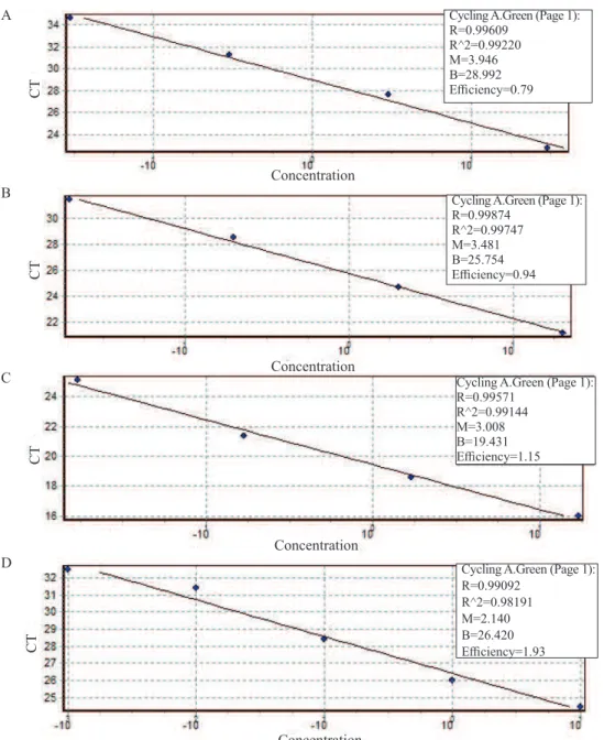
According to the data obtained in the qPCR for S. maydis, the relative efficiency of the curve was 0.79, which Figure 1. Performance of maize seeds inoculated with the isolate of Stenocarpella maydis, CML698 at different inoculum
was determined by the linear regression equation with
correlation coefficient (R²) of 0.99 (Figure 2A). From the
standard curve and the Ct values obtained in the reaction, it
was possible to quantify the DNA present in the inoculated seeds. No DNA of the fungus was found in the study at P0. In the seeds inoculated for 24 h, 2.53 ng/µL of DNA was found, whereas at the P48 potential, this amount was 6.78 ng/µL, increasing to 16.2 ng/µL at the highest potential (P96) (Figure 1D). Botelho et al. (2015) quantified
Figure 2. Standard curves of the real-time PCRs of the DNA concentration in the serial dilution of: (A) Stenocarpella maydis
ranging from 30 ng/μL to 0.03 ng/μL; (B) Stenocarpella macrospora ranging from 20 ng/μL to 0.02 ng/μL; (C)
Colletotrichum gossypii var. cephalosporioides ranging from 17 ng/μL to 0.017 ng/μL; and (D) Corynespora cassiicola ranging from 10 ng/μL to 0.001 ng/μL.
Sclerotinia sclerotiorum in soybean seeds inoculated by
the same technique and obtained results of DNA quantities
proportional to the increase in inoculum potential.
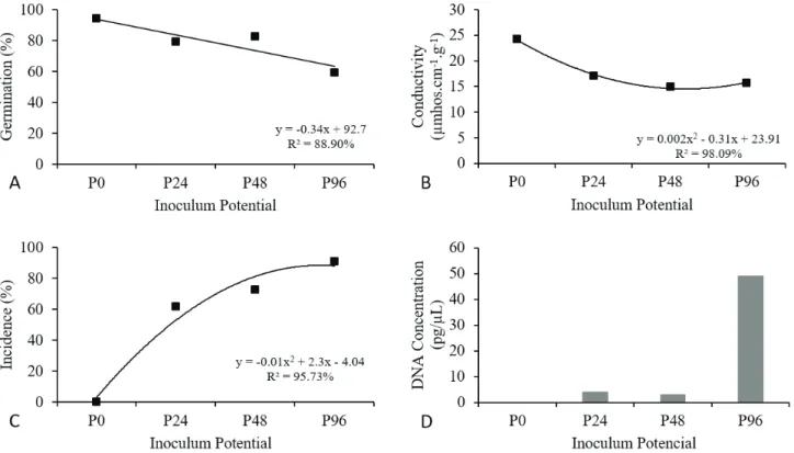
The results in regard to the S. macrospora and maize seed pathosystem show that the action of the fungus in maize seeds led to a gradual, linear reduction in germination percentages, inversely proportional to the values of fungus inoculum potential initially present in the seeds evaluated.
Unlike that which occurred to S. maydis in this study,
Concentration
Concentration
Concentration
Concentration
CT
CT
CT
CT
A
B
C
D
Cycling A.Green (Page 1): R=0.99609
R^2=0.99220 M=3.946 B=28.992 Efficiency=0.79
Cycling A.Green (Page 1): R=0.99874
R^2=0.99747 M=3.481 B=25.754 Efficiency=0.94
Cycling A.Green (Page 1): R=0.99571
R^2=0.99144 M=3.008 B=19.431 Efficiency=1.15
Cycling A.Green (Page 1): R=0.99092
Figure 3. Performance of maize seeds inoculated with the isolate of Stenocarpella macrospora, CMLAPS10, at different
inoculum potentials, P0 (0 h), P24 (24 h), P48 (48 h), and P96 (96 h). (A) Germination percentage values. (B) Mean values of electrical conductivity. (C) Incidence percentage. (D) Fungal DNA concentration in seeds.
reduction in the germination percentages of maize seeds caused by S. macrospora was less accentuated among the inoculum potentials used. Reduction between the lowest and
highest level of inoculum potential was 35%. Without the fungus (P0), the seeds exhibited 94% germination, decreasing to 79% at P24, and, with the increase in inoculum potential, germination decreased to 59% at P96 (Figure 3).
The values of electrical conductivity of the inoculated
seeds were not very different, which shows that S. macrospora
seems to cause less accentuated damage to maize seeds than S. maydis does, and that the highest inoculum potential in the seeds is not able to cause damage proportional to seed performance, as seen from the results of the germination
test (Figure 3). By this vigor test, it was observed that there
was little variation in seed conductivity. In P0, conductivity
was 24.2 µmhos.cm-1.g-1 and with the increase in inoculum
potential, this value fell to 17 µmhos.cm-1.g-1 at P24, 14.96
µmhos.cm-1.g-1 at P48, and 15.69 µmhos.cm-1.g-1 at P96.
It is important to emphasize that from the blotter test, the incidence of S. macrospora in the inoculated seeds was not
total, i.e., 100%, at all the inoculum potentials, contrary to
what occurred with S. maydis. In this study, the percentages of incidence of the fungus in the seeds were higher and linear
in relation to the increase in inoculum potentials (Figure 3). In P24, the incidence of the pathogen in the seeds was 61.5%, increasing at P48 to 72.5%, and, finally, to 91% at P96.
For quantification of S. macrospora by qPCR, with relative
efficiency of the curve of 0.94 (Figure 2B), the quantities of DNA found in the inoculated seeds at the potentials P24 and P48 were not very different. At P24, 4 pg/µL of DNA were detected, not very different from P48, at which 3 pg/µL was detected. At the highest potential (P96), the amount found was 49 pg/µL (Figure 3D).
The DNA concentration of S. macrospora was less
than that of S. maydis, confirming the relationship between inoculum potential and the variables used to evaluate seed performance.
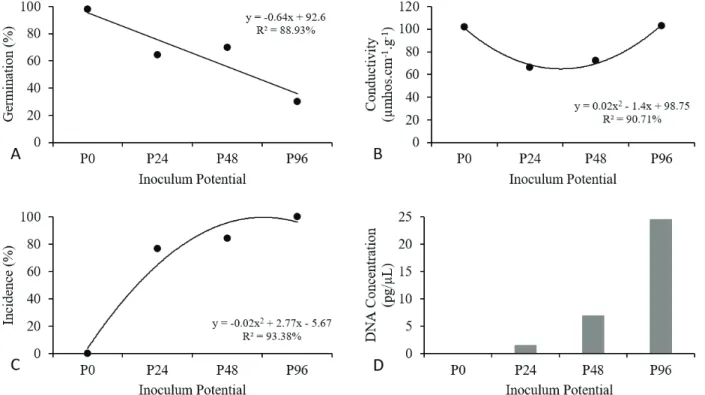
For the pathosystem of C. gossypii var cephalosporioides in cotton, the germination percentage decreased with the increase in
inoculum potential, just as in the pathosystems already mentioned. In the non-inoculated seed (P0), the germination percentage was 98%, with a reduction to 64.5% in those inoculated for 24 h (P24). At the highest potential (P96), germination was only 30%, for a total reduction of 68% (Figure 4).
Figure 4. Performance of cotton seeds inoculated with the isolate of Colletotrichum gossypii var. cephalosporioides, CMLAPS262,
at different inoculum potentials, P0 (0 h), P24 (24 h), P48 (48 h), and P96 (96 h). (A) Germination percentage values. (B) Mean values of electrical conductivity. (C) Incidence percentage. (D) Fungal DNA concentration in seeds.
conductivity reading was 101.66 µmhos.cm-1.g-1. At P24, the
conductivity reading of the solution was 66 µmhos.cm-1.g-1,
increasing to 72.14 µmhos.cm-1.g-1 at P48, and reaching a
value of 102.79 µmhos.cm-1.g-1 at P96 (Figure 4).
A progressive increase in the percentage of C. gossypii var. cephalosporioides present in the inoculated seeds in accordance with the increase in inoculum potential was observed through the seed health test. The incidence of the pathogen in the seeds
at P24 was 76.5%, increasing to 84% at P48, and then to 100% at the highest inoculum potential (P96).
Through the qPCR reactions for C. gossypii var. cephalosporioides, it was found that the curve obtained
relative efficiency of 1.15 (Figure 2C), with quantification at the P24 potential of 1.44 pg/µL of DNA, which increased to 6.89 pg/µL at P48, and 24.5 pg/µL at P96 (Figure 4D).
In the pathosystem of C. cassiicola in soybean seeds,
the germination percentage, just as in all the pathosystem
already analyzed, decreased with an increase in inoculum
potential. In P0, the initial germination percentage was 85%, which reduced to 77.5% at the first inoculum potential (P36). When inoculated at the P108 potential, the seeds exhibited 58% germination, and with an increase in potential to 144 h (P144), this value decreased to 51% (Figure 5).
The values of electrical conductivity increased in the
inoculated seeds as the inoculum potential increased. At P0,
the conductivity reading was 78.43 µmhos.cm-1.g-1. At P36,
this value was 103.6 µmhos.cm-1.g-1, and at P108 and P144,
the values were higher and similar, 112.2 µmhos.cm-1.g-1 and
113.63 µmhos.cm-1.g-1, respectively (Figure 5).
The results obtained from the blotter test showed an increase in the percentage of C. cassiicola in the inoculated seeds as the inoculum potential increased. Presence of the
pathogen in the seeds at P36 was 62.5%, increasing to 69% at P108, and to 89% at the highest potential (P144) (Figure 5).
In the qPCR test for C. cassiicola with determined standard
curve (Figure 2D), in seeds inoculated at P36, 0.09 ng/µL of DNA was detected. At the following potentials, the values found were 0.9 ng/µL - P108 and 2.62 ng/µL - P144 (Figure 5D).
The results of this study showed that the presence of the pathogens S. maydis and S. macrospora in maize seeds, C. gossypii var. cephalosporioides in cotton, and C. cassiicola
in soybean artificially inoculated on seeds is a negative factor
with drastic consequences on seed performance and causes
significant reductions in germination force and vigor (Araújo et al., 2006; Siqueira et al., 2014).
In all the pathosystems studied, there was a sharp fall in
seed germination percentage; the highest percentages occurred at
highest inoculum potential, due to the death or, in some cases, poor formation of the seeds. These results reinforce what was reported in other pathosystems, such as Sclerotinia sclerotiorum and Colletotrichum truncatum in soybean (Botelho et al., 2013; Machado et al., 2001) and Fusarium oxysporum f. sp. phaseoli in dry edible bean seeds(Costa et al., 2003).
The relationship between moisture content, the level of organization of seed cell membranes, and the amount of leachates in the imbibition solution allows association of electrical conductivity to seed vigor, in which high values of electrical conductivity indicate low vigor, and low values indicate high physiological quality of the seeds and,
therefore, high vigor (Hampton and Tekrony, 1995; Vieira
and Krzyzanowski, 1999). Results in regard to electrical conductivity showed that the increase in the period of seed exposure to the pathogen led to reduction in seed vigor at higher inoculum potentials, considering that under this condition there is greater degradation of the cell membrane system, leading to leaching of solutes, which, consequently, increases seed
deterioration (Krzyzanowski et al., 1999; Siqueira et al., 2014).
The lower values of conductivity correspond to lower release of exudates and indicate high seed vigor, as well as greater organization of the cell membrane systems.
Upon comparing the results of the physiological and seed
health test applied in this study and the concentrations of DNA from inoculated fungi at the different inoculum potentials, there
is clearly proportionality between the values of the tests and the
quantities of DNA extracted and evaluated by the qPCR technique. This study also confirms the efficiency of the primers in detection of pathogens in the seeds at different inoculum potentials.
Conclusions
The presence of the pathogens S. maydis and S. macrospora in maize seeds, C. gossypii var. cephalosporioides in cotton seeds, and C. cassiicola in soybean seeds through inoculation by the osmotic conditioning method can lead to gradual and varied reductions in the physiological quality of the seeds at a proportion opposite to the inoculum potentials of these
organisms quantified through qPCR.
The effects of S. macrospora in maize seeds are less than
those observed for S. maydis. The effects of C. cassiicola in association with soybean seeds were less intense, regardless of the inoculum potential used.
For all the pathosystems studied, the primers used in
detection and quantification of the target DNA were specific, with linearity in the standard curve at each level of DNA dilution.
Results showed proportionality between the fungal DNA
Figure 5. Performance of cotton seeds inoculated with the isolate of Corynespora cassiicola, CMLAPS312, at different
extracted from the seeds, the inoculum potentials of each
pathogen, the effects represented by germination percentages,
and seed vigor.
Acknowledgments
Our thanks to the CNPq, CAPES, and FAPEMIG for financial support and to the Riber and Cotton Tecnologia
companies for providing seeds.
References
ABRAHÃO, J. Combate a ramulose tardia do algodoeiro. O Biológico, v.27, p.121-123, 1961.
ABRAHÃO, J.; COSTA, A.S. Instruções para o reconhecimento da ramulose do algodoeiro. O Biológico, v.15, n.3, p.59-60, 1949.
ARAÚJO, D.V.; POZZA, E. A.; MACHADO, J.C.; ZAMBENEDETTI, E. B.; CELANO, F.A.O.; CARVALHO, E.M.; CAMARGOS, V.N. Influência da temperatura e do tempo de inoculação das sementes de algodão na transmissibilidade de Colletotrichum gossypii var. cephalosporioides. Fitopatologia Brasileira, v.31, p.35-40, 2006. http://www.scielo.br/pdf/fb/v31n1/ a06v31n1.pdf
ARAÚJO, A.E.; MENTEN, J.O.M.; FERREIRA, A.C.B.; DIAS, C.T.S.; NÓBREGA, M.B.M.; MORELLO, C.L. Efeito de diferentes níveis de Colletotrichum gossypii South var. cephalosporioides Costa, em plantas de algodão no campo e sua incidência nas sementes. Summa Phytopathologica, v.35, n.4, p.310-315, 2009. http://www.scielo.br/scielo.php?script =sci_ arttext&pid=S010054052009000400009&lng=en&nrm=iso> BARROS, E.; CRAMPTON, M.; MARAIS, G.; LEZAR, S. A DNA-based method to quantify Stenocarpella maydis in maize. Maydica, v.53, p.125-129, 2008. https://www.researchgate.net/ publication/236154879
BOTELHO, L.S.; ZANCAN, W.L.A.; MACHADO, J.C.; BARROCAS, E.N. Performance of common bean seeds infected by the fungus Sclerotinia sclerotiorum. Journal of Seed Science, v.35, n.2, p. 153-160, 2013. http://www.scielo.br/pdf/jss/v35n2/03.pdf BOTELHO, L.S.; BARROCAS, E.N.; MACHADO, J.C.; MARTINS, R.S. Detection of Sclerotinia sclerotiorum in soybean seeds by conventional and quantitative PCR techniques. Journal of Seed Science, v.37, n.1, p.65-62, 2015. http://www.scielo.br/scielo.php?script=sci_ arttext&pid=S2317-15372015000100055&lng=en&nrm=iso
BRASIL. Ministério da Agricultura, Pecuária e Abastecimento. Regras para análise de sementes. Ministério da Agricultura, Pecuária e Abastecimento. Secretaria de Defesa Agropecuária. Brasília: MAPA/ ACS, 2009a. 395p. http://www.agricultura.gov.br/arq_editor/ file/2946_regras_analise__sementes.pdf
BRASIL. Ministério da Agricultura, Pecuária e Abastecimento. Secretaria de Defesa Agropecuária. Manual de Análise Sanitária de Sementes. Brasília: MAPA, 2009b. 200p. http://www.bs.cca.ufsc.br/ publicacoes/manual_analises_sanitarias.pdf
CARVALHO, L.P.; CAVALCANTI, F.B.; LIMA, E.F.; SANTOS, E. O. Influência da ramulose nas características de fibra e produção do algodoeiro. Fitopatologia Brasileira, v.9, p.593-598, 1984.
CASA, R. T.; REIS, E. M.; ZAMBOLIM, L. Doenças do milho causadas por fungos do Gênero Stenocarpella. Fitopatologia Brasileira, v.31, p.427-439, 2006. http://www.scielo.br/pdf/fb/ v31n5/01.pdf
COSTA, M.L.N.; MACHADO, J. C.; GUIMARÃES, R.M.; POZZA, E. A.; ORIDE, D. Inoculação de Fusarium oxysporum f.sp. phaseoli em sementes de feijoeiro através de restrição hídrica. Ciência e Agrotecnologia, v.27, n.5, p.1023-1030, 2003. http://www.scielo.br/ pdf/cagro/v27n5/a08v27n5.pdf
DENTI, E.A.; REIS, E.M. Levantamento de fungos associados às podridões do colmo e quantificação de danos em lavouras de milho do planalto médio gaúcho e dos Campos Gerais do Paraná. Fitopatologia Brasileira, v.28, n.6, p.585-590, 2003. https://www. researchgate.net/profile/Erlei_Melo_Reis/publication/262497313 DIXON, L.J.; SCHLUB, R.L.; PERNEZNY, K.; DATNOFF, L.E. Host specialization and phylogenetic diversity of Corynespora cassiicola. Phytopathology, v.99, p.1015-1027, 2009. http:// apsjournals.apsnet.org/doi/pdf/10.1094/PHYTO-99-9-1015
DURESSA, D.; RAUSCHER, G.; KOIKE, S. T.; MOU, B.; HAYES, R.J.; MARUTHACHALAM, K.; SUBBARAO, K.V.; KLOSTERMAN, S.J. A real-time PCR assay for detection and quantification of Verticillium dahliae in spinach seed. Phytopathology, v.102, n.4, p.243-251, 2012. http://apsjournals. apsnet.org/doi/pdf/10.1094/PHYTO-10-11-0280
FERREIRA, D. F. SISVAR: A computer statistical analysis system. Ciência e Agrotecnologia, v.35, n. 6, p. 1039-1042, 2011. http:// www.scielo.br/pdf/cagro/v35n6/a01v35n6.pdf
GAO, X.; JACKSON, T.A.; LAMBERT, K. N.; LI, S. Detection and quantification of Fusarium solani f. sp. glycines in soybean roots with real-time quantitative polymerase chain reaction. Plant Disease, v. 88, n. 12, p.1372-1380, 2004. http://dx.doi.org/10.1094/ PDIS.2004.88.12.1372
GODOY, C.V.; UTIAMADA, C.M.; MEYER, M.C.; CAMPOS, H.D.; PIMENTA, C.B.; BORGES, E. P. Eficiência de fungicidas para o controle da mancha-alvo, Corynespora cassiicola, na safra 2011/12: resultados sumarizados dos ensaios cooperativos. Circular Técnica Embrapa, Londrina, 2012. 6p.
GODOY, C.V.; UTIAMADA, C.M.; MEYER, M.C.; CAMPOS, H.D.; PIMENTA, C.B.; BORGES, E.P. Eficiência de fungicidas para o controle da mancha-alvo, Corynespora cassiicola, na safra 2012/13: resultados sumarizados dos ensaios cooperativos. Circular Técnica Embrapa. Londrina, 2013. 6p.
HAMPTON, J.G.; TEKRONY, D.M. Handbook of vigor test methods. Zürich. ISTA, 1995. 117p.
KONSTANTINOVA, P.; BONANTS, P.J.M.; GENT-PELZER, M.P.E.; ZOUWEN, P.; BULK, R. Development of specific primers for detection and identification of Alternaria spp. in carrot material by PCR and comparison with blotter and plating assays. Mycological Research, v.106, n.1, p.23-33, 2002. http://dx.doi.org/10.1017/ S0953756201005160
KRZYZANOWSKI, F.C.; VIEIRA, R.D.; FRANÇA-NETO, J.B. (Ed.).Vigor de sementes: conceitos e testes. Londrina: ABRATES, 1999. 218p.
MACHADO, J.C. Patologia de sementes: fundamentos e aplicações. Lavras: ESAL/FAEPE, 1988. 107p.
MACHADO, J.C.; BARROCAS, E.N.; COSTA, L. N.; GUIMARÃES, R.M; MACHADO, C. Uso da técnica de restrição hídrica ou condicionamento osmótico em patologia de sementes. Revisão Anual de Patologia de Plantas, v.20, p.37-63, 2012. http://www.scielo.br/scielo.php?script=sci_ nlinks&ref=000096&pid=S23171537201400010001000016&lng=pt MACHADO, J.C.; OLIVEIRA, J.A.; VIEIRA, M. G.G.C.; ALVES, M.C. Inoculação artificial de sementes de soja por fungos utilizando solução de manitol. Revista Brasileira de Sementes, v.23, n.2, p. 95-101, 2001. http://www.abrates.org.br/revista/artigos/2001/v23n2/ artigo13.pdf
MICHEL, B.E.; RADCLIFFE, D.A. Computer program relating solute potencial to solution composition for five solutes. Agronomy Journal, v.87, n.1, p.131-136, 1995.
ROMERO, M. P.; WISE, K.A. Development of molecular assays for detection of Stenocarpella maydis and Stenocarpella macrospora in corn. Plant Disease, v.99, p.761-769, 2015. http://apsjournals. apsnet.org/doi/pdf/10.1094/PDIS-09-14-0917-RE
SIQUEIRA, C.S.; BARROCAS, E.N.; MACHADO, J.C.; SILVA, U.A.; DIAS, I.E. Effects of Stenocarpella maydis in seeds and in the initial development of corn. Journal of Seed Science, v.36, p.79-86, 2014. http://www.scielo.br/pdf/jss/v36n1/a10v36n1.pdf
SOUSA, M.V.; MACHADO, J.C.; SIMMONS, H. E.; MUNKVOLD, G.P. Real-time quantitative PCR assays for the rapid detection and quantification of Fusarium oxysporum f. sp. phaseoli in Phaseolus vulgaris (common bean) seeds. Plant Pathology, v.64, p.478–488, 2015. http://onlinelibrary.wiley.com/doi/10.1111/ppa.12257/epdf
TANAKA, M.A.S.; MACHADO, J.C. Patologia de sementes. Informe Agropecuário, Belo Horizonte, v. 11, n. 122, p.40-46, 1985. TOLEDO, M.Z; FONSECA, N.R.; CÉSAR, M.L.; SORATTO, R.P.; CAVARIANI, C.; CRUSCIOL, C.A.C. Qualidade fisiológica e armazenamento de sementes de feijão em função da aplicação tardia de nitrogênio em cobertura. Pesquisa Agropecuária Tropical, v.39, n.2, p. 124-133, 2009. https://revistas.ufg.emnuvens.com.br/pat/ article/view/3486/4767
VIEIRA, R.D.; KRZYZANOWSKI, F.C. Teste de condutividade elétrica. In: KRZYZANOWSKI, F.C.; VIEIRA, R.D.; FRANÇA-NETO, J.B. (Ed.). Vigor de sementes: conceitos e testes. Londrina: ABRATES, 1999. cap.4, p.1- 26.