i Licenciatura em Engenharia de Micro e Nanotecnologias
Engineering of hybrid composite systems based on
nanocellulose and magnetic nanoparticles for
biomedical applications
Dissertação para obtenção do Grau de Mestre emEngenharia de Micro e Nanotecnologias
Margarida Seabra Leiria de Matos Glória
Orientador: Doutora Paula Isabel Pereira Soares, Investigadora em Pós-doutoramento, CENIMAT/I3N – Departamento de Ciências dos Materiais, Faculdade de Ciências e Tecnologia da Universidade Nova de Lisboa
Co-orientador: Doutora Susete Maria Brazão Nogueira Fernandes, Investigadora, CENIMAT/I3N - Departamento de Ciência dos Materiais, Faculdade de Ciências e Tecnologia da Universidade Nova de Lisboa
Presidente: Doutor Hugo Manuel Brito Águas, Professor Associado, FCT-UNL
Arguente: Doutor João Paulo Miranda Ribeiro Borges, Professor Associado com Agre-gação, FCT-UNL
Vogal: Doutora Paula Isabel Pereira Soares, Investigadora em Pós-doutoramento, FCT-UNL
iii Engineering of hybrid composite systems based on nanocellulose and magnetic nanoparticles for biomedical applications
Copyright © Margarida Seabra Leiria de Matos Glória, Faculdade de Ciências e Tecnologia, Universi-dade Nova de Lisboa, 2019.
A Faculdade de Ciências e Tecnologia e a Universidade Nova de Lisboa têm o direito, perpétuo e sem limites geográficos, de arquivar e publicar esta dissertação através de exemplares impressos reproduzidos em papel ou de forma digital, ou por qualquer outro meio conhecido ou que venha a ser inventado, e de a divulgar através de repositórios científicos e de admitir a sua cópia e distribuição com objectivos educacionais ou de investigação, não comerciais, desde que seja dado crédito ao autor e editor.
v “If you are going through hell, keep going” Winston Churchill
vii
Acknowledgements
Em primeiro lugar devo agradecer à Faculdade de Ciências e Tecnologia, a instituição que me aco-lheu com tão bom ambiente e ao Departamento de Ciências dos Materiais e Cenimat, onde pude apren-der tanto e desenvolver o meu trabalho.
Quero agradecer à minha orientadora, Doutora Paula Soares, por me ter aceite nesta tese, e por toda a guidance ao longo destes meses. Foi um prazer ter sido aceite num tema que tinha como orien-tadora a oradora de uma certa aula de bionanotecnologia que me deixou boquiaberta. Para além da óbvia orientação e correções muito necessárias, as palavras encorajadoras vieram nos momentos mais críticos e ajudaram bastante.
À minha co-orientadora, Doutora Susete Fernandes, tenho de agradecer pelo interesse que fomen-tou nas aulas de Materiais Celulósicos e Papel, que eventualmente se tornou em interesse acrescido por este tema. A exigência e a atenção ao pormenor foram sempre pertinentes, ainda que os “Não chore!” nem sempre tenham sido eficazes.
Ao Ricardo Chagas, rei do desenrascar, que não tarda será beatificado, pela sua infinita paciência para me ajudar e ensinar. As noções químicas perdidas em mim voltaram à superfície! No inferno, estaremos os dois, lado a lado, hora a hora, a arranjar agitadores orbitais com elásticos de escritório.
Gostava de agradecer também à Professora Célia Henriques e Ana Catarina Pinto por toda ajuda prestada no laboratório de cultura celular, ao Professor João Canejo e ao Pedro Silva pelo auxílio.
É importante agradecer ao Doutor Rodrigo Martins e à Doutora Elvira Fortunato, por terem criado este curso tão especial, que tem um papel tão importante para mim. Costumo dizer, se não fosse este curso, não seria mais nenhum. Por estar tão feliz e realizada nele, tive oportunidade de me envolver numa imensidão de eventos e atividades extracurriculares.
Ao Diogo Saraiva, por me ter dado as boas-vindas no laboratório e nunca dizer que não aos meus pedidos e dúvidas (não deixei nenhum cabelo meu nas tuas celuloses!). Dia bom.
À Bibi, somos duas almas gémeas separadas durante uma vida que se juntaram durante a tese. Ao Miguel Carreto, à Adriana, à Bárbara, à Rita, ao Lavadinho, à Sofia: foi um prazer ter momentos de desespero sincronizados.
Ao meu melhor amigo, João, por estar sempre disponível nos piores momentos, pela honestidade, pelas midnight drives. Viajarei até ao fim do mundo para encontrar caixas de 20 nuggets para ti. No Céu, estaremos a ver vines em loop.
Ao meu grupo de amigos que esteve lá desde o 1º dia, e em tantas noites de estudo na 202: João Ricardo, Ricardo, Joana, Hadassa, Carolina e JP. Aprendi tanto com vocês todos e não teria sido o mesmo enfrentar todos os desafios sem vocês. I had the time of my life fighting dragons with you.
À minha gente que se vestiu de negro comigo, Bernardo, Inês, Neto, Matos, Recife. Aos restantes colegas de curso que não menciono, mas que foram tão boa companhia.
viii Às pessoas da Associação DOS Estudantes do mandato de 2019, encontrei em vocês uma família. Foi um espaço de imenso crescimento pessoal, onde entendi o verdadeiro significado de equipa. Obri-gada por me ensinarem tanto e pelos bons momentos passados, quase que nem sinto o desmontar das festas no frio das 4 da manhã. Agradecimentos em especial à Carolina (mãe!) e ao Pedro, o meu parceiro e fellow batata-casada.
Ao grupo de amigos que somehow consegui manter com muito esporádico contacto ao longo de 5 anos: Filipa, Nunes, Catarina, Ana, Marcelo, Ivan, Kaos, Tiques (ambos), Inês, Rute, Messias. Obrigada pela oportunidade de falar de faculdade, fora da faculdade.
Ao meu Pai (não fiques vaidoso), por me ter dissimuladamente impedido de ir para artes e ter cha-mado à atenção para a existência deste magnífico curso. À minha Mamã, pelo inglês desde pequenina, pelos sacos de água quente durante o estudo, pelas boleias tardias, pelo counseling, pelo apoio incon-dicional. Aos manos, pelos role-models que foram (e são!) e que sempre acreditaram em mim, mesmo quando eu não o fazia.
Ao Miguel, um pedido de desculpas para além do agradecimento, foram 5 anos difíceis e apoiaste-me sempre. Obrigada por simplificares os apoiaste-meus problemas e apoiaste-me atirares comida quando necessário.
Este trabalho foi financiado utilizando fundos concedidos pela FEDER através do Programa COM-PETE 2020 e Fundos Nacionais, através da FCT – Fundação para a Ciência e Tecnologia e também POR Lisboa2020 ao abrigo do projeto POCI – 01-0145-FEDER-007688 (Referência UID/CTM/50025), PTDC/CTM-CTM/30623/2017 e PTDC/CTM-REF/30529/2017 (NanoCell2SEC).
ix
Resumo
Atualmente existe um número crescente de pessoas que sobrevive ao cancro, apesar da incidência e as mortes relacionadas continuarem a aumentar ao longos dos anos. As terapias atuais para o cancro como radioterapia e quimioterapia causam vários efeitos colaterais negativos que afetam significativa-mente o bem-estar do paciente. Em alternativa, a hipertermia magnética induz um aumento da tempe-ratura em células tumorais devido à aplicação de um campo magnético alternado. Este aumento da temperatura é suficiente para desencadear um número de mecanismos que induzem a morte de células cancerígenas, que são mais sensíveis à temperatura, sem prejudicar células saudáveis.
O objetivo desta tese é produzir um novo biomaterial para a aplicação da hipertermia magnética baseado em dois materiais: as já reportadas nanopartículas superparamagnéticas de Fe3O4 (SPIONs)
capazes de induzir calor; e nanocristais de celulose (CNCs), um eficiente reforço mecânico amigo do ambiente. Assim, um novo material compósito utilizando álcool polivinílico (PVA) como matriz foi pro-duzido combinando os dois materiais acima referidos. Dois tipos de estruturas foram propro-duzidos de-pendendo do agente reticulante utilizado: filmes com ácido cítrico e hidrogéis com glutaraldeído. As propriedades físico-químicas, assim como as propriedades estruturais, foram avaliadas por DRX e FTIR. Adicionalmente, a capacidade de inchamento e as propriedades mecânicas foram testadas atra-vés de testes de inchamento e de compressão. Finalmente, foram efetuados testes in vitro para avaliar a performance destes materiais compósitos.
Tanto os filmes como os hidrogéis mostraram resposta quando submetidos a um magneto perma-nente, absorveram o valor da sua massa em água, e apresentaram propriedades mecânicas promis-soras. Os hidrogéis de PVA, PVA/CNC, e PVA/CNC/NP apresentaram um módulo de compressão de 0,76 ± 0,22, 1,45 ± 0,55 e 1,05 ± 0,17 MPa, respetivamente.
Os filmes produziram um aumento de 1.8 °C, enquanto os hidrogéis atingiram 2.1 °C sob aplicação de um campo magnético alternado. Os materiais compósitos são biocompatíveis tendo em conta a composição testada, demonstrando a aplicação promissora destes materiais em condições fisiológicas.
xi
Abstract
Nowadays an increasing number of people survive cancer, still the incidence and related deaths continue to increase over the years. Current cancer therapies like radiation and chemotherapy cause several negative side effects that significantly affect the patient’s wellbeing. In alternative, magnetic hyperthermia induces a temperature increase in tumor cells due to the application of an alternating magnetic field. This rise in temperature is enough to trigger a set of mechanisms that induce the death of the cancerous cells, which are more sensitive to temperature, without harming healthy cells.
The goal of this thesis was to produce a new biomaterial for magnetic hyperthermia application based on two materials: the well-reported heat-inducer Fe3O4 superparamagnetic nanoparticles; and cellulose
nanocrystals (CNCs), an effective green mechanical reinforcement. Thus, a new composite material using polyvinyl alcohol (PVA) as a matrix was produced combining the two above-mentioned materials. Two types of structures were produced depending on the crosslinker used: films with citric acid, and hydrogels with glutaraldehyde. The physicochemical properties, as well as structural properties, were evaluated by XRD and FTIR. Additionally, swelling ability and mechanical properties were assessed with water uptake and compression tests. Finally, in vitro tests were made to evaluate the performance of these composite materials.
Both films and hydrogels were able to respond to a permanent magnet, absorbed their weight in water, and presented promising mechanical properties. PVA, PVA/CNC, and PVA/CNC/NP hydrogels displayed a compressive modulus of 0.76 ± 0.22, 1.45 ± 0.55 and 1.05 ± 0.17 MPa, respectively.
The films managed to produce a 1.8 °C increase in temperature, while the hydrogels reached 2.1 °C under the application of an external alternating magnetic field. The composite materials are biocompat-ible within the tested composition, demonstrating the promising application of these materials in physi-ological conditions.
xiii
Table of contents
ACKNOWLEDGEMENTS ... VII RESUMO... IX ABSTRACT ... 11 LIST OF FIGURES ... 15 LIST OF TABLES ... 19 ACRONYMS ... 21 1 CONTEXT ... 1 2 INTRODUCTION ... 3 Magnetic Hyperthermia ... 3Superparamagnetic Iron Oxide Nanoparticles... 4
Cellulose Nanocrystals ... 4
Polyvinyl Alcohol ... 5
Composite’s state of the art ... 7
3 MATERIALS AND METHODS ... 9
Synthesis of cellulose nanocrystals ... 9
Iron Oxide Nanoparticles Synthesis ... 9
Ternary Composite ... 9
Water Uptake tests ... 10
In vitro tests ... 10
Magnetic Hyperthermia ... 10
Mechanical Tests ... 10
Characterization ... 10
4 RESULTS AND DISCUSSION ... 11
Cellulose nanocrystals’ production ... 11
Hybrid composite system – preliminary work ... 16
Hybrid composite system production ... 19
xiv
5 CONCLUSIONS AND FUTURE PERSPECTIVES ... 32
6 REFERENCES ... 35
7 SUPPORTING INFORMATION ... 41
Cellulose Nanocrystals ... 41
Iron Oxide Nanoparticles’ Synthesis ... 41
In vitro tests ... 42
Characterization Techniques ... 43
xv
List of figures
FIGURE 2.1–REPRESENTATION OF NÉEL AND BROWN RELAXATION MECHANISMS.THE BROWN CIRCLES REPRESENT THE SUPERPARAMAGNETIC IRON OXIDE NANOPARTICLES [19]. ... 3
FIGURE 2.2-REPRESENTATION OF A) CELLULOSE FIBERS, WHERE THE CRYSTALLINE AND AMORPHOUS REGIONS ARE DEPICTED, B) CELLOBIOSE, THE REPETITIVE DIMER OF THE POLYMER CELLULOSE AND THE EFFECT OF SULFURIC ACID HYDROLYSIS IN THE POLYMER ADAPTED FROM [37]. ... 5 FIGURE 2.3-REACTION SCHEME SHOWING BOTH INTERMOLECULAR AND INTRAMOLECULAR CROSSLINKING OF
PVA USING CITRIC ACID [46]. ... 6 FIGURE 2.4-REACTION BETWEEN PVA(1) AND GA(2) IN ACID CONDITIONS PROVIDED BY HYDROCHLORIC
ACID, RESULTING IN CROSSLINKED PVA BY ACETAL BRIDGES (3) WITH THE RELEASE OF WATER [48]. .... 6 FIGURE 4.1-DIAGRAM OF H+ TO NA+ PROTON SUBSTITUTION IN CNCS AFTER CONTACT IN NA PROTONATED
ION-EXCHANGE RESIN ... 12 FIGURE 4.2–ATR-FTIR SPECTROGRAMS OF THE PRECURSOR MATERIAL -MCC,CNC AND SODIUM CNCS
(CNCNA). ... 13 FIGURE 4.3-XRD DIFFRACTOGRAMS OF THE PRECURSOR MATERIAL -MCC,CNC AND SODIUM TREATED
CNCS (CNCNA). ... 13 FIGURE 4.4–AFM-ACQUIRED TOPOGRAPHIC IMAGE, IN AMPLITUDE MODE, SHOWING INDIVIDUALIZED CNC,
AND THE RESPECTIVE DISTRIBUTION OF MEASUREMENTS REGARDING LENGTH AND WIDTH OF THE
NANOCRYSTALS. ... 14
FIGURE 4.5–DIFFERENTIAL THERMAL ANALYSIS OF MICROCRYSTALLINE CELLULOSE,CNC, AND SODIUM PROTONATED CNC. A) THE GRAPH FOR THERMOGRAVIMETRIC MEASUREMENTS. B) THE DIFFERENTIAL SCANNING CALORIMETRY ANALYSIS IS PRESENTED. ... 15 FIGURE 4.6-PHOTOGRAPHS OF HYBRID CNC AND SPION FILMS PREVIOUSLY PRODUCED BY THE SBMG. A)
NO POLARIZER B) LEFT CIRCULAR POLARIZER C) RIGHT CIRCULAR POLARIZER.SAME SCALE APPLIES TO ALL OF THE IMAGES IN THE COMPOSITION. ... 16 FIGURE 4.7-FILM MADE FROM SODIUM TREATED CNCS BY FULL SOLVENT EVAPORATION AT 37°C.
PHOTOGRAPHS OF SAID FILM: A) UNPOLARIZED WHITE LIGHT B) LEFT CIRCULAR POLARIZER C) RIGHT CIRCULAR POLARIZER. D), E) AND F) ARE POLARIZED OPTICAL MICROSCOPY IMAGES OF THE SAME FILMS. ... 17 FIGURE 4.8–SPIONS CHARACTERIZATION:A)TEM IMAGE OF BARE FE3O4 NPS B)TEM IMAGE OF
DMSA-COATED FE3O4 NPS C)XRD SPECTRA OF BOTH BARE AND DMSA-COATED FE3O4 NPS D)
xvi FIGURE 4.9-ADHESION AND PROLIFERATION OF VERO CELLS ON CNC AND CNC/NP FILMS THROUGHOUT 10
DAYS MEASURED USING THE RESAZURIN METHOD.THE LAST COLUMN,CC, REFERS TO THE CELL
CONTROL. ... 18
FIGURE 4.10–FLUORESCENCE MICROSCOPY IMAGES FROM MICROSCOPE NIKON ECLIPSE TI-S OF A)
CONTROL CELLS, B) CELLS ADHERED TO CNC FILMS AND C) CELLS ADHERED TO CNC/SPIONS FILMS. CELL’S CYTOSKELETON IS VISIBLE IN RED AND THE NUCLEI IN BLUE. ... 19 FIGURE 4.11-PHOTOGRAPHS OF PVA FILMS WITH INCREASING CONCENTRATION OF CNC: A)0 WT.%, B)0.5
WT.%, C)1 WT.%, D)2 WT.% E)5 WT.%.SAME SCALE APPLIES TO ALL OF THE IMAGES IN THE
COMPOSITION. ... 19 FIGURE 4.12– A)STUDY OF INCORPORATION OF SPIONS IN PVA FILMS OBTAINED BY SOLVENT
EVAPORATION.FROM LEFT TO RIGHT:0.5 WT.%,5 WT.% AND 7.5 WT.%SPIONS IN RESPECT TO PVA
CONTENT. B)CA-CROSSLINKED FILM AT 130 °C, WITH 10 WT.%SPIONS PRIOR TO WASHING. ... 20
FIGURE 4.13-PRE-STUDY OF THE CONCENTRATIONS OF CROSSLINKER TO BE USED.IN THE LEFT PICTURE A), 1 % OF GA+0.4 % OF HCL.IN THE RIGHT PICTURE B),10 % OF GA+4 %HCL. ... 21 FIGURE 4.14–PHOTOGRAPHS DEPICTING THE OBVIOUS DARKENING OF THE CITRIC ACID-CROSSLINKED FILMS
CATALYZED WITH HCL AT 130 °C.TOP VIEW OF: A)PVA/CA B)PVA/CNC/CA AND C)
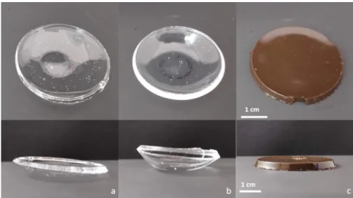
PVA/CNC/NP/CA.SAME SCALE APPLIES TO ALL OF THE IMAGES IN THE COMPOSITION. ... 22 FIGURE 4.15–PHOTOGRAPHS OF THE TOP VIEW OF THE OPTIMIZED CA-CROSSLINKED FILMS AT 130 ºC
DURING 5 H IN THE ABSENCE OF CATALYST (AFTER WASHING AND DRYING): A)PVA, B)PVA/CNC AND
PVA/CNC/NP.SAME SCALE APPLIES TO ALL OF THE IMAGES IN THE COMPOSITION... 22
FIGURE 4.16–PHOTOGRAPHS OF TOP AND SIDE VIEW OF THE OPTIMIZED GA-CROSSLINKED HYDROGELS AT ROOM TEMPERATURE: A)PVA B)PVA/CNC C)PVA/CNC/NP. ... 23 FIGURE 4.17-XRD DIFFRACTOGRAMS FOR PVA, A)CA-CROSSLINKED SAMPLES:PVA,PVA/CNC AND
PVA/CNC/NP, AND B)GA-CROSSLINKED SAMPLES:PVA,PVA/CNC AND PVA/CNC/NP. ... 24 FIGURE 4.18-FTIR SPECTRA OF A) CITRIC ACID-CROSSLINKED PVA FILMS AND B)GA-CROSSLINKED FILMS:
PRISTINE PVA, CROSSLINKED PVA FILM,PVA/CNC CROSSLINKED FILMS, AND PVA/CNC/NP
CROSSLINKED FILMS. ... 25 FIGURE 4.19–THERMOGRAVIMETRIC ANALYSIS OF NEAT PVA, A)CA-CROSSLINKED AND B)GA-CROSSLINKED
SAMPLES:PVA,PVA/CNC,PVA/CNC/NP. ... 26 FIGURE 4.20-WATER UPTAKE VALUES FOR A)CA-CROSSLINKED AND B)GA-CROSSLINKED PVA HYDROGELS:
SIMPLE PVA/CA FILMS, WITH CNC, AND BOTH CNC AND SPIONS ... 27 FIGURE 4.21–ON THE LEFT, TENSILE CURVES FROM NEAT PVA AND CA-CROSSLINKED FILMS.PVA,
PVA/CNC AND PVA/CNC/NP ALL DISPLAY TYPICAL BEHAVIORS.ON THE RIGHT, AN EXAMPLE OF THE TENSILE TEST ON THE FILMS. ... 28
xvii FIGURE 4.22–EXAMPLE OF COMPRESSION CURVE.IN THIS GRAPH OF A GA-CROSSLINKED PVA HYDROGEL, A
STEEP EXPONENTIAL CURVE IS OBSERVED UNTIL FRACTURE.A CLOSE-UP OF THE CURVE BETWEEN 10
AND 20 % DEFORMATION IS PICTURED WITH ITS LINEAR FIT. ... 29
FIGURE 4.23-CYTOTOXICITY OF CA CROSSLINKED FILMS:PVA,PVA/CNC AND PVA/CNC/NP WERE ALL TESTED FOR DIFFERENT CONCENTRATIONS (50,25,12.5 AND 6.25 MG/ML).THE LAST COLUMN,C-,
REFERS TO THE VALUES OF THE NEGATIVE CONTROL... 30 FIGURE 4.24-CYTOTOXICITY OF GA CROSSLINKED GELS:PVA,PVA/CNC AND PVA/CNC/NP WERE ALL
TESTED FOR DIFFERENT CONCENTRATIONS (50,25,12.5 AND 6.25 MG/ML).THE LAST COLUMN,C-,
REFERS TO THE VALUES OF THE NEGATIVE CONTROL... 31 FIGURE 4.25-TEMPERATURE VARIATION DEPENDING ON THE TYPE OF SAMPLE:CA-CROSSLINKED FILM OR
GA-CROSSLINKED HYDROGEL.THE SAME AMOUNT OF SPIONS INCLUDED IN THE SAMPLES WAS TESTED IN SOLUTION FOR COMPARISON SAKE. ... 31
FIGURE 7.1-EXAMPLE OF COMPRESSION CURVE.IN THIS GRAPH OF A GA-CROSSLINKED PVA/CNC
HYDROGEL, A STEEP EXPONENTIAL CURVE IS OBSERVED UNTIL FRACTURE. ... 44 FIGURE 7.2-EXAMPLE OF COMPRESSION CURVE.IN THIS GRAPH OF A GA-CROSSLINKED PVA/CNC/NP
xix
List of tables
TABLE 4.1–VALUES OF THE ELEMENTS C,N,H AND S DETECTED BY ELEMENTAL ANALYSIS ON THE PRECURSOR, FINAL CNC MATERIAL AND VALUES PREDICTED FOR PURE CELLULOSE.THE VALUES ARE PRESENTED IN PERCENTAGE; THE AMOUNT OF OXYGEN (*) WAS CALCULATED BY DIFFERENCE. ... 12 TABLE 4.2–TESTED CONDITIONS TO CROSSLINK THE HYBRID COMPOSITE SYSTEM DEPENDING ON THE
CROSSLINKING AGENT, PRESENCE OF CATALYST, REACTION TEMPERATURE, AND THE RESPECTIVE RESULTS... 21 TABLE 7.1-DATA OBTAINED FROM THE GA CROSSLINKED PVA HYDROGEL FOR THE COMPRESSIVE TEST RUNS
... 44 TABLE 7.2-DATA OBTAINED FROM THE GA CROSSLINKED PVA/CNC HYDROGEL FOR THE COMPRESSIVE TEST RUNS ... 45
TABLE 7.3-DATA OBTAINED FROM THE GA CROSSLINKED PVA/CNC/NP HYDROGEL FOR THE COMPRESSIVE TEST RUNS ... 45
xxi
Acronyms
AFM Atomic Force Microscopy
AMF Alternating Magnetic Field ATR Attenuated Total Reflectance
CA Citric Acid
CNC Cellulose Nanocrystal
DMSA Dimercaptosuccinic Acid
DSC Differential Scanning Calorimetry
EA Elemental Analysis
FTIR Fourier-Transform Infrared
GA Glutaraldehyde
MCC Microcrystalline Cellulose
NP Nanoparticle
PVA Polyvinyl Alcohol
SPION Superparamagnetic Iron Oxide Nanoparticle
TGA Thermogravimetric Analysis
UV Ultraviolet
1
1
Context
In Portugal at the beginning of the 1990s, 18155 deaths could be attributed to malignant neoplasms, and in 2017 this number was closer to 30000 deaths. In fact, this rising trend is not unique in Portugal, since almost every country in the European Union has seen an increase in the incidence of malignant tumor and its related deaths [1], [2]. Although more people survive cancer nowadays due to advances in cancer research, a larger number of people will suffer from cancer as well, due to the increasing life expectancy [3]. Current cancer therapies like radiation and chemotherapy cause several negative side effects that significantly affect the patient’s wellbeing, particularly in the lymphatic system, kidneys and heart, among others [4], [5]. Therefore, effective treatment with reduced side effects becomes a priority in cancer research. Hyperthermia is an ancient way of treating superficial tumors by applying heat to the affected region. Fast-forward a couple millennia and a different form of this therapy is being used: magnetic hyperthermia. Gilchrist et al. [6] in 1957 was the first group to use magnetic iron oxide nano-particles for cancer treatments. Magnetic nanonano-particles were injected into lymphatic channels producing heat when exposed to an alternating magnetic field, thus treating metastatic cancer.
Magnetic nanoparticles can be useful for biomedical applications for the already mentioned hyper-thermia applications, but also for controlled drug delivery and imaging when using magnetic reso-nance [7]. Nevertheless, iron oxide nanoparticles face some limitations, with lack of colloidal stability being the main issue. This can be surpassed by coating the nanoparticles with a surfactant like oleic or dimercaptosuccinic acid (DMSA), avoiding their unwanted aggregation [8].
Cellulose nanocrystals (CNC) are also a nanomaterial receiving attention for its excellent properties, especially when it comes to biomedical applications like tissue engineering [9]. This interesting material is extracted from cellulose, the most abundant biopolymer in nature, and results in very attractive prop-erties: biocompatibility, biodegradability and low toxicity. Nanocellulose-based materials can even be used as a stable environment for cellular growth, adhesion and proliferation: some studies have used cellulose nanowhiskers to grow oriented skeletal muscle cells while others used hydrogels to not only grow cells on the surface, but inside the scaffold as well [10]–[12].
Having this in mind, a previous work developed in the Soft and Biofunctional Materials Group (SBMG) combined the best characteristics of both iron oxide nanoparticles and cellulose nanocrystals in a hybrid composite [13]. The material’s performance was tested in both magnetic hyperthermia and cytotoxicity assays. It was found that the nanoparticles granted CNC’s better thermal properties. The films retained some degree of organization even with the presence of iron oxide nanoparticles, displaying iridescence. Although great results were achieved, it was found that the produced material was fragile, brittle and not easily manageable. This is relevant especially when the application is considered: after all, the ma-terial must withstand the physiological environment and minimal handling. Adding a polymer may be a viable path since it can make the composite more pliable.
Polyvinyl alcohol (PVA) is a non-toxic, biodegradable polymer already employed in drug delivery devices, hemodialysis and bio-separation membranes and soft contact lenses, and has already been used alongside cellulose nanocrystals since the latter is an efficient light-weight reinforcement [14]–[16].
2 Therefore, the goal in this work is to produce a ternary composite with PVA, a widely used polymer for medical devices; CNCs, a renewable and effective mechanical reinforcement; SPIONs, capable of generating heat when subjected to AMF. This composite should be biocompatible and able to generate heat, combining all the attractive properties of its constituents in one device for cancer treatment. To achieve this goal, the following objectives were proposed:
• Study the incorporation of the reinforcement material in the polymer matrix; • Evaluate the maximum concentration of nanoparticles in the composite;
• Optimize the composites crosslinking taking into account the desired application; • Assess the biocompatibility of the new materials through cytotoxicity assays; • Evaluate the heating ability of the composites with magnetic hyperthermia tests;
• Characterize the composites with various techniques, including mechanical and water up-take tests to assess if they’re appropriate for the physiological environment.
Four main parts will structure this work: firstly, there is a theoretical introduction to several relevant aspects of the composite and its functions. Secondly the materials and methods section will describe how and in which way the laboratory techniques were executed. After this, all the results will be pre-sented and discussed. Finally, this work is finished with some final considerations, conclusions of the study and future perspectives.
3
2
Introduction
Magnetic Hyperthermia
The etymology of the word hyperthermia comes from the Greek and means “beyond temperature”. It is a millenary therapy used by ancient Greeks, Romans and Egyptians to treat breast masses with the first evidence being a papyrus dated from 3000 BC [17]. When it is intentionally applied to the human body, it causes the rise of temperature above the typical physiological temperature of 37 °C. Due to their abnormally fast growth, cancer cells have a defective vascular supply which makes them more suscep-tible to temperatures above 42 °C [18]. Different amounts of generated heat are required when the application is considered. For cancer cell death to occur, high heat generation is necessary; as opposed to the mild heat generation required for the activation of specific cellular targets, which bypasses the non-wanted side effects on other cellular components [19]. The mechanism by which cellular death happens is based on heat shock proteins, which alter the cellular walls. Hyperthermia also leads to lack of nutrients in the tumor cells, since it increases the oxygen supply, over-acidifying the cells. This in-creased temperature has the ability to damage, kill and make cancer cells more sensitive to subsequent treatments like radiation or other drugs [20], [21].
Hyperthermia ablation can be applied through microwaves, radiofrequency and high-intensity fo-cused ultrasound but they face limitations: these can cause burns and other negative side effects that hinder their full potential [22]. An interesting mode of applying heat is through magnetic hyperthermia, where an alternating magnetic field is used to induce the temperature increase using magnetic field susceptible materials, like iron oxides nanoparticles. This is advantageous since iron oxide nanoparti-cles can be injected or applied on the cancer site, making it a location-specific treatment [23].
Magnetic spin relaxation is responsible for heat generation when it comes to small single-domain magnetic nanoparticles. The magnetization reversal process happens when an energy barrier is sur-passed, and the magnetic nanoparticles are subjected to an alternating magnetic field. A certain amount of energy is required to rotate the magnetic nanoparticles away from their preferred direction. With re-petitive alignments and relaxations of magnetic spins, heat is generated through energy dissipation. Néel relaxation happens with the flipping of the magnetic spins and is especially relevant when the nanoparticles are internalized and “fixed”. However, this is not the only way magnetic nanoparticles generate heat as depicted in Figure 2.1.
F igur e 2. 1 – Representation of Néel and Brown relaxation mechanisms. The brown circles rep-res ent t he s uperparam agnet ic iron ox ide nanopart ic les [ 19].
4 Brownian relaxation generates heat when whole particle relaxation occurs in a liquid by means of rotational friction, and the viscosity of a medium plays a part. In the case of a small particle, the effective magnetic relaxation time is dominated by the Néel relaxation; on the other hand, the bigger the particle, the more Brownian relaxation contributes to the effective magnetic relaxation time [19].
Superparamagnetic Iron Oxide Nanoparticles
The International Organization for Standardization (ISO) defines a nanoparticle as a material with all three dimensions in the nanoscale, this is, between 1 and 100 nanometers. Magnetic nanoparticles are a specific type of nanoparticles able to respond to a magnetic field, usually composed of an iron oxide, iron, cobalt or nickel core. Iron oxide nanoparticles are stable and biocompatible and when superpara-magnetic, are called SPIONs (superparamagnetic iron oxide nanoparticles), being widely used for bio-medical research in recent years. Various techniques can be employed to produce iron oxide nanopar-ticles including coprecipitation, thermal decomposition, sol-gel and hydrothermal reaction. Chemical co-precipitation is a widely used method to produce iron oxide nanoparticles because it is a simple, fast and low-cost technique. By tailoring the reaction parameters, it is possible to achieve a precise control over the nanoparticle size, which is critical for superparamagnetism to occur. This special behavior means that the magnetite nanoparticles display a single magnetic dipole and magnetic momentum, which does not happen to the same material in greater than 30 nm dimensions. In practical terms, small magnetic nanoparticles are only magnetized when a magnetic field is being applied, which is especially useful for drug delivery applications, since nanoparticles can be guided through the body to the tumor site and control the drug delivery [24]. They are also used for other biomedical applications like en-hanced imaging through magnetic resonance, tissue repair, and cell/tissue targeting [25].
It is therefore expected for biosafety to be an important issue when regarding iron oxide nanoparticles for biomedical applications. It is crucial to know what kind of interactions they have with cells, proteins, etc., but also how they affect essential life mechanisms. One other aspect when considering the bi-osafety of SPIONs is how they biodegrade within the body and their possible bioaccumulation. This is an aspect where the surface groups of the SPIONs are important since some studies have stated that bare SPIONs are toxic [26]. Coatings are also useful to prevent aggregation issues and to link bioactive components, like drugs [27]. Besides natural polysaccharide coatings like the usual dextran, synthetic polymer coatings are also an option to improve these magnetic nanoparticles biocompatibility [19]. Meso-2,3-dimercaptosuccinic acid is widely used to coat magnetite nanoparticles. DMSA is widely used to coat SPIONs and produce a water-stable nanoparticle suspension in a wide range of pH values. Not only does it improve their stability but also the biocompatibility and internalization, making the nanopar-ticles less cytotoxic when compared to their non-coated counterparts [28].
Cellulose Nanocrystals
With the recent years’ climate change and depletion of natural materials resources, cellulose emerges as one of the top raw materials, being the most abundant biopolymer, simultaneously renew-able, biocompatible and biodegradable. It can be found in plants’ cell walls, algae and some bacteria,
5 composed by its repetitive unit, cellobiose. This monomer is made up of two units of anhydroglucopyra-nose alternately rotated 180° linked into linear chains by 1,4-β-glycosidic bonds [29].
Cellulose nanocrystals have an anisotropic nanorod appearance and can be obtained through the strong acid hydrolysis of cellulose fibers. The acid cleaves the more susceptible amorphous regions between the crystalline sections, leaving only the CNC nanorods as seen on Figure 2.2 [30]. These can have 50 to 1160 nm in length and range from 3 to 50 nm in diameter, which strongly depends on the hydrolysis conditions and origin of cellulose [31]. CNCs also present high axial stiffness and tensile strength, low density and good thermal stability [32]. When produced with sulfuric acid, the formed sul-fate ester groups result in negative charges on the surface of the CNCs, enhancing their stability in water due to the electrostatic repulsion [31], [33], [34]. If suspended in water at low concentrations, they will self-assemble into a left-hand lyotropic cholesteric phase. Producing a film from this suspension by full solvent evaporation, will result in a photonic material that presents a short pitch (few hundreds of nanometers), and structural coloration, related to the wavelength of light in the visible region of the electromagnetic spectrum. Through this mechanism, photonically attractive structures can be produced, displaying iridescence and selective circularly polarized light reflection, being even used in security la-beling, cosmetics and coatings [33], [35]. CNCs have received growing attention in recent years, espe-cially when it comes to their biomedical applications. Some interesting works also showed that organized structures like liquid crystalline phases or even spin-coated membranes of cellulose nanowhiskers ex-tracted from tunicates show the ability to grow oriented muscle cells [10], [36].
F igure 2. 2 - Repres entation of a) cellulos e f ibers, w here t he c ryst alline and am orphous regions are depict ed, b) c ellobios e, t he repet it iv e dim er of t he polym er c ellulose and the ef fect of s
ulfu-ric ac id hy drolys is in the polym er adapt ed from [ 37].
Polyvinyl Alcohol
Applications of polyvinyl alcohol can be found in almost every industry sector. Considering its bio-medical applications, it has been made into contact lenses, artificial organs, sensors, membranes, arti-ficial cartilage, wound healing, tissue engineering scaffolds and drug delivery systems [38]. One inter-esting aspect of this material is the possibility to make stimuli-responsive hydrogels in which the water uptake depends on environmental conditions like pH, temperature, magnetic fields, and so on. This trend is justified by the comparable properties of hydrogels and human tissues regarding the retained amount of water and low interfacial tension with fluids [39]. PVA has good thermal and pH stability, it is widely available, inexpensive, nontoxic, biocompatible, easily-processed, and hydrophilic [40]. Because
6 of its hydrophilic nature, it must be modified to minimize swelling in water [41]. This modification to improve mechanical integrity can include freezing, heat treatment, irradiation and chemical crosslinking. Regarding chemical crosslinking, there are numerous crosslinking agents that enable the reaction with glutaraldehyde and citric acid being the most common (Figure 2.3) [42]. Citric acid is an advantageous crosslinker as it is already used as a low-cost food additive (E330) and crosslinking agent for compounds with hydroxyl groups (see Figure 2.3) [14], [43]. Being a metabolic product of the body, it is expected to be biocompatible [15]. The crosslinking with PVA happens through an esterification reaction between the carboxyl groups in citric acid, and the hydroxyl groups in both PVA and CNC, making it a double efficient crosslinker [42], [44]. Wang et al. [45] produced a novel packaging material with PVA and xylan, a polysaccharide and type of hemicellulose. This group found that the interactions between both poly-mers were enhanced by the presence of citric acid, with hydrogen and ester bonds forming between the components.
F igure 2. 3 -R eact ion sc hem e show ing both int ermolec ular and intram olec ular cros slinking of PVA using c it ric ac id [ 46].
Glutaraldehyde reacts with PVA’s -OH functional groups, creating acetal bridges and resulting in a sort of net that is characteristic of crosslinked polymers [39]. Figure 2.4 represents this reaction. Since the hydrophilic -OH groups are consumed in the crosslinking process, an optimum degree of crosslink-ing is favorable [41]. When uscrosslink-ing GA, acidic conditions catalyze the acetalization of hydroxyl and alde-hydic groups. Hydrochloric acid may be a good candidate since it is volatile and will not leave residues that can tamper with the other reagents. Although glutaraldehyde is accessible, economical and effec-tive, it is toxic and must be neutralized [47].
F igure 2. 4 - Reac tion betw een PVA (1) and GA (2) in ac id c onditions prov ided by hydroc hloric ac id, res ulting in c ross linked PVA by ac et al bridges (3) w it h t he r eleas e of w at er [ 48].
7
Composite’s state of the art
In 2004 Lao et al. [49] dispersed magnetic microparticles (Fe3O4) in a polyvinyl alcohol hydrogel and
studied the hybrid material’s potential for hyperthermia, having achieved a stable maximum temperature between 43 and 47 °C within 5 to 6 min. The research group crosslinked this hydrogel via freeze and thaw method. Being a physical crosslinking method, it excuses the usage of chemical crosslinkers; however, it is very time-consuming since it can take at least 3 days. They found that the maximum temperature was a function of the Fe3O4 concentration. Other processes can be less time consuming
regarding the crosslinking of a hydrogel. One can choose a chemical crosslinker, like glutaraldehyde. Twelve years later, Tanpichai et al. reinforced glutaraldehyde crosslinked PVA hydrogels with 1 wt.% CNC and found that the compressive strength was greatly increased. In fact, CNC makes for a great reinforcement due to their high strength and elasticity modulus. A better strain recovery was found as well in the reinforced samples, and it was found that this addition did not affect the swelling ratio or thermal stability of the hydrogels [50], [51].
In their 2013 work, N. Rescignano et al. [52] proposed a ternary bio-nanocomposite using PVA, cellulose nanocrystals and PLGA nanoparticles, with the goal of drug-delivery. The incorporation of 0.5 % of CNC increased the elongation properties without jeopardizing the other mechanical properties. Furthermore, CNCs can also be used to orient particles in a hydrogel: just 3 years later Yoshitake et al. [53] developed a gelatinous-composite from iron oxide nanoparticles and t-carrageenan while using CNC to orient the magnetic nanoparticles in the film. The group observed a specific response in drawn/pulled films to an external magnetic stimulus, attributed to the magneto-anisotropy of the pre-ferred orientation of the iron oxide particle aggregates. Nonetheless, Shen et al. [54] were the only research group to have joined magnetite nanoparticles, cellulose (microcrystalline) and polyvinyl alcohol in a composite. This work was published in 2018 and resulted in an aerogel for dye removal produced in an eco-friendly manner. The article pertinently remarks that there are relatively few studies devoted to developing cellulose and PVA composites as matrix for supporting the NPs. Some groups have com-bined nanocellulose and non-iron-oxide-metallic nanoparticles in polyvinyl alcohol for hyperthermia [55], but also for other purposes, like antimicrobial applications. Zhong et al. developed antimicrobial polyvinyl alcohol films reinforced with hybrid cellulose-copper nanoparticles, while Xu et al. created CNC/silver-NPs functional fillers and produced a PVA blend membrane effective against Escherichia coli and Staph-ylococcus aureus [56], [57]. To date, the combination of CNCs, SPIONs and PVA has not been reported in literature. In this work, this novel composite is extensively studied and characterized for biomedical applications.
9
3
Materials and Methods
Synthesis of cellulose nanocrystals
The reagents utilized in the synthesis of cellulose nanocrystals and its procedure, are described elsewhere [58]. To increase the concentration of CNC in the final suspension, a 15 % PEG aqueous solution was prepared, and the CNC suspension was introduced into a dialysis tubing (SnakeSkin™ with 3.5 K of molecular weight cut-off and 35 mm dry internal diameter membrane) and left in dialysis overnight against the PEG solution, reaching a concentration of 6.64 ± 0.55 wt.% determined by gravi-metric method with 7 replicas. The suspension was stored at 4 °C to avoid degradation and bacterial or fungal growth [29].
To improve the stability of CNCs and increase its initial pH (initial value of 1.59), the proton on the ester sulfate group that are present on the surface of CNC’s was exchanged to Na+ with an ion-exchange
resin. The resin Amberlite® IR120 in the hydrogen form (by Sigma Aldrich), was washed in ultrapure water three times. Afterwards a 10 wt.% NaCl aqueous solution was put in contact with the resin, so the exchange of the ion H+ to Na+ would occur. Since HCl is formed, the pH was measured and monitored
until it was no longer acidic, which indicated the complete exchange. The resin was placed in the CNCs suspension for a week, and a final pH value of 4.6 was achieved. The presence of Na+ was determined
by Inductivity Coupled Plasma Atomic Emission Spectroscopy (ICP-AES) as described in Supporting Information.
Iron Oxide Nanoparticles Synthesis
Iron oxide nanoparticles were synthesized by chemical co-precipitation method as described else-where [59]. The magnetic nanoparticles were stabilized using dimercaptosuccinic acid.
Ternary Composite
A 10 wt.% solution of PVA (95 % hydrolyzed, average M.W. 95,000, Acros Organics) was prepared by dissolving the PVA in ultrapure water with magnetic agitation (500 rpm) in an oil bath at 90 °C, during 1 h until the polymer was visibly dissolved and left to cool. The solution was stored in the fridge for the above-mentioned reasons.
Three types of mixtures were made for each of the crosslinking agents. PVA, PVA with CNCs (PVA/CNC) and PVA with both CNCs and Fe3O4 nanoparticles (PVA/CNC/NP). Glutaraldehyde and
citric acid were used as crosslinkers (5 % m/m in respect to PVA) and hydrochloric acid as a catalyst (2 % wt.) in the glutaraldehyde samples. Hydrochloric acid/glutaraldehyde mixtures were prepared in advance and stored in the fridge. For simple PVA samples, PVA was submitted to magnetic stirring and the crosslinker/catalyst solution was added. The mixture was poured into polystyrene Petri dishes (55 mm of diameter). For the citric acid crosslinked solutions, they were put overnight in an oven at 60 °C, to evaporate water and produce manageable films. In the case of glutaraldehyde crosslinked samples, they were left to dry uncovered at room temperature. In PVA/CNC samples, a 0.5 wt.% amount of CNCs were added with respect to the PVA content. Membranes were obtained as described above
10 for the pristine PVA samples. In PVA/CNC/NP samples, the amount of nanoparticle suspension was added after the CNCs, in order to make 10 wt.% of the PVA content. The citric acid containing samples were submitted to a thermal treatment at 130 ºC for 5 h for best crosslinking results. All samples were washed with PBS and ultrapure water in order to wash out any acidic (CA or HCl) residues.
Water Uptake tests
To evaluate the water uptake capability of the hydrogels, the weight swelling ratio of the samples was analyzed. 10x10 mm squares were cut from the samples PVA, PVA/CNC and PVA/CNC/NP with 3 replicas each. These were put in the oven until dry, reaching the final dimensions of 5x5 mm. For the samples where citric acid was used as a crosslinking agent, 5x5 mm squares were cut.
In vitro tests
Being the goal of this composite its biomedical applications, it is essential to assess its toxicity to cells. Cytotoxicity assays were employed following the extract method and the norm ISO 10993 – Bio-logical evaluation of medical devices, Part 5: Tests for in vitro cytotoxicity and Part 12: Sample prepa-ration and reference materials. Cell adhesion and prolifeprepa-ration assays were done as well. These proce-dures are described in more detail in section 7.3.1 of Supporting Information.
Magnetic Hyperthermia
The thermal behavior of the nanoparticle-containing samples was analyzed with Nb Nanoscale Bio-magnetics’ DM100 Series equipment. The used parameters were 10 minutes, 418.5 kHz of frequency and 24 kA.m-1 of magnetic field strength.
Mechanical Tests
A Hegewald & Peschke inspect micro LC100N equipment was used, with a 100 N load cell. The tests were run with a speed of 2 mm/min and until break or reaching 95 N of load. For tensile tests, 3 replicas were used, and the CA-crosslinked samples were cut in 5x20 mm rectangles (gauge length of 10 mm) with average thickness of 428, 592.5 and 732.5 μm for the PVA, PVA/CNC and PVA/CNC/NP dry samples, respectively. The hydrogel samples were cut in 5 mm diameter circles with half a millimeter of thickness in the case of PVA/GA and PVA/CNC/GA and 1.5 mm for PVA/CNC/NP/GA, tested after extraction from water and patted dry. In the case of compression, 10 replicas were used, and the sample was set on the lower plate and elevated until in contact with the top plate and a preload force of 0.1 N was set to guarantee full contact between the plates and the sample. The compressive modulus was calculated between 10 and 20 % strain and assumed as the compressive modulus for 15% of strain.
Characterization
All the specifications and parameters used in the various characterization techniques employed in the thesis are described in section 7.4 of Supporting Information.
11
4
Results and discussion
This work is focused on the optimization and characterization of a novel ternary composite for mag-netic hyperthermia applications, incorporating three promising materials: cellulose nanocrystals, Fe3O4
superparamagnetic nanoparticles and polyvinyl alcohol.
The first part of the work delves into the synthesis of cellulose nanocrystals from microcrystalline cellulose and its sodium treatment, being subsequently used in the production of both iridescent films and ternary composites. These underwent morphological, structural and chemical characterization. Ad-hesion and proliferation tests were completed with the composite material previously developed by the SBMG, a hybrid composite of CNCs and SPIONs. After this, a new formulation for this material was proposed. The well-studied magnetic nanoparticles were produced via chemical co-precipitation method and incorporated in a PVA matrix. This incorporation was also tested for the cellulose nanocrystals. After this, a study of the crosslinking of the new ternary composite was performed.
Besides structural and chemical characterization of the composites, water uptake tests were per-formed to understand the materials’ behavior in aqueous environments, magnetic hyperthermia tests were carried out to measure the material’s heating ability, and cytotoxicity assays were executed to assess the biocompatibility of the novel composites.
Cellulose nanocrystals’ production
The first part of this thesis focused on the production of cellulose nanocrystals as described else-where [58]. One of the detected problems with the CNCs produced by this methodology was their low pH after synthesis, around 1.59. To make them more suitable for biomedical applications the pH should be around 7, however previous studies have showed that this value did not allow to retain the liquid crystalline organization of the CNCs in suspension [13]. So, CNCs were treated with an ion exchange resin containing sodium (Na+) ions for a week (adapted from [60]). pH values were continuously
meas-ured, and resin was progressively added as needed. The effectiveness of the ion exchange was verified by the obvious increase in pH but also by Inductively Coupled Plasma – Atomic Emission Spectroscopy (ICP-AES). As a result, CNCs suspension increased the pH up to 4.6. This step is important since the lowering of the acidity can avoid degradation and guarantee better biocompatibility results.
The amount of carbon, hydrogen, nitrogen and sulfur was determined through elemental analysis (EA), with 2 replicas for each sample. The results are summarized in Table 4.1, with oxygen values being calculated by difference. The presence of sulfur in the CNC sample is expected and is attributed to the addition of sulfate half-ester groups during the sulfuric acid hydrolysis of microcrystalline cellulose, indicating an effective acid hydrolysis [61].
The obtained numbers can be very useful to calculate the incorporation of sulfate half esters into the surface of the CNCs. The method to do so has been previously reported by Hamad et al. [61], and it allows the determination of the degree of substitution (n) of hydroxyl groups by-OSO3H groups per
12
S (%)=100n× S
6C+10H+(5+3n)O+nS Equat ion 4. 1
The amount of sulfur and calculated n value was 5.35 -OSO3H per 100 AGU units for the
experi-mental acid hydrolysis conditions used. For the same experiexperi-mental conditions Fernandes et al. stated a 0.85 wt. % of sulfur content and subsequently a n value of 4.39. The observed difference is within the elemental analysis’ error, and it is therefore in accordance with the literature [58].
ICP-AES analysis determined a sodium concentration of 63.1 mg/L, confirming the efficient sodium substitution of H+ (nitric acid showed a value of 3.86 mg/L, for reference). The substitution mechanism
is pictured in Figure 4.1. Relative percentages of sulfur and sodium in the sample could be calculated, although the resolution’s limit of EA and ICP-AES are different and could lead to inconsistencies. Thus, both elements should be analyzed via ICP-AES though it should be noted that the latter is a much more expensive technique.
T able 4.1 – Values of the elements C, N, H and S dete cted by elemental analysis on the precur-s or, final CN C m at erial and v alueprecur-s predict ed f or pure celluloprecur-s e. T he v alueprecur-s are preprecur-sent ed in
percent age; t he am ount of ox ygen (* ) w as c alc ulated by dif ferenc e.
C N H S O*
Pure celulose [62] (predicted values) 44.44 0 6.18 0 49.38
Microcrystalline cellulose 43.51 0 6.42 0 50.07
Cellulose nanocrystals 41.14 0 6.05 1.03 51.78
F igure 4. 1 - Diagram of H + t o N a+ prot on subst it ut ion in CNCs aft er c ont act in N a protonated ion-exc hange res in
ATR-FTIR enabled the chemical analysis of the cellulosic samples, and the obtained spectra of the precursor material (microcrystalline cellulose), CNCs and sodium CNCs are presented in Figure 4.2. There are four characteristic IR absorption frequencies attributed to cellulose’s functional groups: the stretching vibration of the O-H bond at 3339 cm-1; the stretching vibration of C-H at 2900 cm- 1 bond and
C-O at 1054 cm-1; and bending of O-H bond at 1650 cm-1. After acid hydrolysis, -OSO
3H groups
substi-tute some of the -OH groups, resulting in a band at 780-860 cm-1 range due to the S-O stretching. No
differences are seen when comparing CNC with the proton ion and sodium protonated CNC. This anal-ysis indicates that CNC Na presents the same chemical linkages as cellulose. All the identifications are in accordance with the values found in literature [63].
13 One expects that the acid hydrolysis process will not influence the structural organization of the cellulose, however it should induce an increase in crystallinity of the sample since it will remove prefer-entially the amorphous regions of the cellulosic fibers. CNCs crystalline structure was analyzed by XRD by comparing the results from the precursor material, CNCs and CNCs treated with sodium (Figure 4.3).
F igure 4. 2 – ATR-FTIR spectrograms of the precursor material - MCC, CNC and sodium CNCs (CNC N a).
F igure 4. 3 - XR D diff ract ograms of t he precurs or m at erial - MC C, C NC and s odium treat ed CN Cs (CN C N a).
XRD diffractograms present the peaks of the type I allomorph cellulose confirming that the hydrolysis process does not affect the structure of cellulose. In order to infer if the reaction changes the crystallinity of the sample, crystallinity “indexes” were calculated with a method presented by Segal et al. [64] , in which one divides the difference between (002) peak’s height and the minimum value between 18 ° and 20 ° by the (002) peak’s height:
14 Ic= (
I(002)-Iam
I(002) ) ×100
Equat ion 4. 2
When analyzing the diffractograms, the most obvious peak is (002) at 22.6 °, surrounded by other smaller and subtle peaks at (101), (101̅) and (004), at 15°, 16.8° and 34.9°, respectively. These results confirm that the hydrolytic process does not alter the cellulose nanocrystals’ structural organization, since they are characteristic of native cellulose, and present in all three samples. The results are within expectations and relevant literature [63].
CNCs presented a crystallinity “index” of 92.1 % while the precursor material, microcrystalline cellu-lose, showed an 88.24 % crystallinity “index”, confirming the removal of amorphous regions. CNC Na exhibited a 92.5% value in respect of “index” of crystallinity. Although this value is higher than the one obtained for CNC, it should be noted that this shift is probably related to the method to determine the index, and not the material itself, since it has not suffered any process that would affect its crystallinity. The morphology of CNCs was visualized by AFM images, as can be seen by the topographic scan (Figure 4.4 a), and the dimensions of 200 nanoparticles were measured from the images. From this it was possible to obtain the length and width distribution curves, Fig. 4.3 b and c, respectively. From this analysis, the length and width of the nanoparticles were determined as 138 ± 49 nm and 5 ± 1 nm respectively leading to an aspect ratio of 27. These results indicate a successful hydrolysis process and are in agreement with the literature [65].
F igure 4. 4 – AFM-acquired topographic image, in amplitude mode, showing individualized CNC, and t he res pect iv e dis tribut ion of m easurem ents regarding lengt h and w idth of t he nanoc ryst als. Differential thermal analysis was performed on microcrystalline cellulose, CNCs and sodium proto-nated CNCs, through both thermogravimetric (TGA) and simultaneously differential scanning calorime-try measurements. Both TGA and DSC curves for the three cellulosic samples are presented in Figure 4.5, in panels a) and b), respectively. When a material is subjected to thermogravimetric analysis, it is
15 decomposed by heat, and the bonds within molecules are broken. This technique can be useful when assessing the thermal stability of materials. The first decrease in mass, observed for all samples until 100-130 °C is usually attributed to evaporation of water and residual moisture in the samples. Several degradation mechanisms like decomposition, dehydration and depolymerization of glycoside units are triggered in the 150-350 °C range. For microcrystalline cellulose, an abrupt loss of mass is noticeable between 285 °C and 345 °C, followed by a much slower and gradual decrease in mass until the end of the experiment, concordant with the trend usually seen in pure cellulose. This follows a first order deg-radation trend, typical of cellulose pyrolysis [66].
Focusing on CNC and CNC Na curves, it can be seen that both samples experience an accentuated loss of mass at 145 °C and 205 °C, respectively. When comparing CNC Na with its counterpart, a 50 °C improvement in thermal endurance is noticeable. This is attributed to the already reported improved thermal stability seen in neutralized and basic pH CNCs These two curves show another fall, although smaller, around 275 °C, until the end of the test, where about 30 % of the initial mass is left. The steadier feature of these curves when compared to the sudden drop of MCC can be attributed to the presence of sulfate groups, preventing the would-be two-step reaction. This presence also explains the higher amount of residue at the end of test: sulfate groups facilitate the extraction of water from the structure, dehydrating it and leading to a subsequent loss of oxygen. With no oxygen, the main mechanism of mass loss during pyrolysis is avoided, with no emission of CO and CO2, preventing mass loss [66].
F igure 4. 5 – Differential thermal analysis of microcrystalline cellulose, CNC, and sodium protonat ed CN C. a) t he gra ph f or t herm ograv im et ric m eas urement s . b) t he diff erent ial sc anning
c alorim et ry analys is is p res ent ed.
Regarding differential scanning calorimetry, CNC Na presents two smaller endothermic peaks around 230 °C and a single more intense peak at 320 °C for MCC, confirming the significant mass losses already mentioned above. The CNC samples do not present this kind of behavior, indicating a gradual loss of mass, which mirrors the slow and multiple-step degradation of the cellulose chains and sulfate derivatives [62], [66].
16
Hybrid composite system – preliminary work
A previous work developed in the Soft and Biofunctional Materials Group (SBMG) combined both superparamagnetic iron oxide nanoparticles and cellulose nanocrystals to produce a novel hybrid com-posite for biomedical applications [13]. Taking advantage of the self-assembly of CNCs into a liquid crystalline phase, the cholesteric organization can be retained upon solvent evaporation and iridescent films were produced with and without SPIONs. The mixture between the two materials was optimized resulting in a final composition of 4 wt.% CNC suspension, and 3 wt. % of SPIONs (in respect to CNC mass). The films retained the cholesteric organization observed for the suspension even with the pres-ence of SPIONs (seen in Figure 4.6). This new composite material was tested in magnetic hyperthermia application resulting in a temperature increase of about 1.5 °C. Additionally, none of the materials tested positive for toxicity by the extract method [13]. Although the group has shown that the produced mate-rials are non-toxic, no information regarding cell proliferation was done, so these tests were performed in the present thesis. In order to do so, it was necessary to produce new materials. Similar iridescent films (without SPIONs) using the CNC treated with sodium were prepared as described in detail in sec-tion 7.1 of Supporting Informasec-tion, with the final result pictured in Figure 4.7.
F igure 4. 6 - Phot ographs of hy brid CNC and SPI ON f ilms prev iously produc ed by t he SBM G. a) N o polariz er b) lef t c irc ular polariz er c ) right circ ular polariz er . Sam e s cale applies to all of t he
im ages in t he c om position.
These iridescent films with 100 μm of thickness present selective reflection of circularly polarized light. This phenomenon indicates that the cholesteric organization is maintained even after full evapo-ration of the solvent. Since CNCs self-assemble themselves (in water, at low enough concentevapo-rations) into a left-handed chiral nematic phase, only left circularly polarized light is reflected. This is confirmed by the photographs as seen in panels b) and e). In panels c) and f) it’s possible to see the extinction of transmitted right circularly polarized light.
Following, superparamagnetic iron oxide nanoparticles were prepared by chemical co-precipitation technique previously optimized by [59], coated with DMSA due to their low stability in physiological conditions, and extensively characterized in a recent study [8]. Figure 4.8 shows the most representative results of SPIONs characterization. To offer some context, the study shows that the uncoated iron oxide nanoparticles are spherical with an average diameter of 9.4 ± 2 nm (when observed by TEM). DMSA coating does not affect the morphology or average diameter. When it comes to its crystallinity, nanopar-ticles were characterized via XRD, having been possible to observe the characteristic peaks of magnet-ite’s cubic crystalline structure. The surfactant’s presence didn’t affect the crystallinity in any way. The
17 dynamic light scattering measurements, employed to assess their stability, showed that are two groups of nanoparticles, classified by its average diameter: 164 ± 13 nm e 101 ± 9 nm.
F igure 4. 7 - Film m ade from s odium treat ed CNCs by f ull s olvent evaporat ion at 37 °C . Phot o-graphs of s aid film : a) unpolariz ed w hit e light b) left circ ular polariz er c ) right c irc ular polariz er .
d), e) and f) are p olariz ed optical micros copy im ages of the same f ilms.
F igure 4. 8 –SPIONs characterization: A) TEM image of bare Fe3O4 NPs B) T EM im age of DM SA -c oat ed F e3O4 N Ps C ) XR D s pect ra of bot h bare and D MSA -c oat ed F e3O4 N Ps D)
Hy drody namic diam et ers of DM SA -coated F e3O4 N Ps [8].
Using the Na and SPIONs, hybrid films were produced with a final composition of 4 wt.% CNC-Na suspension, and 3 wt. % of SPIONs (in respect to CNC mass). Similarly to what was observed for
18 the CNC films, these retained the helicoidal structural organization derived from the liquid crystalline suspensions of CNCs, which might influence cell growth. To test this hypothesis, adhesion and prolifer-ation in vitro tests were completed to evaluate the performance of the hybrid composite system. Figure 4.9 represents the adhesion and proliferation of Vero cells on CNC and CNC/NP films with cell viability as a function of the number of days. Analyzing the results of relative viability (Figure 4.9) for the CNC films, it is possible to notice the prolific growth from the first to the third day, accompanying the trend of cell control. It is followed by a fall in number of cells on the sixth day and subsequent growth until the last time point. For the material containing SPIONs, throughout six days the growth is in accordance with cell control values, demonstrating a fall on the fourth time point (eight and a half days). On the last day, it is noticeable that the growing process recovered. Regarding the cell control values, they are all as predicted although the last day presents a setback, with a lower count of cells when compared to the previous time point. However, these three punctual falls in cell count do not indicate a lack of cell prolif-eration, but rather human errors, like possible aspiration of cells.
F igure 4. 9 - Adhesion and prolif erat ion of Vero c ells on CNC and CN C/N P f ilms t hroughout 10 days meas ured using t he res az urin method. T he las t c olumn, CC , refers t o t he c ell c ont rol.
Dye staining was employed in Vero cells from the adhesion and proliferation test enabling their ob-servation using Phalloidin (red coloration in the images) stains actin, a constituent of the cytoskeleton of cells; DAPI (4’,6-diamidino-2-phenylindole, dihydrochloride) stains the cell’s nuclei, visible in blue in the pictures in Figure 4.10. When comparing all three images, it is possible to assess that the first is more confluent – this is because it concerns the cells in the control well, which are in greater number. Nonetheless, all images confirm the good health of cells in contact with the tested materials.
The obtained results from adhesion and proliferation tests demonstrated promising results concern-ing cell adhesion in the hybrid material of CNC/SPIONs. Even though cell proliferation was demon-strated in the preliminary tests, no influence of the CNC organization was detected. A lower cell count may be needed in order to infer on the orientation of the cells on top of the films. Additionally, it is important to note that the final hybrid composite system was extremely fragile, brittle and not easily manageable. Since this hybrid composite material was developed for biomedical applications, the final
19 structure must endure the physiological environment and minimal handling and therefore it must be optimized.
F igure 4. 10 – Fluorescence m icroscopy images from Microscope Nikon Eclipse Ti-S of a) con-t rol c ells, b) c ells adhered con-t o CN C f ilms and c ) cells adhered con-t o C NC/ SPI ONs f ilm s. Cell’s
cyto-s kelet on icyto-s v icyto-s ible in red and t he nuc lei in blue .
From these preliminary results, a new structure was designed to incorporate CNCs and SPIONs while improving the flexibility of the composite hybrid system. It is well known from literature that CNCs are used as mechanical reinforcements in several types of composites [51], [67], [68]. Therefore, to obtain a more pliable and manageable material, polyvinyl alcohol was used as a matrix. In this work we attempt to associate and optimize CNCs and SPIONs in PVA and study its mechanical improvements, biocompatibility and magnetic hyperthermia ability.
Hybrid composite system production
To produce the hybrid composite system, three core materials were used: CNCs, SPIONs, and PVA as matrix. 10 wt.% PVA solution was produced, since this is a widely used and convenient concentration to use by the scientific community [52]; a suspension of sodium-treated CNCs and DMSA-coated SPI-ONs were used, the latter chosen based on their already extensively studied stability and heating ca-pacity qualities. Different amounts of CNCs and SPIONs were incorporated in the PVA matrix to assess which were originated in the best results.
At first different amounts of CNC’s (0.5, 1, 2 and 5 % m/m related to PVA mass) were incorporated in PVA 10 wt.% and the mixtures left to dry at room temperature overnight, until constant weight. By macroscopic evaluation (see Figure 4.11) it was possible to see white aggregates in concentrations as low as 1 wt.%, resulting in non-homogeneous samples.
F igure 4. 11 - Photographs of PVA f ilms w ith inc reas ing c onc entration of CNC: a) 0 wt.%, b) 0. 5 w t. %, c ) 1 wt. %, d) 2 w t. % e) 5 wt. %. Sam e sc ale applies to all of t he images in t he
20 CNCs are meant to act as a mechanical reinforcement and Rescignano et al. already have showed that an increase in CNCs amount from 0.5 to 1 wt. % does not improve the mechanical properties of the final product, proving that the smaller amount results in the highest value for Young’s modulus without compromising other mechanical properties [52]. Taking the literature and our own macroscopic obser-vations, an amount of 0.5 % wt. of CNCs was selected.
The next goal was to identify the maximum load of SPIONs that could be incorporated in the composite system. Contents of 0.5 and 5 wt.% of SPIONs (in respect to the PVA content) were used in a mixture of 0.5 wt.% CNC/PVA and the solvent allowed to evaporate. Macroscopically, good incorporation and distribution of the SPIONs was verified along the films, so increasing concentrations of SPIONs were tested: 7.5 wt.% and 10 wt.% (see Figure 4.12 a). An incorporation of 10 wt.% SPIONs resulted in flexible and homogenous film, visible in Figure 4.12 b). Additionaly, the composite films incorporating SPIONs were able to respond to a permanent magnet.
F igure 4. 12 – a) Study of incorporation of SPIONs in PVA films obtained by solvent evaporation. F rom left t o right : 0.5 wt. %, 5 wt. % and 7. 5 wt. % SPI ONs in res pect t o PVA c ont ent . b) C
A-c ross link ed f ilm at 130 °C, w it h 10 wt. % SPI ON s prior t o w as hing .
4.3.1 Hybrid composite system crosslinking
PVA is a very hydrophilic material, readily soluble in water. To make this material suitable for bio-medical applications, i.e., insoluble in water-based solutions, it must be crosslinked. Several crosslinking processes can be used, and within chemical crosslinking various options are described in literature: dialdehydes, dicarboxylic and tricarboxylic acids, diisocyanates and boric acid [69]. On one hand there is citric acid which is a non-toxic compound, while glutaraldehyde is a widely used crosslinker, being two acceptable choices for crosslinking this hybrid composite system. Citric acid crosslinks the polymer via esterification between the carboxyl groups of the carboxylic acid and the hydroxyl groups present in the polymer; whereas glutaraldehyde-crosslinking depends on the acetylation of hydroxyl (polymer) and aldehydic groups [39], [46]. It is well known that acidic environments can be advantageous since they catalyze the crosslinking reaction, so hydrochloric acid was used.
Taking these features into account, glutaraldehyde and citric acid were used to crosslink the devel-oped hybrid composite system. Firstly, two concentrations of GA+HCl were tested, 10+4 % and 1+0.1 % m/m in respect to PVA content. This presented two extreme situations that resulted in the hydrogels presented in Figure 4.13: a) shows the hydrogel with the lowest concentration, no consistency is notice-able and it was not easily managenotice-able; for the highest concentration (panel b), as soon as the catalyst came into contact with PVA the crosslinking reaction took place, entrapping the magnetic stirrer within
21 the structure. An intermediate concentration (5 % wt.) was set and the same amount was chosen for citric acid for practicality reasons. When concerning the catalyst, 2 % of HCl was chosen. When cross-linking with GA in the absence of HCl, films were obtained but these were readily soluble in water.
F igure 4. 13 - Pre-s tudy of t he c onc ent rations of c ross link er t o be us ed. In t he lef t picture a), 1 % of GA+ 0. 4 % of HC l. I n t he right pict ure b), 10 % of GA + 4 % H Cl .
For each crosslinker, the presence of catalyst and temperature were tested. In both cases, hydro-chloric acid was used as catalyst, and its efficacy was tested by comparing the crosslinking efficiency with and without the presence of the catalyst. The several attempts and description of the obtained products are summarized in Table 4.2.
T able 4.2 –Tested conditions to crosslink the hybrid composite system depending on the cross-linking agent , pres enc e of c at alyst, react ion t em perature, and t he res pect i v e result s. Crosslinker Catalyst Crosslinking
Tempera-ture Reaction Result
Citric acid
25-37 °C
Films soluble in water, SPIONs release
HCl Films soluble in water, SPIONs release
90 ºC
Slight yellowing, lower swelling degree films
HCl Some browning, lower swelling degree films
130 °C
Good swelling behavior films, yellowing
HCl Good swelling behavior films but severe darkening
(Figure 4.14)
Glutaraldehyde 25 °C
Soluble in water films
HCl Hydrogel (Figure 4.16)
Citric acid did not effectively crosslink the samples at low temperatures (25 and 37 °C), even when the catalyst was added: the produced films were soluble in water, and the films that contained SPIONs turned the water a brown color, revealing a release of SPIONs from the structure. Some studies indi-cated that carboxylic acid crosslinking could only be achieved with temperatures as low as 90 °C, so that was the next approach[41]. This temperature affected the appearance of the films, presenting slight yellowing, and in the cases where catalyst was present, some browning. Although this temperature did