UNIVERSIDADE DE LISBOA
Faculdade de Farmácia
Effects on migration of breast cancer cells
in 3D models upon MMP inhibition
Susana Duarte Lopes Mascarenhas Proença
Orientadora: Doutora Joana Miranda
Co-orientador: Professor Nuno Oliveira
Dissertação
Mestrado em Ciências Biofarmacêuticas
UNIVERSIDADE DE LISBOA
Faculdade de Farmácia
Effects on migration of breast cancer cells
in 3D models upon MMP inhibition
Susana Duarte Lopes Mascarenhas Proença
Orientadora: Doutora Joana Miranda
Co-orientador: Professor Nuno Oliveira
Dissertação
Mestrado em Ciências Biofarmacêuticas
Agradecimentos
Em primeiro lugar quero agradecer aos meus orientadores, Professora Joana Miranda e Professor Nuno Oliveira, por tão bem me integrarem na equipa. Obrigada por me apoiarem, confiarem no meu trabalho e investirem em mim. Ajudaram-me a crescer profissionalmente e pessoalmente.
Quero agradecer também à Madalena Cipriano pelo contínuo apoio e exemplo, essenciais à minha aprendizagem, na implementação de novas técnicas, e pelas discussões científicas. Estou também grata pela tua companhia durante estes longos meses.
Agradeço à Professora Judite Costa por me disponibilizar os compostos [15]pyN5 [16]pyN5, essenciais para o trabalho aqui desenvolvido. À Prof. Fátima Cabral pela paciência em ensinar-me a química dos compostos macrocíclicos, pela disponibilidade e pelo sorriso sempre pronto. À Prof. Matilde Castro por no meio de tantas responsabilidades me ter indicado a Professora Joana Miranda e o Professor Nuno Oliveira para orientadores, e pelo interesse que teve neste trabalho.
Aos restantes membros do laboratório, em especial ao Sérgio Camões, Patrícia Guerreiro e Ana Flórido, por terem participado na minha aprendizagem ao longo desta tese e tornarem a estadia no laboratório tão agradável.
Às minhas amigas cá em Portugal e no estrangeiro pela compreensão e encorajamento, e por estarem sempre disponíveis para me animar.
À minha família pelo apoio neste longo ano, por fazerem tudo ao seu alcance para que eu pudesse concretizar os meus objetivos. Em especial à minha mãe pela inspiração, por desde cedo me ter encorajado a lutar pelos meus sonhos, a ter sempre um espírito crítico e me estimular a dar o meu melhor.
Ao Tiago, por sempre acreditares em mim, por tudo o que aprendi contigo e pela tua paciência.
Publications
The studies included in this thesis were presented in the following publications Abstracts:
Oral Presentation
Proença S, Cipriano M, Cabral MF, Costa J, Fernandes AS, Castro M, Oliveira NG, Miranda J P, Development of 3D breast cancer models for the evaluation of gelatinase inhibition on cell migration, XLVI “Sociedade Portuguesa de Farmacologia” Meeting, 2016, Porto, Portugal
Poster Presentation
Proença S, Cipriano M, Castro M, Oliveira NG, Miranda JP, Development of 3D breast cancer
models for the evaluation of gelatinase inhibition on cell migration and invasion, 7th Post-Graduate iMed.ULisboa Students Meeting, 2015, Lisboa, Portugal
Table of Contents
Abstract ... i
Resumo ... ii
1.Introduction ... 1
1.1. Breast Cancer ... 1
1.2. Tumor Cells Migration and Invasiveness and the Role of Tumor Microenvironment 1 1.3. MMPs ... 3
Gelatinases (MMP-2 and 9) ... 5
1.4. Exogenous MMP Inhibitors (MMPi) ... 7
Macrocycles as MMP inhibitors ... 10
1.5. Three dimensional models in tumor migration/ invasion assays ... 10
2. Aims and experimental models ... 13
3. Methods ... 14
3.1. Chemicals ... 14
3.2. Maintenance of MDA-MB-231 cells in monolayer cultures ... 14
3.3. Multicellular Spheroids Formation and Maintenance ... 14
3.4. Histology ... 15
3.4.1. H&E ... 15
3.4.2. Ki67 ... 15
3.5. MMPi Viability Assays ... 15
3.6. Media Conditioning ... 16
3.7. Modified zymography assay ... 16
3.8. Migration Assays ... 17
3.9. Statistical analysis ... 17
4. Results and Discussion ... 18
4.1 Development of 3D Models of MDA-MB-231 ... 18
4.2. Cell Viability Assays ... 22
4.3.1. Optimization of Zymography technique and Media conditioning ... 23
4.3.2. Modified Zymography Assays for Gelatinolytic activity assessment ... 25
4.4. Migration Assays ... 28
5. Conclusion ... 32
List of Figures
Figure 1. MMPs regulation of Tumor Microenvironment. 3
Figure 2. MMP catalytic center with a modeled Pro-Leu-Gly-Leu-Ala-Gly-amide hexapeptide substrate.
4
Figure 3. Gelatinases basic domain structure. 6
Figure 4. Chemical structure of some hydroxamic acid MMP inhibitors (Marimastat, Prinomastat and ARP-100) and of the macrocyclic compounds containing pyridine ([15]pyN5 and [16]pyN5).
8
Figure 5. Schematic representation of spheroids culture procedure. 14 Figure 6. Optimization of MDA-MB-231 spheroids development conditions. 19 Figure 7. Characterization of MDA-MB-231 spheroids (inoculum 250,000 cell/mL in 10% FBS and 2% MatrigelTM supplemented media) throughout a 4-day culture.
20
Figure 8. Effect of A) [15]pyN5, B) [16]pyN5 and C) ARP-100 on MDA-MB-231 cell viability on 2D and 3D models, evaluated through the MTS assay.
23
Figure 9. Gelatin Zymography Optimization. 25
Figure 10. Schematic representation of the modified zymography technique adopted.
26
Figure 11. Effect of [16]pyN5, [15]pyN5 and ARP-100 on MMP2 and MMP-9 gelatinolytic activity.
27
Figure 12. Values of pZn2+(-log[Zn2+]), calculated using [1], for an aqueous solution of pH 7.4 containing Zn2+(5 μM) and [16]pyN
5 and [15]pyN5 (0.1-40 µM).
28
Figure 13. Scratch Assay with [15]pyN5, [16]pyN5 and ARP-100. 29 Figure 14. Radial migration assays with [15]pyN5, [16]pyN5 and ARP-100. 31
List of Abbreviations
3D- three-dimensional
[15]pyN5- 3,6,9,12,18-pentaazabicyclo[12.3.1]octadeca-1(18),14,16-triene
[16]pyN5- 3,6,10,13,19-pentaazabicyclo[13.3.1]nonadeca-1(19),15,17-triene
AKT- protein kinase B
CM2D- Conditioned medium from 2D models CM3D- Conditioned medium from 3D models CMT- chemically modified tetracycline (s)
DMEM- Dulbecco modified eagle modified medium DMSO- dimethyl sulfoxide
EDTA- ethylenediaminetetraacetic acid ECM- extracellular matrix
EMT- epithelial to mesenchymal transition FBS- fetal bovine serum
FGF- fibroblast growth factor LOX- lysyl oxidase
LTBP1- latent TGF-β-binding protein 1 MMPi- MMP inhibitor(s)
MMPs- matrix metalloproteinases MSS- musculoskeletal syndrome
MTS- tetrazolium salt
3-(4,5-dimethylthiazol-2-yl)-5-(3-carboxymethoxyphenyl)-2-(4-sulfophenyl)-2H-tetrazolium
PAGE- polyacrylamide gel electrophoresis PI3K- phosphoinositide 3-kinase
Py-macrocyles - pyridine containing macrocycles RT- room temperature
SDS- sodium dodecyl sulfate
TIMPs- tissue inhibitors of matrix metalloproteinase TGF-β- transforming growth factor-β
i
Abstract
Breast Cancer lethality is mostly due to metastization, an event that involves cells migration and invasion, with the latter being dependent of extracellular matrix degradation. Due to metalloproteinases (MMPs) role in migration and invasion and correlation of up-regulation of MMP-2 and 9 (gelatinases) with tumor aggressiveness, these enzymes are considered important druggable targets. 3D models, namely spheroids, have been described as more physiological relevant models. Therefore this work aimed to i) implement breast cancer spheroids model for migration studies; and ii) to evaluate the potential of two pyridine-containing macrocyclic compounds [15]pyN5 and [16]pyN5 as MMP inhibitors. The MMP-2 inhibitor ARP-100 was used as control. Spheroids of MDA-MB-231 cells were developed using MatrigelTM in non-adherent culture plates, being monolayer cultures used as controls. During the whole culture time, compact spheroids with diameters of ~200 μm were obtained, containing evenly distributed cells, with different gelatinase secretion when compared to monolayer cultures. The cytotoxicity of the compounds (1-100 µM) was evaluated, through a 24 h tetrazolium-based assay. Cell viability was > 70% for concentrations up to 50 µM of all compounds, in 2D and 3D cultures. The effect of these compounds (1-40 µM) on gelatinases activity was evaluated by a modified zymography technique. For 3D conditioned media both [15]pyN5 and [16]pyN5 at 7.5 µM revealed a 100% inhibition of MMP-2/9, while ARP inhibited 52% and 84% of MMP-9 and MMP-2 activity, respectively. Cell migration was analyzed through radial migration and scratch assay for 3D and 2D cultures, respectively. Migration in 2D models was affected (p<0.05) by both pyridine-containing macrocycles (5 μM), but not significantly by ARP-100 (up to 40 μM). Radial Migration was less extensively affected, having only significant inhibition for [16]pyN5 (20 μM) and ARP-100 (40 μM). In the future, invasion assays will be performed to further explore the therapeutic potential of these promising pyridine-containing macrocycles.
ii
Resumo
O cancro de mama foi, em 2012, a 5ª causa de morte por cancro no mundo [1]. A maioria destas mortes deve-se à metastização, facto que se reflete na taxa de sobrevida a 5 anos que em cancros localizados é de 98,6% enquanto em cancro disseminado de mama é de 25,9% [2]. A metastização envolve migração e invasão celular, capacidades que as células tumorais de mama adquirem através da transição epitelial-mesenquimal. Esta transição consiste numa mudança de célula epitelial para uma célula do tipo mesenquimatosa e é caracterizada pela perda dos marcadores epiteliais, nomeadamente proteínas de interação célula-célula, célula-matriz extracelular (ECM), proteínas do citoesqueleto e perda de polarização. Simultaneamente, as células ganham a plasticidade necessária para a migração/invasão [3]. Este processo depende de proteases, entre as quais as metaloproteases de matriz (MMPs), grupo de enzimas dependentes de zinco que clivam elementos da matriz extracelular, entre outras funções [4]. As MMPs, especialmente o grupo das gelatinases (MMP-2 e 9), têm sido bastante associadas a tumores metastáticos e estão correlacionadas com a agressividade de alguns tumores [5]. Dado que as MMPs foram identificadas como possíveis alvos terapêuticos, foram desenvolvidos inibidores destas enzimas. Estes inibidores demonstraram resultados pré-clínicos promissores, mas já em fase clínica mostraram falta de eficácia e efeitos secundários de síndrome musculosquelético. Uma das hipóteses sugerida para este insucesso responsabilizava o grupo quelante de zinco comum a muitos destes inibidores, o ácido hidroxâmico [6]. No nosso laboratório foram sintetizados dois macrociclos do tipo pentaaza contendo um grupo piridina no anel, o [15]pyN5 e o [16]pyN5. Estes compostos têm a particularidade de apresentarem constantes de estabilidade elevadas para o catião Zn(II), tendo sido avaliado o seu potencial como inibidores de MMPs e consequentemente da migração celular.
Por outro lado a elevada taxa de insucesso no desenvolvimento de fármacos na área de oncologia, revela a necessidade de ter modelos in vitro mais preditivos [7]. Os modelos celulares 3D vêm assim fazer uma ponte entre os modelos animais, mais complexos, e os modelos celulares 2D. Os modelos 3D são fisiologicamente mais relevantes do que os modelos de monocamada por simularem melhor as interações célula-célula, e na presença de ECM, as interações célula-matriz. Estes modelos são especialmente úteis no estudo de cancro dada a importância do microambiente tumoral para a progressão tumoral, incluindo a metastização [8]. Dos vários modelos 3D, os esferoides têm sido dos mais estudados. Estes modelos também são capazes de criar o gradiente de nutrientes e oxigénio que ocorre naturalmente in vivo, gradientes importantes para criar uma heterogeneidade de células proliferativas e por vezes até de centros necróticos [9].
iii
Assim sendo, os objetivos deste trabalho foram utilizar a linha celular metastática de adenocarcinoma mamário, MDA-MB-231 para desenvolver modelos 3D. O modelo clássico 2D, foi utilizado como controlo. Nestes modelos testaram-se os compostos [15]pyN5 e [16]pyN5 como inibidores das MMPs, especificamente das gelatinases, e estudou-se o seu efeito na migração celular, comparativamente com um inibidor comercial da MMP-2, o ARP-100.
O primeiro passo consistiu em implementar e optimizar os modelos 3D, utilizando placas de baixa aderência para a formação de agregados celulares. Por representar o melhor consenso entre esferoides morfologicamente homogéneos e razão célula/meio que permitisse o condicionamento de meio, escolheu-se um inóculo de 250.000 células/mL, obtendo-se esferoides com uma média de diâmetros de 200 μm. A caracterização de cortes histológicos dos esferoides do dia 4 de cultura, por hematoxilina & eosina, revelaram distribuição homogénea de células, a presença de ECM e ausência de centros necróticos. No entanto, a quantificação de proteína total, o ensaio de viabilidade por MTS e a imunomarcação com Ki-67 revelaram reduzida proliferação. O condicionamento de meio de culturas 2D e de esferóides e subsequente análise dos dois revelaram que os esferóides exibiam maior atividade da MMP-9 do que a MMP-2, contrariamente ao observado nas células em monocamada.
A citotoxicidade dos compostos (1-100 µM) foi avaliada em ensaios de 24 horas com MTS, ensaio baseado na bioredução de um tetrazólio, de forma a selecionar as concentrações a serem usadas nos ensaios seguintes. A viabilidade para ambos os macrociclos contendo piridina revelou-se semelhante nos modelos 3D e nos 2D, não sendo inferior a 70% até 50 µM. Já para o ARP-100, a viabilidade celular no modelo 2D revelou-se mais baixa do que no modelo 3D, com concentrações acima de 50 µM.
Seguidamente, o ensaio de zimografia de gelatina para análise da atividade das gelatinases foi implementado e otimizou-se o condicionamento de meio para a sua análise. A utilização de EDTA confirmou a natureza metalo-proteica das bandas observadas. Havendo a hipótese dos compostos se libertarem das gelatinases durante a eletroforese, fenómeno que ocorre com os inibidores endógenos das MMPs, modificou-se o ensaio de zimografia. A modificação consistiu em colocar os compostos ([15]pyN5 [16]pyN5 e ARP-100) em contacto com as gelatinases já após a eletroforese, na fase de degradação gelatinolítica. Desta forma garantiu-se que as diferenças observadas nas bandas resultavam do efeito direto dos compostos.
O teste dos compostos demonstrou que o ARP-100 a 40 μM resultou numa inibição de 100%. No que respeita aos macrociclos, estes não inibiram a actividade gelatinolítica de meio condicionado das culturas 3D a 5 μM, mas atingiram notavelmente, 100% de inibição a
iv
7,5 μM. Esta curva dose-resposta correlaciona com o zinco livre em condições fisiológicas, para ambos os compostos.
O efeito dos compostos na migração celular em 2D foi testado com ensaios de ferida. Este ensaio consiste em fazer um risco numa monocamada confluente e medir a distância entre os extremos da ferida ao tempo 0, e neste caso, às 24 horas, o que indica a migração celular. Para os esferóides, fizeram-se ensaios de migração radial, colocando os esferóides sobre um revestimento de MatrigelTM, onde estes aderem. Após 24 horas mediram-se as distâncias entre as células migradas e a superfície dos esferóides. A migração no modelo 2D foi afetada com os compostos macrocíclicos contendo piridina a 5 μM: [16]pyN5 reduziu 38±2% e [15]pyN5 reduziu 21±4% da migração. No entanto, o efeito de inibição destes compostos não aumentou com a concentração. O ARP-100 pelo contrário, não demonstrou efeitos significativos de inibição até 40 μM. A migração dos modelos 3D revelou-se assim menos afectada pelos compostos. Os compostos macrocíclicos contendo piridina só demonstraram redução da migração aos 20 e 40 μM, só sendo significativa para o [16]pyN5 a 20 μM (20±4%). O ARP-100 apenas diminuiu 23±3% da migração celular a 40 μM (p<0.05).
Concluindo, os modelos 3D de cancro de mama para o estudo de processos envolvidos na metastização, foram implementados e caracterizados com sucesso. Estes modelos demonstraram diferenças relativamente às células em monocamada. Aqui, reportámos também, que os dois compostos [15]pyN5 e [16]pyN5 reduziram a migração em monocamada e, de uma forma mais ligeira, a migração celular nos modelos 3D. Este efeito na migração envolve a inibição da atividade gelatinolítica da MMP-2 e MMP-9, e possivelmente das outras MMPs. Os dois macrociclos contendo piridina aparentaram ser mais potentes nestas funções relativamente ao ARP-100.
Como perspetiva futura, pretende-se testar o [15]pyN5, o [16]pyN5 e o ARP-100 na invasão celular, dado o papel das MMPs ser ainda mais relevante neste processo. Também pretendemos investigar o mecanismo de inibição das MMPs com [15]pyN5 e [16]pyN5 através de estudos in silico.
Palavras-chave: modelos 3D, cancro de mama, migração celular, gelatinases, inibidores de MMPs
1
1.Introduction
1.1. Breast Cancer
Although much progress has been achieved in breast cancer treatment, efforts applied in cancer research should not diminish. In 2012, breast cancer was the 2nd most diagnosed cancer and the 5th leading cause of dead by cancer worldwide [1], and its incidence is continuously increasing by the dissemination of developed countries risk factors such as aging, overweight, tobacco and alcohol consumption and also hormone treatments and reproductive trends [10]. The improvement made in detection methods allows for earlier diagnosis, preventing tumors progression into more aggressive stages and its metastization, explaining the decrease of breast cancer lethality in developed countries. Adjuvant therapy has also aided in decreasing lethality, but therapies targeting specifically tumor cells migration and invasion steps are still of great demand. In breast cancer, the occurrence of distant metastasis is particularly relevant, as nearly all breast cancer–related deaths are not caused by the primary tumor [10, 11].
1.2.
Tumor Cells Migration and Invasiveness and the Role of Tumor
Microenvironment
A crucial step for processes such as embryonic morphogenesis and wound healing is the Epithelial to Mesenchymal Transition (EMT), in which cells gain the ability to migrate. It has been found that tumor cells can also advantageously use EMT, allowing them not to be confined to a specific tissue, thus metastasizing to other sites. EMT comprehends the loss of epithelial markers such as tight and adherent junction proteins, e.g. E-cadherin, desmoplakin, occludins and claudins, and intermediate filaments, e.g. cytokeratin-8, 9 and 18, resulting in loss of basal-apical polarity. Alongside, these cells also gain mesenchymal markers N-cadherin, cytokeratin -5 and 14, vimentin, smooth muscle actin and myosin. Some of them are also markers of basal/myoepithelial breast cells. With the acquired plasticity, tumor cells develop protrusions in their leading edge, through actin filaments polymerization, and then form focal contacts by integrin binding extracellular matrix (ECM) proteins. ECM Proteases, such as matrix metalloproteinases (MMPs), are recruited to the leading edge for localized proteolysis of ECM net, clearing away for cell movement which unfolds by myosin-mediated actin filament contraction and simultaneously detachment of the trailing edge by focal contact
2
disassembly. Cells may migrate individually or collectively, depending on the state of cell-cell adhesions (e.g. cadherins and gap junctions) [reviewed in [3, 12] .
The loss of E-cadherin, which compromises cell-cell-aggregation, is one of the most important events in EMT, such that simply silencing E-cadherin induces this process. Nevertheless, the role of E-cadherin cannot be reduced to a structural function as full EMT was not observed in E-cadherin mutants lacking only its ectodomain [13]. In vivo, E-cadherin loss can be due to mutations in its promoter or downregulation by transcriptional factors such as Slug, Twist and SnaiI [14]. During the E-cadherin depletion, β-catenin, which is sequestered in its cytoplasmic domain, is released. If β-catenin is unphosphorylated, it can activate the expression of EMTinducing transcriptional factors [13]. GSK3β (Glycogen synthase kinase -3β), the protein responsible for the phosphorylation of β-catenin, has its activity inhibited by hypoxia. In fact, it is long established the importance of hypoxia for tumor progression for its role as an inducer of several EMT-transcriptional factors (e.g. Hypoxia-inducible factor 1-α, SnaiI and Twist1). Moreover, hypoxia also increases chemotherapeutics resistance [14, 15].
ECM has a major role in mesenchymal migration, not only as a barrier, but also as substrate [3]. Besides its structural function, ECM has been shown to have essential roles in normal and malignant breast tissue development by inducing specific signaling pathways. Indeed, the ECM undergoes structural and composition changes during tumor development, particularly in breast tumor, ECM gains a characteristic stiffness. Levental et al. [16] showed that this stiffness can be caused by lysyl oxidase (LOX)-induced collagen crosslinking. This event induces tension-dependent integrin clustering and consequently promotes focal adhesions which increase growth and invasion of premalignant epithelium through Phosphoinositide 3-kinase (PI3K). This increased malignancy was not observed with the inhibition of Lox, PI3K or clusterisation of integrins. Among LOX regulators are transforming growth factor-β (TGF-β) [16] and hypoxia [17].
These examples are just a glimpse of the complex influence that tumor microenvironment has in tumor development. It is also noteworthy that much remains to be clarified about EMT, as there are doubts about its relevance in vivo, for not all circulating tumor cells exhibit a mesenchymal phenotype. But again as stated by Li et al. [18], EMT “is not a binary process, but instead includes a spectrum of intermediate states that have different degrees of simultaneous expression of epithelial and mesenchymal markers,” and which is probably regulated by redundant pathways. Also, it has been reported a cancer cell line performing “amoeboid” migration, a proteolytic-independent migration, characterized by very few focal contacts cell-matrix [19], which role in vivo is still unknown.
3
1.3.
MMPs
As aforementioned, MMPs also have an important role in tumor metastization. In fact, some of these proteinases correlate with increased tumor aggressiveness in primary breast cancer cells and cell lines [5, 20, 21], suggesting a potential role as biomarkers. Furthermore, MMPs have been connected with diseases in the nervous, cardiovascular and respiratory systems, and disorders of the immune system, such as the abnormal tissue proteolysis typical of arthritis [6, 21]. Moreover, MMPs also participate in matrix-cell signal transduction modulation as during ECM degradation, peptides with chemotactic functions are generated, further inducing tumor migration [3, 22]. Also some growth factors, such as fibroblast growth factors (FGF) and vascular endothelial growth factors (VEGF), which are sequestered by the ECM protein, heparin, are released, inducing tumor proliferation, angiogenesis and/or migration [24, 25]. Some of MMPs’ roles in regulation of tumor microenvironment, including migration/invasion, are depicted in Fig. 1 [26].
Figure 1. MMPs’ regulation of Tumor Microenvironment (EBM-endothelial basement membrane; BM-bone marrow) [26].
MMP constitutes a family of 24 zinc (Zn2+) dependent endopeptidases, which are currently grouped by their ECM substrate and domain organization: collagenases (MMP-1, 8 and 13); gelatinases (MMP-2 and 9); stromelysin (MMP-3, 10 and 11); matrylisins (MMP-7 and 26); membrane type MMPs (MT-MMPs), etc. Despite this nomenclature, it has been shown that MMPs have a substantial substrate overlap and can virtually cleave any ECM
4
component and in some cases growth factors, cell adhesion molecules and other proteases precursors as well [23, 27].
MMPs catalytic domain is quite conserved, containing a catalytic Zn2+ ion (Fig. 2 [22]). They also contain a structural Zn2+ ion and one to three Ca2+ ions [28]. The catalytic Zn2+ ion is coordinated by three histidine residues contained in the conserved motif HExGHxxGxxH and a fixed water molecule, which is simultaneously hydrogen bonded to the protonated carboxyl group of the catalytic glutamic acid residue [29]. The cleavage of the peptide bond, as illustrated in Fig. 2, is mediated by the zinc ion, water molecule and a nearby residue which functions as an hydrogen donor [30]. This catalytic site is flanked by three pockets on the right and left side [31].
Figure 2. MMP catalytic center with a modeled Pro-Leu-Gly-Leu-Ala-Gly-amide hexapeptide substrate [22].
Another common structure to MMPs is the hemopexin-like C-terminal domain which is essential for non-proteolytic functions of MMPs [26]. Finally, most MMPs also have a highly conserved signal peptide for secretion, and are produced as zymogens, requiring a pro-peptide cleavage for activation, which depending on the MMP can occur intracellularly or extracellularly, and can include different proteins for its activation [20, 23]. This pro-peptide domain contains a conserved “cysteine switch” motif at the N-terminal, in which the sulfhydryl group of the cysteine residue integrates in a tetrahedral coordination sphere with the Zn2+ ion, blocking its activity and maintaining enzyme latency [31].
MMP activity is not directly correlated to its expression/secretion as they have endogenous inhibitors, the tissue inhibitors of matrix metalloproteinase (TIMPs). Four TIMPs
5
have been described in humans with different localization and substrate specificity. Besides this specificity, they still have a considerable substrate overlapping. TIMPS are homologous proteins highly conserved in their immediate N-terminal region through which interacts with the MMPs catalytic domain. A key event in this inhibition is the bidentate coordination of the Zn2+ by N-terminal cysteine residue of TIMPs, which displaces the water molecule coordinated by Zn2+ from the enzyme [32].However, because TIMPs can target other proteins than MMPs, their effects are not restricted to the ones directly related to MMP inhibition, having either pro or anti-proliferative, and even anti-apoptotic effects which makes their role in cancer ambiguous. In fact, TIMP-1 correlates with lung metastasis and breast cancer stage [21, 33]. As neither the expression of TIMPs can be completely correlated to MMP activity, nor their effects are solely anti-tumorigenic, they are not greatly considered as a possible therapy target.
Similarly, although initially was thought all MMPs had a detrimental effect on cancer, the opposite was shown, with some MMPs presenting beneficial roles in cancer [6]. Hence, targeting MMPs’ for therapy should be carefully addressed as further studies on their functions should be made.
Gelatinases (MMP-2 and 9)
MMP-2 and MMP-9, gelatinases A and B, respectively, are some of MMPs that have a higher correlation with metastatic cancer and are indicative of worse prognosis [34, 35]. MMP-9 correlation with the stage of cancer is particularly strong, being shown in one epidemiological study that the concentration of this protein decreased after surgical removal of the tumor. This concentration remained low increasing again only 1 to 8 months before diagnosis of recurrence for 20% of patients. It even correlated to the cases in which there was a lack of response to therapy [36]. However, in tumor microenvironment, not just tumor cells are responsible for MMP-2/9 production. Breast cancer cells MDA-MB-231 were able to release MMP-2 bound to non-malignant fibroblasts membrane [37] and it was shown that neutrophils synthesis and secretion of MMP-9, has an important role in tumor progression [38]. Although MMPs are highly homologous proteins, gelatinases have a unique domain, the fibronectin type II repeats (Fig. 3 [23]), which aids in the binding to fibronectin, gelatin or collagen for their cleavage. Selectivity has been a major focus in terms of MMP inhibitors design. In this sense, a big emphasis to the S1’ pocket was given as it was the one differing the most among MMPs. This pocket is hydrophobic, more in MMP-2 than 9, and is open with no significant interactions. The major difference between MMP-9 and 2 is a loop present in the former and not in the last [29].
6
Figure 3. Gelatinases basic domain structure [23]. Common to MMPs: Pre-peptide signal,
Pro-propetide domain; Zn- highly conserved zinc-binding site; H- Hinge region linking the Hemopexin-like domain to the catalytic domain (not existing in MMP-2); Specific for gelatinases: II-fibronectin type II repeats.
2 pro-form of 72 kDa can be proteolytically activated to a 66 kDa form, by MMP-1, 7, thrombin and TIMP-2. TIMP-2 interacts through its C-terminal with MMP-2 hemopexin domain and through its N-terminal with MT-MMP1 (MMP-14). The MMP-2 pro-domain is then cleaved when a second MT-MMP-1 joins the complex and interacts with the first MT-MMP1 hemopexin domain. Although TIMP-3 and TIMP-4 can also form these complexes, by not allowing cleavage of pro-MMP-2, they act as inhibitors of the complex. The activated MMP-2 in its turn can activate other MMPs such as MMP-9, MMP-1 and MMP-13 [27, 39, 40]. Moreover, the 92 kDa pro-MMP-9 can be activated to a 83 kDa form by plasmin, trypsin 2, MMP-3 and MMP-13 [23, 40].
The MMP-9 is also the only MMP known to form homodimers, by their hemopexin domains binding. These homodimers have been associated to CD44 complex, which is thought to protect MMP-9 from inhibition by TIMP-1 and to have a role in migration/invasion perhaps more relevant than MMP-9 monomers [41].
Despite their name, gelatinases also cleave other ECM proteins, such as collagen type IV, V, VII, IX and X [23]. The gelatinase substrates also include molecules with signaling functions. For example, gelatinases were shown to activate TGF-β by proteolytic activation of pro-TGF-β, and also by releasing it from latent TGF-β-binding protein 1 (LTBP1), an ECM protein [42][43]. Although TGF-β has dual roles in cancer, some cancer cells gain mutations that turn them insensitive to TGF-β tumor suppressive arm, but keep TGF-β pro-tumor functions involved in EMT, tumor growth, etc. [44]. In a similar manner, TIMP-free MMP- 9 releases VEGF and FGF-2 bound to the ECM [38, 45] promoting angiogenesis. MMP-2 induces VEGF expression, by interacting with integrin αVβ3 that activates PI3K/Protein Kinase B (AKT) pathway and transcription factors downstream, such as HIF-1α.VEGF in turn, further induces MMP synthesis [46]. Indeed, Nakamura et al. [47] showed that administration of a MMP-9 and 2 inhibitor MMPi270, decreased lymph node metastasis and in vitro presented inhibition of lymphangiogenesis properties such as invasion and tube formation. For migration/invasion specific pathways it was reported that MMP-9 induced expression by
7
Epithelial Growth Factor Receptor (EGFR) caused the loss of E-cadherin [48] which, as herein earlier described, is a fundamental step in EMT. Also, during ECM proteolysis, MMP-9 and MMP-2 produce peptides with chemotactic effects, such as the N-terminal ectodomain of Ninjurin1 [49] and Laminin 5γ2 fragment [50], respectively. However, it is noteworthy the ambiguous role that gelatinases, as other MMPs and TIMPs have. Potent suppressors of angiogenesis, angiostatin and a monomeric NC1 domain, result from the degradation of plasminogen and collagen IVα3, respectively, by MMP-9 [51, 52]. Tumors with sizes over 500 mm3, in MMP-9-deficient mice had accelerated growth relative to control, correlating to low tumstatin [52]. Moreover, MMP-9 promoted an anti-tumor immune response, including massive neutrophils infiltration and significantly induced tumor regression in mice models [53].
1.4.
Exogenous MMP Inhibitors (MMPi)
The role of MMPs in tumor development lead to the creation of several MMP inhibitors. The first class of inhibitors consisted of peptidomimetics, compounds which contained pseudopeptides that mimic peptidic bonds, inhibiting MMP action by reversible competitive inhibition, occupying the substrate site and chelating zinc. Due to the MMP promiscuity, by mimicking the collagen MMP’s cleavage site, these compounds have a relatively broad action [23, 54].
Many zinc-binding groups such as aminocarboxylates, sulfhydryls and derivates of phosphoric acid have been tested for MMP inhibition [54]. However, by having a stronger binding with MMPs, creating hydrogen bonds with residues of Glu and Ala, conserved aminoacids of MMPs catalytic site, the hydroxamates derivates were the ones receiving more focus with more of these compounds entering clinical trials [55]. Batimastat was the first MMPi entering clinical trials but had limitations in its administration route due to solubility issues [56]. The next generation of these compounds, such as marimastat, in Fig. 4, were soluble enough to allow oral administration [57, 58]. Despite showing very promising pre-clinical data, when used in clinical trials marimastat failed to increase overall survival and induced musculoskeletal syndrome (MSS) side effect that also characterized treatments based on other MMPi [59]. Although specific inhibition of some MMPs such as MMP-1 was indicated as a possible cause for MSS, it was latter disproven [60, 61]. The next generation of inhibitors was developed taken into account these proteins whose structures were then resolved by crystallographic methods. The aim was to develop more specific compounds that could surpass the first generation problems, regarding lack of efficiency and harmful side effects [6, 31]. Being chelating agents, MMPi may off-target non-MMP metal-dependent proteinases [61], hampering a full understand of their effects. Besides, as within MMPs, gelatinases have been indicated as especially important for metastization, molecules more specific for MMP-2 and 9, were
8
developed. CGS27023A (Novartis) was designed containing an arylsulfonyl group adjacent to hydroxamate, which increased the affinity for gelatinases S1 pocket, and thus, its specificity. Prinomastat (Pfizer) (Fig. 4) was developed following CGS27023A rational, with increased specificity as it contained a diphenylether more suitable for “deep pocket” MMPs [62]. Yet, both compounds still maintain some of the first generation MMPi MSS side effects and therefore, a translation of the pre-clinical success to clinical trials was not observed.
Figure 4. Chemical structure of some hydroxamic acid MMP inhibitors (Marimastat, Prinomastat and ARP-100) and of the macrocyclic compounds containing pyridine ([15]pyN5 and [16]pyN5).
Brown et al [63] developed a mechanism-based inhibitor, SB-3CT, with increased specificity for gelatinases. Its specificity for MMP-2 may be due to the induction of pro-MMP-2 like conformation by directly coordinating the catalytic zinc ion to the sulfur atom of the bound inhibitor, forming a tetrahedral coordination at the zinc ion [64]. This mechanism-based inhibitor was shown to successfully inhibit ethanol induced in vitro invasion of breast cancer cell lines [65], liver metastasis and to increase survival in an aggressive mouse model of T-cell lymphoma [66].
Using as structural model the arylsulfonamide group contained in prinomastat, CGS-27023A and retrohydroxymate-based ABT-518, Rosselo et al. [67, 68] developed a set of new molecules screened for MMPs inhibitory activity. ARP-100 (compound then denominated 10a), which is illustrated in Fig. 4, showed to be the most promising considering both its
[15]pyN5 [16]pyN5 Marimastat ARP-100 Prinomastat Collagen α- chain Hydroxamic Acid
9
potency for MMP-2 inhibition but also selectivity for this MMP over MMP-1, 3 and 7. This selectivity was obtained by adding an alkyl substituent at the carbon atom adjacent to the hydroxamic acid, which added lipophilic interactions to the S1 region of the active site. Consequently, ARP-100 was tested in HT1080 (fibrosarcoma cell line), inhibiting in ~60% of invasive cells (observed by formation of protrusions) in MatrigelTM layered plates (50 nM) [67]. ARP-100 was also tested as a gelatinase inhibitor in A549 lung cancer cells (50 and 100 µM) as a pharmacological control of MMP-2 siRNA [46], and in a co-culture of SKOV3 cell line with primary non-tumor fibroblasts (20-100 nM), where it inhibited adhesion and invasion [69].
Besides these specifically designed compounds, some other old drugs, such as biphosphonates [4, 70] and disulfiram [71] revealed MMPi activity. For disulfiram, mechanisms such as decreasing the DNA binding activity of AP-1 and NF-κB, that diminish tumor invasiveness, have been shown [72].
Amongst natural compounds, curcumin has shown anti-carcinogenic properties. Such properties have been attributed to the inhibition of MMP-2 and 9 in MDA-MB-231 cells through TIMP-1 and 4 up-regulation, and by inducting apoptosis and p21 expression’s regulation, through a p53-dependent pathway [73]. Also neovastat (AE-941), a water soluble extract from shark cartilage, was shown to inhibit tumor growth and progression in vivo through several mechanisms including MMP-2, 9 and 12 inhibition and VEGF receptor competitive binding. Although in some clinical trials neovastat has improved survival (16.3 versus 7.1 months) in refractory renal cancer cell patients [74], no difference was observed between neovastat and a placebo in incurable breast or colorectal carcinoma [75].
Moreover, after being discovered that tetracyclines and their semisynthetic analogues could inhibit collagenases [76], derivates of these molecules were produced for just this effect. Their antimicrobial activity was chemically removed and these chemically modified tetracyclines (CMT) [77] were evaluated for tumor therapy, with CMT-3 (COL-3 or metastat) having the highest anti-tumorigenic potential. Besides gelatinases’ activity and expression inhibition [78], CMT-3 can induce apoptosis in tumor cells from 2 to 8-fold more cytotoxic than doxycycline, both in a caspase dependent and independent pathway and arrest cells at G0/G1 phase [79, 80]. After showing promising pre-clinical results, by decreased the number of metastasis [79] and delaying tumor growth in rat models [81], CMT-3 entered clinical trials. Currently, it is in phase III for Kaposi’s sarcoma and in phase I for advanced cancer. No musculoskeletal side effects have been reported yet [21] .
Finally, one of the long lasting problems that has cursed MMPi clinical trials is the design of the clinical trials itself [82]. As aforementioned, MMPs influence in increasing malignancy is a quite early event in tumor development and thus pre-clinical data in early-stage tumors was overall successful. As an example, treatment with batimastat in rats, starting 2 days prior to tumor surgery until 70th day, resulted in 100% survival after the death of all rats
10
in control group [83]; contrasting with the clinical trials which are based on treatment of patients with advance staged tumors.
Macrocycles as MMP inhibitors
Macrocycles have been potentially described as more specific than open chain chelators, for its ring rigidity which constrains the type of metal it can coordinate [84]. The macrocycle [12]aneN4 (cyclen) has such a specificity for zinc, that coupled to a fluorophore it works as zinc sensor to detect apoptosis [85]. Therefore, in order to develop new compounds with MMP inhibitory activity, nitrogen-based zinc binding groups, such as macrocycles, have been described. Jacobsen et al. [86] showed that relatively to one of the most common zinc binding groups, acetohydroxamic acid (AHA), macrocycles [9]aneN3, [12]aneN4 and [14]aneN4 have improved MMP-3 inhibition potency by 185, 139 and 19-fold (IC50), respectively. Besides, macrocycles were also much more selective than AHA, as they did not inhibit the non-heme iron enzyme soybean lipoxygenase. In contrast, coupling macrocycle compound cyclam to marimastat showed no advantages [87].
In our laboratory, (Chemical Biology and Toxicology, Faculty of Pharmacy, Universidade de Lisboa) [88], two pentaaza-macrocycles containing pyridine in their backbone, [15]pyN5 and [16]pyN5 (Fig. 4), were synthesized and have shown high stability constants for Zn2+. Moreover, [15]pyN
5 complexed with Cu2+, has been studied as a superoxide mimetic modulating ROS. This activity was shown to be very promising for cancer treatment as it protected MCF-10A (non-malignant cell line) from oxiplatin treatment in detriment of further inducing death in cancer cell line MCF-7 [89], and to inhibit cell migration and invasion when co-administered with doxorubicin [90].
1.5.
Three dimensional models in tumor migration/ invasion assays
With oncology being one of the areas with higher attritions in drug development, one should take a critical look on the models mostly used in pre-clinical phases as they may not be translating the in vivo situation [7, 91].
It has been broadly described microenvironment factors (e.g. ECM, stromal cells and gradients of nutrients/ gases) playing crucial roles in tumor development. Therefore, it is reasonable that the development of models, which better represent the interaction of these elements, are essential to obtain more accurate predictions of in vivo cells behavior. Although animal models partly represent this complexity, they are also expensive, time consuming, raise ethical issues and do not represent human systems idiosyncrasies. Besides, specifically for tumor context, animal models do not contain the natural tumor cells heterogeneity, and the
11
animals are frequently immunodeficient. As for in vitro models, the current gold standard are cell culture monolayers, in which cells adhere to plastic surfaces, have no gradients of nutrients and oxygen, and have minimal cell-cell interactions. In this context, three dimensional (3D) models of tumor cells emerged as a better alternative by making a bridge between in vivo and in vitro monolayers models [8, 92]. The most widely studied and characterized 3D model are spheroids which consist in compact cell aggregates that recapitulate in vivo cell-adhesion profile inducing reduced proliferative rates relative to monolayers [92]. Besides the three-dimensionality of cell-cell adhesion, spheroids frequently contain ECM elements either by tumor cells intrinsic production or by addition reconstituted extracellular matrix such as hydrogels derived from natural sources [92]. Among these, cell adhesion molecules β-integrins and E-cadherin have critical roles in resistance to chemotherapeutics [93, 94] and overall, in malignancy. Weaver et al. [95] has shown that a tumor cell line overexpressing β1-integrin, had its phenotype reversed to a less-malignant one both in 3D and in vivo models, by treatment with anti-β1 integrin antibody.
Moreover, spheroids mimic the gradient of nutrients and oxygen that occurs in avascular tumor nodules. By being able to develop an hypoxic region and even necrotic cores, spheroids can give rise to distinct cell populations: proliferating, quiescent and necrotic [96]. Heterogeneity is of extremely importance for chemotherapeutics screenings as the quiescent population often is the cause of chemoresistance. It has been shown by Mellor et al. [97] that tumor spheroids of quiescent cells exhibited more resistance to common chemotherapeutics cisplatin, doxorrubicin, 5-fluorouracil and vinblastine than tumor spheroids composed of proliferating cells.
All together these microenvironment factors differentiate cells in spheroids from cells in monolayers. Spheroids of human and mouse tumor cell lines demonstrated transcriptomic differences in two enzymes involved in mitochondrial metabolism and redox reactions enzymes and downregulation of a DNA mismatch repair enzyme (PMS2 gene), enzymes associated with acquired resistance to alkylating agents [98]. Transmembrane proteins also suffer changes, as spheroids of MDA-MB-231 cells show β1-integrins [99] and CD44 [100] upregulated. Moreover breast cancer spheroid models have shown higher expression of ALDH1, MMP1 [100] and urokinase (Upa) [94]. For presenting more stemness-related traits and proteins associated to metastization, spheroids respond differently than monolayers to compounds targeting EMT [101]. Spheroids of non-malignant breast cancer cells which develop an acinar morphology also showed MMP-9 important role in the epithelial polarization disruption [102], possibly by interfering with laminin 111, a molecule previously shown to be essential for normal acinar morphogenesis [103]. This MMP-9 activity was translated to in vivo as its silencing in malignant cells T4-2, hampered their tumorigenic ability in mice [102].
12
Therefore, spheroids are considered very suitable models also for migration/invasion studies [94, 104, 105].
Although 3D models offer more technical difficulties than 2D, they have in fact been fulfilling their potential. Bissel et al. [106, 107] have shown the importance of microenvironment factors on normal and malignant breast tissue development, a groundbreaking work that could hardly be done without 3D models. Moreover, in spite of their complexity, they are still in vitro models and so high-throughput platforms of 3D models have been developed for chemotherapeutic compounds screenings [101, 104, 108].
13
2. Aims and experimental models
The global aim of this work was to develop more physiologically relevant 3D model of metastatic breast cancer, to evaluate the effect of potential MMP inhibitors (MMPi) on cell migration.
MDA-MB-231, a triple negative cell line, obtained from a pleural effusion of an breast adenocarcinoma was herein used. MDA-MB-231 cells are metastatic, having both migration and invasion potential, and detectable MMP-2 and MMP-9 secretion [5]. Although there are a variety of methods to create spheroids, herein non-adhesive plates seemed the most suitable. In parallel, monolayers were used as controls.
Following the hypothesis that the MSS side effects and lack of efficiency from the earlier MMPi were due to the binding group, hydroxamic acid, testing alternative zinc-binding compound was the next step. Therefore, in this work two pyridine-containing macrocyclic compounds, [15]pyN5 and [16]pyN5, were suggested as MMP inhibitors. The inhibitory activity of these compounds was evaluated by applying a zymography gel technique with gelatin substrate. These compounds were compared with the commercially available MMP-2 pharmacological inhibitor, ARP-100, which contains the classical zinc-binding group. Moreover, [15]pyN5, [16]pyN5 and ARP-100 effect on cell migration was evaluated through a radial and scratch migration, for 3D and 2D cultures, respectively.
14
3. Methods
3.1. Chemicals
A 40 mM stock solution of ARP-100 (Santa Cruz Biotechnology) was prepared in DMSO (Sigma-Aldrich®). Working solutions were performed so that in each final solution, the DMSO concentration was kept at 0.25% (v/v). Macrocyclic compounds [15]pyN5 and [16]pyN5 were dissolved in water at a concentration of 2 mM.
3.2. Maintenance of MDA-MB-231 cells in monolayer cultures
MDA-MB-231 cells, a human breast carcinoma cell line (ATCC), were inoculated at cell densities ranging from 30,000-150,000 cells/ml and cultured in Dulbecco’s modified Eagle’s medium (DMEM) (Sigma-Aldrich®) supplemented with 10% fetal bovine serum (FBS) (Gibco®, ThermoFisher Scientific). Cultures were kept at 37 °C, under a humidified atmosphere at 37 °C, 5% CO2-in air.
3.3. Multicellular Spheroids Formation and Maintenance
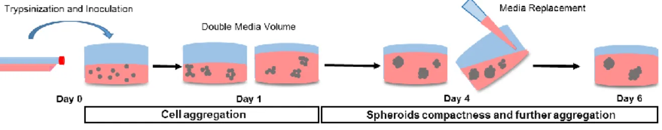
For spheroid generation, 6-well, 24-wells and 96-wells ULA flat-bottomed plates (Corning® Costar®) were inoculated with 1500 μL/well, 300 μL/well and 50 μL/well, respectively, of cell suspension at the optimized density of 250,000 cells/mL Spheroids culture medium consisted in DMEM supplemented with 10% FBS and 2% Growth Factor Reduced MatrigelTM (Corning®). Twenty-four hours for 6 and 24-wells plates and 2 h for 96-wells plates after inoculation, the volume of culture medium was doubled. Spheroids were incubated for up to 6 days with medium replacement every 3 days, in a humidified chamber at 37°C, 5% CO2. A scheme of spheroids formation is depicted in Fig. 5.
15
3.4. Histology
3.4.1. H&E
Day-4 spheroids were resuspended in Tissue Tek® O.C.T.™ (Sakura®) for preparing cryosections of 5-7 μm. Slides were first stained with Harris's haematoxylin (Sigma-Aldrich®) for 20 min, followed by Eosin Y (Sigma-Aldrich®) staining for 2 min. Slides were then submitted to increasing concentrations of Ethanol and finally incubated in xylene (EMD Chemicals). Samples were mounted with Entellan® (Merck). Images were acquired on an Olympus CK30 inverted microscope using Motic Images Version 2.0 software.
3.4.2. Ki67
Spheroid cryosections were fixed with pre-cooled acetone for 30 min at -20 °C, left drying and then, permeabilized with 0.08% Tween 20® for 2 min at room temperature (RT) and blocked for 30 min with 2% bovine serum albumin (BSA) in PBS. Incubation with primary antibody was performed overnight (ON) in a humidified chamber at 4°C. The primary antibody used was: Ki67 (Rabbit IgG, AB16667, Abcam®) diluted 1:100 In 1% BSA in PBS. The incubation with the secondary antibody goat anti-rabbit 594 (1:500) (Invitrogen™) was carried out for 1h at RT. Sections were mounted using ProLong gold antifade with DAPI (4',6-diamidino-2-phenylindole, Invitrogen™) and observed on an inverted fluorescence microscope (Axiovert 200M, Carl Zeiss) coupled with a monochrome camera (AxioCam MNC, Carl Zeiss). Sample fluorescence was examined at excitation/emission wavelengths of 590/617 nm (Alexa Fluor 594) and 358/461 (DAPI). Images were collected using AxioVision Rel. 4.7 software.
3.5. MMPi Viability Assays
The cytotoxicity of ARP-100, [15]pyN5 and [16]pyN5 was evaluated in MDA-MB-231 cells in 2D and 3D culture conditions using CellTiter 96® AQueous One Solution Cell Proliferation Assay (Promega). In 2D cultures,8x103 cells were seeded in 96-well plates and maintained for 24 h in DMEM supplemented with 10%FBS. Thereafter, cells were exposed to 100 μL of ARP-100, [15]pyN5 and [16]pyN5 in final concentrations 100, 75, 50, 25, 10, 5, and 1 µM, in serum free DMEM. After 24 h incubation, 20 μL MTS was added to the wells and left incubating for 1 h. The absorbance was recorded by spectrophotometry at 490 nm and 690 nm (SPECTROstar Omega, BMG LABTECH).
For 3D culture conditions, spheroids cultured in 96-welll ULA plates were used. Four-day spheroids’ culture medium was replaced by 100 µL of DMEM with 1,25% FBS, 2% MatrigelTM and ARP-100, [15]pyN
16
spheroids were incubated 1 h with MTS. Spheroids were pelleted and the supernatant read by spectrophotometry at 490 nm and 690 nm.
The number of surviving cells was assessed by the determination of the difference of absorbance, A490 nm– A690 nm and cell viability was expressed as percentage to control. Experiments were performed in triplicates.
3.6. Media Conditioning
For medium conditioning under 2D conditions (CM2D), 1,500,000 cells were seeded in 75 cm2 T-flask for 24h reaching a 50% confluency. After washing twice with PBS, 15 mL serum-free DMEM was added to the cells. 3D medium conditioning (CM3D) was performed at day 4 of culture, in 6-well ULA plates. Spheroids were washed twice with serum-free DMEM, and medium substituted by 2 mL serum free DMEM supplemented with 2% MatrigelTM. For both culture conditions, medium was conditioned for 24 h after which, it was harvested and concentrated about 100X in 10 kDa centrifugal concentrators (Millipore). Total protein was quantified by spectrophotometry (SPECTROstar Omega, BMG LABTECH) in a low volume microplate (Lvis plate, BMG LABTECH). Total protein concentration was determined through Lambert-Beer equation, with absorbance 280-340 nm and extinction coefficient of bovine serum albumin.
3.7. Modified zymography assay
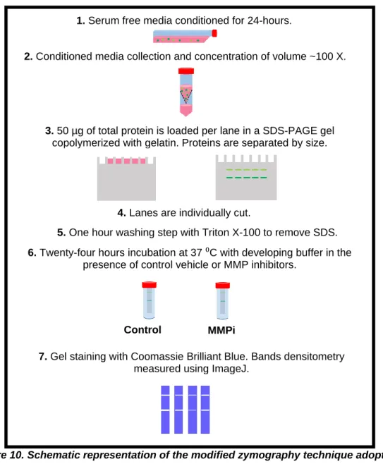
To screen MMP-2 and 9 total gelatinase activity, a sodium dodecyl sulfate Polyacrylamide gel electrophoresis (SDS-PAGE), with the gel containing gelatin as substrate, was performed using the methodology described in [109] with modifications (Fig. 10). CM concentrated samples were resolved by electrophoresis under non-reducing conditions, in a 10% Polyacrylamide containing 0.1% SDS and 0.1% (w/v) Gelatin. After electrophoresis, lanes were individualized and washed with 2.5% Triton X-100 in 3 steps of 20 min, to remove SDS. Lane gels were incubated for 24 h at 37 °C in, the Developing Buffer (50 mM Tris Base, 200 mM NaCl, 5 μM ZnCl2, 5 mM CaCl2.2H2O and 0.02% NaN3) which restores the MMPs protease activity. ARP-100, [15]PyN5 and [16]pyN5 were tested by dissolving them in the Developing Buffer. Gels were revealed in 0,1% Coomassie Blue R-250 (National Diagnostics). MMP activity was detected as a clear band in the background of uniform staining. Band quantification was performed using ImageJ software (National Institutes of Health, USA).
17
3.8. Migration Assays
3.8.1. Scratch Assay
In vitro scratch assays were performed according to Liang et al. [110]. Briefly, 200,000 MDA-MB-231 cells were seeded in 24-well plates. After 24 h incubation, medium was removed and a scratch performed with a 200-µL pipette tip, leaving a gap of approximately 0.7 mm in width. Cells were then rinsed twice with PBS and kept in serum-free medium containing 5-40 µM of ARP-100, [15]pyN5 and [16]pyN5. The scratch was evaluated microscopically (Motic AE 2000 inverted microscope), and four images of each scratch were recorded using Moticam 2500 at defined time-points: 0, 5, and 10 and 24 h. Non-invaded distance was measured using Motic Images PLUS v2.0 software. Experiments were performed in triplicates.
3.8.2. Radial Migration
This assay was performed according to Vinci et al [104]. Flat-bottomed, 24-well plates (Sarstedt) were coated with 0.01 mg/mL with Poly-D-Lysine (Sigma-Aldrich®) in sterile water for 2 h at RT, followed by 1:30 diluted MatrigelTM coating for 30 min. Four-day spheroids were transferred to pre-coated plates and maintained in 500 μL of medium containing 10% FBS and 2% MatrigelTM and the compounds, ARP-100, [15]pyN
5 and [16]pyN5 in concentrations ranging from 5-40 µM. Images were captured at 5, 10 and 24 h Motic AE 2000 inverted microscope. Effects of compounds were analyzed by measuring the distance between cells in the migration front and the perimeter of the spheroid. More than 10 spheroids per condition were analyzed, with 3 measures per spheroid.
3.9. Statistical analysis
Statistical analyses were carried by GraphPad Prism v6.0 software, (La Jolla, CA, USA). ANOVA with Tukey’s multiple comparison post-test was performed for 3 or 4 independent assays. Anova repeated measures tool was used when there were values matching. Results are presented as means ±standard error of the mean (SEM), except where indicated. Statistical significance was represented as probability (p) value *<0.05, **<0.01 and ***<0.001.
18
4. Results and Discussion
4.1 Development of 3D Models of MDA-MB-231
A suspension culture system using Ultra-Low Attachment (ULA) plates was developed and optimized in order to obtain MDA-MB-231 3D spheroids. ULA plates consist of plates coated with a hydrophilic and neutrally charged hydrogel that prevents cell adherence to the wells surface, therefore promoting cell aggregation. For this purpose, different culture parameters such as inoculum concentration, FBS concentration and ECM supplementation were evaluated. MDA-MB-231 cells were cultured in 24-well plates for 6 days in a batch mode consisting of culture medium replacement every 3 days (Fig. 5).
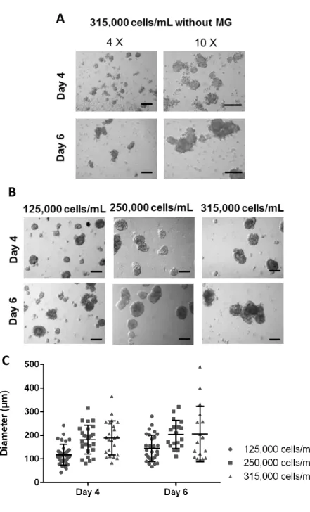
Medium supplementation with both 10% and 20% FBS, did allow formation of cell aggregates. However, these aggregates were easily disrupted and were heterogeneous in size and morphology (Fig. 6 A). To obtain more compact spheroids of MDA-MB-231 cell line, previous reports [111] recommend the addition of a reconstituted basement membrane. MatrigelTM [112], which is a commercially available basement membrane preparation extracted from the Engelbreth-Holm-Swarm (EHS) mouse sarcoma, rich in ECM proteins (e.g. laminin, collagen IV and heparin sulfate proteoglycans), was therefore added to the 10% FBS cultures at the inoculation step. Indeed, as shown in Fig. 6 B, 2% MatrigelTM allowed the formation of more compact and homogenous spheroids. Consequently, the 10% FBS condition with 2% MatrigelTM was further adopted.
According to previous reports [96, 111] cell densities ranging from 125,000 to 315,000 cell/mL were initially tested (Fig. 6 B). Twenty-four hours were given for cells aggregation, after which media volume was doubled. Measurements of spheroids were based on phase-contrast images up to day 6 of culture, yielding a size-distribution plot (Fig. 6 C).
With the higher inoculums (315,000 cells/mL), larger but more heterogeneous spheroids were obtained. As for the inoculum concentrations of 125,000 and 250,000 cells/mL, homogeneous spheroids were observed from day 4 onwards. However, in order to obtain a conditioning medium volume per cell in 3D cultures as in the two-dimensional system (further explored in Chapter 4.3.1.), the inoculum of 125,000 cells/mL was considered inadequate as it would require a very low cell confluence for medium conditioning in the 2D model. Therefore, for MDA-MB-231 spheroids development, the condition with cell density of 250,000 cells/mL in media supplemented with 10% FBS and 2% MatrigelTM was adopted and further characterized up to day 4.
19
Figure 6. Optimization of MDA-MB-231 spheroids development conditions. A) Contrast phase
pictures of aggregates formed without MatrigelTM (MG), from inoculum 315,000 cells/mL, at day 4
and 6 of culture (magnification 4X, scale bars=200 μm; magnification 10X, scale bar=100 μm). B) Contrast phase pictures of spheroids with 2% MatrigelTM from inoculums 125,000; 250,000 and
315,000 cells/mL at day 4 and 6 of culture (magnification 4X, scale bars=200 μm) Pictures taken with inverted microscope. C) Distribution plot for spheroids diameters (µm) at day 4 and 6 of culture of cells inoculated at 125,000; 250,000 and 315,000 cells/mL.
20
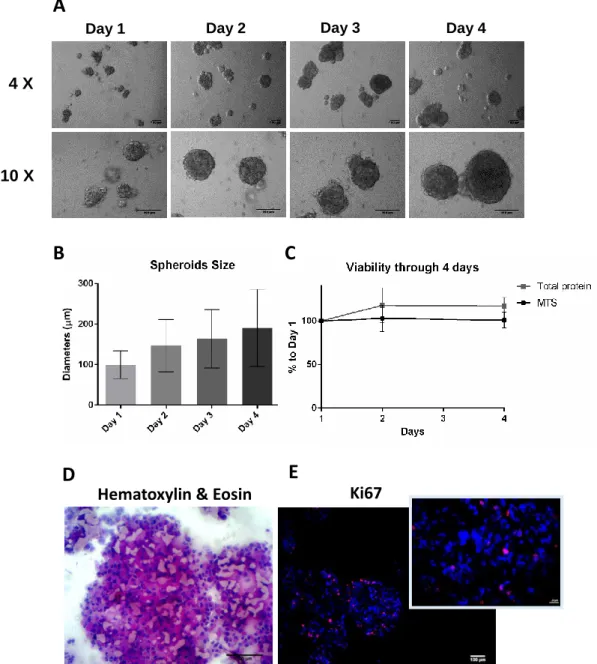
Figure 7. Characterization of MDA-MB-231 spheroids (inoculum 250,000 cell/mL in 10% FBS and 2% MatrigelTM supplemented media). A) Representative phase contrast images of the
spheroids from day 1 to day 4. Pictures taken with inverted microscope (scale bars=100 μm for magnification 4X, and 10X); B) Spheroids diameters (µm) over culture time represented as mean ± SD). C) Percentage of MTS and total protein of days 2 and 4 relative to day 1 of culture. (mean ± SEM, n=2-3). D) Cryosections stained with Hematoxylin & Eosin. Scale bar=100 μm. E) Cryosection immunolabeled with Ki-67(pink) and Dapi (blue). Major Figure scale bar= 100 μm. Minor Figure scale bar=20 μm.
B
D
Hematoxylin & Eosin
Ki67
E
C
Day 4 Day 3 Day 1 Day 2A
4 X 10 X21
Phase contrast microscopy shows that at day 1, MDA-MB-231 cells inoculated at a cell density of 250.000 cells/mL,form small aggregates of approximately 90 µm diameter (Fig. 7 A and B). These aggregates further gathered, forming spheroids with increasing sizes and compactness over culture time. On average, spheroid diameters were 147 ± 64 µm (mean ± SD) at day 2 and 191 ± 95 µm (mean ± SD) at day 4 of culture (Fig. 7 B). From day 4 onwards the spheroids diameter was maintained (data not shown). To further monitor the morphology of the spheroids, histological analysis of spheroid cryosections were performed at day 4. H&E staining of sections revealed that spheroids were solid with cells homogeneously distributed and embedded in extracellular matrix presenting no evidence of necrotic cores (Fig. 7 D). Immunolabeling with the proliferative marker Ki-67 (Fig. 7 E) shows 15% proliferating cells, including cells within the spheroid. This proliferation percentage is in agreement with the observed 1.2-fold increase in total protein (Fig. 7 C). These results together with the MTS assay data (Fig. 7 C), show reduced proliferation in 3D cultures when compared with monolayer models, a pattern which has been broadly reported for many cancer cell lines including MDA-MB-231 [111, 113, 114]. Several factors, which induce this change in proliferation have been described, such as hypoxia and nutrients hindered diffusion, ECM elements and their stiffness [114–116]. Proliferation marker Ki-67 has been used as a prognostic marker for breast cancer, with most in vivo tumors having under 50% Ki-67+cells. This contrasts with a very different percentage,100% Ki-67+cells, reported in MDA-MB-231 monolayer [117].
Obtaining a realistic proliferation is vital for the assessment of potential chemotherapeutic agents, and also important for better understanding of breast cancer cell heterogeneity and classification. MDA-MB-231 cell line has been described as a highly proliferative triple-negative cells belonging to Basal-like tumors’ type. Recently, a new subtype of breast tumors was identified, claudin-low [118]. These subtype is characterized for low or no expression of epithelial cell-cell adhesion molecules E-cadherin and Claudin-3,4 and 7 and high expression of EMT markers such as Vimentin and EMT transcription factors (e.g. Twist and SnaiI). The claudin-low subtype is also described to have a stemness signature by having high CD44/CD24 and CD49f/Epithelial cell adhesion molecule (EpCAM) ratios, and by having a low percentage of Ki-67+ cells, indicating lower proliferation when compared to other subtypes, such as Basal-like. A screening of several breast cancer cell lines identified nine cell lines, including MDA-MB-231, with the same expression profiles as claudin-low subtypes [119]. The main difference between cell lines and the primary tumor identified as claudin-low was the proliferation gene cluster, which could be a result of the selection imposed on cell lines or of the standard in vitro conditions. Thus, 3D models may give further insight about these cell lines by being able to reveal information that has been hiding under non-physiological culture conditions [120]. Moreover, MDA-MB-231 not expressing E-cadherin
22
could explain why these cells do not form spheroids spontaneously as they have their cell-cell-adhesion ability hampered. Hence, these cells require the addition of extrinsic ECM to form compact spheroids as they only tightly aggregate through integrin-β1 (cell-matrix interactions molecule) [99].
4.2. Cell Viability Assays
In order to rule out direct cytotoxic effects that could somehow conflict with migration assays, dose-response curves were performed to determine concentrations of the compounds [15]pyN5, [16]pyN5 and ARP-100, with no, or limited, effect on cells viability. The cytotoxicity activity of these compounds in both 2D and 3D models of MDA-MB-231 was assessed through MTS assay that detects viable and metabolic active cells. Since some of the cellular assays are performed in serum free conditions (e.g. scratch-assay), the cytotoxicity assays were also performed in serum-free conditions. Following the concentrations of ARP-100 used in literature [46,69], concentrations ranging 1-100 μM were chosen (Fig. 8). As for [15]pyN5, [16]pyN5 the same range ofconcentrations was adopted.
As depicted in Fig. 8 A and B, both pyridine containing macrocycles (py-macrocycles) induced a slight decrease on cell viability in 2D cultures at concentrations higher than 10 and 25 μM for [15]pyN5and [16]pyN5, respectively. Up to 75 μM, viability was higher than 74%, decreasing to ~60% at 100 μM concentrations for both [15]pyN5and [16]pyN5. The cytotoxicity profile observed in spheroids cultures was very similar between both py-macrocycles, with viability values never below 80% (Fig. 8 A and B).
Concentrations of ARP-100 up to 25 μM, showed no cytotoxicity in monolayers (Fig. 8 C). At a concentration of 50 μM, cell viability was 75% and decreased in a concentration dependent manner, attaining the value of 44% at 100 μM of ARP-100. In spheroid cultures, ARP-100 did not exhibit the same dose-response curve. Cell viability was never lower than 78%. In particular, for the ARP-100 concentrations of 75 and 100 µM, significantly higher (p<0.05 and p<0.01, respectively) cell viabilities were obtained in 3D cultures when compared to monolayer. These results suggest that the 3D model is more resistant to ARP-100 than the monolayer one. The toxicity mechanism for ARP-100 is not known and it is beyond our scope to explain the phenomenon in this specific case. Nevertheless, higher resistance of 3D models has been broadly reported [104, 108, 121], with the exception of some cell lines and drugs. As an example, it has been shown that in HT080 in 3D collagen fibers [121] and MCF-7 spheroids [94], proliferation and viability, respectively, were less affected by doxorubicin than monolayer cultures. The overall higher resistance of the 3D models has been related to the quiescence as aforementioned, ECM-mediated signaling pathways [122, 123] (e.g. apoptotic pathways) and even hypoxia-mediated P-glycoprotein up-expression [124]. Furthermore,
23
similarly to the gradient of nutrients and oxygen, drug penetration is also somewhat hindered in spheroids, once again mimicking the hindered diffusion in solid tumor [125, 126].
Following the viability assay studies, compounds’ concentrations ranging 5-40 µM were adopted, in order to have a cell viability above 70 %.
Figure 8. Effect of A) [15]pyN5, B) [16]pyN5 and C) ARP-100 on MDA-MB-231 cell viability on 2D and 3D models, evaluated through the MTS assay. Results are expressed in percentage
(mean± SEM, n=3;4) to respective controls, non-treated condition for [15]pyN5 and [16]pyN5 and
0.25% DMSO for ARP-100. Statistical significance is expressed relatively to respective controls as *p<0.05, **p<0.01, ***p<0.001.
4.3.1. Optimization of Zymography technique and Media conditioning
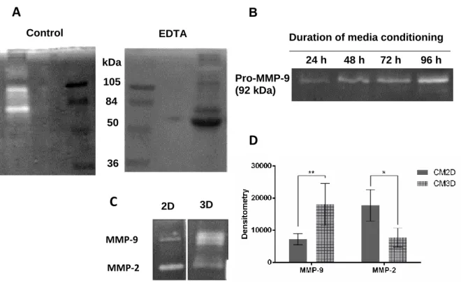
A zymography technique, in which gelatin is used as substrate for MMP-2/9 and pro-MMP-2/9, was implemented to screen both enzyme activities. The first step is equivalent to a common SDS-PAGE, except for that it must be performed under non-reducing conditions. After electrophoresis, the gel is washed to remove SDS and then incubated with a buffer
![Figure 1. MMPs’ regulation of Tumor Microenvironment (EBM-endothelial basement membrane; BM-bone marrow) [26]](https://thumb-eu.123doks.com/thumbv2/123dok_br/19201765.954073/14.892.245.652.553.866/figure-regulation-tumor-microenvironment-endothelial-basement-membrane-marrow.webp)
![Figure 2. MMP catalytic center with a modeled Pro-Leu-Gly-Leu-Ala-Gly-amide hexapeptide substrate [22]](https://thumb-eu.123doks.com/thumbv2/123dok_br/19201765.954073/15.892.272.631.423.753/figure-mmp-catalytic-center-modeled-amide-hexapeptide-substrate.webp)
![Figure 3. Gelatinases basic domain structure [23]. Common to MMPs: Pre-peptide signal, Pro- Pro-propetide domain; Zn- highly conserved zinc-binding site; H- Hinge region linking the Hemopexin-like domain to the catalytic domain (not existing i](https://thumb-eu.123doks.com/thumbv2/123dok_br/19201765.954073/17.892.197.710.124.250/figure-gelatinases-structure-propetide-conserved-hemopexin-catalytic-existing.webp)
![Figure 4. Chemical structure of some hydroxamic acid MMP inhibitors (Marimastat, Prinomastat and ARP-100) and of the macrocyclic compounds containing pyridine ([15]pyN 5 and [16]pyN 5 ).](https://thumb-eu.123doks.com/thumbv2/123dok_br/19201765.954073/19.892.226.631.317.722/chemical-structure-hydroxamic-inhibitors-marimastat-prinomastat-macrocyclic-containing.webp)
![Figure 8. Effect of A) [15]pyN5, B) [16]pyN5 and C) ARP-100 on MDA-MB-231 cell viability on 2D and 3D models, evaluated through the MTS assay](https://thumb-eu.123doks.com/thumbv2/123dok_br/19201765.954073/34.892.135.751.260.746/figure-effect-arp-mda-viability-models-evaluated-assay.webp)