UNIVERSIDADE DE LISBOA
FACULDADE DE CIÊNCIAS
DEPARTAMENTO DE BIOLOGIA VEGETAL
Embryonic thymic epithelium differentiation in
chicken: study of molecular signals involved in
lymphoid progenitor cells colonization
Carlota Bobone Lucena
DISSERTAÇÃO
MESTRADO EM BIOLOGIA MOLECULAR E GENÉTICA
2012
UNIVERSIDADE DE LISBOA
FACULDADE DE CIÊNCIAS
DEPARTAMENTO DE BIOLOGIA VEGETAL
Embryonic thymic epithelium differentiation in
chicken: study of molecular signals involved in
lymphoid progenitor cells colonization
Dissertação orientada por
Professora Dra. Hélia Cristina de Oliveira Neves (FMUL)
Professora Dra. Rita Maria Pulido Garcia Zilhão (FCUL)
Carlota Bobone Lucena
MESTRADO EM BIOLOGIA MOLECULAR E GENÉTICA
2012
Agradecimentos
À Hélia, por me ter recebido no seu laboratório e pela dedicação e preocupação que sempre mostrou comigo e com o meu trabalho; pela autonomia e liberdade me proporcionou e por tudo o que aprendi durante este ano.
À Professora Rita, pela oportunidade de a ter como orientadora, pela disponibilidade e apoio que sempre demonstrou e sobretudo pela contribuição para o meu gosto pela investigação desde os tempos da licenciatura.
À Unidade de Biologia da Hematopoiese do Instituto de Histologia e Biologia do Desenvolvimento da FMUL, pelo acolhimento e ajuda na realização desta tese. Em especial à Isabel pelo exemplo de boa vontade, pelos conselhos, e especialmente pela ajuda essencial para a análise de citometria, e ao Vítor, pela sua dedicação e disponibilidade, por toda a ajuda sem a qual o trabalho não estaria completo.
À Marta pelo dia-a-dia no laboratório, pelos grandes momentos (em tempo e qualidade) que passámos juntas, pela sua boa disposição contagiante, e “espírito de equipa”: essencialmente pela amizade que se criou.
A todos os que passaram pelo laboratório, especialmente ao Rafael, à Marta e mais recentemente à Joana, pela ajuda indispensável para a realização deste trabalho.
À minha família e aos meus amigos, pela presença constante e apoio em todos os momentos, pelo exemplo que são e que me estimula a ser sempre mais.
Resumo
O timo é o órgão linfóide primário responsável pela diferenciação de progenitores hematopoiéticos linfóides (PHL) em linfócitos T maduros. Após o nascimento, os PHL entram no timo através dos vasos sanguíneos e iniciam a timopoiese, processo complexo de diferenciação em linfócitos T. Os PHL começam por especificar-se na linhagem T e depois iniciam o seu longo processo de diferenciação adquirindo coreceptores de membrana característicos das células T maduras, como os marcadores CD3, CD4 e CD8. A timopoiese é essencial para a constituição de um sistema imunitário saudável. De facto, os linfócitos T são o componente principal do sistema imunitário adaptativo, capazes de responder a agentes infecciosos, e aumentando a capacidade de defesa do organismo com cada exposição a antigénios específicos.
O timo é um órgão constituído por células epiteliais tímicas (CET), células mesênquimais derivadas da crista neural (CN), células dos vasos sanguíneos e células hematopoiéticas. O seu desenvolvimento está intimamente ligado ao das glândulas paratiróides, uma vez que os seus epitélios partilham a mesma origem embrionária: a endoderme das terceira e quarta bolsas faríngicas (3/4 BF)1,2. A origem endodérmica das células epiteliais foi pela primeira
vez demonstrada por Le Douarin e Jotereau utilizando o modelo de quimeras galinha-codorniz3. Neste trabalho também mostraram que distintos mesênquimas ectópicos, são
capazes de suportar (mesênquima permissivo, da somatopleura) ou não (mesênquima não permissivo, do sómito ou do botão do membro) o desenvolvimento da endoderme na formação dum timo funcional, revelando a importância das interações celulares entre a endoderme e o mesênquima adjacente, nas fases iniciais do desenvolvimento tímico3. Durante esta primeira fase do desenvolvimento tímico, ocorre a especificação das CET2.
Os territórios presuntivos do timo e das glândulas paratiróides são definidos pela expressão de factores de transcrição distintos nas bolsas faríngicas, o gene Foxn1 (forkhead box N1) e o gene Gcm2 (glial cells missing-2), respectivamente4. Assim, em galinha, o rudimento do
timo foi identificado ao dia 4,5 de desenvolvimento embrionário (E4,5) (E4 em codorniz) na região dorsal das 3/4 BF enquanto que o domínio das paratiróides ocupa um território mais anterior e ventral das mesmas5.
A segunda fase do desenvolvimento do timo depende da sua colonização por PHL e de interações entre estes e as células epiteliais tímicas. Estas interações são essenciais para a diferenciação das CET em duas linhagens celulares, cortical e medular, originando consequentemente a formação de dois compartimentos tímicos: o córtex e a medula6–9.
Durante a embriogénese, Le Douarin descreveu três ondas de colonização do epitélio tímico (ET) por PHL, no modelo de galinha10. A primeira onda ocorre ao E6,5 (E6 em codorniz),
antes da vascularização do rudimento tímico, entrando os progenitores hematopoiéticos no rudimento tímico através da sua migração pelo mesênquima adjacente3,11,12. As restantes
ondas realizam-se através dos vasos sanguíneos, ao E12 e E1810.
As interações entre os PHL e as TEC são mediadas por vários factores solúveis, como citocinas (estimulam a proliferação dos timócitos) e quimiocinas (importantes na migração dos PHL para o rudimento tímico), e por vias de sinalização como a sinalização Notch. A importância do microambiente Notch para a correcta especificação de progenitores hematopoiéticos nas diferentes linhagens linfóides foi demonstrada pela primeira vez em 2001 pelo grupo de L. Parreira13. Além disso, os genes envolvidos na sinalização Notch
(receptores, ligandos e genes-alvo) são expressos de forma distinta nos diferentes territórios do timo adulto, reforçando a importância desta via de sinalização na função do mesmo14. Recentemente, o grupo de H. Neves observou em embriões de galinha que os genes envolvidos na sinalização Notch estão diferencialmente expressos na endoderme das bolsas faríngicas, em estádios prévios à formação do rudimento tímico.
Em estudos recentes, este mesmo grupo, utilizando o modelo de quimeras galinha-codorniz, observou que células da endoderme das 3/4 BF de codorniz (dador; E3) quando enxertadas num ambiente permissivo (mesênquima da somatopleura) dum embrião de galinha (receptor; E2,5), especificam em ET (Foxn1+). Também constataram que, 5 dias após o enxerto, o epitélio tímico se encontra colonizado por PHL com origem no embrião receptor (galinha)5. Contudo, quando a endoderme é enxertada num ambiente não permissivo (mesênquima do botão embrionário do membro posterior), nas mesmas janelas temporais, esta expressa Foxn1 mas não é colonizada por células hematopoiéticas5. Estes resultados
sugerem que, quando enxertada num ambiente não permissivo, a endoderme não possui e/ou não recebe os sinais do mesmo (mesênquima adjacente) necessários à sua colonização por PHL. De facto, em codorniz, a primeira vaga de colonização do rudimento tímico por PHL (E6) ocorre durante a janela temporal em análise. Assim, este sistema representa um novo modelo de estudo para identificar os factores envolvidos na migração dos PHL e na colonização do rudimento tímico pelos mesmos, etapas fundamentais ao desenvolvimento do timo.
Este trabalho teve como primeiro objectivo identificar os sinais moleculares da via de sinalização Notch envolvidos na fase de colonização do rudimento tímico (CET) pelos PHL. Para este efeito utilizou-se o modelo de quimeras galinha-codorniz acima descrito. Embriões quiméricos, com 3 e 5 dias de desenvolvimento após o enxerto da endoderme, foram sacrificados e estudados por hibridação in situ com genes envolvidos na sinalização Notch, especificamente os ligandos-Notch, Delta1 e Delta4. Os resultados obtidos mostram que a endoderme, quando enxertada no ambiente permissivo, expressa ambos os ligandos, nos
dias 3 e 5 após o enxerto. Em contraste, quando a endoderme é enxertada num ambiente não permissivo, embora expresse Delta4, apresenta uma expressão diminuída ou inexistente de Delta1 (3 e 5 dias após o enxerto). Assim, os resultados sugerem que Delta1 poderá ser o ligando Notch específico envolvido na mediação do sinal Notch na interação entre o epitélio (ligando Delta1) e os PHL (receptor Notch) nas fases iniciais de colonização do epitélio tímico. Em conformidade, no timo adulto de ratinho foi descrita a expressão de
Delta1 na junção cortico-medular, local de entrada dos PHL após o nascimento14.
Atualmente, decorrem estudos para aprofundar o papel deste ligando e caracterizar a sua sinalização durante o processo de colonização do epitélio tímico. De futuro, também serão estudadas outras moléculas possivelmente envolvidas neste processo, tais como quimiocinas, e componentes de outras vias de sinalização.
Em paralelo, neste trabalho caracterizou-se o desenvolvimento do timo e das glândulas paratiróides em galinha (entre os dias 5 e 18 de desenvolvimento), ao nível histológico e ao nível da diferenciação celular dos linfócitos T no rudimento tímico (por análise de citometria de fluxo).
A análise histológica foi realizada através da expressão in situ dos genes Foxn1 e Gcm2. Em cortes seriados de parafina, ao longo dos estádios estudados (E5 a E13), observou-se a expressão de Foxn1 e Gcm2 nos rudimentos do timo e das glândulas paratiróides, respectivamente. Concluiu-se que a expressão destes factores de transcrição é mantida até ao dia E13. Mostrou-se também que as glândulas paratiróides apresentam uma morfologia adulta a partir de E9, e que o timo ao dia 13 de desenvolvimento se encontra ao longo do pescoço e diferenciado em dois compartimentos (córtex e medula).
Analisou-se a dinâmica de diferenciação das populações de timócitos presentes no rudimento tímico, entre E11 e E18, por citometria de fluxo, utilizando os marcadores de membrana CD3, CD4 e CD8. Os resultados desta análise mostram que até E13 existem duas populações imaturas de timócitos, CD8+CD4-CD3- e CD3+CD8-CD4-, pouco proliferativas, e derivadas da primeira onda de colonização, sugerindo que podem ter um papel na especificação do epitélio tímico. De facto, no momento da segunda onda de colonização (E12-13) a medula começa a ser definida, evidência morfológica do processo prévio de especificação das células epiteliais em linhagens cortical e medular. Paralelamente, após a segunda onda de colonização (>E15) observa-se uma forte expansão de células duplas positivas, CD4+CD8+, sugerindo que estes timócitos têm uma
maior capacidade de se diferenciarem (e expandirem) e possivelmente de contribuir para a maturação dos dois compartimentos tímicos.
Palavras-chave: organogénese do timo, epitélio tímico, colonização por progenitores hematopoieticos linfóides, Foxn1, Delta1.
Abstract
The thymus is the primary lymphoid organ where maturation of lymphoid progenitors cells (LPCs) into T-cell occurs. This maturation depends on interactions between the LPCs and thymic epithelium (TE). TE derives from endoderm of the 3rd and 4th Pharyngeal Pouches
(3/4 PPs) (in chicken) and depends on epithelial-mesenchymal interactions and on LPCs colonization to become functional.
Using the quail-chick model, our group showed that distinct mesenchymal tissues are permissive (somatopleure) or non-permissive (limb bud) to 3/4 PPs endoderm specification and early development. In addition, only in the permissive environment the endoderm is colonized by LPCs.
In this work, we aimed to identify the molecular cues, namely Notch signaling ligands, involved in TE colonization by LPCs. For that, we used the quail-chick chimeric model. Using immunohistochemistry combined with in situ hybridization techniques we showed that Delta1, as opposed to Delta4, is down-regulated in the endoderm developed in the non-permissive mesenchyme (with no LPCs colonization). Together, these results suggest that mesenchymal-epithelial interactions are important to establish a proper environment to thymus formation and that Delta1 is involved in TE colonization by LPCs.
In parallel, we described thymus and parathyroid glands development through the in situ expression analysis of specific transcription factors for each organ (Foxn1 and Gcm2, respectively). We observed that the expression of both transcription factors is maintained from E5 to E13 in the respective developing organs.
We then characterized thymocyte populations during thymic organogenesis by flow cytometry analysis. We identified two immature populations, single positive for CD3 or CD8, at early thymic development (<E13), suggesting that these thymocytes may be important in TECs specification into different lineages (cortical and medullary), and subsequent maturation of thymic compartments.
With this work, we hope to clarify important events in thymus organogenesis: colonization by LPCs and TECs specification.
Keywords: thymus organogenesis, thymic epithelium, lymphoid progenitors cells colonization, Foxn1, Delta1.
Index
Agradecimentos III Resumo IV Abstract VII Abbreviations 1 Introduction 2I. The Thymus: Structure and Function 2
II. The Thymus and Parathyroid Glands Development 5 II.1 Thymus Development: thymocyte-independent phase 7 II.2 Thymus Development: thymocyte-dependent phase 7
III. Notch Signaling Pathway 9
III.1 Notch signaling in thymopoiesis 9
Materials and Methods 11
Results 15
I. Notch signaling in thymus epithelium colonization by LPCs 15 II. Formation of chicken pharyngeal organs: thymus and parathyroid glands 17 III. Thymocytes population kinetics during chicken embryo development 20
Discussion 23
I. Delta-1 ligand is involved in early colonization of the thymic rudiment by LPCs 23 II. Ontogeny of chicken thymus and parathyroid glands: histological analysis 25 III. Ontogeny of chicken thymus: characterization of thymocytes maturation 25
Bibliography 28
Appendix I – BUFFERS, MEDIA AND OTHER SOLUTIONS 32
Appendix II – PROTOCOLS 34
Abbreviations
APC – Antigen Presenting Cells DC – Dendritic Cells
DMEM – Dulbecco's Modified Eagle Medium DN – Double Negative
DNA – Deoxyribonucleic Acid DP – Double Positive
E – Embryonic day
EDTA – Ethylenediaminetetraacetic Acid FACS – Fluorescence Activated Cell Sorter FBS – Fetal Bovine Serum
h, min, sec, ms – hour, minute, second, millisecond HE – Hematoxylin-Eosin
IHC – Immunohistochemistry ISH – In Situ Hybridization LPC – Lymphoid Progenitor Cell
MHC – Major Histocompatibility Complex NC – Neural Crest
NS – Negative Selection ON – overnight
PA – Pharyngeal Arch
PBS – Phosphate Buffered Saline Pen/Strep – Penicillin/Streptomycin PFA – Paraformaldehyde
PP – Pharyngeal Pouch PS – Positive selection PTE – Parathyroid Epithelium PTH – Parathyroid Hormone RNA – Ribonucleic Acid
RT-PCR – Reverse Transcriptase-Polymerase Chain Reaction SP – Single Positive
TCR – T-cell Receptor TE – Thymic Epithelium TEC – Thymic Epithelial Cell TN – Triple Negative
Introduction
I. The Thymus: Structure and Function
The thymus is the primary lymphoid organ that supports the differentiation of Lymphoid Progenitor Cells (LPCs) along the T-cell pathway. T-cells are the main component of the adaptive immune system, capable to respond to infectious agents and to increase in magnitude and defensive capabilities with each successive exposure to antigens.
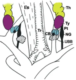
In mammals, the thymus is a bilobed organ, located in the central compartment of the thoracic cavity, above the heart and behind the sternum. In birds two thymi exist, located bilaterally near the jugular vein, along the neck, subdivided in seven lobes each (Figure 1), divided themselves in lobules1,5. In mammals, located in the vicinity of the thymus, there are
four parathyroid glands. In birds, there are also four glands located under the thyroid glands, near the nodose ganglion and the ultimobranchial body5 (Figure 1). These glands are small
endocrine glands that produce the parathyroid hormone (PTH), regulating calcium and phosphate homeostasis in the organism15.
Figure 1. Schematic representation of the anatomic location of foregut endoderm-derived glands in adult chicken. (CB, carotid body; Es, esophagus; NG, nodose ganglion; Pt, parathyroid glands; Th, thymus; Tr,
trachea; Ty, thyroid; UBB, ultimobranchial body). (Adapted from Neves et al. 20125)
The thymus is an epithelial organ surrounded by a capsule, consisting of a thick layer of connective tissue, and each lobule consists of an outer cortex and an inner medulla. The cortex and medulla are composed by distinct and specific Thymic Epithelial Cells (TECs), blood vessels cells, bone marrow derived cells (mostly macrophages and dendritic cells) and mesenchymal cells. Cortical-TECs (cTECs) and medullar-TECs (mTECs) form a
three-
dimensional network densely packed. Together these elements form the stromal component of the thymus that supports the maturation of LPCs into T-cells.
In the mature thymus, developing T-lymphocytes (called thymocytes) make up more than 95% of its cellularity11, where the cortex contains a dense population whereas the medulla is more sparsely populated. With age, the thymus involutes (invasion of the parenchyma by adipose tissue) and is virtually undetectable in post-pubertal humans16. In chicken, the size and weight of the thymus reach their maximum at the second week of life, and thymic involution is related to an increase in the rate of the sex hormones at the time of puberty17.
The main function of the thymus is to support maturation of LPCs into T-cells, a process called thymopoiesis. It is a complex process that starts with the colonization of the thymus primordium by LPCs, followed by their commitment to T-cell lineage, and subsequent differentiation to mature T-lymphocytes. These are characterized by the expression of T-cell receptors (TCR), associated with the co-receptor CD3, and the expression of accessory molecules such as CD4 and CD8.
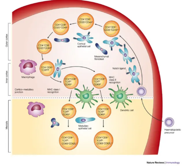
In the postnatal thymus, LPCs enter at the cortico-medullary junction through blood vessels11. LPCs are Triple Negative (TN) cells, as they do not express the co-receptor CD3
or the accessory molecules CD4 and CD8. These TN cells can be further discriminated according to surface expression of CD44 and CD25. Sequential gain of CD25 and loss of CD44 expression, respectively, serve to delineate early differentiation steps within this subset of thymocytes14 (Figure 2). The first maturation steps occur in the cortex where early thymocytes undergo proliferative expansion, are committed to the T-cell lineage, and start rearranging the TCR genes, the major event in thymocytes development. This process, called VDJ recombination, consists in the rearrangements in individual lymphocytes of different variable (V) region gene segments with diversity (D) and/or joining (J) gene segments. This creates a unique antigen-binding structure in each T-lymphocyte clone, leading to a highly diverse T-repertoire. In association with the TCR expression, thymocytes express the co-receptor CD3. In this phase, LPCs mature from TN to Double Positive (DP) cells, expressing CD4 and CD8. Then the DP cells selected to proceed maturation migrate into the medulla, to become CD4+ or CD8+ Single Positive (SP) T-lymphocytes. These cells
Figure 2. Thymocytes maturation pathway. The thymic architecture is organized into discrete cortical and
medullary areas, each of which is characterized by the presence of particular stromal cell types, as well as thymocyte precursors at defined maturation stages. Thymocyte differentiation is characterized by the expression of well-defined cell-surface markers, including CD4, CD8, CD44 (or CD117) and CD25, as well as the status of the TCR. Interactions between Notch receptor-expressing thymocytes and thymic stromal cells that express Notch ligands induce a complex programme of T-cell maturation in the thymus, which ultimately results in the generation of self-tolerant CD4+ helper T-cells and CD8+ cytotoxic T-cells, which emigrate from the thymus to establish the peripheral T-cell pool. (Adapted from Zúñiga-Pflücker 200418)
During their maturation, thymocytes are submitted to high-stringency selection processes, where potentially harmful cells that avidly recognize self-structures may be eliminated or induced to alter their antigen receptors. In the DP stage of thymocytes maturation, Positive Selection (PS), also called death by neglect, ensures that mature T-cells are self-MHC (Major Histocompatibility Complex) restricted. In this process, thymocytes whose TCR binds to self-MHC molecules are rescued from programmed cell death, whereas thymocytes whose receptors do not recognize self-MHC die by default.
DP thymocytes in the medulla are also subjected to Negative Selection (NS) by which developing thymocytes that express self-reactive antigen receptors are eliminated, thereby contributing to the maintenance of self-tolerance. NS involves high-avidity binding of a
thymocyte to self-MHC molecules with bound peptides in thymic Antigen Presenting Cells (APCs) leading to apoptotic cell death (a phenomenon known as clonal deletion). This is an important mechanism for maintaining tolerance to many self-antigens: it is called central tolerance16.
These selection processes, drive maturation of TCR-expressing thymocytes and shape the TCR repertoire toward self-MHC restriction and self-tolerance.
In the periphery, T-cells become activated when the TCR recognizes the complex of foreign antigens bound to a MHC molecule on the surface of APCs. CD4+ T-cells, called T-helper,
are class-II MHC restricted and are important in cell-mediated immunity (macrophage activation) and humoural immunity (B-cell differentiation). CD8+ cells are the cytotoxic
T-cells, class-I MHC restricted, whose function is to kill cells infected with microbes, and tumor cells16.
The major molecules (TCR, CD3, CD4 and CD8 co-receptors) and major events in the maturation pathway of LPCs into T-cells appear to be conserved between birds and mammals, although some differences have been described19,20.
II. The Thymus and Parathyroid Glands Development
Thymus development is intimately linked to that of the parathyroid glands as they share a common embryonic origin. These pharyngeal organs derive from the pharyngeal pouches (PPs), bilateral transient structures arising from the endoderm of the most anterior region of the foregut: the pharynx1. The PPs, along with the opposing pharyngeal clefts (invaginations
of surface ectoderm) form the separation between pharyngeal arches, the bilateral bulges that involve the pharyngeal region1,2 (Figure 3).
The endodermic origin of TECs was demonstrated for the first time, using the quail-chick chimeric model by Le Douarin and Jotereau3. In chicken and in human, thymic and
parathyroid organ rudiments derive from the third and fourth PPs (3/4 PPs)3,5 (In mouse these glands derive from the 3rd PP).
Figure 3. Scheme representing coronal section through the pharyngeal zone. Pharyngeal arches, consist of
mesenchymal and mesodermal cells bounded by an outer layer of surface ectoderm (blue) and inner layer of pharyngeal endoderm (yellow). The ectoderm forms invaginations, the pharyngeal clefts, which separate the arches, whereas the endoderm forms the opposing outpocketings, the pharyngeal pouches.
The common primordium is patterned into organ-specific domains that can be identified by the regionalized expression of molecular markers early in organogenesis, before morphological distinctions are present4.
In mouse, the parathyroid domain is defined by the expression of the transcription factor glial
cells missing-2 (Gcm2) as early as embryonic day 9.5 (E9.5), before the formation of the
organ primordium4. Thymic Epithelium (TE), is identified in a ventral position in the pouch, by
the expression of the transcription factor forkhead box N1 (Foxn1), detected from E11.25, coinciding with the budding off and outgrowth of the common primordium4. Once the
primordium forms, Gcm2 and Foxn1 are expressed in a complementary fashion in the parathyroid- and thymus-specific regions before the division into morphologically distinct organs4.
Foxn1 loss-of-function mutation leads to congenital athymia and hairlessness in nude
mouse. In this mouse, LPCs fail to enter the thymic primordium and instead remain in the surrounding perithymic mesenchyme. In addition, the epithelial cells within the primordium fail to expand after E12.5 and vascularization of the primordium does not occur12. Thus,
Foxn1 is not required for initiation of thymus organogenesis6, but is required cell-autonomously for TECs differentiation21.
Gcm2 may be the determining factor for parathyroid development, since, when deleted, no parathyroid glands are formed, (without interfering with thymus development)22. In Gcm2
-/-mutant mouse, parathyroid hormone (PTH) expression is undetectable at E11.5, indicating that Gcm2 plays a crucial role very early in parathyroid organogenesis4. Gcm2 is exclusively
expressed in the parathyroid and its embryonic rudiment, in mammals and avians22, and is an excellent marker of the early parathyroid domain4,15.
In quail, Gcm2 and Foxn1 expression are detected at E2.5 and E3.5 respectively by RT-PCR, indicating that endoderm specification into Parathyroid Epithelium (PTE) occurs earlier than into TE5. In chicken, Foxn1 and Gcm2 transcripts occupy mutually exclusive and
adjacent domains of 3/4 PPs at E5, with Foxn1 expression located in the dorsal/anterior domain and Gcm2 in a more ventral one5.
Although these data are similar to those reported in the mouse, the domain of Foxn1 expression is inverted along the dorsal-ventral axis of the pouches in chicken and quail, when compared to mouse embryos. These distinct positions during embryogenesis might contribute to the different anatomical locations of the adult thymus between mammals and birds5.
Thymus organogenesis can be divided in two main temporal phases; an early thymocyte-independent phase, where cellular interactions between the endoderm and the surrounding mesenchyme direct TECs specification2, followed by a thymocyte-dependent phase. At this later stage, the thymic rudiment depends on colonization by LPCs and on lympho-epithelial (LPC-TEC) interactions for further maturation of thymic epithelium into cortical and medullar compartments6–9.
II.1 Thymus Development: thymocyte-independent phase
Using the quail-chick chimeric model, Le Douarin and Jotereau showed that the 3/4 PPs endoderm isolated from early quail embryos (E1.5 - E2.5) was able to develop into thymic epithelium (TE) with the cooperation of a heterologous mesenchyme such as the somatopleure or splanchnopleure of 3 days-old chicken embryos, which thus could be considered “permissive” to endoderm development23. Furthermore, the grafted endoderm
was capable of inducing the heterologous mesenchyme to participate in the formation of a fully developed thymus3,23,24. In contrast, mesenchymal environments of the somite and limb
bud were non-permissive to 3/4 PPs endoderm development23,24. These data provided the first evidence that the development of a functionally competent thymus depends initially on a series of epithelial-mesenchymal interactions, that direct TECs specification2. Moreover, they revealed that some heterologous mesenchymal tissues are able to mimic the role played by NC-derived mesenchyme during normal development of the thymus in the pharyngeal region. NC-cells are a transient population formed between the neural tube and surface ectoderm, which colonizes the branchial arches, surrounds each primordium, and later forms the perivascular mesenchyme and the capsule of the thymus3.
II.2 Thymus Development: thymocyte-dependent phase
As pointed out above, in a second phase, thymic development depends on colonization by LPCs and on lympho-epithelial (LPC-TEC) interactions, essential for differentiation of TECs
in two different lineages, cTECs and mTECs, and for T-cell maturation6–8. In mouse, it was
shown that this colonization step and subsequent differentiation of cTECs and mTECs are also dependent on Foxn1 activity6.
Le Douarin first described three different waves of colonization of LPCs in avian embryogenesis10 while in mouse only two waves are described25. In mouse, the first wave of
colonization occurs before thymus vascularization, at E1211, and LPCs enter the rudiment by migration trough the surrounding perithymic mesenchyme. After vascularization, LPCs enter the thymus through blood vessels (in the second wave of colonization and in adult)26. As the
two embryonic waves behave differently, one may wonder if different molecular interactions between LPCs and TE may occur during the distinct colonization events12. In chicken, the
first wave of colonization occurs at E6 - E6.53 and the pathway by which LPCs enter the thymus has not been described yet.
It was suggested that chemokines secreted by the TE and PTE guided LPCs homing into the fetal thymus. Chemokine/chemokine-receptor pairs can be categorized in two families. “Inflammatory” chemokines regulate migration of leukocytes in inflammatory conditions, and “homeostatic” chemokines, constitutively expressed, regulate migration of leukocytes and their precursors under steady-state conditions27.
Chemokine/chemokine-receptor interactions, such as Cxcl12/Cxcr4 and Ccl25/Ccr9 pairs, have been implicated in the migration of thymocytes within the thymus11. In mouse, Liu et al
showed that the coordination between the chemokines Ccl21 (a Ccr7 ligand) and Cccl25 (a Ccr9 ligand) is essential for guiding LPCs colonization before thymus vascularization, but not after26. They showed that Ccl25 is expressed both by the Foxn1-dependent domain and the
Gcm2-dependent domain of the 3rd PP, whereas Ccl21 is expressed in the 3rd PP region
exclusively by the Gcm2-dependent domain26.
In Foxn1-/- mouse, there is a loss of expression of the chemokines Ccl25 and Cxcl12
(expressed by the fetal thymus and a ligand of Cxcr4), possibly explaining the observed lack of LPCs colonization of the thymic rudiment2,12,28. Moreover, when Ccl25 expression is
induced in Foxn1-/- thymic rudiment an increase in the lymphocytes number is observed in
the thymic niche, compared to nude thymic epithelium29.
Candidate molecules that could mediate the crosstalk between thymocytes and the thymic stroma are molecules reciprocally expressed by TECs and thymocytes, such as adhesion molecules and cytokines and their receptors. A potential example is Notch signaling pathway, since Notch-1 is expressed on early thymocytes, and Notch ligands are expressed in TECs.
III. Notch Signaling Pathway
Notch signaling is a major signaling-pathway, highly conserved between the animal kingdom, which regulates many biological processes in embryonic development, as well as differentiation and tissue homeostasis in many adult organ systems. In the last years, it became evident that Notch plays an essential role in the development of embryonic hematopoietic stem cells, and influences multiple lineage decisions of developing lymphoid and myeloid cells30. Notch receptors and ligands have been implicated in multiple
checkpoints in thymocyte development and peripheral T-cell differentiation.
Mammals possess four Notch receptors, activated by two families of membrane-bound ligands named Jagged-1 and -2, and Delta1, 3, and 431. In chicken, there are two Notch
receptors, activated by Serrate-1 and -2 (Jagged homologues), and Delta ligands (Delta1, -3 and -4).
Notch activation is initiated when Notch receptors interact with ligands on neighboring cells, which results in the proteolytic release of the Notch intracellular cytoplasmic (ICN) domain of Notch receptors, catalyzed by the γ-secretase complex. The Notch intracellular domain translocates to the nucleus and binds to the transcription factor CSL. Once bound to CSL, ICN recruits other co-activators, including mastermind proteins, to form a transcriptional activation complex that induces the expression of downstream target genes. This Notch activation complex is short-lived, as nuclear ICN is targeted for degradation31,32. The effects of Notch signaling are dependent on dose, timing and context, and regulate cell-fate decisions (inhibit, delay or induce differentiation), cell numbers and positions through effects on proliferation and survival31,32.
III.1 Notch signaling in thymopoiesis
Notch signaling is implicated in lymphopoiesis, particularly in thymocytes development. Specifically, Notch1 inactivation causes a complete block in T-lineage development, which suggests that Notch1 is the key Notch receptor involved in T-lineage commitment and thymic T-cell maturation in vivo32–34. The ligands responsible for Notch activation and T-cell commitment have already been identified. Jaleco et al, showed that Delta1, and not Jagged-1, completely blocks the differentiation of lymphoid progenitor cells into the B-cell lineage, while promoting the emergence of a population of cells with characteristics of a T/NK-cell precursor13. Moreover, in vivo over-expression of Delta4 results in extra-thymic T-cell
development32, and in vivo inactivation of Delta4 in mouse TECs leads to a complete block of thymopoiesis, and ectopic appearance of immature B-cells in the thymus34,35. Furthermore,
signaling through activated Notch has been implicated in the decision processes underlying the functional bifurcation of CD4/CD8 T-lymphocytes, the choice between αβ and γδ TCR
and in maturation processes of CD4+ and CD8+ cells13,36,37.
In the mouse model, Notch-related genes are differentially expressed in the adult thymic microenvironments, stressing the importance of this signaling pathway in thymic function38,39.
TE microenvironment expresses all Notch ligands (with the exception of Delta3, which is hardly found in this organ34). Moreover, it has been shown that Delta4 transcripts are highly
expressed in the subcapsule and outer cortex regions of the thymus14 while, Delta1 is most abundant at the cortico-medullary junction, the point of entry of LPCs14. The importance of
Delta1 in thymic microenvironment was highlighted when Masuda and collaborators showed that Delta1-mediated Notch signaling activation induced the appearance of a normal thymic architecture in murine fetal thymic organ cultures40. Jagged-1 expression is relatively uniform
in the adult thymus, showing its highest level in the central medulla, whereas Jagged-2 displays two peaks of expression within the inner cortex and outer medulla regions14.
As described above, when Ccl25 expression is induced in nude mice thymic rudiment there is an increase in lymphocyte homing. Interestingly, when combined with induction of Delta4 expression, thymopoiesis is restored29.
Altogether these data suggest that both Delta1 and Delta4 may be the main Notch ligands involved in thymopoiesis.
During chicken development, preliminary data from our group showed that Notch signaling-related genes (receptors, ligands and target genes) are expressed in the prospective thymic domain and surrounding mesenchyme suggesting a role of Notch signaling at early thymic development.
IV. Objectives
In this work we studied chicken thymus (and parathyroid glands) organogenesis. Specifically, we aimed to:
1.
Identify the molecular cues, namely Notch signaling related molecules, involved in thymus colonization by LPCs. For that we used chimeric embryos having quail 3/4 PP endoderm grafted into either permissive or non-permissive mesenchymal territories of chicken embryos.2. Characterize the in situ expression of the transcription factors, Foxn1 and Gcm2 during early development of thymic and parathyroid glands rudiments (from E5 to E13).
3. Unravel the ontogenic T-cell development during the dynamic waves of thymic colonization using flow cytometry analysis of CD markers (from E11 to E18).
Materials and Methods
Avian Embryos
Fertilized chicken (Gallus gallus) eggs obtained from Sociedade Agrícola Quinta da Freiria, S.A., Portugal, and quail (Coturnix coturniz japonica) eggs obtained from Interaves, Sociedade Agro-Pecuária, S.A. Portugal, were stored at 16ºC and incubated at 38ºC in a humidified incubator to initiate development. Embryos age was estimated by the duration of incubation.
Riboprobes synthesis
Plasmid DNA MIDI-Preparation – Single colonies of transformed bacteria with TOPO II PCR
plasmids cloned with the gene of interest were collected and inoculated into a pre-culture of 5mL of liquid LB medium supplemented with ampicillin (100µg/mL) grown at 37ºC with vigorous shaking (225rpm) up to mid-log exponential growth phase. 1mL of the pre-culture was then inoculated into 50mL of liquid LB supplemented with ampicillin (100µg/mL) and incubated overnight at 37ºC with vigorous shaking (225rpm). To purify the plasmids we used the QIAfilter Plasmid Midi kit (QIAGEN) according to the protocol recommended by the manufacturer. DNA samples were stored at -20ºC.
Riboprobes synthesis – Recombinant TOPO II PCR plasmids containing the sequence of the
genes of interest were digested with the appropriate restriction enzyme in order to be linearized (Table 1): 20µg of DNA, 15µL of 10X enzyme buffer, 5µL of restriction enzyme (6U-20U/µL) and RNase-free water up to 150µL. Purification of the linearized plasmid was performed using phenol:chloroform extraction and ethanol precipitation (see appendix II). Anti-sense and sense RNA labeled probes were synthesized by “run-off” in vitro transcription: 8µL of Transcription Optimized 5X Buffer (Promega), 4µL of 0.1M DTT (Promega), 2µL of each rGTP, rATP, rCTP (10mM) (Roche), 1,3µL of rUTP (10mM) (Roche), 0,7µL of Digoxigenin-11-UTP (10mM) (Roche), 2µL of RNasin® Ribonuclease Inhibitor (Promega) 14µL of RNAse free water, 2µL of the appropriate RNA polymerase (see Table 1), and 2µL (2µg) of the linearized DNA template. This reaction solution was then incubated for 2h at 37ºC, and afterwards treated with 6µL of DNase I recombinant RNase-free (10U/µL) (Roche) at 37°C for 15min. The digoxigenin (DIG) labeled probe was purified using Illustra MicroSpin G-50 Columns (GE Healthcare) according to manufacturer instructions. The samples were stored at -20ºC.
Table 1. Restriction enzymes and RNA Polymerases used to linearize and synthesize the riboprobes.
Gene Specie Restriction
Enzime
RNA
Polymerase Plasmid Source
Foxn1 Chicken NotI SP6 Neves et al, 20125
Gcm2 Chicken NotI SP6 Neves et al, 20125
Delta1 Chicken EcoR1 T3 D. Henrique
Delta4 Chicken Xho1 T7 D. Henrique
Agarose gel electrophoresis – Restriction reactions and riboprobes integrity were confirmed
by agarose gel electrophoresis. UltraPureTM Agarose (Invitrogen) was dissolved by heating in 1X TAE buffer (appendix I) to a final concentration of 0.8-1.5%, depending on the required resolution for the DNA fragment or RNA.
In Situ Hybridization, Immunohistochemistry and Hematoxylin-Eosin staining
Paraffin sections – At specific developmental stages the embryos were dissected, and after
removing the extra-embryonic, were fixed overnight with 3.7% paraformaldehyde at 4ºC, processed and included in paraffin. Sections were obtained in a Manual Rotary Microtome (Leica RM2235), in series of three slides with four 7µm sections each, thus allowing the comparison of different stainings in regions in close proximity in the embryo.
In Situ Hybridization (ISH) in paraffin sections – ISH with different riboprobes (Table 1) were performed following the protocol previously described in Neves et al. 20125. Pictures were
taken using an Upright Brightfield Microscope Leica DM2500, equipped with a color camera for brightfield and differential interference contrast imaging.
Immunohistochemistry (IHC) in paraffin sections – Paraffin sections were obtained as
described above. Protein expression detection in paraffin sections was performed using the protocol described by Neves et al. 20125, and optimized for each antibody. The antibody
against HNK1 (Human Natural Killer 1) antigen was used to detect cells of the peripheral nervous system41. HNK1 is expressed in migrating neural crest cells and in other cell types. It
is a neuronally expressed adhesion molecule with a carbohydrate epitope that contains a sulfoglucuronyl residue. The QCPN (Quail non-Chick PeriNuclear antibody) antibody used targets a nuclear antigenic determinant specific to quail cells5. Pictures were taken using an Upright Brightfield microscope Leica DM2500, equipped with a color camera for brightfield and differential interference contrast imaging.
Hematoxylin-Eosin (HE) staining – HE staining in paraffin sections was done using Harris
Hematoxylin (Merck Millipore) and Eosin Y alcoholic (Thermo scientific) according to manufacturers instructions. Pictures were taken using an Upright Brightfield microscope Leica DM2500, equipped with a color camera for brightfield and differential interference contrast imaging.
Flow Cytometry
Cells preparation - Chicken embryos were dissected at specific time points: E11, E12, E13,
E15, E17 and E18. The thymi were isolated and maintained overnight at 4ºC in PBS (1X)/ 2% FBS/ Penstrep (1X). Thymocytes suspensions, pooled from at least five embryos for each developmental stage, were prepared by mechanically disrupting the thymus by gently pressing the tissue through a fine nylon mesh (pore size 70µm) to obtain a single cell suspension. The suspension was then purified by Ficoll density gradient separation (Ficoll-Paque PLUS GE Healthcare Life Sciences), to remove dead cells and red blood cells. Cell counts were performed using a haemocytometer and we used 105 cells for each staining
(see Appendix II). Each time point analysis was repeated independently twice.
Cells staining - Cells were stained following the protocol described in Appendix II. Cells were
stained using a panel of mouse anti-chicken monoclonal antibodies from Southern Biotech: anti-CD4 FITC (clone CT-4), anti-CD8α FITC (clone CT-8), anti-CD8α PE (clone CT-8) and anti-CD3 PE (clone CT-3). Antibodies concentrations used were as suggested by the manufacturer. Single stainings for all antibodies were analyzed at each time point and the results were consistent with the results obtained with combinations of antibodies (Data not shown). The combinations of antibodies used are described in Table 2.
Table 2. Flow cytometry staining combinations Double Stainings
FITC CD4 CD4 CD8 CD4/CD8
PE CD8 CD3 CD3 CD3
Analysis - Data was acquired in a BD LSRFortessa cell analyzer. Different settings were
needed to analyze stainings with CD3-PE or CD8-PE, and for thymocytes populations from older embryos (E17 and E18). For each staining condition a minimum of 10000 events was acquired, and data was analyzed with FlowJo (Tree Star, Inc. Ashland, OR). A cell gate was used to exclude debris in the events acquired, and then with a singlet gate we defined the population of thymocytes to study. Afterwards, cut-offs between negative and positive populations were defined individually. The proportions of CD3, CD4 and CD8 defined cell
populations are expressed as a percentages of thymocytes, and the mean and standard deviation between the two independent experiments for each condition were calculated. Quail-Chick Chimeras
Quail embryos were dissected at E3, and the pharyngeal region isolated and treated with a solution of pancreatin (8mg/ml, Sigma) for 60 min on ice, allowing full separation of the endoderm of the 3/4 PP from the mesenchyme. The isolated endoderm was grafted in chicken embryos either in the somatopleural mesenchyme, or in the limb bud mesenchyme, at E2.5 - E3 (Figure 4). The chimeric embryos were incubated at 38ºC in a humidified incubator for further development. Embryos were sacrificed 3 or 5 days post-grafting and processed according to the method used afterwards.
Figure 4. Quail-chick chimeric model:.Quail endoderm (E3) is grafted in the mesenchymal territories of the
somatopleure or of the limb bud of chicken embryos (E2.5 - E3).
Results
I. Notch signaling in thymus epithelium colonization by LPCs
In 1967, Le Douarin using the quail-chick model showed that distinct mesenchymal tissues are permissive (somatopleural mesenchyme) or non-permissive (limb bud mesenchyme) to sustain 3/4 PPs endoderm development in a functional thymus23.
Recently, our group showed that 3/4 PPs endoderm specified into TE when grown in permissive or in non-permissive mesenchymal territories. However, TE colonization by LPCs occurred only when endoderm is developed in permissive environments (somatopleural mesenchyme)5.
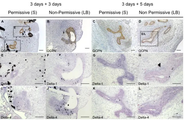
Here, our aim was to investigate the ligands of Notch signaling involved in thymic epithelium colonization. For that we developed quail-chick chimeras as previously described. Briefly, E3 quail 3/4 PPs endoderm was isolated and grafted into distinct mesenchymal territories, either in the somatopleural or the limb bud territory, of E2.5 chicken embryos. Due to the complex manipulation of grafting procedures, a slight delay in the endoderm development was observed in chimeric embryos. The chimeric embryos were allowed to develop for 3 or 5 days, stages corresponding to E5 and E7 of the grafted endoderm. The first wave of colonization by LPCs occurs in the middle of this time-window (at E6 in quail). We analyzed the expression of Notch ligands, Delta1 and Delta4, known to be expressed in the thymus and to support T-cell differentiation13,38,42. The grafted endoderm was identified by screening
paraffin sections of chimeric embryos stained by immunohistochemistry with QCPN (specific for quail cells) (Figures 5A-D). Sequential section slides were then used to analyze the expression of Foxn1, Delta1 and Delta4, by in situ hybridization. The neural tube expression of Delta1 and Delta4 of the chimeric embryos was used as an internal control. Controls for
Foxn1 expression were performed in parallel using sections with thymi of E13 chicken
embryos (data not shown).
In 3- and 5-days chimeric embryos, we observed Delta1 expression in the endoderm grafted in the permissive mesenchyme (n=1 for each condition; Figures 5E, 5G). In contrast, when grafted into the non-permissive limb bud mesenchymal territory, low or no expression of
Delta1 was observed in the developing endoderm (n=1 for each condition; Figures 5F, 5H).
Moreover, Delta1 expression was up-regulated in the limb bud mesenchyme in the vicinity of the grafted endoderm in chimeric embryos with 5 days of development (n=1; Figure 5H), but not in the somatopleural mesenchyme (Figures 5E, 5G).
The other Notch-ligand studied, Delta4, was strongly expressed in the endoderm grafted in either mesenchymal environments, both after 3 and 5 days of chimeric embryos development (n=1 for each condition; Figures 5I-L). No Delta4 expression was observed in the surrounding mesenchyme in any condition (Figures 5I-L).
We confirmed that Foxn1 is faintly expressed in the endoderm of the chimeric embryos analyzed, in both permissive and non-permissive environments (n=1 for each condition; data not shown).
Figure 5. Delta-1 and Delta-4 expression in the grafted quail endoderm and surrounding chick mesenchyme of chimeric embryos. Transverse sections of chicken host embryos after grafting quail E3
endoderm in the Somatopleural (A,E,I; C,G,K) or in the Limb Bud (B,F,J; D,H,L) mesenchyme of chicken E2.5. Chimeric embryos developed for 3 days (A,E,I; B,F,J) or 5 days (C,G,K; D,H,L). Sequential paraffin sections were stained with QCPN antibody (A-D), or processed for in situ hybridization with Delta1 probe (E-H) and Delta4 probe (I-L). (S: Somatopleural mesenchyme; LB: Limb Bud mesenchyme). Scale bars 20µm.
Together our data showed that Delta1, and not Delta4, is specifically down-regulated in the endoderm developed in the non-permissive mesenchymal territory (limb bud), where no LPCs colonization is observed, and up-regulated in the adjacent mesenchyme.
II. Formation of chicken pharyngeal organs: thymus and parathyroid glands
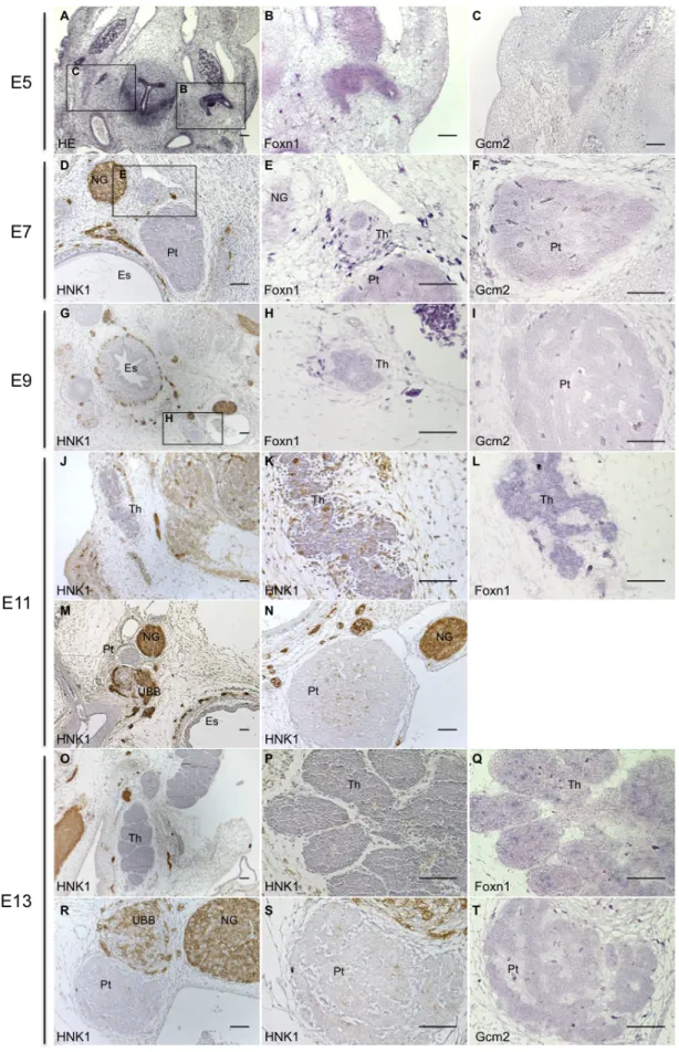
In chicken, the thymus and parathyroid glands share a common embryonic origin, the endoderm of the 3/4 PPs. Their development is initiated with the budding-off of the pouches, followed by their patterning into organ-specific domains. These domains can be identified by the expression of the specific transcription factors Foxn1 and Gcm2, for the thymus and parathyroid, respectively5. Afterwards, the rudiments detach from the pharynx and migrate to their final locations.We studied the development of these glands in chicken embryos from E5 onwards, by in situ hybridization technique. We analysed the expression of Foxn1 and Gcm2, in sequential slide sections of the neck region of chicken embryos.
First, we identified the area of interest (the glands rudiments) by Hematoxylin-Eosin (HE) staining in slide sections of E5 embryos (Figure 6A). The adjacent slides were then used to detect Foxn1 and Gcm2 expression. We observed that these genes were faintly expressed in the pouches (Figures 6B, 6C).
At later stages, organs primordia were identified by morphological analysis combined with HNK1 (Human Natural Killer 1) detection by immunohistochemistry. This marker is expressed in migrating neural crest cells and derivatives and in other cell types (like NK-cells) allowing a better determination of the area of interest (the glands rudiments)41. From E5 onwards, Foxn1 and Gcm2 were detected in the epithelia of the organs primordia, of the thymus and parathyroid glands, respectively, at all developmental stages studied (Figures 6B, 6E, 6H, 6L, 6Q and 6C, 6F, 6I and 6T).
As observed in Figures 6D-F, at E7, organs primordia were detached from the pharynx, separated from each other and near the nodose ganglion, as previously described2,5. Two days later (E9), the parathyroid glands had already a cordonal/tubular structure, characteristic of the mature organ (Figure 6I). The thymus formed a consistent cord-like structure, expressing Foxn1 transcripts in epithelial cells, intermixed with very few non-expressing large cells, probably corresponding to homing of hematopoietic progenitors cells (Figure 6H).
Figure 6. The expression of Foxn1 and Gcm2 in developing epithelia of the thymus and parathyroid glands in chicken embryos. Transverse sections of chicken embryos of E5 (A-C), E7 (D-F), E9 (G-I), E11 (J-N)
and E13 (O-T), stained with HE (A), HNK1 (D,G,J, K, M, N, O, P, R and S). B, E, H, L and Q were hybridized with
Foxn1 riboprobe; and C, F, I and T with Gcm2 riboprobe. (Es, esophagus; NG, nodose ganglion; Pt, parathyroid
At E11, the thymus was a tiny cord-like structure elongated along the neck of the embryos, and already lobulated, as seen in Figures 6J and 6K. Again, between the epithelial Foxn1 positive cells, we observed few negative cells, suggesting that there were still very few LPCs at this stage in the thymus (Figure 6L). As observed in Figures 6O-Q, the different thymic lobes at E13 were very lobulated and exhibit an increased number of Foxn1 negative cells when compared to E11. Moreover the separation between cortex and medulla was discernible at this stage of thymic development. At these two later stages, HNK1 staining in the thymus was observed (Figure 6J, 6K, 6O, 6P), suggesting the presence of Natural-Killer (NK)-cells.
Gcm2 expression was observed at E11 (data not shown) and at E13 (Figure 6T). At these
later stages, the parathyroid glands maintained their anatomical position, near the nodose ganglion and the ultimobranchial body (Figures 6M, 6R), and increased in size (data not shown).
With this study, we confirmed that at E7 the rudiments have separated from the pharynx and from each other. We observed that at E9 the thymus forms a consistent cord-like structure that starts elongating along the neck. At E11, it has already subdivided into several lobes, that two days latter had clear lobules. In contrast, the parathyroid glands, at E9, are already located at their final anatomical position exhibiting the morphology of the mature organ. Moreover, we observed Foxn1 and Gcm2 expression in thymic and parathyroid epithelia of the rudiments until E13, respectively.
III. Thymocytes population kinetics during chicken embryo development
During chicken embryonic development, the thymus is colonized by LPCs in cyclic waves, at E6.5, E12 and E1810,43,44. Once inside the thymus, the progenitors adopt T-cell lineage fate,
and undergo a sequence of developmental events leading to the generation of different functional lineages45. These developmental stages of thymocytes can be discriminated
according to surface expression of molecular markers.
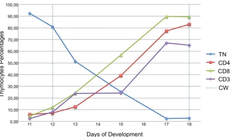
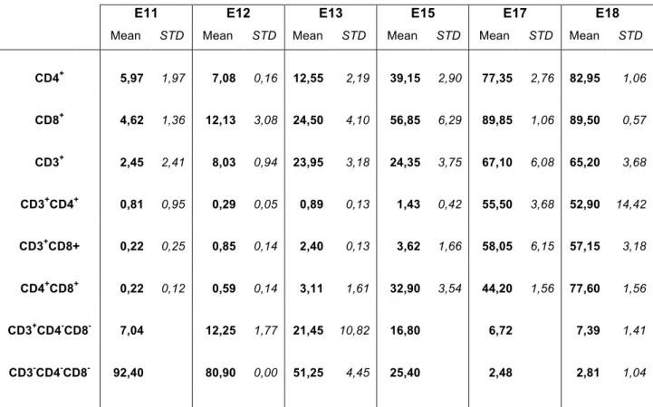
To study the dynamics of thymopoiesis, during chicken thymus organogenesis, we analysed the expression of the co-receptor CD3 and accessory molecules CD4 and CD8, by flow cytometry, using different antibodies combinations (Table 2 in Materials and Methods; dot blots representing the percentages of thymocytes co-expressing CD3 and CD4 or CD8 shown in Appendix III, Figure 11). The study was performed from E11 onwards, since very few hematopoietic cells could be harvested from the thymic rudiment at earlier stages, preventing a representative cytometry analysis. The results are presented in Figure 7 and Table 3, as percentages of thymocytes expressing the molecular markers studied.
At E11, after the first wave (at E6) and before the second wave (at E12)10 of colonization,
each of the three markers studied is expressed by a small percentage of cells. The percentage of thymocytes expressing CD8 showed a sharp increase from E13 onwards, while CD4+ cells presented a similar increase with a short delay (Figure 7: green and red curves, respectively). We observed that the percentage of CD3+ cells rose at two different
time-points, between E12 and E13 (up to 23.95%), and between E15 and E17 (up to 67.1%) (Figure 7: purple curve; Table 3).
At E18, the oldest time-point analyzed, 89.50% of the population expressed CD8, 82.95% expressed CD4 and 65.20% expressed the CD3 co-receptor (Table 3). As expected, the proportion of TN cells decreases during development, from more than 90% at E11 to less than 5% at E18 (Figure 7: blue curve).
Figure 7. Percentages of thymocytes expressing CD3, CD4 and CD8 during chicken thymus ontogeny.
Flow cytometry analysis of CD3, CD4 and CD8 expression in thymocytes harvested from E11, E12, E13, E15, E17 and E18 thymi. Triple Negative (TN) thymocytes (CD3-CD4-CD8-); and single staining for CD4, CD8 and CD3. CW: Colonization Waves (timing of the 2nd and 3rd colonization waves by LPCs at E12 and E18).
Distinct populations were discriminated according to CD3 expression levels, CD3low and
CD3high. Until E15, all CD3+ thymocytes had a low expression of CD3. In contrast, at later
stages of development (E17 and E18), when CD3 expression reaches its maximum (over 60%), 10-15% of the thymocytes presented a high level of CD3 expression (Figure 8).
Figure 8. Percentages of thymocytes expressing different CD3 levels at E17 and E18. In red the unstained
thymocytes curve, and in blue the CD3-PE staining.
Moreover, between E13 and E15, we observed an increase of DP thymocytes, CD4+CD8+, from 3.11% to 32.90%. In the case of double positive populations for CD4+CD3+ or
CD8+CD3+ an increase in cells percentage was observed between E15 (less than 5%) and E17 (above 50%) (Table 3).
E17 E18 CD3 Co u n t Unstained CD3-PE staining
Table 3. Percentages in thymocytes population expressing CD3, CD4 and CD8.
E11 E12 E13 E15 E17 E18
Mean STD Mean STD Mean STD Mean STD Mean STD Mean STD
CD4+ 5,97 1,97 7,08 0,16 12,55 2,19 39,15 2,90 77,35 2,76 82,95 1,06 CD8+ 4,62 1,36 12,13 3,08 24,50 4,10 56,85 6,29 89,85 1,06 89,50 0,57 CD3+ 2,45 2,41 8,03 0,94 23,95 3,18 24,35 3,75 67,10 6,08 65,20 3,68 CD3+CD4+ 0,81 0,95 0,29 0,05 0,89 0,13 1,43 0,42 55,50 3,68 52,90 14,42 CD3+CD8+ 0,22 0,25 0,85 0,14 2,40 0,13 3,62 1,66 58,05 6,15 57,15 3,18 CD4+CD8+ 0,22 0,12 0,59 0,14 3,11 1,61 32,90 3,54 44,20 1,56 77,60 1,56 CD3+CD4-CD8- 7,04 12,25 1,77 21,45 10,82 16,80 6,72 7,39 1,41 CD3-CD4-CD8- 92,40 80,90 0,00 51,25 4,45 25,40 2,48 2,81 1,04 Percentages of thymocytes expressing CD3, CD4 and CD8 at all stages analyzed (STD: standard deviation).
In summary, before E13 we observed two immature thymocyte populations (CD8+CD4-CD3 -and CD3+CD8-CD4+) derived from the first wave of colonization. After E13, the percentage of
CD4+CD8+ (DP) cells increases. Moreover, at E17 and E18, a small percentage of CD3+ thymocytes expressed high levels of CD3 co-receptor.
Discussion
Understanding the steps that lead to the development of a functional thymus, a complex process crucial for correct thymopoiesis, is of major importance to understand the building of a healthy immune system.
In this work, the first aim was to study the molecular cues involved in TE colonization by LPCs using the quail-chick chimeric model. Specifically, we studied the involvement of distinct Notch-ligands in this process. We showed that, in conditions without TE colonization by LPCs, Delta1, and not Delta4, was down-regulated in the epithelium.
Secondly, we described histologically the development of the thymus and parathyroid glands by in situ expression of the transcription factors, Foxn1 and Gcm2, respectively. We observed that, from E5 to E13, the expression of both transcription factors was maintained in the thymic and parathyroid rudiments.
Finally, we analysed by flow cytometry the maturation of the population of thymocytes during chicken thymus organogenesis. The analysis showed that at early stages (before E13) of thymic development, two immature populations emerged (CD8+CD4-CD3- and CD3+CD8
-CD4-), while after E13 a strong increase of a more mature population of DP cells (CD4+CD8+) was observed. This shift corresponds to the timing of the second colonization
wave and the establishment of cortical and medullar compartments.
I. Delta-1 ligand is involved in early colonization of the thymic rudiment by
LPCs
The colonization of thymic epithelium is a crucial and early event in thymus organogenesis. Recent results from our lab, using the quail-chick chimeric model, showed that 3/4 PPs endoderm when developed in a non-permissive environment was able to specify into TE (Foxn1+), but not to be colonized by LPCs5. In this study, we used this avian chimeric model
approach to identify the molecular cues involved in thymic epithelium colonization. We identified Delta1, as the specific Notch-ligand of the Delta-family involved in this process.
Delta1, but not Delta4, was down-regulated in the endoderm grown in the non-permissive
environment, whereas in the endoderm grown in the permissive environment, where LPCs colonization occurred, Delta1 expression was maintained (Figure 9).
Figure 9. Delta1 expression in the endoderm and surrounding mesenchyme in chimeras developed for 5 days. Schematic representation of Delta1 expression in chimeric embryos developed for further 5 days.
In agreement, expression of Delta1 was reported in the cortico-medullary junction of adult mice thymus, the entry region of LPCs. Other reports showed that in vitro expression of
Delta1 in stromal cells induces LPCs commitment to T-lineage13. Together, these data
suggest that Delta1 ligand expressed by TECs might be necessary to a successful seeding of LPCs when entering the thymus.
However, we cannot exclude that other Notch ligands of the Serrate family may also be involved in this event. In fact, we observed the expression of Serrate-1 in the 3rd PP
endoderm at E3 and E4, and of Serrate-2 in the mesenchyme surrounding the 3rd PP (3rd
PA) (unpublished data), suggesting that these ligands may be involved in early stages of thymus organogenesis.
In the 5-days post-grafting chimeric embryos, we observed that the down-regulation of
Delta1 in the developing endoderm was accompanied by its up-regulation in the surrounding
non-permissive mesenchyme (limb bud). This ectopic expression of Delta1 in the neighboring mesenchyme further suggests distinct epithelial-mesenchymal interactions that might prevent thymic colonization. This interaction (between TE - Delta1- with non-permissive mesenchyme - Delta1+) may prevent either thymus vascularization and/or expression of
chemokines involved in directing LPCs migration towards the thymic rudiment12,26.
In the future, it will be of interest to study TE vascularization in both environments, and to analyze the expression of other molecular cues that could be involved in the process, such as Ephrin receptors, vascular endothelial growth factor families and other ligands of Notch signaling, and chemokines. Furthermore, the presence of LPCs in the surrounding mesenchyme should be assessed to help understand the possible role of chemokines and vascularization.
It is noteworthy that these results should be confirmed by the analysis of more experimental cases. The impossibility to show more cases was due to the time-consuming intensive train