R E S E A R C H A R T I C L E
Open Access
Optimization of heterologous protein production in
Chinese hamster ovary cells under overexpression
of spliced form of human X-box binding protein
Galina Gulis
*, Kelly Cristina Rodrigues Simi, Renata Rodrigues de Toledo, Andrea Queiroz Maranhao
and Marcelo Macedo Brigido
Abstract
Background:The optimization of protein production is a complex and challenging problem in biotechnology. Different techniques for transcription, translation engineering and the optimization of cell culture conditions have been used to improve protein secretion, but there remain many open problems involving post-translational modifications of the secreted protein and cell line stability.
Results:In this work, we focus on the regulation of secreted protein specific productivity (using a recombinant human immunoglobulin G (IgG)) by controlling the expression of the spliced form of human X-box binding protein (XBP-(s)) in Chinese hamster ovary cells (CHO-K1) under doxycycline (DOX) induction at different temperatures. We observed a four-fold increase in specific IgG productivity by CHO cells under elevated concentrations of DOX at 30°C compared to 37°C, without detectable differences in binding activityin vitroor changes in the structural integrity of IgG. In addition, we found a correlation between the overexpression of human XBP-1(s) (and, as a consequence, endoplasmic reticulum (ER) size expansion) and the specific IgG productivity under DOX induction.
Conclusions:Our data suggest the T-REx system overexpressing human XBP-1(s) can be successfully used in CHO-K1 cells for human immunoglobulin production.
Keywords:CHO cells, Heterologous protein production, X-box binding protein, T-REx™system, Doxycycline
Background
The optimization of the production of secreted proteins, such as therapeutic monoclonal antibodies (mAbs), is still a challenging problem in pharmaceutical biotech-nology. Although biopharmaceutical products can be produced by many host cell systems, eukaryotic cells are preferred due to their ability to correctly process and modify human proteins. The primary goal is to establish the ideal combination of a rapid accumulation of pro-ductive biomass and the maintenance of cell viability for as long as possible. Many different strategies have been considered for improving both cell viability and the productivity of recombinant proteins, including mAbs. These strategies include physiological optimization and genetic and metabolic engineering [1,2].
The most common problem during the optimization of protein production is an error in protein folding in the endoplasmic reticulum (ER). The inhibition of pro-tein folding activates the unfolded propro-tein response (UPR), which is a signal transduction network. Over-coming UPR is one of the many strategies for optimizing protein productivity. For instance, protein production has been tested under the expression of survival proteins that play important roles in UPR, including B-cell lymphoma protein 2 (bcl-2), B-cell lymphoma-extra-large protein (bcl-XL) [3-5], caspase inhibitors [6] and molecular chaperones/heat shock proteins (HSP70) [7]. The role of the spliced form of X-box binding protein (XBP-1(s)) (which plays an important role in regulation processes, such as physical expansion of the ER, increas-ing the mitochondrial mass and function, increasincreas-ing the cell size and enhancing total protein synthesis) in opti-mizing protein production has also been studied [8]. * Correspondence:[email protected]
Institute of Biological Sciences, Department of Cell Biology, University of Brasilia, Campus Universitário Darcy Ribeiro, Brasília, DF 70910-900, Brazil
This approach to increasing the secretion capacity of mammalian cells by overexpressing the transcription factor XBP-1(s) was successful in CHO cells; the pro-duction of the secreted proteins alkaline phosphatase (SEAP) and alpha-amylase (SAMY) was enhanced upon XBP-1(s) overexpression [9], as was the production of antibody [10]. However, these studies have shown that using overexpression systems without regulation leads to cell apoptosis due to the accumulation of the produced proteins [11].
To overcome accumulation-induced apoptosis, other strategies have been applied to regulate the protein production, such as the use of induction systems. For instance, tetracycline has been used to optimize the overexpression of glycosyltransferases under the control of the Tet on/off system in CHO cells, but unfortunately, a high expression of glycosyltransferases still led to growth inhibition [12]. Furthermore, interesting work using the same expression system has been conducted to control the overexpression of human transferrin (hTf) in human embryonic kidney (HEK-293) cells. That study found fa-vorable concentrations of tetracycline at which the overex-pression of hTf was optimal, but again, the high levels of expression limited the cell viability. Such impairment might have been a consequence of the overexpression of the protein of interest, which might have altered the qual-ity of this cell product or even been toxic to the cells [13]. Some studies have attempted to investigate the effect of the expression of an ER-resident molecular chaperone, protein disulfide isomerase (PDI), on the specific produc-tion levels of thrombopoietin (TPO) and antibody (Ab) in Chinese hamster ovary cells. Mohan and colleagues used the Tet-off system (in the absence of tetracycline) to regu-late PDI, TPO and Ab expression in CHO cells under doxycycline (DOX; a chemical analogue of tetracycline) induction. However, only a small increase in antibody production was observed, and the production of TPO was not affected by PDI expression [14].
Moreover, the optimization of protein production in CHO cells cultured at different temperatures has been addressed. For instance, lowering the temperature from 37°C to 33°C increased the production of erythropoietin (EPO) by approximately four-fold, but at the same time, a low cultivation temperature suppressed cell growth [15]. In addition, a temperature reduction from 37°C to 33°C in the culture of a CHO cell line producing recombinant human granulocyte/macrophage colony-stimulating fac-tor (CHO-K1-hGM-CSF) led to a reduced growth rate, increased cell viability, improved cellular protein pro-duction and decreased cell metabolism [16]. One study on the optimization of protein production at 32°C also demonstrated that the specific growth rate of CHO cells producing human mAb decreased by 30–63% at 32°C compared to 37°C. However, the specific antibody
productivity of these cells was significantly enhanced at 32°C [17]. Lowering the cultivation temperature even more, from 37°C to 30°C, caused growth arrest associated with a 1.7-fold increase in the specific production of se-creted alkaline phosphatase (SEAP) in CHO cells [18].
In this context, we attempted to optimize the specific IgG productivity under different culture temperatures and by regulating the overexpression of apoptotic human protein XBP-1(s) using the T-REx™ system (Invitrogen, Carlsbad, CA, USA). The applied T-REx™ system con-tains a regulatory plasmid (pcDNA6/TR), which encodes the tetracycline repressor, and an inducible expression plasmid (pcDNA™4/TO/myc-His A) with a tetracycline inductor for expression of the gene of the interest ( xbp-1(s)). Co-transfected together, these plasmids created a network to regulate XBP-1(s) expression under DOX induction. We cloned xbp-1(s)into the T-REx™ system to control its expression with DOX. Then, we trans-fected the obtained T-REx™-XBP-1(s) system into stably IgG-producing CHO cells and selected stable clones of this system expressing IgG-T-REx-XBP-1(s) to control specific IgG productivity under DOX induction (Figure 1). We determined the optimal concentration of DOX and the temperature at which IgG-T-REx-XBP-1(s) cells pro-duced the maximal amount of IgG without a significant inhibition of cell growth. Moreover, cells treated with DOX for seven days recovered viable cell density to the level of non-treated cells after DOX was washed out from the cell system, and their specific IgG productivity dropped to the basal level. Furthermore, we studied the dependence of spe-cific IgG productivity and viable cell density on the overex-pression of XBP-1(s) and ER size expansion.
Methods
Cell lines and media
The CHO-K1 (ATCC®CCL-61™) and Raji (ATCC®CCL-86™) cell lines were purchased from American Type Culture Collection (ATCC, Manassas, VA, USA). CHO-K1 cells were grown and maintained at 37°C or 30°C
with 70% humidity and 5% CO2 in HAM F12 media
(Gibco, Big Cabin, OK, USA) supplemented with 2% fetal bovine serum (FBS, Gibco, Big Cabin, OK, USA) and were used in experiments on protein production. Raji cells were grown and maintained at 37°C with70%
humidity and 5% CO2 in RAMP media (Gibco, Big
Cabin, OK, USA) supplemented with 10% FBS and were used in FACS direct ligation experiments.
Plasmids and cloning
(anti-CD20)-producing cells. The human xbp-1(s) coding sequence was chemically synthesized by GeneScript (Piscat-away, NJ, USA). The restriction enzymes Hind III and
BamH I (Fermentas, Ontario, Canada) were used to obtain the xbp-1(s) insert and then clone it into the inducible expression plasmid pcDNA™4/TO/myc-His A from the Invitrogen T-REx™system (Invitrogen, Carlsbad, CA, USA). This plasmid was used to co-transfect IgG-producing stable clones of CHO cells along with the regulatory plasmid pcDNA6/TR (Invitrogen, Carlsbad, CA, USA). To confirm
xbp-1(s) cloning, XL1-blue bacterial cells (Stratagene, La Jolla, CA, USA) were transformed with ligated DNA. Ampicillin (Sigma, Ronkonkoma, NY, USA)-selected colonies were isolated and processed for DNA extrac-tion and purificaextrac-tion, which was performed using a QIAprep Miniprep Kit (Qiagen, Valencia, CA, USA). Restriction analysis and sequencing (using CMV
for-ward primer 5′-CGCAAATGGGCGGTAGGCGTG-3′
and BGH reverse primer 5′-TAGAAGGCACAGTCG
AGG-3′) confirmed the cloning of thexbp-1(s)insert.
Transfection with pCOMIRES anti-CD20 DNA (IgG-encoding plasmid) into CHO cells and generation of stable IgG-producing cells
The transfection of pCOMIRES HIL anti-CD20 plasmid (encoding an anti-CD 20 (IgG) antibody, a secretable
protein with molecular weight 150 kDa (two light chains, each with molecular weight 25 kDa, and two heavy chains, each with molecular weight 50 kDa)) into CHO cells was performed using a PolyPlus (JetPrime, New York, NY, USA) kit in six-well test plates (TPP, San Diego, CA, USA) according to the manufacturer’s instructions. The clones harboring the pCOMIRES HIL anti-CD20 transgene were selected from a mixed popula-tion by the single-cell dilupopula-tion method. Geneticin (Roche, Gaillard, France) was used for selection at 800μg/mL.
Transfection with the T-REx™-XBP-1(s) system into stable IgG-producing clones of CHO cells and generation of stable double clones (IgG-T-REx-XBP-1(s) cells)
The co-transfection of T-REx-xbp-1(s)plasmid (encoding a spliced form of human apoptotic XBP-1 protein with pre-dicted molecular weight 40 kDa) along with regulatory plasmid pcDNA6/TR into one of the stable IgG-producing clones was performed using a PolyPlus (JetPrime, New York, NY, USA) kit according to the manufacturer’s instructions in six-well test plates (TPP, San Diego, CA, USA). Blasticidin (Sigma, Ronkonkoma, NY, USA) and Zeocin (Sigma, Ronkonkoma, NY, USA) were added to a final concentration of 0.5 μg/mL and 50 μg/mL,
respectively. The selective markers encoded by regulatory plasmid pcDNA6/TR and expression plasmid pcDNA™4/
TO/myc-His A are against blasticidin and Zeocin, respectively.
Doxycycline induction
Selected IgG-T-REx-XBP-1(s) cells (after the first trans-fection, IgG clones; after the second, co-transtrans-fection, T-REx-XBP-1(s) clones) were induced by DOX at differ-ent concdiffer-entrations: 0 μg/mL for control, 0.1 μg/mL,
0.5 μg/mL and 1μg/mL. We used these concentrations
because we found out that 5 μg/ml and 7.5 μg/ml of
doxycycline completely inhibits cells growth for clones and wild type CHO-K1 cells. DOX induction was per-formed 24 hr after IgG-T-REx-XBP-1(s) cells seeding at a uniform cell density (0.5 × 10⁵cells/mL) in tissue culture flasks (75 cm2, TPP, San Diego, CA, USA) and then incu-bated for seven days at 37°C or 30°C. All cultures reached at least 80% under these conditions. Samples were col-lected for viable cell density, Ab detection by ELISA, nuclear extract isolation and ER staining. Half of the cells in each group continued to grow for seven more days in DOX-free medium after DOX wash-out. Inde-pendently, IgG-T-REx-XBP-1(s) cells were incubated for 42 days at 30°C (150 cm2 flasks, TPP, San Diego, CA, USA) with or without 1μg/mL DOX. In all DOX
induc-tion experiments, DOX was added (at an appropriate concentration) every three days to the cell culture. Induction experiments were performed twice in dupli-cate (four independent culture samples per group).
Viability assay
The viable cell density of the IgG-T-REx-XBP-1(s) cells were tested under different DOX concentrations (0 μg/
mL, 0.1μg/mL, 0.5μg/mL or 1μg/mL) every day during
seven days of cell growth at 37°C and 30°C. Seeding was performed at a uniform cell density (0.06 × 10⁵ cells/ mL) in six-well tissue culture plates (TPP, San Diego, CA, USA). At the seventh day all cultures reached at least 80% under these conditions. In addition, the viable cell density of IgG-T-REx-XBP-1(s) cells was tested on the seventh day of growth with DOX and on the seventh day after wash-out in DOX-free medium. In addition, IgG-T-REx-XBP-1(s) cells were tested every seventh day during 42 days of cell growth under 1 μg/mL DOX (or
0 μg/mL as control) at 30°C. The viable cell density was
measured using the trypan blue (Sigma, Ronkonkoma, NY, USA) exclusion method with a hemocytometer and light microscope for manual cell counting. Every viable cell density experiment was performed twice in duplicate (single determination from each of two independent cul-ture samples per group in two independent experiments).
ELISA
The supernatants of IgG-T-REx-XBP-1(s) cells in the pres-ence or abspres-ence of DOX were collected every seventh day
of 37°C or 30°C growth for two weeks or every seventh day for six weeks and processed for analysis by enzyme-linked immunosorbent assay (ELISA) (duplicate deter-mination from each of two independent culture samples per group in two independent experiments). The Lunc/ Maxisorp Immunoplate (Thermo Scientific, Waltham, MA, USA) was incubated with primary antibody (goat anti-human IgG (H + L), 1:3000 dilution; Thermo Scien-tific, Waltham, MA, USA) and blocked with 3% fat-free dehydrated milk solution. After blocking and washing the plate, the supernatants were applied to the plate and incubated for 2 hr. The plate was washed again, and secondary antibody (anti-human IgG Fc-specific, alka-line phosphatase-conjugated, produced in goat, 1:3000 dilution; Sigma, Ronkonkoma, NY, USA) was applied for 1 hr. The plate was washed again, and at the end of the procedure, the signal of absorbance was read at 405 nm by a microplate reader (ELx800 96-well Micro-plate Reader, MTX Lab Systems, Inc., Vienna, VA, USA) after 4-Nitrophenyl phosphate disodium salt solution (pNPP) (Invitrogen, Carlsbad, CA, USA) addition. In addition, human IgG (whole molecule; Thermo Scien-tific, Waltham, MA, USA) was used in different concen-trations as a control on the same plate.
Isolation and purification of produced proteins
Western blotting
Anti-CD20 antibody was also detected by western blot-ting. Five hundred nanograms of IgG sample was loaded in each well of a Bis-Tris gel (NuPAGE® Novex 4-12% Bis-Tris Gel, Invitrogen, Carlsbad, CA, USA) and sepa-rated by sodium dodecyl sulfate–polyacrylamide gel electrophoresis (SDS-PAGE) according to the manu-facturer’s instructions. The proteins were transferred to the Hybond-C Extra nitrocellulose membrane (Amersham® Bioscience, Piscataway, NJ, USA) and blocked in 3% fat-free milk PBS solution. The immunodetection was performed using anti-human IgG (Fc-specific, alka-line phosphatase-conjugated, produced in goat (1:3000 dilution) (Sigma, Ronkonkoma, NY, USA) with a BCIP/ NBT substrate Kit™(Invitrogen, Carlsbad, CA, USA).
XBP-1(s) was also probed by western blotting. The nu-clear extracts from the IgG-T-REx-XBP-1(s) cells were prepared as described by Becker and colleagues [10]. Briefly, the nuclear extracts were prepared from 5×10⁶ cells/per sample and equal volumes of nuclear extracts were loaded into a Bis-Tris gel (NuPAGE® Novex 4-12% Bis-Tris Gel, Invitrogen, Carlsbad, CA, USA), and SDS-PAGE was performed according to the manufacturer’s instructions. Samples were transferred to the Hybond-C Extra nitrocellulose membrane (Amersham® Bioscience, Piscataway, NJ, USA), and after blocking with 3% fat-free milk PBS solution, rabbit anti-human-XBP-1(s) (1:1000 dilution; Sigma, Ronkonkoma, NY, USA) was added, followed by alkaline phosphatase-conjugated anti-rabbit IgG incubation (1:1000 dilution; Sigma, Ronkonkoma, NY, USA). The proteins were revealed using a BCIP/NBT substrate Kit™(Invitrogen, Carlsbad, CA, USA).
Fluorescence-activated cell sorting (FACS)-ER staining The IgG-T-REx-XBP-1(s) cells that were grown for seven days under DOX induction and those that were grown for one more week after wash-out were collected at 3×10⁵ cells/per staining and washed with HBSS buffer
(140 mM NaCl, 4.7 mM KCl, 1 mM MgCl2, 1.5 mM
CaCl2, 10 mM glucose, 10 mM HEPES, pH 7.4). After
washing with HBSS buffer, the cells were labeled with 250 nM of ER-Tracker™Green Dye (ER-Tracker™Green Dye for Live-Cell Endoplasmic Reticulum, Molecular Probes, Invitrogen, Carlsbad, CA, USA) according to the manufacturer’s manual. The samples were washed again with HBSS buffer and analyzed using a BD FACS Verse flow cytometer (BD Bioscience, San Jose, CA, USA). Ten thousand events were collected per sample using no gate for acquisition. The dead cells were not excluded in the analysis. We used BD FACSuite to data acquisition. The experiment was performed twice in duplicate.
FACS direct ligation assay
Raji cells were grown for five passages as described above, collected and resuspended in 1 part RAMP media with 10% FBS and 1 part FACS buffer (PBS supple-mented with 2% FBS) at 3×10⁶ cells/well in a 96-well plate (TPP, San Diego, CA, USA). After centrifugation, the cells were blocked with FcR blocking reagent (MACS, Biotec, Bergisch Gladbach, Germany) on ice for 30 minutes according to the manufacturer’s instructions. Purified and dialyzed IgG samples, which were produced by IgG-T-REx-XBP-1(s) cells at 37°C and 30°C, and com-mercial IgG (rituximab, MabThera, Genetech Inc., South San Francisco, CA, USA) as a positive control were added at 100 ng per well. Samples were incubated on ice for 1 hr and centrifuged after the addition of FACS buf-fer. The cells were washed twice with FACS buffer and incubated with mouse FITC anti-human IgG (BD Phar-mingen™, BD Biosciences, San Jose, CA, USA) according to the manufacturer’s manual. The cells were incubated on ice for 30 minutes in the dark, washed again twice with FACS buffer and processed for fluorescence inten-sity measurements using a BD FACS Verse flow cyt-ometer (BD Bioscience, San Jose, CA, USA). Each experiment was performed twice in duplicate.
Results and discussion
Viability and IgG production under induction with DOX in IgG-T-REx-XBP-1(s) cells cultivated at 37°C and 30°C To establish a DOX-regulated XBP-1(s) cell line, we first created stably IgG-producing clones of CHO cells by transfecting the pCOMIRES HIL anti-CD20 plasmid into CHO-K1 cells. Then, IgG-CHO clones selected with 800 μg/mL of Geneticin were co-transfected with the
T-REx-XBP-1(s) system and processed for second selec-tion using 0.5 μg/mL of blasticidin and 50 μg/mL of
Zeocin. From these double clones harboring both pCO-MIRES HIL anti-CD20 and T-REx-XBP-1(s) system plas-mids, we chose one out of 20 for DOX induction at different concentrations (0 μg/mL (control), 0.1μg/mL,
0.5μg/mL or 1μg/mL) and grew them at 37°C or 30°C.
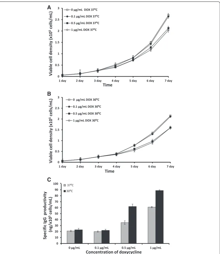
Every day, cells were collected to monitor viable cell density. We found that the viable cell density of the IgG-T-REx-XBP-1(s) cells grown at 37°C under 1μg/mL
or 0.5 μg/mL DOX was slightly lower compared to the
control and to the cells with 0.1 μg/mL DOX induction
(Figure 2A). Moreover, the viable cell density of IgG-T-REx-XBP-1(s) cells grown at 37°C without DOX or with 0.1 μg/mL DOX was 1.25-fold higher compared to the
cells grown at 30°C (Figure 2A and 2B). These data agree with several previous studies [15-18]. Moreover, the viable cell density of IgG-T-REx-XBP-1(s) cells grown at 37°C under 1 μg/mL DOX induction was increased by
IgG-T-REx-XBP-1(s) cells grew more slowly at 30°C with or without DOX compared to those at 37°C.
The supernatants from all cells were collected after seven days of induction and tested by ELISA to determine
their IgG yields. The specific IgG productivity depended on the concentration of DOX: under 0.5μg/mL and 1μg/
mL DOX, the increase of specific IgG productivity was 40% and 66%, respectively, compared to the basal level of
A
B
C
Figure 2Viable cell density of IgG-T-Rex-XBP-1(s) cells cultured at 37°C (A) or 30°C (B) and their specific IgG productivity at 37°C and 30°C (C) under induction with 0μg/mL (control), 0.1μg/mL, 0.5μg/mL and 1μg/mL DOX.Error bars represent the standard deviation of
specific IgG productivity (0 or 0.1 μg/mL DOX) at 37°C
(Figure 2C). These data demonstrate that IgG-T-REx-XBP-1(s) cells produced three-fold more IgG compared to un-treated cells, even at low viable cell density. At 0.1μg/mL
DOX, there was no improvement in specific IgG productiv-ity at 37°C. Moreover, the data from ELISA indicate that protein production in the cells incubated at 30°C increased four-fold and three-fold under 1 μg/mL and 0.5 μg/mL
DOX, respectively. Once more, induction at a low concen-tration of DOX (0.1 μg/mL) did not increase specific IgG
productivity at 30°C, as above at 37°C. In contrast, the specific IgG productivity by IgG-T-REx-XBP-1(s) cells at 30°C increased by 31.5% and 43.5% compared to in-duction at 37°C under 1 μg/mL and 0.5 μg/mL DOX
concentrations, respectively (Figure 2C). However, we did not detect any effect of low temperature on specific IgG productivityper se(without the induction of DOX). Tigges and Fussenegger [9] reported the same lack of effect in CHO cells expressing SEAP, whereas other authors reported an increase in the production of differ-ent proteins at low temperature and with no inductor [15-18]. These deviations in experimental results may be due to differences in the proteins and cell lines used in these studies. In conclusion, our data demonstrate a successful improvement of specific IgG productivity using 1μg/mL DOX in IgG-T-REx-XBP-1(s) cells at 30°C.
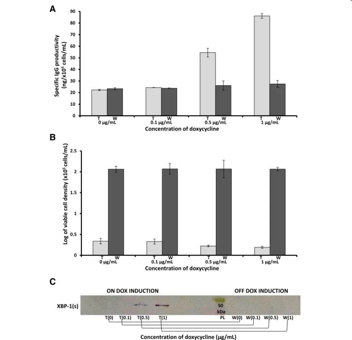
Effect of XBP-1(s) expression and ER size expansion on protein production in IgG-T-REx-XBP-1(s) cells
To test the hypothesis that XBP-1(s) expression and ER size expansion indirectly regulate protein production, IgG-T-REx-XBP-1(s) cells were incubated under different concentrations of DOX for seven days at 30°C, and then the same cells were washed with DOX-free medium and incubated for seven more days at the same temperature in fresh DOX-free medium. The supernatant was collected before wash-out and after seven days of incubation in DOX-free medium and processed for ELISA. ELISA showed that specific IgG productivity by cells with DOX (first seven days) increased in a DOX concentration-dependent manner. The specific IgG productivity by cells
incubated with 1 μg/mL DOX and 0.5 μg/mL DOX
reached four-fold and three-fold that of the untreated cells (treated (t), Figure 3A). In addition, ELISA demonstrated that after DOX removal, the specific IgG productivity returned to the basal level (washed, (w) Figure 3A). More-over, viability analysis indicated that the growth of the IgG-T-REx-XBP-1(s) cells under different concentrations of DOX was slightly inhibited (first seven days, treated (t), Figure 3B) and then restored to the same level (washed (w), Figure 3B) as cells that had never been exposed to DOX (0 μg/mL (t or w), Figure 3B). In addition,
IgG-T-REx-XBP-1(s) cells were used to prepare nuclear extracts, which were analyzed by western blotting for the immune
detection of XBP (s). Human XBP-1 (s) was overexpressed in a DOX concentration-dependent manner (first seven days, on DOX induction, treated (t) Figure 3C), but it was absent in cells that were washed and incubated in DOX-free medium for seven days (last seven days, off DOX induction, washed (w), Figure 3C).
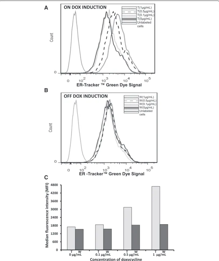
ER expansion was also observed by flow cytometry. The fluorescence change of ER-Tracker™ was used as a measure of ER size expansion. In this analysis, cell staining was more intense in samples treated with 1μg/
mL or 0.5 μg/mL DOX for seven days (on DOX
induc-tion, treated (t), Figure 4A) than in those treated with 0.1 μg/mL (on DOX induction, treated (t), Figure 4A)
or 0 μg/mL or those that were washed out (w),
Figure 4B). Moreover, the signal from the washed out cells was equal among different conditions (off DOX induction, washed (w), Figure 4B), which indicated that DOX was responsible for a cascade of processes leading to ER size expansion. In addition, measurements of the median fluorescence intensity (MFI, Figure 4C) obtained from FACS analysis showed that the MFI of the cells treated with 1μg/mL or 0.5μg/mL DOX was 2.7-fold or
1.85-fold higher, respectively, than the MFI of non-treated cells or washed out cells (Figure 4C). Thus, cells under DOX induction and low temperature grew more slowly but, at the same time, exhibited a greater increase in specific IgG productivity. Our data also demonstrate that the wash-out of DOX from the cells restored their viable cell density but reduced their specific IgG prod-uctivity to basal levels. Taken together, our results indi-cate the optimal conditions for specific IgG productivity under DOX induction. XBP-1(s) was overexpressed under induction with DOX, which led to the ER size expansion, and this resulted in an increase of specific IgG productivity/secretion. Our findings corroborate the data obtained by Tigges and Fussenegger, who reported an increased production of SEAP and SAMY under the expression of XBP-1(s) and the expansion of ER and Golgi [9].
Binding activity of the recombinant proteins produced at different temperatures
of IgG produced at different temperatures (Figure 5B). These data support the use of low-temperature culture conditions under induction by DOX to increase protein production without eliminating the binding activity and structural integrity of the protein of interest.
Establishing a stable protein-producing cell line
To establish a stable cell line, IgG-T-REx-XBP-1(s) cells were seeded and grown at 30°C under 1μg/mL DOX (or
0 μg/mL DOX as control) for 42 days. The supernatant
was collected every seventh day and submitted to ELISA, which showed stable specific IgG productivity around an average of 80.2 ng/105cells/mL by cells incubated with
DOX compared to the basal level (25.7 ng/105cells/mL in cells without DOX) (Figure 6A). Moreover, the viable cell density of IgG-T-REx-XBP-1(s) cells under 1 μg/mL
DOX was lower than cells without DOX for 42 days (Figure 6B), which allowed cells to be kept in the same
A
B
C
A
B
C
Figure 4FACS analysis.The samples were run through the flow cytometer until 10.000 events were collected using no gate. The IgG-T-Rex-XBP-1(s) cells were grown under induction with 0μg/mL, 0.1μg/mL, 0.5μg/mL or 1μg/mL DOX (on DOX induction, treated (t)) for seven days at 30°C. Then,
culture flask without passaging to new flasks. The de-scribed approach might be useful for the production of recombinant secreted proteins, as cells growing under the combination of DOX induction and low temperature multiply more slowly but are more productive.
Conclusion
Many studies have been published on improving recom-binant protein production. In general, the published data suggest that the optimization of the production of spe-cific target proteins requires spespe-cifically adjusted growth
A
B
Figure 5FACS and Western analyses.Median fluorescence intensity measurements of mouse FITC anti-human IgG ligated to the tested IgG (produced by IgG-T-REx-XBP-1(s) cells under DOX induction at 37°C and 30°C (and rituximab, control) that were previously incubated with Raji cells)(A). Western blot analysis of IgG produced by IgG-T-REx-XBP-1(s) cells at 37°C and 30°C under DOX induction (PL, protein ladder)(B).
A
B
Figure 6Specific IgG productivity (A) and viable cell density (B) of the IgG-T-REx-XBP-1(s) cells under 1μg/mL DOX induction (0μg/mL
conditions and a carefully chosen cell line. In the present study, we optimized the conditions for IgG (human anti-CD20) specific productivity in CHO-K1 cells. We showed that the combination of low temperature (30°C) and XBP-1(s) overexpression regulated by DOX induction significantly improved anti-CD20 specific productivity: under 1μg/mL DOX treatment, specific IgG
productiv-ity was increased by 32% compared to the cells grown under the same concentration at 37°C and 74% compared to the cells grown without DOX induction at 37°C or 30°C. Moreover, the results of our study indicate the direct dependence of specific IgG productivity on the concen-tration of DOX (under 0.5μg/mL, the increase was
2.7-fold, and under 1 μg/mL, the increase was 3.9-fold),
which allows for the precise regulation of specific IgG productivity in CHO-K1 cells. In addition to the con-centration dependence, we demonstrated the possibility of returning the specific IgG over productivity to the basal level of specific productivity by removing DOX. This step also restored the viable cell density, which permitted the cells to overcome the problem of accu-mulation of the target protein. In the production of pro-teins, it may be possible to use the T-Rex-XBP-1(s) system to turn up and down the production of protein, repeating this cycle several times to accumulate higher amounts of target protein without a loss of cell viability. We also observed a DOX concentration-dependent relationship involving XBP-1(s) overexpression (western analysis), ER size expansion (FACS measurements) and specific IgG productivity (ELISA). Finally, our data dem-onstrate that it is possible, under DOX induction at low temperature, to produce a target protein for an extended period of time. Taken together, our data suggest the T-REx-XBP-1(s) system can be used in CHO-K1 cells for human immunoglobulin production.
Abbreviations
Ab:Antibody; bcl-2: B-cell lymphoma protein 2; bcl-XL: B-cell lymphoma-extra-large protein; CHO-K1: Chinese hamster ovary cells; CHO-K1-hGM-CSF: CHO cell line producing recombinant human granulocyte/ macrophage colony-stimulating factor; DOX: Doxycycline; ELISA: Enzyme-linked immunosorbent assay; EPO: Erythropoietin; ER: Endoplasmic reticulum; FACS: Fluorescence-activated cell sorting; HEK-293: Human embryonic kidney cells 293; HSP70: Heat shock proteins 70; hTf: Human transferrin; IgG: Human immunoglobulin G; mAbs: Monoclonal antibodies; MFI: Median fluorescence intensity; PDI: Protein disulfide isomerase; pNPP: 4-Nitrophenyl phosphate disodium salt solution; SAMY: alpha-amylase; SEAP: Secreted alkaline phosphatase proteins; SDS-PAGE: Sodium dodecyl sulfate–polyacrylamide gel
electrophoresis; Tet: Tetracycline; TPO: Thrombopoietin; UPR: Unfolded protein response; XBP-(s): Spliced form of human X-box binding protein.
Competing interests
The authors declare that they have no competing interests.
Authors’contributions
GG, AQM and MMB designed the study, interpreted the results and wrote the manuscript. GG, KCRS and RRT performed the experiments and interpreted the results. All authors read and approved the final manuscript.
Acknowledgments
The work was funded by grant from BNDES, Brazil. GG is a fellow of PNPD postdoctoral program from CAPES. KCRS is a fellow of the CAPES graduate program.
Received: 23 December 2013 Accepted: 8 April 2014 Published: 11 April 2014
References
1. Costa AR, Rodrigues ME, Henriques M, Azeredo J, Oliveira R:Guidelines to cell engineering for monoclonal antibody production.Eur J Pharm Biopharm2010,74:127–138.
2. Frenzel A, Hust M, Schirrmann T:Expression of recombinant antibodies.
Front Immunol2013,4(217):1–20.
3. Fussenegger M, Fassnacht D, Schwartz R, Zanghi JA, Graf M, Bailey JE, Pörtner R:Regulated overexpression of the survival factor bcl-2 in CHO cells increases viable cell density in batch culture and decreases DNA release in extended fixed-bed cultivation.Cytotechnology2000,
32(1):45–61.
4. Itoh Y, Ueda H, Suzuki E:Overexpression of bcl-2, apoptosis suppressing gene: Prolonged viable culture period of hybridoma and enhanced antibody production.Biotechnol Bioeng1995,
48(2):118–122.
5. Meents H, Enenkel B, Eppenberger HM, Werner RG, Fussenegger M:Impact of coexpression and coamplification of sICAM and antiapoptosis determinants bcl-2/bcl-x (L) on productivity, cell survival, and mitochondria number in CHO-DG44 grown in suspension and serum-free media.Biotechnol Bioeng2002,80(6):706–716.
6. Sauerwald TM, Betenbaugh MJ, Oyler GA:Inhibiting apoptosis in mammalian cell culture using the caspase inhibitor XIAP and deletion mutants.Biotechnol Bioeng2002,77(6):704–716.
7. Lasunskaia EB, Fridlianskaia II, Darieva ZA, da Silva MS, Kanashiro MM, Margulis BA:Transfection of NS0 myeloma fusion partner cells with HSP70 gene results in higher hybridoma yield by improving cellular resistance to apoptosis.Biotechnol Bioeng2003,81(4):496–504.
8. Shaffer AL, Shapiro-Shelef M, Iwakoshi NN, Lee AH, Qian SB, Zhao H, Yu X, Yang L, Tan BK, Rosenwald A, Hurt EM, Petroulakis E, Sonenberg N, Yewdell JW, Calame K, Glimcher LH, Staudt LM:XBP1, downstream of Blimp-1, expands the secretory apparatus and other organelles, and increases protein synthesis in plasma cell differentiation.Immunity2004,
21(1):81–93.
9. Tigges M, Fussenegger M:Xbp1-based engineering of secretory capacity enhances the productivity of Chinese hamster ovary cells.Metab Eng 2006,8(3):264–272.
10. Becker E, Florin L, Pfizenmaier K, Kaufmann H:An XBP-1(s) dependent bottle-neck in production of IgG subtype antibodies in chemically defined serum-free Chinese hamster ovary (CHO) fed-batch processes.
J Biotechnol2008,135(2):217–223.
11. Becker E, Florin L, Pfizenmaier K, Kaufmann H:Evaluation of a combinatorial cell engineering approach to overcome apoptotic effects in XBP-1(s) expressing cells.J Biotechnol2010,146(4):198–206.
12. Umaña P, Jean-Mairet J, Bailey JE:Tetracycline-regulated overexpression of glycosyltransferases in Chinese hamster ovary cells.Biotechnol Bioeng 1999,65(5):542–549.
13. Jones J, Nivitchanyong T, Giblin C, Ciccarone V, Judd D, Gorfien S, Krag SS, Betenbaugh MJ:Optimization of tetracycline-responsive recombinant protein production and effect on cell growth and ER stress in mammalian cells.Biotechnol Bioeng2005,91(6):722–732.
14. Mohan C, Park SH, Chung JY, Lee GM:Effect of doxycycline-regulated protein disulfide isomerase expression on the specific productivity of recombinant CHO cells: thrombopoietin and antibody.Biotechnol Bioeng 2007,98(3):611–615.
15. Yoon SK, Song JY, Lee GM:Effect of low culture temperature on specific productivity, transcription level, and heterogeneity of erythropoietin in Chinese hamster ovary cells.Biotechnol Bioeng2003,
82(3):289–298.
16. Bollati-Fogolín M, Forno G, Nimtz M, Conradt H, Etcheverrigaray M, Kratje R:
Temperature reduction in cultures of hGM-CSF-expressing CHO cells: effect on productivity and product quality.Biotechnol Prog2005,
17. Yoon SK, Hwang SO, Lee GM:Enhancing effect of low culture temperature on specific antibody productivity of recombinant Chinese hamster ovary cells: clonal variation.Biotechnol Prog2004,
20(6):1683–1688.
18. Kaufmann H, Mazur X, Fussenegger M, Bailey J:Influence of low temperature on productivity, proteome and protein phosphorylation of CHO cells.Biotechnol Bioeng1999,63(5):573–582.
doi:10.1186/1472-6750-14-26
Cite this article as:Guliset al.:Optimization of heterologous protein production in Chinese hamster ovary cells under overexpression of spliced form of human X-box binding protein.BMC Biotechnology201414:26.
Submit your next manuscript to BioMed Central and take full advantage of:
• Convenient online submission
• Thorough peer review
• No space constraints or color figure charges
• Immediate publication on acceptance
• Inclusion in PubMed, CAS, Scopus and Google Scholar
• Research which is freely available for redistribution