Catarina Barbeiro Afonso
abril de 2016
The role of endoplasmic reticulum-mitochondria
contact sites and mitochondrial phospholipid
composition in Saccharomyces cerevisiae
acetic acid-induced apoptosis
Catarina Barbeir
o Afonso
The role of endoplasmic reticulum-mitochondria cont
act sites and mitochondrial phospholipid composition in
Sacchar
omyces cer
evisiae
acetic acid-induced apop
UMinho|20
Catarina Barbeiro Afonso
abril de 2016
The role of endoplasmic reticulum-mitochondria
contact sites and mitochondrial phospholipid
composition in Saccharomyces cerevisiae
acetic acid-induced apoptosis
Trabalho realizado sob orientação do
Professora Doutora Maria João Marques Ferreira
Sousa Moreira
e da
Professora Doutora Maria Manuela Sansonetty
Gonçalves Côrte-Real
Dissertação de Mestrado
i
Nome: Catarina Barbeiro Afonso
Endereço eletrónico: [email protected] Telefone: +351 96 120 78 97 Bilhete de Identidade/Cartão do Cidadão: 13897149 8 ZX7
Título da dissertação:
The role of endoplasmic reticulum-mitochondria contact sites and mitochondrial phospholipid composition in Saccharomyces cerevisiae acetic acid-induced apoptosis
Orientadoras:
Professor Doutora Maria João Marques Ferreira Sousa Moreira Professora Doutora Maria Manuela Sansonetty Gonçalves Côrte-Real
Ano de conclusão: 2016
Designação do Mestrado: Mestrado em Genética Molecular
É AUTORIZADA A REPRODUÇÃO INTEGRAL DESTA DISSERTAÇÃO APENAS PARA EFEITOS DE INVESTIGAÇÃO, MEDIANTE DECLARAÇÃO ESCRITA DO INTERESSADO, QUE A TAL SE COMPROMETE.
Universidade do Minho, _____/_____/_________
iii
Ás minhas orientadoras, as Professoras Maria João Sousa e Manuela Côrte-Real, um grande obrigado por me guiarem nesta etapa do meu percurso. Não só pela oportunidade de realizar este trabalho no seu grupo de investigação, mas também por todos os conhecimentos transmitidos ao longo deste ano de trabalho e pela dedicação nesta etapa final, o meu sincero agradecimento.
Á professora Rosário, que permitiu a realização de parte fundamental do meu trabalho no seu laboratório na Universidade de Aveiro, pela hospitalidade e pelas oportunidades que colocou no meu caminho um muito obrigado.
Aos meus colegas da Micro I, pelas boas horas de trabalho que partilhamos no laboratório. Um obrigado especial à Lisandra, ao Dário, e à Cátia por me ajudarem a ultrapassar os vários obstáculos que se apresentaram á minha frente, sempre com um sorriso amigo.
A todos os que me ajudaram durante a minha estadia no laboratório de Lipidómica em Aveiro, em especial á Beta, pela ajuda e paciência que teve em todas as minhas idas e vindas.
Aos amigos de outros laboratórios e á que foi fazer a tese fora, pela amizade e pelo riso que aliviou tanto stress quando nem tudo corria bem.
Aos meus pais, pela educação, e pelas oportunidades que me deram, e por todo o apoio, e ás minhas irmãs, que mesmo sendo tão chatas, conseguem pôr-me um sorriso na cara com um estalar de dedos.
Á Rita, pela amizade e compreensão, pelas portas abertas – as de casa quando ia para Aveiro, e as do coração, todos estes anos (para lá de muitos); e acima de tudo, pelos abanões para eu acordar para a vida e pelas palavras certas sempre que precisava.
E finalmente, ao João. O teu apoio foi fundamental para conseguir chegar aqui. Obrigado por toda a paciência, ouvido atento, compreensão e carinho que me deste.
v
Title:
The role of endoplasmic reticulum-mitochondria contact sites and mitochondrial phospholipid composition in Saccharomyces cerevisiae acetic acid-induced apoptosisEndoplasmic reticulum-mitochondria contact sites (ERM-CS), found from yeast to mammals, are interface structures where membranes of the two organelles are maintained in close proximity by tethering protein complexes. These structures play a prominent role in the communication between the two organelles, including the transport of molecules and transmission of regulatory signals, and have been implicated in apoptotic signaling, though the exact mechanism is still unclear. In the present work we questioned if perturbation of ERM-CS could have a role in Saccharomyces cerevisiae acetic acid-induced apoptosis. We also aimed to assess if perturbation of mitochondrial phospholipid content resulting from deficiency in ERM-CS components or in proteins involved in phospholipid synthesis or transport could influence acetic acid-induced apoptosis. Cell viability assays and assessment of cytochrome c (cyt c) release carried in S. cerevisiae and in mutants affected in the above mentioned processes show that all the mutant strains, except those involved in cardiolipin biosynthesis and remodeling, display a delay in apoptotic cell death in response to acetic acid treatment related with an hindrance of cyt c release. Analysis of the phospholipid composition of mitochondria isolated from the strains under study, grown in glucose or in a less repressive carbon source, and treated or not with acetic acid, show different profiles between carbon sources and the different strains, and also after treatment with acetic acid. Knowing the important role of the ERMES complex in the mitochondrial metabolism, and to uncover if this complex could be involved in Bax mediated cell death, we transformed mutant strains deficient in each protein of this complex with Bax. Assessment of cell death induced by expression of Bax, suggested that there is no alteration in any of the transformed strains in comparison with the BY4741 phenotype, with the exception of one mutant, which showed a slight decrease in cell survival. However, further analysis is necessary to confirm these results.
vii
Título: O papel dos locais de contacto entre o retículo endoplasmático e a mitocôndria, e da
composição fosfolipídica mitocondrial na apoptose induzida por ácido acético em Saccharomyces cerevisiae.Os locais de contacto entre o retículo endoplasmático e a mitocôndria (ERM-CS), presentes na levedura e conservados em células de mamífero, estabelecem uma interface de proximidade entre estes dois compartimentos celulares através de complexos proteicos específicos (ex. ERMES em leveduras). Estas estruturas têm um papel proeminente na comunicação entre estes dois organelos, incluindo o transporte de moléculas e sinais regulatórios, e foram também implicados na sinalização apoptótica, embora este mecanismo ainda seja elusivo. Neste trabalho questionamos se perturbações dos ERM-CS poderiam ter implicações na apoptose induzida por ácido acético na levedura S. cerevisiae. Foi ainda objetivo perceber se perturbações do perfil fosfolipídico mitocondrial, causado por deficiências em componentes do ERMES ou em proteínas envolvidas na síntese ou transporte de fosfolípidos afectavam a apoptose induzida por ácido acético. Ensaios de viabilidade celular e avaliação da libertação de citocromo c (cit c) realizados na estirpe selvagem de S. cerevisiae e em mutantes deficientes nos processos acima mencionados mostraram que todas as estirpes mutantes, exceto as afectadas na biossíntese e remodelação de cardiolipina, exibem um atraso na morte celular relacionado com uma inibição da liberação cit c. A análise da composição de fosfolípidos de mitocôndrias isoladas a partir das estirpes em estudo, cultivadas em glucose ou em numa fonte de carbono menos repressora, e após tratamento com ácido acético, mostrou diferentes perfis fosfolipídicos. Conhecendo o papel importante do complexo ERMES no metabolismo mitocondrial, e com vista a descobrir se este complexo pode estar envolvido na morte celular mediada por Bax, as estirpes mutantes deficientes em cada proteína deste complexo foram transformadas com Bax. Avaliação da morte celular induzida por expressão da Bax, sugere que não há nenhuma alteração em qualquer das estirpes transformadas em comparação com a estirpe selvagem, com a exceção de um mutante, que mostrou um ligeiro decréscimo na sobrevivência de células. No entanto, uma análise mais aprofundada é necessária para confirmar estes resultados.
ix
αH α-helixes
AAC ADP/ATP Carrier
AIF Apoptosis Inducing Factor ANT Adenine Nucleotide Translocator BH Bcl-2 Homolog
BMP Bis(monoacylglycero)phosphate BSA Bovine Serum Albumin
Calpains Ca2+ Sensitive Cysteine
Proteases
Caspases Cys-Asp acid Proteases CDP-DG Cytidine
Diphosphate-diacylglycerol
CFU Colony Forming Units Cho-P Choline-phosphate cyt c cytochrome c CL Cardiolipin CoA Coenzyme A DG Diacylglycerol
DNA Deoxyribonucleic Acid
DISC Death Inducing Signaling Complex Drp1 Dynamin-related Protein 1
EDTA Ethylenediaminetetraacetic Acid EMC Endoplasmic Reticulum Membrane
Complex
Endo G Endonuclease G ER Endoplasmic Reticulum
ERMES ER-Mitochondria Encounter
Structure
Etn-P Ethanolamine-phosphate FA Fatty Acids
Facl4 Fatty acid CoA ligase 4 Hex Hexadecenal
HOG High Osmolarity Glycerol IAP Inhibitors of Apoptosis Protein IMM Inner Mitochondrial Membrane IP3R IP3 Receptor
LC Liquid Chromatography
MAC Mitochondrial Apoptosis Channel MAM Mitochondria Associated
Membrane
MAPK Mitogen-activated Protein Kinase MCU Mitochondrial Calcium Uniporter Mfn Mitofusin
MLCL Monolysocardiolipin
MOMP Mitochondrial Outer Membrane
Permeabilization
MS Mass Spectrometry mtDNA Mitochondrial DNA
OMM Outer Mitochondrial Membrane OD Optical Density
PA Phosphatidic acid PC Phosphatidylcholine PE Phosphatidylethanolamine PEG Polyethylene Glycol PG Phosphatidylglycerol
PGK1 Phosphoglycerate Kinase PI Phosphatidylinositol
Pis1 Phosphatidylinositol Synthase PS Phosphatidylserine
x
RNA Ribonucleic Acid
ROS Reactive Oxygen Species SAM S-adenosylmethionine
Smac/Diablo Second Mitochondria
Derived Activator of Caspase/Direct IAP Binding protein with low pI
tBid Truncated Bid TCA Trichloroacetic Acid TG Triglycerides
TIM Translocase of the Inner
Mitochondrial Membrane
TLC Thin-Layer Chromatography TNF Tumor Necrosis Factor TOM Translocase of the Outer
Mitochondrial Membrane
XI
Index
Acknowledgements ... iii Abstract... v Resumo... vii List of Abbreviations ... ixChapter I – General Introduction ... 1
1. Apoptosis ... 3
1.1 The BCL-2 protein family ... 5
1.1.1 Pro-apoptotic vs anti-apoptotic members ... 5
1.2 Role of mitochondria in apoptosis ... 6
1.2.1 Bax role in apoptosis and MOMP ... 8
1.3 Yeast as a model of apoptosis ... 12
1.4 Acetic acid as an inducer of yeast apoptosis ... 14
2. Mitochondrial Phospholipids ... 15
2.1 Endoplasmic Reticulum - Mitochondrial (ER-M) contact sites: MAMs, ERMES and EMC... 15
2.2 Yeast Phospholipid Metabolism ... 19
2.2.1 Phospholipid biosynthesis in S. cerevisiae ... 20
2.2.2 The Kennedy pathway ... 20
2.2.3 The CDP-DG pathway ... 21
2.2.4 Cardiolipin synthesis and remodeling ... 22
2.3 Phospholipids during apoptosis: role and alterations ... 23
3. Main aims and study overview ... 24
Chapter II - Materials and Methods... 27
1. Assessment of cyt c release during acetic acid-induced cell death in the strains Δcrd1, Δtaz1, Δups1, Δmdm10, Δmdm12 and Δcho1 ... 29
1.1 Yeast strains ... 29
XII
1.5 Assessment of total cytochrome c content ... 30
1.6 Mitochondria isolation ... 30
1.7 Assessment of cyt c release upon treatment with acetic acid ... 31
1.8 Assessment of cell wall resistance ... 32
2. Assessment of alterations in the mitochondiral phospholipid composition of Δcrd1, Δtaz1, Δups1, Δmdm10, Δmdm12 and Δcho1 strains with and without acetic acid treatment ... 32
2.1 Yeast strains ... 32
2.2 Mitochondria isolation ... 32
2.3 Lipid extraction ... 33
2.4 Phospholipid quantification ... 33
2.5 Separation and quantification of phospholipid classes ... 34
2.6 LC-MS characterization of cardiolipin and phosphatidylglycerol species in BY4741 and Δcrd1 strains ... 34
3. Assessment of the cell survival in the strains Δups1, Δmdm10, Δmdm12, Δmdm34 and Δmmm1 upon expression of human Bax and Bax P168A ... 35
3.1 Yeast strains and plasmids ... 35
3.2 Growth conditions and cell death assays ... 36
Chapter III – Results ... 37
1. Assessment of cyt c release during acetic acid-induced apoptosis in the strains Δcrd1, Δtaz1, Δups1, Δmdm10, Δmdm12 and Δcho1 ... 39
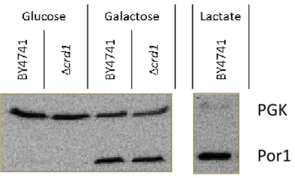
1.1. Assessment of mitochondrial content in Δcrd1, Δtaz1, Δups1, Δmdm10, Δmdm12 and Δcho1 strains ... 39
1.2 Growth of Δcrd1, Δtaz1, Δups1, Δmdm10, Δmdm12 and Δcho1 strains in galactose medium 40 1.3 Survival of galactose grown cells of Δcrd1, Δtaz1, Δups1, Δmdm10, Δmdm12 and Δcho1 strains during acetic acid-induced apoptosis ... 41
1.4 Assessment of cytochrome c release in galactose grown cells of Δcrd1, Δtaz1, Δups1, Δmdm10, Δmdm12 and Δcho1 during acetic acid-induced apoptosis ... 42
XIII
1.4.1 Assessment of cyt c content in galactose grown cells of Δcrd1, Δtaz1, Δups1, Δmdm10, Δmdm12 and Δcho1 strains ... 42
1.4.2 Optimizatin of the cell wall enzymatic digestion step of the mitochondrial isolation protocol ... 42
1.4.3 Evaluation of cytochrome c release ... 43 1.5 Assessment of the cell wall resistance of Δcrd1, Δtaz1, Δups1, Δmdm10, Δmdm12 and Δcho1 strains after acetic acid treatment ... 44 2. Assessment of alterations in the mitochondrial phospholipid composition of Δcrd1, Δtaz1, Δups1, Δmdm10, Δmdm12 and Δcho1 strains in response to acetic acid treatment ... 45 3. Assessment of the cell survival of Δups1, Δmdm10, Δmdm12, Δmdm34 and Δmmm1 strains upon expression of human Bax and Bax P168A ... 53 Chapter IV – Discussion ... 55 References ... 63
Chapter I – General Introduction
3
1. Apoptosis
The term apoptosis was coined by Kerr et al. in 1972 as an active form of programmed cell death triggered by an environmental stimulus (Kerr, Wyllie, & Currie, 1972). Hitherto, apoptosis is the most studied form of programmed cell death, and is characterized by the ordered appearance of several morphological or biochemical features. It occurs primarily as a mean of controlling cell population in a given tissue, during development or aging, but it can also occur as a defense mechanism by eliminating cells damaged by deleterious agents. This process is highly coordinated, normally genetically determined and energy-dependent (Elmore, 2007).
This type of programmed cell death involves single or small cells clusters. Its predominant morphological features are, in an early stage, cell shrinkage and pyknosis (irreversible chromatin condensation) and, in later stages, blebbing of the plasma membrane, along with karyorrhexis and karyolysis (fragmentation and dissolution of the nucleus), loss of plasma membrane phospholipid asymmetry and budding. The fragmentation of the cell into apoptotic bodies occurs without loss of the cell membrane integrity, these structures being then phagocytized and degraded in cells such as macrophages (Elmore, 2007). This feature distinguishes apoptotic from necrotic cell death where loss of membrane integrity occurs with the subsequent release of several cell constituents that cause an inflammatory response.
There are two main apoptotic pathways, the extrinsic and the intrinsic pathways. The extrinsic pathway, also known as the death receptor pathway, acts mainly as a response to external stimuli. This pathway acts by involvement of death receptors from the Tumor Necrosis Factor (TNF) receptor protein superfamily (such as FasR or TNFR1), which upon binding of the respective ligands, trimerizes and stimulates the Death Inducing Signaling Complex (DISC), leading to the recruitment and activation of the Caspase pathway (Elmore, 2007; Gómez-Sintes, Hernández, Lucas, & Avila, 2011; Zacks, Zheng, Han, Bakhru, & Miller, 2004).
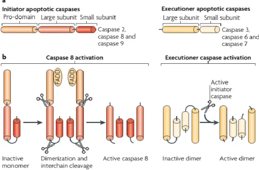
The Caspase pathway involves the activation of a cascade of different caspases (Cys Asp acid proteases), which are divided into two groups, the initiators and the executioners (Figure 1). Upon activation, the initiators caspases (like C8, C9 and C10) cleave certain substrates, the more important of which are the executioner caspases, (C3, C6 and C7). After cleavage, these activated caspases are translocated to their cellular targets, leading to several of the phenotypic characteristics of apoptosis (Tait & Green, 2010).
4
The intrinsic pathway, also known as the mitochondrial pathway, is activated by intracellular stimuli caused by cell stress (e.g. DNA damage, increase of cytosolic calcium concentration, oxidative stress). This pathway is mitochondria-dependent, contrary to the extrinsic pathway, in which the mitochondria may be involved solely in the amplification of cell death. The activation of this pathway leads to the permeabilization of the mitochondrial outer membrane (MOMP), causing the release of pro-apoptotic proteins, such as cytochrome c (cyt c) and Smac/Diablo (Second Mitochondria Derived Activator of Caspase/Direct IAP Binding protein with low pI). These factors will activate the caspase-dependent mitochondrial pathway by activating Apaf-1 and procaspase-9, resulting in the formation of the apoptosome. In later stages of apoptosis there is also the release of AIF, endonuclease G (Nuc1) and CAD, that will cause nuclear DNA fragmentation and condensation (Elmore, 2007).
Despite the differences between them, the two pathways are linked, sharing several molecules, in a way that the activation of one can enhance and amplify, or even provoke the activation of the other (Figure 2) (Crompton, 2000; Elmore, 2007). Cells can be divided into two types, according to the necessity of involving the intrinsic pathway. In Type I cells, such as lymphocytes, the caspase pathway is enough to effectively execute cell death, whereas in type II cells, such as hepatocytes, there is a need to provoke MOMP through Bid activation (Crompton, 2000; Galluzzi, Kepp, Trojel-Hansen, & Kroemer, 2012; Hao & Mak, 2010).
Figure 1 - Caspase (Cys Asp acid proteases) classification and activation. a) caspases are divided into two
5
1.1 The BCL-2 protein family
1.1.1 Pro-apoptotic vs anti-apoptotic members
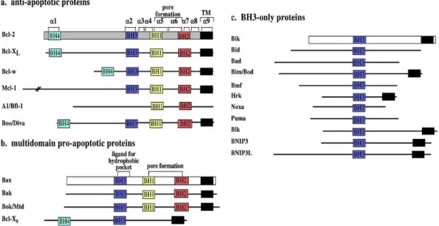
Apoptosis is a well-regulated process, and key intervenients in that regulation, particularly in MOMP, are several proteins of the Bcl-2 family. According to the activity and the number of Bcl-2 homolog (BH) domains, the family members are divided into three classes: anti-apoptotic, pro-apoptotic, and BH3-only proteins (Figure 3). The first class includes proteins that have four BH-domains, from BH1 to BH4. Bcl-2, Bcl-xL and Mcl-1, among others, are part of this class. Pro-apoptotic proteins can also have the four BH-domains, but BH4 domain is less conserved in this class. Bax, Bak and Bcl-Xs are included in the pro-apoptotic class (Adams & Cory, 2007; Andersen & Kornbluth, 2013; Chipuk, Moldoveanu, Llambi, Parsons, & Green, 2011; Er et al., 2006; García-Sáez, 2012; Ghibelli & Diederich, 2010).
The last class is the most heterogeneous class and includes proteins that only have the BH3 domain, such as Bid, Bad, Noxa and Puma. The BH3-only proteins are sub-divided further into two
Figure 2 - Intrinsic and extrinsic pathways of apoptosis. a) Intrinsic pathway, initiated by internal stimuli; b) extrinsic pathway, initiated by death
receptor activation (Tait & Green,
6
subgroups: the activators, which directly bind to the pro-apoptotic proteins, activating them, and the sensitizers, which bind to the anti-apoptotic proteins, liberating the pro-apoptotic proteins attached to them, thus blocking the inhibitors (Adams & Cory, 2007; Andersen & Kornbluth, 2013; Chipuk et al., 2011; Er et al., 2006; García-Sáez, 2012; Ghibelli & Diederich, 2010).
1.2 Role of mitochondria in apoptosis
Mitochondria have an essential role “deciding” if the cell lives or dies when subjected to stress. These organelles, which are believed to originate from a symbiotic relation between prokaryotes and early eukaryotes, are the energy production base of the cell. Their shapes vary between several vesicular structures to tubular networks with ramifications across the cell (Cosentino & García-Sáez, 2014; Friedman & Nunnari, 2014).
Mitochondria present two membranes: the outer mitochondrial membrane (OMM), which forms a smooth envelope, rich in lipids that provide fluidity, and transmembrane channels that allow the passage of molecules of small size (3-5 kDa); and the inner mitochondrial membrane (IMM) that presents several invaginations, the cristae, is rich in proteins essential for mitochondrial functions, and is impermeable even to small molecules. Between both membranes, in the intermembrane space, lie several proteins involved in both the respiratory chain and apoptosis. The most abundant protein in this region is cyt c. Inside the IMM, the matrix is where many catabolic and anabolic pathways occur, such
Figure 3 - Mammalian Bcl-2 family members, with highlighted BH domains, and important secondary structures. A)
7
as Krebs cycle or amino acids synthesis, and also where the mitochondrial DNA (mtDNA) is located, which encodes the genes for some of the enzymes involved in oxidative phosphorylation, and RNA and proteins necessary for the synthesis of those enzymes (Cosentino & García-Sáez, 2014; Friedman & Nunnari, 2014; Herrmann & Riemer, 2010; Horvath & Daum, 2013).
Upon apoptotic stimuli, several alterations of the mitochondria lead to cell death. Some of these alterations include: MOMP; alterations of the mitochondrial membranes phospholipidic composition; loss of mitochondrial membrane potential, which consequently blocks oxidative phosphorylation and leads to an accumulation of reactive oxygen species (ROS); cristae remodeling; and mitochondrial fragmentation (Cosentino & García-Sáez, 2014).
MOMP is considered a point of no return in the cell death process. Although there has been a lot of breakthroughs in the past few years, there are still many theories about the mechanism responsible for this permeabilization, and the means by which it works are still elusive.
There are several channels known to open during apoptosis. The mitochondrial permeability transition pore (PTP), is believed to be a complex of several proteins such as VDAC (Voltage Dependent Anion Channel), a channel that allows the passage of several cytosolic molecules and ions, ANT (Adenine nucleotide translocator), and possibly cyclophilin D and other factors, though the lack of any of these proteins does not appear to impair the formation of this complex (Büttner et al., 2011; Vieira et al., 2000; Vyssokikh & Brdiczka, 2003). Although studies report opening of these channels, they do not directly allow the passage of cyt c or other pro-apoptotic factors, due to the small opening and only communicating between the matrix and the cytosol (Er et al., 2006; García-Sáez, 2012; Ghibelli & Diederich, 2010).
It was believed that the opening of these channels could lead to swelling of the mitochondrial matrix with the rupture of the OMM, freeing the pro-apoptotic factors present in the intermembrane space into the cytosol, but further studies determined that this permeabilization could occur due to the opening of newly formed pores in the OMM, the MACs (Kuwana et al., 2002; Lin et al., 2011; Peixoto et al., 2011). Studies showed that these channels are formed through oligomerization of Bax and/or Bak. These channels would be big enough to allow the efflux of molecules such as cyt c, SMAC and AIF to the cytosol (Gillies & Kuwana, 2014; Peixoto et al., 2011; Tait & Green, 2010).
Even with the knowledge that Bax is involved in the formation of these channels, the process by which this protein is stimulated to oligomerize and form channels is still unknown, although some theories have been proposed.
8
1.2.1 Bax role in apoptosis and MOMP
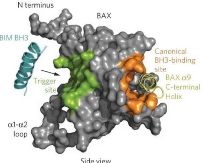
Bax is a pro-apoptotic protein of 21kD, with 192 amino-acids. In 2000, Suzuki et al. defined the three-dimensional structure of this protein, identifying several domains important in its characterization (Figure 4) (Suzuki, Youle, & Tjandra, 2000). The localization of this protein is mainly dictated by its activation. Although normally cytosolic when inactive, a small part of the protein is constitutively bound to the mitochondria by a weak link through a C-terminal transmembrane domain, possibly interacting with VDAC (Li & Dewson, 2015). Upon binding of the activator, Bax undergoes conformational changes, exposing certain domains such as the α-helixes (αH) 5/6, that are putative pore-forming transmembrane domains, and αH 9, an anchoring domain. All three are hydrophobic and involved in the translocation and insertion of the protein in the mitochondrial membrane (Suzuki et al., 2000). Other relevant domains of this protein are: αH 1, a site of interaction with the activators truncated Bid (tBid) and Puma; and the N-terminal, an unstructured domain used in assays to discriminate between the active/inactive form of Bax, because it hides the αH 9 in the inactive form of the protein, and exposes it when Bax is activated (Suzuki et al., 2000).
Bax has several functions within the cell. It is implicated in the dynamics of mitochondria, more precisely in mitochondrial fission, due to its presence in the membrane sites of fission and capacity to
Figure 4 - Modelling of Bax protein with highlighted central domains for its activity, along with the Bim
9
bind to the proteins involved, increasing the fission rate and leading to the collapse of the mitochondrial network (Er et al., 2006; Ghibelli & Diederich, 2010).
Along with fission, Bax promotes MOMP, through the activation of MAC (Mitochondrial Apoptosis Channel), a channel formed by its homo- or hetero-oligomerization– this subject will be further analyzed below in this introduction (Ghibelli & Diederich, 2010). MOMP causes the release of mitochondrial proteins such as SMAC/Diablo, a protein dimer that competes with Inhibitors of Apoptosis Proteins (IAPs) bound to caspases, liberating them; Apoptosis Inducing Factor (AIF), a protein that promotes the activation of caspase-independent apoptotic pathways, along with Endo G, an endonuclease that is included in this pathway; and more importantly, cyt c (Ghibelli & Diederich, 2010).
Besides contributing to the release of cyt c by MOMP, Bax also indirectly amplifies its release. After activation, Bax is translocated to the ER membrane, where Bcl-2 is inhibiting IP3R (IP3 receptor). Once there, Bax interferes with this inhibition, increasing the Ca2+ efflux to the cytosol (Decuypere et al.,
2011). In the zones of close proximity between the ER and mitochondria, the high concentrations of Ca2+ induce the oxidation of cardiolipin, which leads to further release of cyt c (Er et al., 2006;
García-Sáez, 2012; Ghibelli & Diederich, 2010).
Bax is activated by various signals both from the intrinsic and the extrinsic apoptosis pathways, and also by physicochemical alterations. After activation from the extrinsic pathway, Bid is recruited to the mitochondria via PACS-2, a multifunctional sorting protein, and is cleaved into tBid by the activated caspase-8. Then, tBid recruits Bax from the cytosol, and by binding to the αH 1 of the protein, it will activate it, and expose the domains required for targeting the protein to the mitochondrial membrane (Ghibelli & Diederich, 2010; Korsmeyer et al., 2000; Ott, Norberg, Zhivotovsky, & Orrenius, 2009). Bax can also be activated by other BH3-only proteins such as Bim (Er et al., 2006).
Bax activation can be triggered by alterations of the physicochemical properties of the cytosol. In the case of oxidative stress, the MAP kinases JNK and p38 are activated, causing up-regulation of Bax, and its liberation from chaperones like Ku70 or 14-3-3 by acetylation and phosphorylation, respectively. These proteins are able to sequestrate cytosolic Bax. The 14-3-3 chaperone is also important for Bax translocation to the mitochondria. Oxidative stress may also activate Bax, directly, through the oxidation of the protein and consequent dimerization. The dimer would acquire the ability to translocate and insert into the mitochondrial membrane (Er et al., 2006; Ghibelli & Diederich, 2010; Smaili et al., 2003).
The increase of Ca2+ concentrations in the cytosol indirectly causes Bax activation, through the
10
concentrations, cleaving Bax’s N-terminal region and exposing the domains of the protein required for its activity. Calpains also cleave Bid, and though in a different site than it is normally cleaved, it maintains its role as a Bax activator (Ghibelli & Diederich, 2010; Smaili et al., 2003).
Differences in the pH, can also activate this protein, by either exposing the BH3 domain, allowing for heterodimerization (pH 6.0-6.8 and 7.6-8.0), or exposing the N-terminal, resulting in translocation of the protein to the mitochondria (pH <6 and >8). Similarly, heat induces the conformational changes necessary for this translocation (Er et al., 2006; Kelekar & Thompson, 1998). In cancer cells there is expression of a glycoprotein, clusterin, that is able to specifically bind to the active Bax, inhibiting its oligomerization, an essential step for its role in apoptosis (Er et al., 2006).
One of the models currently being discussed for the activation of the MOMP channels is the conjugation of two previously proposed models, the direct activator model and the depressor model, and thus is referred to as “embedded together” model. This model discusses the role of the sub-division of the BH3-only proteins, where the roles of the activators and the sensitizers work together to allow activation and insertion of Bax in the outer mitochondrial membrane, where oligomerization takes place (Figure 5) (Bender & Martinou, 2013; Hagberg & Mallard, 2009; Hollville & Martin, 2012; Korsmeyer et al., 2000; Ripple, Abajian, & Springett, 2010; Walensky & Gavathiotis, 2011).
Studies show that the activation of Bax leads to the exposure of the BH3 domain. There are two models for its oligomerization, the asymmetric autoactivation and the symmetric dimer formation. The first model believes that the Bax’s exposed BH3 domain could, along with the BH3-only activators, recruit more Bax, through an autoactivating mechanism, which would be propagated linearly throughout the oligomer. The second and more accepted model, proposes the formation of dimers by disulfide cross-linking. These dimers would then connect between them, by dimer oligomerization (Bender & Martinou, 2013; Chipuk et al., 2008; Kelekar & Thompson, 1998; Korsmeyer et al., 2000).
11
The channel that is formed, MAC, shows an increase in the conductance according to the increased number of Bax homodimers that become part of the structure. Also, it was reported that lipids are involved in the formation of this channel, since the observation of these structures with electron microscopy showed that the edges of the channel do not present proteins and are, in fact, of phospholipidic nature. One of the lipids most discussed in this subject is cardiolipin (CL), as it was shown that its presence is required for the formation of MAC (Bender & Martinou, 2013; Tait & Green, 2010). Also, there are several studies that link MACs not to CL, but to a sphingolipid, ceramide, due to its capacity to form channels through phospholipidic bilayers. Studies show that after apoptotic stimuli, the ceramide concentrations in the OMM increase, and that this lipid after being translocated to the mitochondria is transformed into hexadecenal (hex) that acts synergistically with Bax forming a channel big enough for the release of the pro-apoptotic factors (Chipuk et al. 2012; Stiban & Perera 2015). There are also studies that suggest the formation of pores composed only by ceramide, or that ceramide acts solely as Bax activators (Chipuk et al., 2012; Patwardhan et al., 2015). Other studies suggest that VDAC oligomerizes when induced by phospholipids present in mitochondrial membrane, specially phosphatidylglycerol (PG), when the ratio PG/CL is high, forming channels big enough to allow cyt c release (Betaneli, Petrov, & Schwille, 2012; Zheng et al., 2010).
Cyt c is a protein of around 12 kD, found in mitochondria, more specifically bound to the outer face of the Inner Mitochondrial Membrane (IMM) by cardiolipin (CL), where it has an important role in the production of energy, as an intermediate of the electron transport chain (Ascenzi, Polticelli, Marino, Santucci, & Coletta, 2011). Since 85% of this molecule is present within the cristae, disorganization and loosening of these structures caused by the disruption of the Opa1 (Mgm1 in yeast) complexes is needed for its significant release from mitochondria. The dysfunction of these complexes is known to impair replication of mtDNA and the electron transport chain. The disassembly of Opa1 complexes is induced by pro-apoptotic BH3-only proteins, while still requiring the presence of Bax (Otera & Mihara, 2012; Yamaguchi, Lartigue, Perkins, & Scott, 2008) and is also aided by the decreased mitochondrial membrane potential. Although essential, this remodeling of the cristae is still not enough to release cyt c, further events regarding CL being necessary (Cosentino & García-Sáez, 2014).
Along with the pro-apoptotic factors, other enzymes present in the intermembrane space are released, such as adenylate cyclase, a controller of the adenine levels (Burkart, Shi, Chouinard, & Corvera, 2011), whereas proteins from the matrix remain in the mitochondria (such as citrate synthase) (Leek, Mudaliar, Henry, Mathieu-Costello, & Richardson, 2001), indicating that the IMM remains intact.
12
In later stages of apoptosis, fragmentation of this organelle occurs by overexpression and translocation of Drp1 (dynamin-related protein 1) (Dnm1 in yeast) to the OMM. The Ca2+ accumulation
in the cytoplasm due to mitochondrial and ER dysfunction leads to the dephosphorylation of Drp1 by Ca2+-activated calcineurin, inducing its translocation to the mitochondria, where it interacts with Fis1 to
carry out mitochondrial fragmentation (Cosentino & García-Sáez, 2014; Scorrano, 2013). Although the mechanism of the MOMP is not yet well understood, its impact in apoptosis is undoubtedly of great importance (Belizário, Alves, Occhiucci, Garay-Malpartida, & Sesso, 2007; Bender & Martinou, 2013; Montero et al., 2010; Tait & Green, 2010).
1.3 Yeast as a model of apoptosis
Until less than 20 years ago, the idea that unicellular organisms could undergo apoptosis was discarded because it was considered illogical for a cell to willingly commit suicide. However, Madeo and his colleagues, in 1997 found proof that a possible form of apoptosis was exhibited by a cell cycle mutant cdc48S565G of S. cerevisiae. This mutant presented exposure of phosphatidylserine, DNA
fragmentation and chromatin condensation and fragmentation, all text-book markers of programmed cell death (Madeo, 1997). This created controversy, but ultimately, the idea that yeast populations should not be thought of as a group of independent unicellular organisms but as a multicellular community, with interaction between its members has prevailed. With this idea in mind, the thought of a cell committing to a programmed cell death process because it is damaged or old, and it would be of greater benefit as a resource for healthy cells in the colony, is not so odd.
Still, yeast lacks several of the major apoptotic regulators. However, this fact can have its advantages, since yeast can become a basic and more simple model that can be used to study not only yeast apoptosis, but also interactions between main mammalian apoptosis regulators through heterologous expression (Fröhlich, Madeo, & Frohlich, 2000; Weinberger, Ramachandran, & Burhans, 2003).
Some of the orthologue proteins important for yeast apoptosis are the metacaspase Yca1, the mitochondrial proteins Nuc1 and Aif1 (orthologues of mammalian Endo G and AIF respectively) and the protease Nma111 (orthologue of HtrA2/Omi) (Carmona-Gutierrez et al., 2010; Mazzoni & Falcone, 2008). Yca1 is the only known orthologue of mammalian caspases, and is involved in apoptosis induced by several triggers (Carmona-Gutierrez et al., 2010; Mazzoni & Falcone, 2008). Although it seems to be an important protein for yeast apoptosis, it is not essential. Studies show that apoptotic cell
13
death mediated by Nuc1 and Aif1 does not require Yca1, which could imply that, as for mammalian, yeast apoptosis may not always require caspases (Carmona-Gutierrez et al., 2010).
Another protein that might be involved in yeast apoptosis is Nma111, orthologue of HtrA2/Omi, a mitochondrial protease in humans, that in yeast is located in the nucleus and acts through cleavage of Bir1, a yeast inhibitor of apoptosis (AIP). Besides Nuc1 and Aif1, there are other mitochondrial proteins involved in yeast apoptosis, such as Ndi1, a NADH dehydrogenase located in the IMM; cyt c, although its mammalian role as an activator of caspase function is still unclear in yeast; and Drp1, which, as in mammalian apoptosis, causes mitochondrial fragmentation upon death stimuli (Carmona-Gutierrez et al., 2010). The mitochondrial ADP/ATP carrier (AAC) and the vacuolar protease Pep4, the yeast orthologue of human cathepsin D, have also been linked to yeast apoptosis, enhancing mitochondrial degradation (Pereira et al., 2010).
As previously stated, proteins from the Bcl-2 family are essential as apoptosis regulators in mammalian cells. Until recently, obvious orthologues of these proteins have not been found in yeast, which made yeast the perfect model for their study upon heterologous expression, and for the elucidation of their involvement in apoptosis. For example, heterologous expression of the human pro-apoptotic protein Bax has been found to induce yeast cell death (Greenhalf, Stephan, & Chaudhuri, 1996), with impaired growth, accumulation of ROS, MOMP and cyt c release (Greenhalf et al., 1996; Priault, Camougrand, Kinnally, Vallette, & Manon, 2003). Other apoptosis markers, such as plasma membrane blebbing, phosphatidylserine exposure at the outer leaflet of the plasma membrane, chromatin condensation, DNA fragmentation, characteristic of apoptosis have been identified (Ligr et al., 1998). Also, a study using a patch clamping technique for analysis of a high conductance channel, in a Bax expressing strain, found a mitochondrial channel in the OMM with MAC characteristics, which would allow the release of the pro-apoptotic factors from mitochondria (Pavlov et al., 2001; Polcic, Jaka, & Mentel, 2015; Priault et al., 2003). These channels differed from the ones artificially made solely from Bax proteins in in vitro systems, a fact that would lead to believe the MAC channels require a component other than Bax, found in both yeast and mammalian mitochondria (Polcic et al., 2015). Still, further studies using the same system suggest that the cell death induced by Bax expression may not be yeast apoptosis, but instead, this protein activates autophagy that culminate in cell death (Kiššova et al., 2006).
In 2011, Büttner et al. found a yeast protein with a BH3 domain, the Ybh3, which has been shown to regulate apoptosis in yeast, more specifically the mitochondria mediated pathway of apoptosis. It was demonstrated that this protein, when overexpressed, sensitizes the cells to apoptotic stimuli, by
14
translocating to the mitochondria and disrupting mitochondrial membrane potential. It was also shown that this protein is inhibited by Bcl-XL and that, once in the mitochondria, through interaction with Mir1p
and Cor1p, it may physically interact with putative components of PTP, although this contact is not essential for cell death mediated by Ybh3p (Büttner et al., 2011).
Yeast apoptosis has been studied extensively since it was first described, and it has been shown to be induced by several factors, including oxygen stress (such as H2O2), valproic and acetic acid,
hyperosmotic stress, arsenic, mRNA instability, loss of ubiquitination control, impaired DNA replication and chronological aging, among others (Carmona-Gutierrez et al., 2010).
1.4 Acetic acid as an inducer of yeast apoptosis
Acetic acid (CH3COOH) is a byproduct of alcoholic fermentation carried by S. cerevisiae. It was
previously described that in certain concentrations it can lead to an impaired fermentation. In most S. cerevisiae strains repression by glucose inhibits acetic acid metabolism. In this condition, at low pH, its non-dissociated form (pKa = 4.75) enters the cell by simple diffusion and through the aquaglyceroporin
channel Fps1, and its dissociation when inside may cause intracellular acidification, which in turn affects several pathways essential for the cell activity. However, in the presence of acetic acid, this channel is phosphorylated by Hog1 (high osmolarity glycerol (HOG) mitogen-activated protein kinase (MAPK)), leading to the translocation and degradation of this channel in the vacuole, as a way of protecting the cell from stress. The MAPK Slt2, a protein that is part of the PKC pathway, which has a role in maintaining cell wall integrity, is also activated by acetic acid stress. However, Fps1-facilitated entry of acetic acid inhibits the activation of Slt2 phosphorylation (Mollapour, Shepherd, & Piper, 2009). The effect acetic acid has in the cell depends on the concentration used, and can cause cell death either by necrosis, when at high concentrations, or with some of the characteristic features of mammalian apoptosis. Some of the cell alterations occurring during acetic acid-induced apoptotic cell death are exposure of phosphatidylserine in the outer leaflet of the cytoplasmic membrane, chromatin condensation and DNA fragmentation (P. Ludovico, Sousa, Silva, Leão, & Côrte-Real, 2001; M. J. Sousa, Ludovico, Rodrigues, Leão, & Côrte-Real, 2012). Also, this cell death process involved mitochondrial dysfunction including accumulation of reactive oxygen species (ROS), initial hyperpolarization followed by loss of mitochondrial membrane potential, impaired mitochondrial respiration and cyt c release (Paula Ludovico et al., 2002).
The yeast metacaspase, Yca1, is also activated by acetic acid stress, and this activation is dependent of culture growth phase, being maximal at the very beginning of exponential growth phase
15
and decreasing along growth (Pereira, Camougrand, Manon, Sousa, & Côrte-Real, 2007). Also, acetic acid-induced apoptosis can occur without cyt c release, though to a smaller extent (Guaragnella, Bobba, Passarella, Marra, & Giannattasio, 2010). The presence of an apoptosome is still unclear, as are any events downstream of cyt c release. Acetic acid-induced apoptosis also provokes the release of the other pro-apoptotic factors from the intermembrane space, such as Nuc1 and Aif1, which undergo translocation to the nucleus to exert their functions (Guaragnella et al., 2012; M. J. Sousa et al., 2012). Mitochondrial dynamics is also of great importance in acetic acid-induced cell death in yeast, as deletion of DRP1 gene increases acetic acid resistance, and impairs mitochondrial fragmentation (Giannattasio, Guaragnella, Zdralević, & Marra, 2013; M. J. Sousa et al., 2012).
2. Mitochondrial Phospholipids
Lipids are a group of organic molecules which are hydrophobic, that is soluble in organic solvents but not in water. These compounds can be characterized according to their structure and functions, being divided into 8 classes, as established by the International Lipid Classification and Nomenclature Committee in 2005: fatty acids (FA), glycerolipids, glycerophospholipids, sphingolipids, prenol lipids, glycolipids, sterols and its derivatives, and polyketides (Fahy E, Subramaniam S, Brown HA, Glass CK, Merrill AH Jr, Murphy RC, Raetz CRH et al., 2005; Klug & Daum, 2014).
The endoplasmic reticulum, along with the Golgi apparatus and the mitochondria, are the main lipid synthesizing organelles. As some of the newly synthesized lipids are needed in other locations within the cell, the cell’s phospholipid transport systems are also of major importance in lipid metabolism. In this context, mitochondria associated membranes (MAMs), an ER membrane sub fraction, play key functions in the synthesis of some phospholipids. Yeast, besides being used as a model for the study of programmed cell death, is also used as a research model for the elucidation of lipid metabolism, since apart from the more practical advantages already covered, the metabolic pathways are very well-conserved among all eukaryotes. (Daum, Lees, Bard, & Dickson, 1998; Klug & Daum, 2014; Santos & Riezman, 2012; Zhang et al., 2014)
2.1 Endoplasmic Reticulum - Mitochondrial (ER-M) contact sites: MAMs, ERMES and EMC
There are different optimal conditions for the several metabolic pathways within the cell, which evolutionarily shaped the organelles, each with a specific controlled milieu. Due to this
16
compartmentalization of the cell, a way of connecting the different organelles, such as vesicular trafficking, is needed for exchange of nutrients and metabolites. The connection between the ER and the mitochondria, however, does not use this form of communication, but rather a tether between their membranes that can be observed by electron microscopy, and is also evidenced in mitochondria isolates with ER membrane contamination. Tethering of the mitochondrial to the ER is achieved by several protein complexes and is present in about 5-20% of the mitochondrial surface. In this zone of close apposition, both organelles are within 30 nm of each other (De Vos et al., 2012; Kornmann, 2013; Michel & Kornmann, 2012; Raturi & Simmen, 2013). This zone of close apposition is controlled by proteins like PACS-2 in mammals, where the lack of this protein results in disruption of the tethering complexes and fragmentation of the mitochondria (Simmen et al., 2005).
There are several protein complexes that are involved in the interactions between ER and mitochondria in metazoans (Figure 6):
In the ER membrane and the OMM there is a protein Mmr1, that can dimerize and anchor both organelles during budding, allowing proper mitochondrial inheritance after their delivery through Myo2, across the actin cables to the bud (Kornmann, 2013; McBride, 2011);
The mitochondrial Fis1 binds to the ER protein Bap31, and the activation of this complex through the recruitment of procaspase-8 forming an ARCosome leads to the release of Ca2+. This release amplifies the death signals, inducing apoptosis (Iwasawa, Mahul-Mellier,
Datler, Pazarentzos, & Grimm, 2011; Kornmann, 2013; Vance, 2014);
The ER protein VAPB interacts with the mitochondrial PTPIP51, a complex involved in the regulation of intracellular Ca2+ homeostasis (De Vos et al., 2012; Kornmann, 2013);
The mitochondrial VDAC, a nonselective pore for ions and hydrophilic molecules, and the IP3R localized in ER membrane are bound by the chaperone Grp75. This chaperone also regulates Ca2+ flux between these organelles, by catalyzing its release from the ER by IP3R,
and its transport through VDAC into the mitochondria (Decuypere et al., 2011; Hayashi, Rizzuto, Hajnoczky, & Su, 2009; Kornmann, 2013; Malhotra & Kaufman, 2011);
In mammalian cells, the mitochondrial proteins mitofusin (Mfn) 1 and 2, bind to the ER Mfn2, linking the two organelles (Kornmann, 2013);
The PACS-2 is also required for the close apposition of the organelles, possibly through links with IP3R (Simmen et al., 2005).
17
The contact between mitochondria and the endoplasmic reticulum has been associated with several functions. One of those functions is the involvement in mitochondrial dynamics and its inheritance. Studies showed that the mitochondrial fission events are in greater numbers in the sites where there is close apposition between the two organelles, possibly because ER plays a role in the constricting stages. Evidence of ER-determined mitochondria inheritance during division of yeast and mammalian cells was also provided. In yeasts, the ERMES complex may also be involved in replication of the mtDNA (mitochondrial DNA), as strains deficient in proteins from this complex showed loss of mtDNA (English & Voeltz, 2013; Kornmann, 2013; Michel & Kornmann, 2012; Otera & Mihara, 2012; Raturi & Simmen, 2013).
Facilitating calcium (Ca2+) exchange is also a function of ER-mitochondria contact sites. This ion is
essential in cell metabolism, being involved in several processes, such as regulation of cellular bioenergetics, intracellular signaling or motility. Ca2+ ions entering mitochondria through these ER-M
contact sites serve various proposes, acting as co-factors for enzymes of the citric acid cycle in the mitochondrial matrix and other proteins related to the mitochondrial division and motility. In addition, high Ca2+ concentrations in the mitochondrial matrix enhances the onset of apoptosis, by activation of
the PTP, and possibly amplifying pro-apoptotic factors leakage to the cytosol (English & Voeltz, 2013; Kornmann, 2013; Malhotra & Kaufman, 2011; Michel & Kornmann, 2012).
As previously described, the chaperone protein Grp75 binds to the IP3R in the ER membrane, and to the VDAC channel in the OMM, so that both proteins are “mouth-to-mouth”. This way, when the IP3R is activated by a physiological stimulus, the Ca2+ transported to the cytosol flows directly through the
channel into the mitochondrial intermembrane space, and then through the mitochondrial calcium uniporter (MCU) to enter the matrix. The release of this ion from the ER is regulated by several
Figure 6 - Representation of the several protein interactions in MAMs in yeast (c) and metazoans ((d) to (h))
18
mechanisms, namely by the cytosolic calcium concentration: low or very high Ca2+ concentrations inhibit
the IP3R, emphasizing the control of the ER-M contact sites in the calcium homeostasis (Decuypere et al., 2011; Kornmann, 2013; Raturi & Simmen, 2013).
One of the first functions described for the connections between ER and mitochondria was its role in lipid metabolism, more precisely in its synthesis. ER-M contact sites are enriched with both enzymes involved in lipid biosynthesis and in proteins responsible for its transport between the membranes of the two organelles. This allows the use of these proteins as reliable marker proteins, such as fatty acid coenzyme A (CoA) ligase 4 (Facl4), an enzyme that mediates the catalysis of the bond between fatty acids and CoA for triacylglycerol synthesis (Kornmann, 2013; Osman, Voelker, & Langer, 2011; Raturi & Simmen, 2013).
In yeast, the best characterized tethering complex is ERMES. The ERMES complex includes 4 different proteins: Mmm1, an ER Protein, Mdm12, a cytosolic protein, and Mdm34 and Mdm10, both mitochondrial proteins (Figure 7). This complex has an array of functions, from aiding the lipid exchange, regulation of mtDNA replication and assisting mitochondrial protein import (Kornmann, 2013; Lahiri et al., 2014; Michel & Kornmann, 2012).
The wide range of processes in which this complex is involved implies the presence of regulation of some sort for it to respond correctly to the right stimuli. During the purification of this complex, a new protein was discovered, identified later as the Miro GTPase Gem1. Gem1 has two GTPase domains, being suggested that they have different roles, the first domain focusing in localization, and the second regulating the ERMES, specifically in the lipid exchange and synthesis (Michel & Kornmann, 2012).
Until recently, ERMES was the only characterized tethering complex between ER and the mitochondria in yeast, but the deletion of this complex only reduced the phospholipid exchange present in the mutants, and the transfer of PS to the mitochondria was not impaired (Lahiri et al., 2014; Zinser et al., 1991). These studies suggested that another complex would exist tethering the two organelles, since the proximity between them seems vital to the cell.
In 2014, Lahiri et. al showed a relation between the Endoplasmic Reticulum Membrane Complex (EMC) and Cho2, responsible by transport of PE from the mitochondria to the ER. The yeast EMC is formed of 6 proteins (EMC1-6), and is involved in ER-associated degradation (Wideman, 2015). The
Figure 7 - The ERMES complex at the ER–
mitochondria connection (Michel &
19
genetic analysis of the cluster uncovered a possible function associated with mitochondria and lipid metabolism. The authors also tested PS transfer from ER to mitochondria in mutants without EMC proteins 1/2/3/5 and 6, which showed a decreased transport (about 50%), and found that the mitochondria in these mutants are abnormal, showing a 50% decrease in PE levels, an increase of PA and CL, and as it was previously demonstrated for cells with MMM1 deletion, these cells cannot grow in nonfermentable carbon sources. Ultimately the same group showed that cells missing either EMC or ERMES complex display decreased ER-mitochondrial tethering, and that cells missing both EMC and Mmm1p were not viable (Lahiri et al., 2014).
2.2 Yeast Phospholipid Metabolism
Although there is not one molecular monomer for lipids, fatty acids (FA) are basic structures common to the majority of complex lipids. They are composed of a carboxyl group attached to a linear hydrocarbon chain, which can vary in length and saturation. Usually used as energy storage, they can also regulate transcription, be part of the signaling pathways, or be involved in the synthesis of several other lipids species, such as membrane lipids. This great demand calls for several ways for the cell to produce them, either by de novo synthesis which takes place in the cytosol and mitochondria, with consequent elongation in the ER; by uptake from the outside of the cell; or by hydrolysis of complex lipids within the cell. The FA species present in yeast vary according to the growth conditions and the subcellular membrane of focus (Daum et al., 1998; Klug & Daum, 2014). Due to their toxicity at high concentrations, they are stored in lipid droplets in the cytosol, and are rapidly consumed in phospholipid formation, explaining why there is always a steady state low concentration of this lipids (Cosentino & García-Sáez, 2014).
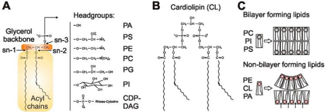
Glycerophospholipids, or as they are most commonly called phospholipids, are formed by a polar hydrophilic head and a hydrophobic tail which provide an amphipathic character and help the formation of the characteristic bilayer composition of most cell membranes. The tail is composed of a diacylglycerol (DG) backbone, formed by a glycerol molecule to which fatty acids link through ester bonds in the sn1 and sn2 carbons. The glycerol sn3 carbon is where the head forms a bond through a phosphate group, which links with an alcohol. The most common head groups, which separate phospholipids in several classes, are serine, ethanolamine, choline, glycerol or inositol, and form respectively phosphatidylserine (PS), phosphatidylethanolamine (PE), phosphatidylcholine (PC), phosphatidylglycerol (PG) and phosphatidylinositol (PI). Also, phospholipids differ according to the saturation of the fatty acid chain (Figure 8) (Daum et al., 1999; Horvath, Wagner, Steyrer, & Daum,
20
2011; Klug & Daum, 2014). The phospholipid membrane composition depends on the organelle, and is different in both inner and outer layer of the membrane, these phospholipid specificity having an important function in membrane structure (Horvath et al., 2011; Klug & Daum, 2014).
2.2.1 Phospholipid biosynthesis in S. cerevisiae
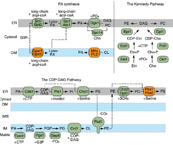
The central metabolite of the biosynthesis pathways for the formation of the several phospholipid classes is phosphatidic acid (PA). This lipid is the divergence point of two major pathways of lipid biosynthesis: first, its cleavage into DG allows its entry into the storage pathway, where it is transformed in triglycerides (TG) and is then stored in lipid droplets; secondly, its conversion to cytidine diphosphate-diacylglycerol (CDP-DG), by Cds1 in the ER and Tam41 in the mitochondria, contributes to the formation of most phospholipids as their precursor. From this precursor, all other phospholipids are formed, by either the CDP-DG pathway or the Kennedy pathway (Horvath et al., 2011; Klug & Daum, 2014; Zhang et al., 2014).
2.2.2 The Kennedy pathway
The Kennedy pathway is one of the ways to produce PC and PE, two of the most abundant phospholipids in yeast. It occurs in the ER, and the de novo synthesis happens through a series of reactions catalyzed by enzymes that are significantly conserved on eukaryotes (Figure 9) (Gibellini & Smith, 2010; Zhang et al., 2014).
The first step consists of a phosphorylation of choline and ethanolamine transported from external sources through Hnm1 (a plasma membrane transporter), by ATP-dependent choline and ethanolamine kinases (CKi1 and EKi1, respectively), which form choline-phosphate (Cho-P) and ethanolamine-phosphate (Etn-P). These enzymes differ in specificity, since CKi1 can use both substrates (although
Figure 8 - Phospholipids in mitochondrial membranes. (A) The central structural element of phospholipids is a glycerol backbone with different hydrophilic head groups (B) CL structure (C) Bilayer and non-bilayer
21
choline preferably), where EKi1 can only use ethanolamine (Gibellini & Smith, 2010; Klug & Daum, 2014; Zhang et al., 2014). Afterwards, cytidylyltransferases specific to both Cho-P and Etn-P (PCT1 and ECT, respectively) combine CTP to form CDP-choline and CDP-ethanolamine. These enzymes are considered rate limiting, and thus control PC and PE synthesis through this pathway.
The final step of this pathway involves the ER-enzymes DAG-cholinephosphotransferase (Cpt1) and DAG-ethanolaminephosphotransferase (Ept1), which catalyze the transfer of head groups choline-phosphate and ethanolamine-choline-phosphate to DAG, produced by the dephosphorylation of PA by Pah1, forming PC and PE. PC can be further transformed in PS in the ER through the enzymes Pss1 and Pss2 (phosphatidylserine synthase) (Gibellini & Smith, 2010; Klug & Daum, 2014; Zhang et al., 2014).
2.2.3 The CDP-DG pathway
The CDP-DG pathway is the other branch of phospholipid biosynthesis, by which PI, PS, PG, and consequently CL, PE and PC, are formed, in a pathway that includes both the ER and mitochondria (Figure 9). The CDP-DG present at the ER membrane can be transformed into PI through the ER protein phosphatidylinositol synthase (Pis1) that places an inositol group where the cytidine monophosphate (CMP) is inserted. In the mitochondria, CDP-DG is transformed into PGP, through the phosphatidylglycerophosphate synthase (Pgs1), which is in turn transformed in PG by the phosphatidylglycerophosphate phosphatase, Gep4. The phospholipid PG can be further transformed into cardiolipin through a process described further in this introduction (Klug & Daum, 2014; Zhang et al., 2014).
The same precursor (CDP-DG) also allows the formation of PS, through a reaction catalyzed by the ER enzyme PS synthase Cho1, which replaces the CMP with a serine group. This phospholipid is then translocated to the mitochondrial inner membrane, where it suffers decarboxylation into PE in a reaction catalyzed by Psd1 or Psd2 (Flis & Daum, 2013; Klug & Daum, 2014; Tatsuta, Scharwey, & Langer, 2014; Zhang et al., 2014). PE is essential for mitochondrial function, due to its capacity to generate negative membrane curvature and form nonbilayer structures (Aguilar et al., 1999; Osman et al., 2011).
For the formation of PC, the transport of PE back to the ER is required, and the PE head group undergoes sequential methylations, using methyl groups from S-adenosylmethionine (SAM). The first reaction is catalyzed by phosphatidylethanolamine methyltransferase Cho2 and the other two by the phospholipid methyltransferase, Opi3. As the enzymes required for this pathway are intercalated between the two membranes, the flipping the phospholipids back and forth needs to be assured, not
22
only to obtain the final products, but also to disperse these important phospholipids throughout the various cell membranes (Gaynor et al., 1991; Klug & Daum, 2014; Osman et al., 2011; Zhang et al., 2014).
Evidence was found in yeast that this phospholipid exchange between the two membranes happens mostly in the ER-mitochondria contact sites, due to the reduced phospholipid transport present in strains with deletions in proteins of the tethering complexes already described (Kornmann, 2013; Osman et al., 2011; Prinz, 2010; Tamura, Sesaki, & Endo, 2014; Tatsuta et al., 2014). In mammals, the levels of these enzymes are highly controlled by other proteins, such as PACS-2, that mediates the membrane associated quantities of Facl4 and Pss1 (Simmen et al., 2005).
2.2.4 Cardiolipin synthesis and remodeling
Cardiolipin (CL), is a phospholipid that makes up to 20% of the mitochondrial membranes, mostly present in the IMM, with a role in the stability and activity of some of IMM proteins, and involved in apoptosis. The cascade of enzymes required for its synthesis is found in the IMM, but the precursor of this lipid, phosphatidic acid (PA), can be found either in the ER membrane, or, to a lesser extent, be synthesized de novo in mitochondria by phospholipases, as already described (Figure 9) (Ascenzi et al., 2011; Osman et al., 2011).
Synthesis of CL is catalyzed by the Crd1, the cardiolipin synthase, that uses PG and CDP-DG as substrates. Then, CL enters a cycle of remodeling, but the pathways involved in mammalians are still rather unclear, and the proteins involved still unknown. Firstly, CL is remodeled into monolysocardiolipin (MLCL), by Cld1 through removal of an acyl chain, and MLCL in again transformed in CL through a transacylation reaction, where Taz1 transfers an acyl chain from another phospholipid (Baile, Lu, & Claypool, 2014; Baile, Sathappa, et al., 2014; Klug & Daum, 2014; Zhang et al., 2014). Although this mechanism is observed in both eukaryotes and prokaryotes, remodeled and unremodeled CL appear to be indistinguishable in yeast, and remodeling may only be a repair mechanism of CL acyl chain damages caused by reaction with ROS (Baile, Sathappa, et al., 2014).
Cardiolipin, found predominantly in the mitochondria, is, as mentioned above, important for several processes within this organelle. The presence of this cross-linked dimer in the IMM acts as an integrity stabilizer, which helps protecting the membrane from damage. This stabilization is essential for the activity of the respiratory complexes III and IV of the respiratory chain and also of the AAC (ATP/ADP carrier) (Houtkooper & Vaz, 2008; Osman et al., 2011).
23
This important mitochondrial phospholipid also has a role in protein import from the cytosol into the mitochondrial matrix. TIM (translocase of the inner mitochondrial membrane) and TOM (translocase of the outer mitochondrial membrane) are the complexes responsible for the protein transport, and studies show that cells lacking the proteins responsible for the CL synthesis have an impaired protein import into mitochondria (Osman et al., 2011). It is also believed that CL is involved in regulation of fusion/fission of mitochondria. This phospholipid binds to the positively charged lysines in inter-mitochondrial membranes Opa1 and its yeast orthologue Mgm1, stimulating its fusion activity. Also, translocation of Opa1/Mgm1 to the IMM is dependent in cardiolipin concentration (Gonzalvez & Gottlieb, 2007; Houtkooper & Vaz, 2008; Zhang et al., 2014).
2.3 Phospholipids during apoptosis: role and alterations
Some of the events that occur during apoptosis lead to changes in lipid composition, of both the plasma and intracellular membranes. One of the phospholipids identified as having a role in this form of
Figure 9 - Biosynthetic pathways for mitochondrial phospholipids. The major precursor PA is synthesized in the ER and mitochondrial outer membranes. The CDP-DAG pathway is located in the ER and mitochondrial inner membranes and produces a variety of phospholipids including PI, PS, PE, PC and CL. The ER-located
24
cell death is PS, since, as already described in this introduction, its externalization to the outer leaflet of the plasmatic membrane is the signal for clearance by the macrophages (Fadok & Henson, 2003). In the mitochondrial membrane other alterations regarding the oxidation of the phospholipids or of their ratio among the two mitochondrial membranes may be at play. MOMP, as already described, may be aided by these alterations, since changes in membrane organization facilitate the formation of the pores through which the pro-apoptotic proteins may be released. Peroxidation of mitochondrial phospholipids by ROS may even change the favored curvatures of certain phospholipids, such as PA. Also, the increase of certain phospholipid species, namely LPC and DG, which favor opposite membrane curvatures, is observed during apoptosis (Cosentino & García-Sáez, 2014; Crimi & Esposti, 2011). The release of Ca2+ by the ER, and the consequent increased concentration in ER-M contact sites will also aid
converting the bilayered disposition into hexagonal phases, due to the conical shape of CL and PE (Cosentino & García-Sáez, 2014; Crimi & Esposti, 2011; Flis & Daum, 2013; Osman et al., 2011).
Cardiolipin has an important role in different stages of apoptosis. Firstly, this lipid can activate the Fas receptor in the plasma membrane, thus being capable of inducing apoptosis. Also, as previously stated, cyt c is linked to CL in the IMM, and when the degree of unsaturation of the CL acyl chains is high, cyt c is more susceptible to undergo a conformational change and act as a CL-specific peroxidase. The modified cyt c, along with the elevated ROS levels, will result in CL oxidation, which leads to a reorganization of the lipids in microdomains with hexagonal HII configuration, with a core of polar phospholipid heads (Firsov, Kotova, Korepanova, Osipov, & Antonenko, 2015; Montero et al., 2010; Osman et al., 2011; Unsay, Cosentino, Subburaj, & García-Sáez, 2013). This alteration in CL structure causes an alteration of the curvature of the membrane. This may be involved in the increase of specificity in the recruitment of truncated-Bid to the inner and outer mitochondrial membranes, in the efficiency of membrane insertion and oligomerization of Bax, and in the formation of MAC (Gonzalvez & Gottlieb, 2007; Hollville & Martin, 2012; Kagan et al., 2005; Lutter et al., 2000; Montero et al., 2010; Osman et al., 2011; Raemy & Martinou, 2014). Also, cyt c itself may be able to cause disorganization of the OMM and cause MOMP, but only upon translocation of CL to the OMM (Xu, Vanderlick, & Beales, 2013).
3. Main aims and study overview
In 2013, Sousa et al. performed a genome-wide screening in S. cerevisiae to uncover genes involved in the regulation of apoptosis induced by acetic acid. Besides reinforcing the exploitation of