Mestre em Ciências das Zonas Costeiras
Contribution to drug discovery and
development for tauopathies using yeast
as a model
Dissertação para obtenção do Grau de Doutor em Bioengenharia
Orientador: Prof. Doutora Ana Cristina Carvalho Rego,
Professora Auxiliar com Agregação, Faculdade
de Medicina e Centro de Neurociências e Biologia
Celular, Universidade de Coimbra
Co-orientador: Prof. Doutora Helena Margarida Moreira de
Oliveira Vieira, Professora Associada Convidada,
Faculdade de Ciências, BioISI
–
Biosystems &
Integrative Sciences Institute, Universidade de
Lisboa
Co-orientador: Prof. Doutor Manuel Nunes da Ponte, Professor
Catedrático, Faculdade de Ciências e Tecnologia,
Universidade Nova de Lisboa
Júri:
Presidente: Prof. Doutora Ana Isabel N. M. A. de Oliveira Ricardo Arguentes: Prof. Doutor Vítor Manuel Vieira da Costa
Prof. Doutora Cláudia Maria Fragão Pereira Vogais: Prof. Doutor Tiago Fleming de Oliveira Outeiro
Prof. Doutora Paula Cristina da Costa A. M. Ludovico ..Prof Doutor Christophe François Aimé Roca
Contribution to drug discovery and development for
tauopathies using yeast as a model
Copyright
Marta Isabel Heitor Cerejo, Faculdade de Ciências e Tecnologia da Universidade Nova de Lisboa e Universidade Nova de Lisboa
No PhD is easy. Mine was a roller-coaster. From the birth of my first child to the insolvency of BIOALVO, Life found a way to push me beyond what I thought my limits were. It was an incredible journey and it wouldn’t have been possible without the contribution of amazing people to whom I wish to thank.
To BIOALVO and Fundação para a Ciência e Tecnologia for granting me the opportunity and financial support to develop my PhD work.
To Professor Helena Vieira, my CEO/supervisor/mentor/friend/sister, for trusting in my ability to do this work at BIOALVO. Thank you for teaching me about biotech and about how we can bring science closer to people. Thank you for your support, even during the most difficult times of your life.
To Professor Ana Cristina Rego, for your scientific supervision, comprehension, trust and friendship. Thank you for receiving me at your lab in CNC-UC. I have never been received so well in a lab and, although it was a short time, I learned a lot about cell biology and of what science in academia is. To the MIT-Portugal Doctoral Program that allowed me to learn and experience so many new things, but especially to meet inspiring people, including my teachers and my colleagues. Particularly, to Professor Manuel Nunes da Ponte, my co-supervisor, for all the support provided during the toughest circumstances and to Professor José Silva Lopes, for support, kind words and availability to answer all my questions.
To Professor Tiago Outeiro and Professor Paula Ludovico, advisors of my Thesis Advisory Committee, for listening to all my progress presentations, for helpful scientific discussion, constructive critic and valuable insights.
To BioISI-FCUL team, particularly to Professor Rogério Tenreiro and Cláudia Luís, for scientific discussions and technical tips. Thank you for receiving me in your lab, when everything else was collapsing. Likewise, thank you Sandra Tenreiro and Tiago Mendes from IMM, for your support and for receiving me at your lab. I will never forget that you were there for me.
To my work family at BIOALVO, particularly Ricardo Pinheiro, Ana Almeida, Ana Martins, Maria Antónia Pereira and Gianmario Ciaccioli. To my master student, Maria Fernandes, always willing to help and participate and with a smile to spare. To my friend Cátia Rodrigues, my perfect work partner, always there. To all my colleagues at CNC, particularly Carla Lopes (thank you for being a part of my life so easily and so quickly!), Luana Naia, Ana Oliveira, Sandra Mota, Luísa Ferreira and Isabel Dantas, that made me feel so at home away from home.
comprehension and care.
Este trabalho pretendeu contribuir para a descoberta e desenvolvimento de drogas (DDD) para tauopatias, enquanto expandia o conhecimento sobre este grupo de doenças neurodegenerativas, incluindo a doença de Alzheimer (DA). Utilizando a levedura, um modelo reconhecido em estudos de neurodegenerescência, foram produzidos modelos úteis para o estudo da interação entre tau e beta-amilóide (Aβ1-42), características de DA. A caracterização destes modelos sugere que estas proteínas co-localizam e que Aβ1-42, tóxica para a levedura, está envolvida na fosforilação de tau (Ser396/404), via o ortólogo de GSK-3β de levedura, enquanto tau facilita a oligomerização de Aβ1-42. O mapeamento do interactoma de tau, conseguido através de um rastreio da colecção de leveduras de genes knockout, constitui uma ferramenta nova, constituída por 31 genes, para identificar novos mecanismos de toxicidade de tau e para identificar novos alvos terapêuticos ou biomarcadores. Este estudo genómico também selecionou a levedura mir1Δ-tau40 para o desenvolvimento de um sistema de rastreio de drogas GPSD2TM. Uma biblioteca de 138 extratos únicos de bactérias marinhas, recolhidas nas fontes hidrotermais da Crista Meso-Atlântica, foi rastreada utilizando mir1Δ-tau40. Foram identificados 3 extratos supressores da toxicidade de tau, que constituem bons pontos de partida para DDD. A estirpe mir1Δ é suscetível à toxicidade de tau, relacionando a patologia de tau com a função mitocondrial.
SLC25A3 é o gene humano homólogo de MIR1 e codifica a proteína mitocondrial transportadora de fosfato (PiC). Utilizando iRNA, a expressão de SLC25A3 foi silenciada em células neurais. Este foi o primeiro passo para a construção de um modelo que, futuramente, permitirá estudar a relação entre tau e a mitocôndria e validar PiC como um alvo terapêutico. O conjunto de ferramentas de DDD aqui apresentado contribui para o desenvolvimento de terapias inovadoras e eficazes, urgentemente necessárias para lidar com estas doenças neurodegenerativas, de elevado impacto humano e socioeconómico.
Palavras-chave: tau, beta-amilóide, tauopatias, doença de Alzheimer, S. cerevisiae, descoberta e desenvolvimento de drogas
This work aimed to contribute to drug discovery and development (DDD) for tauopathies, while expanding our knowledge on this group of neurodegenerative disorders, including Alzheimer’s disease (AD). Using yeast, a recognized model for neurodegeneration studies, useful models were produced for the study of tau interaction with beta-amyloid (Aβ), both AD hallmark proteins. The characterization of these models suggests that these proteins co-localize and that Aβ1-42, which is toxic to yeast, is involved in tau40 phosphorylation (Ser396/404) via the GSK-3β yeast orthologue, whereas tau seems to facilitate Aβ1-42 oligomerization. The mapping of tau’s interactome in yeast, achieved with a tau toxicity enhancer screen using the yeast deletion collection, provided a novel framework, composed of 31 genes, to identify new mechanisms associated with tau pathology, as well as to identify new drug targets or biomarkers. This genomic screen also allowed to select the yeast strain mir1Δ-tau40 for development of a new GPSD2TM drug discovery screening system. A library of unique 138 marine bacteria extracts,obtained from the Mid-Atlantic Ridge hydrothermal vents, was screened with mir1Δ -tau40. Three extracts were identified as suppressors of tau toxicity and constitute good starting points for DDD programs. mir1Δ strain was sensitive to tau toxicity, relating tau pathology with mitochondrial function. SLC25A3, the human homologue of MIR1, codes for the mitochondrial phosphate carrier protein (PiC). Resorting to iRNA, SLC25A3 expression was silenced in human neuroglioma cells, as a first step towards the engineering of a neural model for replicating the results obtained in yeast. This model is essential to understand the mechanisms of tau toxicity at the mitochondrial level and to validate PiC as a relevant drug target. The set of DDD tools here presented will foster the development of innovative and efficacious therapies, urgently needed to cope with tau-related disorders of high human and social-economic impact.
Keywords: tau, beta-amyloid, tauopathies, Alzheimer’s disease, S. cerevisiae, drug discovery and development
Acknowledgements ... vii
Resumo ... ix
Abstract ... xi
List of Figures ... xix
List of Tables ... xxi
List of Equations ... xxiii
Abbreviations ... xxv
Chapter 1. Introduction ... 1
1.1. Proteinopathies... 3
1.2. Tauopathies ... 4
1.3. Tau protein – state-of'-the-art ... 5
1.3.1. Tau biology ... 5
1.3.1.1. Gene structure, transcripts and isoforms of tau ... 5
1.3.1.2. Tau protein structure, expression and post-translational modifications ... 6
1.3.1.3. Tau binding partners and functions... 8
1.3.2. Tau in disease ... 11
1.3.2.1. Alzheimer’s disease ... 12
1.3.2.2. Frontotemporal dementia ... 14
1.3.3. Loss vs. gain of function ... 15
1.3.3.1. Loss of normal function of tau protein in disease ... 16
1.3.3.2. Toxic gain of function of tau protein in disease ... 16
1.4. Tau as a drug target ... 18
1.4.1. Therapeutic strategies targeting tau ... 18
1.5. BIOALVO SA ... 20
1.6. Yeast as a model and a screening tool ... 21
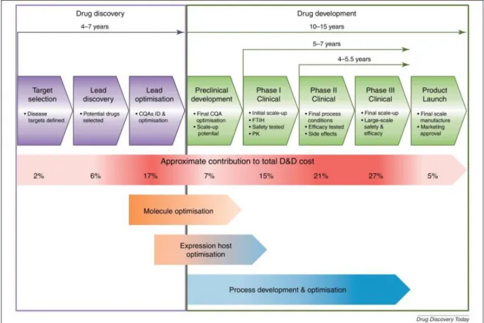
1.7. Pharmaceutical drug discovery and development ... 24
1.7.1. The drug discovery phase ... 26
1.7.1.1. Target identification and validation ... 26
1.8. Main aim and specific objectives ... 30
Chapter 2. Material and Methods ... 31
2.1. Material ... 33
2.1.1. Reagents ... 33
2.1.2. Cells ... 34
2.1.2.1. Escherichia coli strains ... 34
2.1.2.2. Saccharomyces cerevisiae strains... 34
2.1.2.3. H4 neuronal mammalian cells ... 35
2.1.3. Plasmids ... 35
2.1.3.1. Yeast plasmids... 35
2.1.3.2. Mammalian cell plasmids ... 36
2.2. Methods ... 37
2.2.1. Cells media, growth and storage ... 37
2.2.1.1. Escherichia coli media and growth ... 37
2.2.1.2. Yeast media and growth ... 38
2.2.1.3. H4 neuronal mammalian cells ... 38
2.2.2. Molecular biology methods... 39
2.2.2.1. DNA extraction ... 39
2.2.2.2. Quantification of DNA concentration ... 39
2.2.2.3. Agarose gel electrophoresis and DNA gel extraction and purification ... 40
2.2.2.4. Polymerase chain reaction (PCR)... 40
2.2.2.5. Restriction digestion ... 41
2.2.2.6. DNA ligation ... 41
2.2.2.7. Competent E. coli ... 41
2.2.2.8. Introduction of plasmid DNA into E. coli ... 42
2.2.2.9. Selection of positive clones and DNA sequencing ... 42
2.2.3. Genetically engineered yeast strains to express human proteins ... 42
2.2.3.1. Yeast episomal plasmids for Aβ1-42 and tau40 expression ... 42
2.2.3.2. Yeast integrative plasmids for Aβ1-42 and tau40 expression... 43
2.2.3.3. Yeast transformation ... 44
2.2.4.3. Sarkosyl protein fractionation ... 47
2.2.4.4. Fluorescence microscopy and counting of cells with protein inclusions ... 48
2.2.4.5. Statistical analysis ... 48
2.2.5. Screen for gene enhancers of tau40 toxicity with the YKO collection ... 49
2.2.5.1. Preparations of high quality and purified pESC-Leu_Gal10-tau40 plasmid ... 49
2.2.5.2. YKO collection replication ... 49
2.2.5.3. Transformation of YKO strains ... 49
2.2.5.4. Screening yeast gene deletions enhancers of tau40 toxicity ... 50
2.2.5.5. Confirmation of yeast ORF deletion ... 52
2.2.6. Identification of bacterial natural extracts suppressors of tau toxicity in yeast ... 53
2.2.6.1. Validation of the platform mir1Δ-tau40 ... 53
2.2.6.2. SEAVENTbugs marine prokaryotic collection ... 54
2.2.6.3. Screening of 138 natural aqueous extracts from the SEAVENTbugs marine prokaryotic collection ... 55
2.2.7. Genetically engineered PiC knockdown (KD) H4 cells ... 57
2.2.7.1. H4 cells transient transfection... 57
2.2.7.2. PiC knockdown in H4 cells ... 57
2.2.8. Characterization of PiC KD H4 cells ... 58
2.2.8.1. Cell viability analysis: LDH assay ... 58
2.2.8.2. Protein expression analysis ... 58
2.2.8.3. Assessment of mitochondrial function... 59
Chapter 3. A yeast model for studying tau and beta-amyloid interaction ... 63
3.1. Summary ... 65
3.2. Introduction... 66
3.3. Results ... 68
3.3.1. Yeast strains produced in this study ... 68
3.3.2. Single copy beta-amyloid and tau40 integrated into W303-1A genome did not cause toxicity to yeast growth ... 68
3.3.3. Beta-amyloid mCherry fusion protein was toxic to yeast growth at 37ºC ... 70
3.4. Discussion ... 78
Chapter 4. A genome-wide screening to identify yeast gene deletions that enhance tau toxicity ... 83
4.1. Summary ... 85
4.2. Introduction... 86
4.3. Results ... 88
4.3.1. Human tau40 expression was phosphorylated by Rim11, the yeast orthologue of GSK-3β ... 88
4.3.2. Tau40 toxicity enhancer screen ... 89
4.4. Discussion ... 99
Chapter 5. Bacterial natural extracts suppressors of tau toxicity in yeast ... 103
5.1. Abstract ... 105
5.2. Introduction... 106
5.3. Results ... 108
5.3.1. The yeast strain mir1Δ-tau40 was suitable for drug discovery screenings ... 108
5.3.2. Eleven natural extracts were able to rescue mir1Δ-tau40 yeast growth in the primary screening ... 110
5.3.3. Three natural extracts were classified as good candidates for future development in the dose-response confirmation assay ... 112
5.4. Discussion ... 113
Chapter 6. Initial characterization of a mammalian cell model of PiC silencing ... 119
6.1. Abstract ... 121
6.2. Introduction... 122
6.3. Results ... 124
6.3.1. PiC knockdown apparently was not toxic to cells ... 124
6.3.2. Tau phosphorylation at Ser202/Thr205 was not altered by PiC knockdown ... 126
6.3.3. PiC knockdown cells presented apparent compromised mitochondrial function ... 127
6.3.3.1. PiC knockdown did not affect intracellular calcium levels or mitochondrial membrane potential ... 127
Chapter 7. Conclusions ... 133
7.1. Conclusions ... 135
7.2. Go-to-market strategy ... 138
Chapter 8. References ... 141
8.1. References ... 143
List of Figures
Figure 1.1. Tau gene, mRNA and protein isoforms in the human brain. ...6
Figure 1.2.Tau in healthy and diseased neurons. ... 12
Figure 1.3. Alzheimer’s disease predicted progression. ... 13
Figure 1.4. Alzheimer’s disease hallmark post-mortem lesions. ... 14
Figure 1.5. MAPT gene mutations. ... 15
Figure 1.6. Putative sequence of events in tau aggregation into neurofibrillary tangles... 16
Figure 1.7. Tau-based therapeutic strategies. ... 19
Figure 1.8. Cellular processes conserved in yeast, relevant for neurodegeneration... 22
Figure 1.9. Drug discovery and development process phases, with reference to average time and approximate cost of development. ... 24
Figure 2.1. pESC-LEU vector map (Stratagene). ... 35
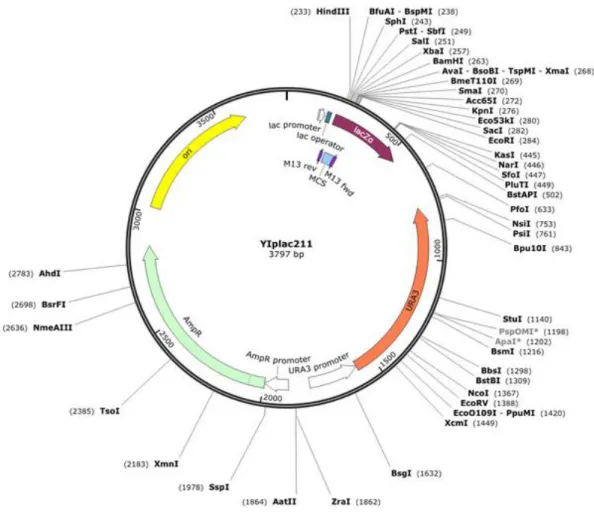
Figure 2.2. YIp211 vector map (http://www.snapgene.com/). ... 36
Figure 2.3. pCDNA3_eGFP vector map. ... 36
Figure 2.4. Map of the pLKO.1 vector. ... 37
Figure 2.5. Schematic diagram of YIp211_GAL (BIOALVO). ... 44
Figure 2.6. Example of a screening plate set. ... 50
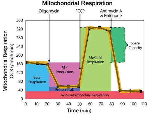
Figure 2.7. Representative OCR profile obtained with the XF Cell Mito Stress test... 61
Figure 3.1 Expression of untagged Aβ1-42 in the cytoplasm of S. cerevisiae BY4741. ... 69
Figure 3.2. Integrative model of co-expression of Aβ1-42-mCh and tau40 in the cytoplasm of S. cerevisiae W303-1A... 70
Figure 3.3. Episomal model of co-expression of Aβ1-42-mCh and tau40 in the cytoplasm of S. cerevisiae BY4741. ... 71
Figure 3.4. Overexpression of Aβ1-42-mCh in S. cerevisiae (BY4741) induces growth delay at 37ºC ... 72
Figure 3.5. Expression of Aβ1-42-mCh and tau40 fluorescent proteins in the cytoplasm of S. cerevisiae BY4741. ... 74
Figure 3.6. Accumulation of Aβ1-42-mCh in S. cerevisiae BY4741. ... 75
Figure 3.8. Tau40 phosphorylation at the AD-related epitopes S396/404 by Rim11, the GSK-3β yeast
orthologue. ... 77
Figure 4.1. Tau40 expression in the cytoplasm of Saccharomyces cerevisiae is non-toxic to yeast. Tau40 is phosphorylated in the pathology-related epitopes Ser396/404... 88
Figure 4.2. Tau40 toxicity enhancer screen high-throughput strategy. ... 89
Figure 4.3. Dot spot assays of yeast knockout strains ski7Δ, gsh1Δ, pes4Δ and ckb1Δ after induction of tau40 expression for 3 days incubation at 30ºC. ... 92
Figure 4.4. Dot spot assays of yeast knockout strains atp23Δ, atp4Δ, etr1Δ and iki3Δ after induction of tau40 expression for 2-3 and 6 days incubation at 30ºC. ... 94
Figure 4.5. Dot spot assays of yeast knockout strains atp11Δ, rrd1Δ, vps15Δ and aim10Δ after induction of tau40 expression for 2-3 and 6 days incubation at 30ºC. ... 95
Figure 4.6. Dot spot assays of yeast knockout strains coq9Δ, mrpl10Δ and yke2Δ after induction of tau40 expression for 6 days incubation at 30ºC. ... 96
Figure 4.7. Dot spot assays of yeast knockout strains pep3Δ and zap1Δ after induction of tau40 expression for 6 days incubation at 30ºC. ... 97
Figure 4.8. Dot spot assays of yeast knockout strains htb2Δ, mrp4Δ and mir1Δ after induction of tau40 expression for 6 days incubation at 30ºC. ... 97
Figure 4.9. ORF deletion confirmation of yeast strains identified as sensitive to tau40 toxicity by dot spot assays. ... 98
Figure 4.10. Dot spot assays of yeast knockout strains mir1Δ, pep3Δ, yke2Δ, coq9Δ, htb2Δ, aim10Δ, mrpl10Δ, zap1Δ and mrp4Δ after induction of mCherry expression for 6 days incubation at 30ºC. ... 99
Figure 5.1. Yeast strain mir1Δ-tau40. ... 108
Figure 5.2. Validation of the mir1Δ-tau40 drug discovery platform. ... 109
Figure 5.3. mir1Δ-tau40 primary screening. ... 110
Figure 5.4. Hit example of the primary screening with mir1Δ-tau40 drug discovery platform. ... 112
Figure 6.1. Optimization of immunoblot for detection of the mitochondrial phosphate carrier (PiC). .. 124
Figure 6.2. Knockdown of SLC25A3 in H4 cells. ... 125
Figure 6.3. PiC knockdown effect on cell viability. ... 126
Figure 6.4. Levels of tau phosphorylation at Ser202/Thr205 (AT8-tau). ... 127
Figure 6.5. Variation of intracellular Ca2+ and mitochondrial membrane potential (ΔΨm) in PiC knockdown H4 cells. ... 128
List of Tables
List of Equations
Abbreviations
Aβ Beta-amyloid
Aβ1-42 Beta-amyloid peptide residues 1-42 Abl Tyrosine-protein kinase ABL1 AD Alzheimer's disease
AD2 Phosphorylation-dependent monoclonal antibody directed against tau proteins found in Alzheimer's disease ADI Alzheimer's disease international
ADME Absorption, Distribution, Metabolism, Excretion
ADMET Absorption, Distribution, Metabolism, Excretion, Toxicity ALS Amyotrophic Lateral Sclerosis
ANOVA Analysis of Variance APP Amyloid precursor protein ATP Adenosine triphosphate
ATPAF1 ATP synthase mitochondrial F1 complex assembly factor 1 β-ME Beta mercaptoethanol
BSA Bovine serum albumin Ca2+i Intracellular Ca2+
CAPS N-cyclohexyl-3-aminopropanesulfonic acid CDK5 Cyclin-dependent kinase 5
cDNA Complementary DNA
CHIP-Seq Chromatin immunoprecipitation sequencing CIAP Calf Intestinal Alkaline Phosphatase CK1 Casein kinase 1
CSNK2B Casein kinase 2, beta polypeptide DDD Drug discovery and development DMEM Dulbecco’s modified Eagle’s medium DMSO Dimethyl sulfoxide
DNA Deoxyribonucleic acid dNTP Deoxynucleotide DRP-1 Dynamin 1-like protein dsDNA Double stranded DNA DTT Dithiothreitol
EDTA Ethylenediamine tetraacetic acid eGFP Enhanced green fluorescent protein EGTA Ethylene glycol tetraacetic acid ETC Electron transport chain
EV Empty vector
FAD Familial Alzheimer's disease
FAP Familial amyloidotic polyneuropathy FBS Foetal bovine serum
FTDP-17 Frontotemporal dementia and parkinsonism linked to chromosome 17 FTLDU Frontotemporal lobar degeneration with ubiquitin-positive pathology Fura-2AM Fura-2 acetoxy-methyl-ester
FUS Fused in Sarcoma protein
Fyn Proto-oncogene tyrosine-protein kinase Fyn
G418 Geneticin
GADPH Glyceraldehyde 3-phosphate dehydrogenase
GAL Galactose
GAL1 Galactose inducible promoter 1 GAL10 Galactose inducible promoter 10
GCLc Glutamate-cysteine ligase, catalytic subunit
gDNA Genomic DNA
GLU Glucose
GPS D2TM Global Platform Screening for Drug Discovery GSK-3β Glycogen synthase kinase-3 beta subunit GSPT1 G1 to S phase transition 1 protein
GTO Granular tau oligomers
h Hour
HD Huntington's disease
HEPES 4-(2-hydroxyethyl)-1-piperazineethanesulfonic acid hERG Human ether-a-go-go related gene
HIST1H2BB Histone cluster 1, H2bb HRP Horseradish peroxidase HSP60 Heat shock protein 60 HSP70 Heat shock protein 70 HTS High-throughput screening
IgG Immunoglobulin G
IKBKAP Kinase complex-associated protein IND Investigative New Drug
INT Iodonitrotetrazolium chloride iRNA Interference RNA
KanMX Kanamycin selector module conferring kanamycin resistance in yeast kb Kilo nucleotide bases
KD knockdown
LB Luria Broth media
LDH Lactate dehydrogenase
Leu Leucine
LEU2 Leucine locus LiAc Lithium acetate
MAPT Microtubule associated protein tau gene MARK Microtubule affinity-regulating kinase mCh mCherry fluorescent protein
MCI Mild cognitive impairment
min Minute
mPTP Mitochondrial permeability transition pore mRNA Messenger ribonucleic acid
MRPL15 Mitochondrial ribosomal protein L15 MRPS2 Mitochondrial ribosomal protein S2 MTBD Microtubule binding domain NAD+/NADH Nicotinamide adenine dinucleotide NCE New chemical entity
NDA New drug application NFT Neurofibrillary tangle NMDA N-methyl-D-aspartate
NP Natural product
NRF1 Nuclear respiratory factor-1 OCR Oxygen consumption rate OD600 Optical density at 600 nm
ON Overnight
ORF Open reading frame OXPHOS Oxidative phosphorylation
PARS2 Mitochondrial prolyl-tRNA synthetase 2 PBS Phosphate buffered saline
PCR Polymerase chain reaction PD Parkinson's disease
PDR5 Plasma membrane ATP-binding cassette (ABC) yeast transporter PEG Polyethylene glycol
PFDN6 Prefoldin subunit 6
PGK-1 Phosphoglycerate kinase 1 PHF Paired helical filaments
Pi Inorganic phosphate
PiC Mitochondrial phosphate carrier protein
PIK3R4 Phosphoinositide-3-kinase, regulatory subunit 4 PK/PD Pharmacokinetics/pharmacodynamics
PMS N-methylphenazonium methyl sulfate PP2A Protein phosphatase 2A
PPP2R4 Protein phosphatase 2A activator, regulatory subunit 4 PSD95 Postsynaptic density protein 95
p-tau Phosphorylated tau PVDF Polyvinylidene difluoride R&D Research and development
RAF Raffinose
RBMX RNA Binding Motif Protein, X-Linked
RIM11 Yeast gene coding for a protein kinase homologue to human GSK-3β rim11Δ Yeast strain carrying a deletion of RIM11 ORF
Rim11 Protein kinase homologue to human GSK-3β RIPA Radio-immunoprecipitation assay
RNA Ribonucleic acid
rpm Rotations per minute
RT Room temperature
SAD Sporadic Alzheimer’s disease SC Synthetic complete media SDS Sodium dodecyl sulfate
SDS-PAGE Sodium dodecyl sulfate polyacrylamide gel electrophoresis SEM Standard error of the mean
Ser Serine
shRNA Short harpin RNA
SLC25A3 Solute carrier family 25 member 3 gene
SNQ2 Plasma membrane ATP-binding cassette (ABC) yeast transporter SOD Superoxide dismutase
ssDNA Single stranded DNA TAE Tris-acetate-EDTA buffer
tau Microtubule associated protein tau tau40 441 amino acid long tau isoform TBS Tris buffered saline
TBST Tris buffered saline supplemented with Tween 20 TCA Tricarboxylic acid
TDP-43 TAR DNA-binding protein 43
TMRM Tetramethylrhodamine methyl ester perchlorate TRIS Tris(hydroxymethyl)aminomethane
UPS Ubiquitin-proteasome system
Ura Uracil
URA3 Uracil locus UV Ultraviolet light
VDAC Voltage-dependent anion channel proteins VPS18 Vacuole protein sorting 18 homologue
WT Wild-type
YKO Yeast knockout collection YPD Yeast extract peptone dextrose ZNF70 Zinc finger protein 70 gene
Chapter 1.
1.1. Proteinopathies
Protein misfolded disorders are triggered by changes in three-dimensional structure of proteins that lead to their self-association and precipitation (Bayer, 2013). Genetic defects, changes in the physical-chemical properties of proteins and/or failure of the protein quality control are processes that influence protein misfolding and formation of small order oligomers that tend to aggregate in higher order structures. These changes in conformation make proteins pathologically active, either by acquiring toxic functions or by losing their physiological functions (Bayer, 2013; Wolfe, 2012).
The aggregation of misfolded proteins may occur in different cells and regions of the body, originating a variety of disorders. When affecting the central nervous system (CNS), proteinopathies are often neurodegenerative disorders, and can be characterized by one or more proteinaceous aggregates (Bayer, 2013). Neurons are quite sensitive to the effects of misfolded proteins due to their post-mitotic nature and structure (Wolfe, 2012). Indeed, the long and narrow axonal projections of neurons can be easily clogged by accumulating proteins or by inefficient transport of nutrients and organelles (Wolfe, 2012). Additionally, accumulated misfolded proteins cannot be diluted through cell division, thereby turning neuron’s integrity highly dependent on the protein homeostasis processes that usually start to fail during ageing (Bayer, 2013; Chen et al., 2011; Wolfe, 2012). These processes involve different yet interconnected cellular strategies that aim at refolding, degrading, or sequestering misfolded proteins. A network of molecular chaperones is central to all these processes, being able to recognize misfolded proteins, actively promoting its refolding or, if not possible, promoting their degradation via the ubiquitin-proteasome system (UPS) (Chen et al., 2011). Another pathway of misfolded protein degradation is autophagy, namely macroautophagy, a process mediating bulk degradation of long-lived proteins or organelles (Rami, 2009).
Neurodegeneration following intra- or extracellular deposition of misfolded aggregated proteins is a common feature of disorders such as Alzheimer’s disease (AD), Parkinson’s disease (PD) and Huntington’s disease (HD). Despite the diversity of proteins involved in these disorders, all seem to adopt a similar, insoluble structure, consisting in fibrils with crossed β-pleated sheet structures (Skovronsky, Lee & Trojanowski, 2006). These disorders are associated with dementia that either occurs in the beginning of the disease, as in AD, or during its progression, as in PD or HD. These age-dependent syndromes are associated with the loss of neuronal function, ultimately leading to the impairment of several cognitive functions, such as memory, thinking, orientation, comprehension and learning capacity (Prince & Jackson, 2009).
healthcare. In 2010, the total worldwide costs of dementia were US$604 billion dollars (Wimo & Prince, 2010). Based on demographics, ADI estimates that by 2030 these costs have increased by 85%, with developing countries bearing the highest share of this economic burden (Wimo & Prince, 2010). For all these reasons, dementia is considered a global health priority (Wortmann, 2012).
A significative progress has been made towards the understanding of the aetiology of many dementias in the last decades, but so far there are no mechanism-based treatments for most disorders. It is therefore imperative that new and better therapeutic solutions are promptly found and made available. Several international cooperative programmes tackle this health threat in several fronts, including (1) raising population awareness and identifying forms of prevention; (2) defining biomarkers to improve early diagnosis and clinical trial assessment; (3) developing drugs and vaccines; and (4) identifying new risk genes and factors that will help define the exact mechanism of disease, essential for the development of effective therapies (Prince & Jackson, 2009).
The most common neurodegenerative disorder is AD, accounting to 50-70% of all cases of dementia. Clinically, AD is characterized by progressive memory loss and cognitive decline due to synapse loss and neuronal cell death (Weintraub, Wicklund & Salmon, 2012). Histopathologically, AD is characterized by two types of post-mortem protein deposits: extracellular amyloid plaques composed of beta-amyloid (Aβ), and neurofibrillary tangles (NFTs) composed by hyperphosphorylated microtubule-associated protein tau (tau) (Goedert & Spillantini, 2006; Wolfe, 2012).
1.2. Tauopathies
The presence of NFTs is a unifying characteristic of a group of heterogeneous dementias and movement disorders known as tauopathies, listed in Table 1.1 (Spillantini & Goedert, 2013b).
Table 1.1. Diseases with tau pathology.
Alzheimer’s disease Hallervorden-Spatz disease Amyotrophic lateral
sclerosis/parkinsonism-dementia complex Myotonic dystrophy
Argyrophilic grain disease Niemann-Pick disease type C
Chronic traumatic encephalopathy Non-Guamanian motor neuron disease with neurofibrillary tangles
Corticobasal degeneration (CBD) Pantothenate kinase-associated neurodegeneration
Creutzfeldt-Jakob disease Pick’s disease
Dementia pugilistic Postencephalitic parkinsonism
Diffuse neurofibrillary tangles with calcification Prion protein cerebral amyloid angiopathy Down’s syndrome Progressive subcortical gliosis
Familial British dementia Progressive supranuclear palsy (PSP) Familial Danish dementia SLC9A6-related mental retardation Frontotemporal dementia and parkinsonism linked
to chromosome 17 (FTDP-17) Subacute sclerosing panencephalitis Gerstmann–Sträussler–Scheinker disease Tangle-only dementia
Guadeloupean parkinsonism White matter tauopathy with globular glial inclusions
Some of the disorders listed above, such as CBD and PSP, are characterized by hyperphosphorylated misfolded tau and formation of NFTs in the absence of other neuropathological abnormalities, clearly involving tau in neurodegeneration. However, other disorders, such as AD, are called secondary tauopathies, due to the involvement of other aggregating proteins in the pathology (Ballatore, Lee & Trojanowski, 2007; Spillantini & Goedert, 2013b).
1.3. Tau protein
–
state-of'-the-art
1.3.1. Tau biology
1.3.1.1. Gene structure, transcripts and isoforms of tau
Tau is encoded in the human brain by a single gene (MAPT) located over 100kb on the long arm of chromosome 17 at band position 17q21.1 (Figure 1.1, top panel) (Gendron & Petrucelli, 2009; Neve et al., 1986; Spillantini & Goedert, 2013b).Two main haplotypes have been identified (H1 and H2), being H1 the most common and overexpressed in some tauopathies (Ávila et al., 2004; Spillantini & Goedert, 2013b).
Figure 1.1. Tau gene, mRNA and protein isoforms in the human brain.
Top panel depicts the MAPT gene, composed of 16 exons (E). Coloured boxes represent alternative spliced exons, white boxes represent untranslated boxes and black boxes represent the exons coding for the repeat domain. In the middle panel, alternative mRNA splicing of E2, E3 and E10 (in colour), produces 6 tau isoforms that are expressed in the adult human brain. The commonly used terms to designate each tau isoform are listed and schematized in the bottom panel, with the number of amino acids and corresponding molecular weight (adapted from Brunden, Trojanowski & Lee, 2009; Martin, Latypova & Terro, 2011).
1.3.1.2. Tau protein structure, expression and post-translational modifications
150-240), which contains seven PxxP motifs, an interaction motif for binding proteins with SH31 domains (Zempel & Mandelkow, 2014).
Tau is a natively unfolded protein, highly soluble, heat and acid-stable, and therefore, does not precipitate during boiling or acid treatment (Mandelkow et al., 2007). Its high solubility and unfolded nature are explained by an enrichment in polar and charged amino acids, being a highly hydrophilic protein (Mandelkow et al., 2007). Despite these characteristics, in disease, tau forms amyloid-like deposits (paired helical filaments, PHF), due to the existence of short hexapeptide motifs in the MTBD 2 and 3 (275VQIINK280 and 306VQIVYK311). These motifs are hydrophobic and interact via a cross-β structure that contributes to the core of PHFs, while the rest of the protein remains highly disordered (Mandelkow et al., 2007).
Tau proteins have been found to be mainly expressed in the central and peripheral nervous systems, but relatively high levels have been detected also in heart, skeletal muscle, kidney, lung and testis and lower levels in adrenal gland, stomach and liver (Morris et al., 2011; Wolfe, 2012). In the CNS, tau is mainly expressed in neurons, but it also occurs in astrocytes and perineuronal glial cells (Gendron & Petrucelli, 2009). In neurons, tau localizes predominantly to axons (Gendron & Petrucelli, 2009), being also found in dendrites (Ittner et al., 2010) and in the nucleus (Shea & Cressman, 1998; Sultan et al., 2011).
Tau proteins are subjected to a high number of post-translational modifications, such as phosphorylation, glycosylation, glycation, prolyl-isomerization, cleavage or truncation, nitration, polyamination, ubiquitination, sumoylation, oxidation and aggregation (reviewed in Martin et al., 2011). The diversity of these modifications suggests that tau biology is highly regulated (Morris et al., 2011). Phosphorylation is the most common and extensively studied tau post-translational modification, because it is widely accepted that (i) phosphorylation level regulates tau binding to the microtubules and (ii) abnormal phosphorylation of tau occurs before the onset of NFTs (Martin et al., 2011; Noble et al., 2013). Tau isoforms can be phosphorylated in more than 80 serine, threonine and tyrosine residues by a variety of kinases (Noble et al., 2013). Kinases that phosphorylate tau at serine/threonine residues include proline-directed kinases, such as glycogen synthase kinase-3 (GSK-3) and cyclin-dependent kinase 5 (CDK5), non-proline-directed kinases such as casein kinase 1 (CK1) and microtubule affinity-regulating kinases (MARKs) (Noble et al., 2013). Tau tyrosine kinases include Fyn, Abl and Syk (Noble et al., 2013). A complete list of tau phosphorylation sites can be found at
http://cnr.iop.kcl.ac.uk/hangerlab/tautable. The level of tau phosphorylation is also regulated by phosphatases that dephosphorylate tau. Indeed, protein phosphatase A (PP2A), the major cell phosphatase, has been implicated in the regulation of tau phosphorylation level and its activity is decreased by about 50% in AD brains (Martin et al., 2011; Noble et al., 2013). The balance between kinase and phosphatase activity is critical for tau function and dysfunction (Wolfe, 2012).
1.3.1.3. Tau binding partners and functions
Tau most widely accepted function is the regulation of microtubule assembly and stability (Weingarten et al., 1975). In vivo, tau may be more involved in microtubules dynamics, participating in processes such as establishment of neuronal polarity, axonal outgrowth and transport of cellular cargoes along axons and dendrites (Gendron & Petrucelli, 2009; Wolfe, 2012). The interaction of the N-projection domain of tau with the plasma membrane (Brandt, Leger & Lee, 1995) and the actin cytoskeleton (Fulga et al., 2007) suggests that tau serves as a mediator between microtubules and the plasma membrane and the actin network (Morris et al., 2011).
Due to intense study of tau biology in the last decade, many dogmas have been challenged and new functions of tau are being established. Although many studies point to a critical function of tau in cytoskeleton-related processes, four independently generated tau knockout mice strains were shown to be viable, fertile and relatively normal (Ke et al., 2012b). Moreover, knockdown of tau with small interference RNA (siRNA) is not cytotoxic to primary cultured neurons and does not prevent axon formation (Qiang et al., 2006). These results indicate that tau is not essential to neurons or microtubule formation. This can be explained by mechanisms of compensation and/or redundant functions of other microtubule-binding proteins, such as MAP1A and MAP1B (Ke et al., 2012b; Morris et al., 2011; Wolfe, 2012).
Other tau functions have been reported as a result of interactions with other cellular structures and enzymes (Morris et al., 2011). Table 1.2 presents a list (non-exhaustive) of several tau-binding partners, placing the protein in many other cell processes.
Table 1.2. Partial list of tau interactors.
Gene Protein Function References
AATF
apoptosis antagonizing transcription factor
Interacts with MAP3K12/DLK, a protein kinase known to be involved in the induction of cell apoptosis
(Barbato et al., 2003)
AKT1
RAC-alpha serine/threonine-protein kinase
Regulate many processes including metabolism, proliferation, cell survival, growth and
angiogenesis (Sadik et al., 2009)
APOE apolipoprotein E3 Mediates the binding, internalization, and catabolism of lipoprotein particles (Huang & Jiang, 2009)
APP Amyloid beta A4 protein
Cell surface receptor and performs physiological functions on the surface of neurons relevant to neurite growth, neuronal adhesion and axonogenesis
(Guo et al., 2006)
ASYN alpha-synuclein
May integrate presynaptic signalling and membrane trafficking. Involved in Parkinson's disease
(Kawakami et al.,
2011)
BAG1 BCL2-associated athanogene
Binds to BCL2 enhancing its anti-apoptotic effects, representing a link between growth factor receptors and anti-apoptotic mechanisms
Gene Protein Function References
BIN1 Myc
box-dependent-interacting protein 1
May be involved in regulation of synaptic vesicle endocytosis. May act as a tumour suppressor and inhibits malignant cell transformation
(Chapuis et al.,
2013)
CAPN2 calpain 2, (m/II) large subunit Calcium-activated nonlysosomal, intracellular cysteine proteases neutral proteases, are (Glading et al., 2004)
CDK5 Cyclin-dependent-like kinase 5
Proline-directed serine/threonine-protein kinase essential for neuronal cell cycle arrest and differentiation and may be involved in apoptotic cell death in neuronal diseases by triggering abortive cell cycle re-entry
(Liu et al., 2002)
DCTN1 dynactin 1
Required for the cytoplasmic dynein-driven retrograde movement of vesicles and organelles along microtubules. Dynein-dynactin interaction is a key component of the mechanism of axonal transport of vesicles and organelles
(Magnani et al., 2007)
DNAAF2 dynein, assembly factor 2 axonemal,
Highly conserved protein involved in the preassembly of dynein arm complexes that power cilia
(Scholz & Mandelkow, 2014)
EP300
histone
acetyltransferase p300
Regulates transcription via chromatin remodelling and is important in the processes of cell
proliferation and differentiation (Min et al., 2010)
FYN Tyrosine-protein
kinase Fyn
Non-receptor tyrosine-protein kinase that plays a role in many biological processes including regulation of cell growth and survival, cell adhesion, integrin-mediated signalling, cytoskeletal remodelling, cell motility, immune response and axon guidance
(Usardi et al., 2011)
GSK-3b Glycogen synthase kinase-3 beta Constitutively active protein kinase involved in many signalling pathways (Kawakami2014) et al., HDAC6 histone deacetylase 6
Plays a critical role in transcriptional regulation, cell cycle progression, and developmental events. Histone acetylation/deacetylation alters chromosome structure and affects transcription factor access to DNA
(Ding, Dolan & Johnson, 2008)
HSP90AB1 Heat shock protein HSP 90-beta
Chaperone that promotes the maturation, structural maintenance and proper regulation of specific target proteins involved, for instance, in cell cycle control and signal transduction
(Karagoz et al., 2014)
HSPA1A Heat shock 70 kDa
protein 1A/1B
Stabilizes existing proteins against aggregation and mediates the folding of newly translated proteins in the cytosol and in organelles. It is also involved in the ubiquitin-proteasome pathway
(Jinwal et al., 2013)
HSPA4 Heat shock 70 kDa protein 4
Chaperone-mediated protein complex assembly; Protein import into mitochondrial outer
membrane; response to unfolded protein (Jinwal et al., 2013)
HSPA8 Heat shock 70kDa protein 8
Chaperone: binds to nascent polypeptides to facilitate correct folding. It also functions as an ATPase in the disassembly of clathrin-coated vesicles during transport of membrane components through the cell
Gene Protein Function References
LRKK2
Leucine-rich repeat serine/threonine-protein kinase 2
Regulates autophagy, plays a role in retrograde trafficking pathway for recycling proteins, regulates neuronal process morphology in the intact CNS. Involved in Parkinson's disease
(Kawakami et al.,
2014)
NUB1
Negative regulator of ubiquitin-like proteins 1
Negative regulator of NEDD8, a ubiquitin-like protein that conjugates with cullin family members
in order to regulate vital biological events (Richet et al., 2012)
PEG10
Embryonal carcinoma differentiation-regulated protein
Reported to have a role in cell proliferation,
differentiation and apoptosis (Gu et al., 2013)
PINCH
LIM and senescent cell antigen-like-containing domain protein 1
Adapter protein in a cytoplasmic complex linking beta-integrins to the actin cytoskeleton, bridges the complex to cell surface receptor tyrosine kinases and growth factor receptors. Involved in the regulation of cell survival, cell proliferation and cell differentiation
(Ozdemir et al.,
2013)
PRNP Major prion protein May play a role in neuronal development and synaptic plasticity (Schmitz2014) et al.,
PSEN1 Presenilin 1
Mutations in this gene cause AD. Presenilins are postulated to regulate APP processing through their effects on gamma-secretase, an enzyme that cleaves APP. Also, it is thought that the presenilins are involved in the cleavage of the Notch receptor, such that they either directly regulate gamma-secretase activity or are protease enzymes
(Takashima et al., 1998)
PSMC2 Proteasome subunit, ATPase, 2 26S
Part of multicatalytic proteinase complex; this subunit interacts with several basal transcription factors-; so, in addition to participation in proteasome functions, participates in the regulation of transcription
(Babu, Geetha & Wooten, 2005)
RPS6KB1 Ribosomal protein S6 kinase beta-1
Serine/threonine-protein kinase that acts downstream of mTOR signalling in response to growth factors and nutrients to promote cell proliferation, cell growth and cell cycle progression
(Pei et al., 2006)
S100B S100 calcium-binding protein, beta (neural) Ion-binding protein (Yu & Fraser, 2001)
SIRT1 NAD-dependent deacetylase sirtuin-1
Studies suggest that the human sirtuins may function as intracellular regulatory proteins with
mono-ADP-ribosyltransferase activity (Min et al., 2010)
SLC1A2 Excitatory amino acid transporter 2
Transports L-glutamate and also L- and D-aspartate. Essential for terminating the postsynaptic action of glutamate by rapidly removing released glutamate from the synaptic cleft. Acts as a symport by co-transporting sodium
(Sasaki et al., 2009)
SQSTM1 Sequestosome 1 Multifunctional protein that binds ubiquitin and regulates activation of the nuclear factor kappa-B
(NF-kB) signalling pathway (Babu et al., 2005)
STUB1 (CHIP)
STIP1 homology and U-box containing protein 1
E3 ubiquitin-protein ligase which targets misfolded chaperone substrates towards proteasomal degradation
(Petrucelli et al.,
Gene Protein Function References
STXBP1 Syntaxin binding
protein 1
Appears to play a role in release of neurotransmitters via regulation of syntaxin, a transmembrane attachment protein receptor
(Bhaskar et al.,
2004)
TRAF6 TNF associated factor 6 receptor- E3 ubiquitin protein ligase, acts as a signalling molecule (Babu et al., 2005)
TTLL6
Tubulin
polyglutamylase TTLL6
Polyglutamylase that preferentially modifies alpha-tubulin, by generating side chains of glycine on the gamma-carboxyl groups of specific glutamate residues
(Zempel et al., 2013)
UBC Polyubiquitin-C
Ubiquitination has been associated with protein degradation, DNA repair, cell cycle regulation, kinase modification, endocytosis, and regulation of other cell signalling pathways
(Petrucelli et al.,
2004)
UBE2D2 Ubiquitin-conjugating enzyme E2D 2
Regulated degradation of misfolded, damaged or short-lived proteins in eukaryotes occurs via the ubiquitin (Ub)-proteasome system (UPS)
(Shimura et al., 2004)
YWHAB 14-3-3-zeta Adapter protein implicated in the regulation of a large spectrum of both general and specialized
signalling pathways (Luong et al., 2000)
YWHAQ 14-3-3 protein theta
Adapter protein implicated in the regulation of a large spectrum of both general and specialized
signalling pathways (Chun et al., 2004) Most of tau kinases and phosphatases are not listed.
Tau binding partners include cytoskeletal proteins, as expected, signalling molecules, proteins involved in the heat shock response and protein folding pathways, regulation of cell cycle and apoptosis. Taking into consideration some of these interactors, tau can act as a protein scaffold, regulating many signalling pathways. One of the most studied of such pathways, in neurons, involves tau interaction with the tyrosine kinase Fyn, establishing tau as a post-synaptic protein (Ittner et al., 2010). The authors hypothesize that tau acts as scaffold protein bringing together Fyn and postsynaptic density protein 95 (PSD95), localizing Fyn at synapses, enabling its activation through N-methyl-D-aspartate (NMDA) receptors. Indeed, tau is required for phosphorylation of NMDA receptor subunit GluN2B in dendrites and mediates Aβ toxicity at dendrites in a mice model of AD (Ittner et al., 2010). Functional roles for nuclear tau have been also proposed (Sjoberg et al., 2006). Moreover, the high degree of tau post-translational modifications, which significance has not been fully characterized yet, further contributes to the complexity of tau biological and pathological roles (Ballatore et al., 2007).
1.3.2. Tau in disease
Abnormal tau hyperphosphorylation is common between human tauopathies, reducing its normal association with microtubules and axonal distribution and, eventually, leading to its aggregation in intracellular filamentous deposits (Figure 1.2). The morphology, isoform content and intracellular localization of these deposits differs depending on the tauopathy (Ballatore et al., 2007). How exactly these morphological changes lead to neurodegeneration is still not fully understood and is a matter of intense debate in the field.
Figure 1.2.Tau in healthy and diseased neurons.
Tau binds and stabilizes microtubules in healthy neurons. In disease, tau becomes hyperphosphorylated, detaching from the microtubules and losing its normal distribution, accumulating in the neuron cytosol (Brunden et al., 2009).
1.3.2.1. Alzheimer
’s
disease
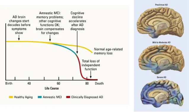
First described in 1907 by Alois Alzheimer, AD is clinically characterized by progressive memory loss and cognitive decline, mood swings, personality changes and loss of independence. The main risk factor for developing AD is age, with prevalence increasing exponentially every 5 years over the age of 65. Early onset is more uncommon and usually suggests a genetic cause (Prince & Jackson, 2009).Death usually occurs 3 or 4 years after diagnosis in people older than 80, or 10 to more years when the disease is diagnosed in younger people.
(Severe AD). Definitive diagnostic of AD is only obtained at authopsy (Hampel et al., 2011; Rodgers et al., 2008).
Figure 1.3. Alzheimer’s disease predicted progression.
The left panel correlates dementia symptoms between AD and normal age-related memory loss. The right panel, presents a schematics of AD brain morphology, depicting the characteristic brain atrophy (Rodgers et al., 2008).
The most common form of AD is called sporadic AD (SAD) presumed to occur due to a complex combination between genetic and environmental causes. The major risk factor for developing SAD is aging, presence of AD risk genes and other environmental factors, as diabetes and cholesterol (Hampel et al., 2011). The hereditary form of AD, usually of early onset, is designated as familial AD (FAD), accounting for less than 5% of all AD cases. FAD occurrence has been associated with mutations in the gene coding for amyloid precursor protein (APP), or its duplication, as occurs in Down Syndrome, and in the presenilin genes (PSEN1 and PSEN2), coding for gamma-secretase subunits, responsible for the cleavage of APP and formation of Aβ. Mutations in the MAPT gene, coding for tau proteins, have not been identified in AD (Medina & Avila, 2014).
Despite the progress in AD detection using cerebral spinal fluid biomarkers and brain imaging scans, the definite confirmation of diagnostics occurs only at autopsy, with the histopathological detection of the two hallmark protein aggregates, the extracellular amyloid plaques and intracellular NFTs (Figure 1.4) (Goedert & Spillantini, 2006).
Figure 1.4. Alzheimer’s disease hallmark post-mortem lesions.
Senile plaques formed by beta-amyloid and neurofibrillary tangles, the latter composed of hyperphosphorylated microtubule-associated protein tau (Goedert & Spillantini, 2006).
Aβ peptides are produced by sequential proteolytic cleavage of APP, by beta- and gamma-secretases. Aβ peptides of 40-42 amino acids (3-4 kDa) are produced at a ratio 10:1, being the peptide Aβ1-42 the most amyloidogenic (Goedert & Spillantini, 2006; LaFerla et al., 2007). The source of intraneuronal Aβ is still debatable, since it can be internalized by the cell from the extracellular plaques or, depending on the site of its production, not secreted to the extracellular space and, hence, intracellular. In principle, intraneuronal Aβ can be produced whenever APP and beta- and gamma-secretases co-localize, and this includes the plasma membrane, trans-Golgi network, endoplasmic reticulum, and endosomal, lysosomal and mitochondrial membranes (LaFerla et al., 2007). If Aβ is produced in the plasma membrane or in the secretory pathway it will be extracellular; if it occurs within the cell, then it will be located intracellularly (LaFerla et al., 2007). The high neurotoxicity of intraneuronal Aβ has been demonstrated in in vitro and in vivo studies (Billings et al., 2005; LaFerla et al., 2007; Oddo et al., 2003) and several reports suggest a direct link between Aβ and tau in causing toxicity in AD (reviewed in Ittner & Gotz, 2011). The mechanism of such interplay, however, is not fully understood and three main modes of interaction have been proposed. Briefly, Aβ may be the trigger of AD, driving tau pathology, probably by activating tau kinases, such as GSK-3β or CDK5; conversely, tau may mediate Aβ toxicity, through its recently established interaction with Fyn kinase; and finally, both proteins may have synergistic toxic effects, as occurs at the level of mitochondria (Ittner & Gotz, 2011).
Other evidences opening new areas of investigation are the reports of the existence of extracellular tau that can induce pathology in surrounding neurons, thus contributing for the spreading of tauopathy throughout the brain (Clavaguera et al., 2009; Guo & Lee, 2011).
1.3.2.2. Frontotemporal dementia
Frontotemporal lobar degeneration (FTLD) is a heterogeneous group of disorders, characterized by frontal and temporal brain atrophy and neuronal loss (Pan & Chen, 2013; Rademakers, Neumann & Mackenzie, 2012). A subset of FTLD disorders arise from fully penetrant, autosomal dominant point mutations in the MAPT gene coding for the microtubule associated protein tau (FTLD-tau), such as FTDP-17, associated with P301L mutation (Rademakers et al., 2012). These genetic tauopathies are
Tangle
accompanied by complex behavioural and cognitive disturbances, including compromised executive function (Pan & Chen, 2013; Rademakers et al., 2012).
Since the discovery of mutations in the MAPT gene associated with FTDP-17, in 1998, over 40 mutations have been identified (Figure 1.5) (Goedert, 2005). Many mutations cluster in and around the regions encoding the microtubule binding domain, suggesting that perturbations in the ability of tau to bind microtubules could be involved in neurodegeneration (Wolfe, 2012). The discovery of these mutations associated with disease demonstrated without doubt that tau dysfunction alone is capable of causing disease (Ballatore et al., 2007; Goedert, 2005). There are mainly two classes of mutations: missense mutations within the coding region of the gene that may conduce to decreased ability of tau to bind microtubules or increase the propensity to form insoluble fibrils; and mutations that affect the splicing of exon 10, leading to an increase of 4R isoforms and therefore to a disequilibrium of the molar ratio between 3R and 4R tau isoforms (Brunden et al., 2009; Goedert, 2005).
Figure 1.5. MAPT gene mutations.
Schematic diagram of tau gene with mutations in the coding region indicated using the numbering of the 441-amino-acid tau isoform (Goedert, 2005).
The clinical and pathological phenotype differs between the different tau mutations (reviewed in Goedert, 2005). Some lead to tau accumulation in both neurons and glial cells, whereas others are exclusive to neurons. Tau filaments also present ultrastructural differences depending on mutations, with some presenting twisted helical filaments; others paired helical filaments (similar to what occurs in AD) and others straight helical filaments (Goedert, 2005).
1.3.3. Loss
vs
. gain of function
As in other proteins associated with different neurodegenerative diseases, tau-induced neurodegeneration is thought to be a consequence of mechanisms of gain of toxic function combined with mechanisms of loss of normal function (Frost, Gotz & Feany, 2015; Medina & Avila, 2014; Noble, Pooler & Hanger, 2011).
0 1 2 3 4 4A 5 6 7 8 9 10 11 12 13 14
1.3.3.1. Loss of normal function of tau protein in disease
Under normal physiological conditions, the binding of tau to microtubules, and consequently its function as a cytoskeleton protein, is regulated by a balance between phosphorylation and dephosphorylation (Noble et al., 2013). In disease, hyperphosphorylation or mutations that decrease tau’s ability to bind microtubules lead to tau detachment from microtubules and missorting from the axon to the somatodendritic compartment. This would reduce the functionality of microtubules and disruption of the structure of the neuronal cytoskeleton, interfering with neuronal polarity, synaptic plasticity, transport of nutrients and organelles along the axon to the synapse, leading to synapse dysfunction and neuronal loss. In this sense, tau mechanism of neurodegeneration would be associated with loss of its normal function (Frost et al., 2015; Medina & Avila, 2014; Noble et al., 2011).
1.3.3.2. Toxic gain of function of tau protein in disease
Hyperphosphorylation, misfolding and missorting of tau from the axon to the cytoplasm leads to increased propensity of tau for suffering additional conformational changes that ultimately lead to formation of soluble oligomers, aggregates and fibrils in the cell body and dendrites of neurons (Ballatore et al., 2007; Ittner et al., 2011). The exact mechanism of tau aggregation is still not fully understood, but there are evidences suggesting that hyperphosphorylation and other post-translational modifications, such as proteolysis, precede tau aggregation (Noble et al., 2013). Also, larger tau aggregates appear to evolve from the successive aggregation of smaller tau species, such as monomers, dimers and soluble oligomers (Figure 1.6) (reviewed in Cowan & Mudher, 2013).
Figure 1.6. Putative sequence of events in tau aggregation into neurofibrillary tangles.
Tau filaments can originate directly from soluble oligomers or from granular tau oligomers (GTO) and can have three forms, as mentioned in the previous section: paired helical filaments (PHF’s), predominant in AD, straight filaments and twisted helical filaments (Cowan & Mudher, 2013; Goedert, 2005). These filaments exhibit β-sheet structure and can be considered amyloid. The bundling of tau filaments originates NFTs that may fill the entire neuron cytosol. The accumulation of tau filaments also in dendrites originates neuropil threads (Cowan & Mudher, 2013). These aberrant species would be, per se, the cause of neuronal dysfunction and degeneration, acting in a variety of cellular processes (reviewed in Frost et al., 2015). Increasing evidences demonstrate that small tau oligomers are the most toxic form of tau, since filamentous and fibrillary tau are not necessary or sufficient to cause tau toxicity and may even be considered a neuroprotective strategy, as suggested by studies with other aggregating proteins such as Aβ, huntingtin or alpha-synuclein (Cowan & Mudher, 2013; Wolfe, 2012). With disease progression, larger tau aggregates will physically impair protein homeostasis and disrupt normal cell functioning (Yoshiyama, Lee & Trojanowski, 2013).
Roberson et al., 2007). Other study suggested a feedback mechanism with tau regulating Aβ, since tau removal resulted in reduced plaque load (Leroy et al., 2012). Finally, pathological tau has been found to activate cell cycle re-entry in post mitotic neurons, initiating a cascade of events resulting from tau-induced actin-stabilization, mitochondrial dysfunction, oxidative stress, DNA damage, heterochromatin relaxation and aberrant gene expression that ultimately leads to neuronal cell death (Frost et al., 2015).
1.4. Tau as a drug target
As mentioned in the previous section, some tauopathies are characterized by accumulation of hyperphosphorylated misfolded tau in the absence of deposition of other proteins, clearly demonstrating the role of tau in disease onset and progression. Moreover, the discovery of mutations in tau gene (MAPT) in FTDP-17 has proved unequivocally that tau dysfunction is sufficient to cause neurodegeneration and dementia (Ballatore et al., 2007; Goedert, 2005).
Increasing evidences also advocate to a more central role of tau in AD pathogenesis and neurotoxicity, albeit the established amyloid cascade hypothesis that postulates Aβ as the disease trigger (Hardy & Allsop, 1991). One of such evidences is the high correlation between cognitive decline and tau pathology, rather than with extracellular Aβ deposition (Medina & Avila, 2014). Additionally, it is becoming widely accepted that tau interacts with Aβ in causing neurotoxicity in AD, although the mechanism of such interaction is not fully understood (Ittner & Gotz, 2011). Further support of tau causative role in neurodegeneration is given by evidences of tauopathy spreading to neighbouring neurons in AD (Clavaguera et al., 2009). Moreover, drug discovery and development programmes focused on Aβ pathology have shown limited efficacy in late stage clinical studies for AD. For example, active immunisation with Aβ resulted in the clearance of the peptide but did not prevent tau pathology or neurodegeneration (Yoshiyama et al., 2013).
The failure of Aβ-based therapies together with increasing understanding of the role of tau in neuropathogenesis, contributed to the focus on tau as a potential target for therapeutic intervention in a wide-range of neurodegenerative disorders (Davidowitz & Moe, 2012; Medina & Avila, 2014). Tau-based therapeutic strategies have, therefore, become a priority and will benefit from the clarification of tau biology and tau-mediated mechanisms of disease (Davidowitz & Moe, 2012; Noble et al., 2011).
1.4.1. Therapeutic strategies targeting tau
To date, there is no effective disease-modifying therapy for tauopathies. Regarding AD, the 5 marketed drugs used for treating symptoms include: four acetylcholinesterase inhibitors and one NMDA-receptor antagonist (Calcul et al., 2012; Noble et al., 2011).
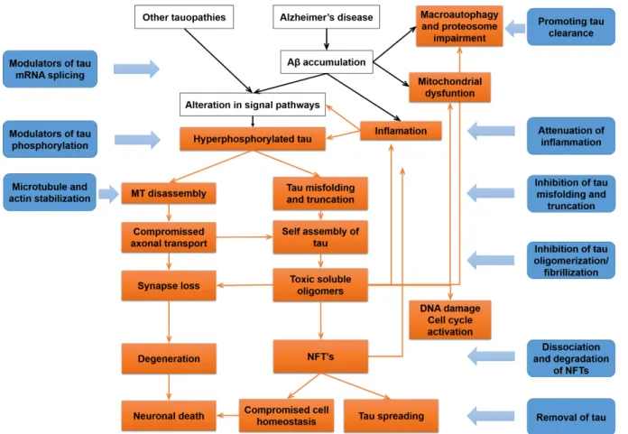
different approaches allows covering all aspects of tau dysfunction at different stages of disease progression (Figure 1.7) (Noble et al., 2011; Yoshiyama et al., 2013).
Figure 1.7. Tau-based therapeutic strategies.
Representation of several aspects of tau dysfunction at different stages of disease (orange squares), correlated with the different therapeutic strategies under development (blue squares) (adapted from Yoshiyama et al., 2013).
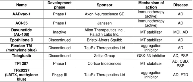
The therapeutic strategies available include inhibitors of tau phosphorylation and misfolding, aggregation blockers, promoters of tau clearance, tau immunotherapies, inhibitors of tau propagation, attenuation of inflammation and mitochondrial dysfunction and oxidative stress, and approaches targeting the regulation of tau pre-mRNA splicing, cell cycle activation, DNA damage and heterochromatin relaxation. There are also strategies that tackle loss of function of tau, such as microtubule-stabilizing agents (Frost et al., 2015; Medina & Avila, 2014; Noble et al., 2011; Yoshiyama et al., 2013). Most of the studies are still in pre-clinical stage, however several small molecules have reached the early stages of clinical development (Medina & Avila, 2014; Noble et al., 2011). Table 1.3 summarizes the small molecules that have reached the clinical stage of development (based on ALZFORUM therapeutics database available at http://www.alzforum.org/).
Table 1.3. Tau-based therapeutics in development.
Name Development phase Sponsor Mechanism of action Disease
AADvac-1 Phase I Axon Neuroscience SE Immunotherapy (active) AD
ACI-35 Phase I Janssen Immunotherapy (active) AD
Davunetide
(AL-108) Inactive
Allon Therapeutics Inc.,
Paladin Labs Inc. MT stabilizer MCI, AD
Epothilone D Discontinued Bristol-Myers Squibb MT stabilizer AD Rember TM
(methylene blue) Discontinued TauRx Therapeutics Ltd
aggregation
inhibitor AD
Tideglusib Discontinued Zeltia Group GSK-3β inhibitor AD, PSP TPI 287 Phase I Cortice Biosciences MT stabilizer AD, CBD, PSP TRx0237
(LMTX, methylene blue)
Phase III TauRx Therapeutics Ltd aggregation inhibitor AD, FTD
Source http://www.alzforum.org/therapeutics.
1.5. BIOALVO SA
Aiming to discover innovative drugs against neurological disorders, BIOALVO was founded in 2006 as a biopharmaceutical company. Its platform technology – GPS D2TM (Global Platform Screening for Drug Discovery) was patented and demonstrated true potential to identify active compounds against different targets. In a constant search for innovative molecules and compounds, BIOALVO turned into the sea and natural sources of new bioactives. This powerful combination gave very positive results in identifying new compounds and activities. In 2010, the company started to slowly enter into other pharmaceutical and cosmetics areas through work with its partners/clients. In 2011, BIOALVO made a deep repositioning strategy, focusing on the exploitation of its assets and uniqueness: the combination of unique and proprietary libraries of extracts with its patented GPS D2TM technology to maximize the applications of natural ingredients in all possible industries. BIOALVO became the Biotech for Natural Products, dedicated to providing fully-integrated biotech solutions to maximize natural products market applications.