TIAGO MANUEL SANTOS JUSTO
Role of Ccbe1 during cardiac differentiation of
mouse ESCs
Doutoramento em Ciências Biomédicas
Trabalho efetuado sob a orientação de:
Professor Doutor José António Henriques de Conde Belo
UNIVERSIDADE DO ALGARVE
Departamento de Ciências Biomédicas e Medicina
2016
Role of Ccbe1 during cardiac differentiation of mouse
ESCs
Declaração de autoria de trabalho
Declaro ser o autor deste trabalho, que é original e inédito. Autores e trabalhos consultados estão devidamente citados no texto e constam da listagem de referências incluída.
iv
Copyright – Tiago Manuel Santos Justo. Universidade do Algarve.
Departamento de Ciências Biomédicas e Medicina.
A Universidade do Algarve reserva para si o direito, em conformidade com o disposto no Código do Direito de Autor e dos Direitos Conexos, de arquivar, reproduzir e publicar a obra, independentemente do meio utilizado, bem como de a divulgar através de repositórios científicos e de admitir a sua cópia e distribuição para fins meramente educacionais ou de investigação e não comerciais, conquanto seja dado o devido crédito ao autor e editor respetivos'.
xi
Acknowledgements
I hereby acknowledge all the support given to me throughout these pays years from all the people in my life. It has been a pleasure to meet and work closely with a wonderful lab crew, whether in normal days or heavy load ones. I reckon all the patience and kindness shown to me by everyone around me, in and out of campus. There are a few names that I will always bear closely in mind and thought from those who worked together with me, helping me to achieve this important mark on my academic life and perhaps professional life. Starting with my co-supervisor Paulo Pereira, who many times had to deal with a quite a lot of naive questions and had them explained to me through several perspectives. That was quite a job, and another PhD itself! Anyways I got to quit poetic writing in scientific manuscripts! I thank Professor José Belo for his support, for giving me the chance of developing this work in his team, and for being a great person in real life. I thank Fernando for his friendship, and sharing a house in a Lisbon adventure during our last PhD year was a remarkable experience, and so it says the oven!
There are quite a few other good friends worth mentioning by name for their incredible support. I thank Ana Tátá for having collaborated with me, but specially for being an amazing person, knowing always how to cheer someone. I also thank Professor José Bragança for all instructive discussions throughout my work. I thank all the Belinhos for being cheerful and amazing people: to Zé Inácio, to Rubi, to Carol, to the 2 Martinhas, to Margaret (mostly because of the wine), to Oriol, to the 2 Joões and to Sara. I thank all the CEDOC crew and the afternoon toast buddies (Inês was the main bread source). Also the weekly cake society!
My pre-PhD reallife mates also deserve a good shout of thanks. I thank João Nuno for being a great friend and adviser, Renato, Nagila, Marta, Sara, Angelina, Joaninha Cristo, my fav cous Abe, my brother Luke and to all in my loong next of kin list: Marta, Rute Tiago, Matias, Adriana, Rui, São, Eduardo, Paulo, Sara, Eduardo II, Samuel, Isaac, Débora, Joana, Jonas, Mariline, Daniel, Leonardo, Alexandre, Pedro, Marco, lil Alice, Isa, Elsa, Zé Manel, Henrique (the policeman), avó Lila, Rosa, Justo, mom and dad (the trumpet guy). Nothing would mean a thing without any of you! I hope we can still live many adventures together and I hope I can learn a lot from all of you.
I have learnt endurance and tenderness with avó Lila, which will always have an important special place in my heart, no matter how long it will take to see her beautiful smile again. Thank you for that, and I miss all of our adventures and late night talks!
I thank again my parents Ana and Manuel, and to my four grandparents for providing me all the education I needed to come this far in life, because above everyone it was thanks to them, and I will be for them!
xiii
Resumo
Das suas quatro cavidades à sua síncrona rede elétrica, o coração foi perfeitamente projetado para servir de interface entre cada órgão presente no corpo humano. Devido à sua complexidade, as doenças cardiovasculares englobam também um grande conjunto de manifestações clínicas incluindo miocardites, hipertensão arterial, defeitos congénitos cardíacos e doenças isquémicas. Muitas destas patologias traduzem-se geralmente na perda de tecido cardíaco funcional e por outro lado pela formação de tecido fibrótico não funcional. Similarmente ao que ocorre nos países desenvolvidos, em Portugal também as doenças cardiovasculares continuam a ser uma das maiores causas de morbidade e mortalidade.
Devido à limitada capacidade regenerativa do coração e ao facto das terapias existentes para tratar doenças cardiovasculares serem ineficientes ou implicarem enormes riscos para o paciente, é urgente desenvolver novas terapias mais eficazes. Nesse sentido, o uso de células multi e pluripotentes tem contribuído na última década para um franco avanço nesta área. Muitos ensaios clínicos têm sido feitos, ou decorrem ainda, onde se avalia a capacidade regenerativa de células estaminais de diferentes origens na reposição dos tecidos cardíacos danificados. Além disto pensa-se que certos nichos de células progenitoras de cardiomiócitos residentes no coração adulto possam representar um mecanismo endógeno de regeneração. De modo a explorar este mecanismo tem-se recorrido a técnicas de isolamento destas células para transplante em doentes cardíacos. No entanto, até agora as melhorias evidenciadas por essas terapias celulares parecem estar associadas a efeitos parácrinos que as células transplantadas exercem sobre os tecidos envolventes, em detrimento da sua implantação no tecido danificado e consequente diferenciação em novo tecido cardíaco. Em paralelo às terapias celulares tem-se feito um esforço para desenvolver patches e scaffolds que possam complementar estas terapias por facilitar o homing de células transplantadas ao constituírem uma matriz onde estas células possam ser envolvidas e desempenhar a sua função.
xiv
regeneração cardíaca é o uso de células já diferenciadas com identidade semelhante à do tecido a ser substituído. No caso do miocárdio, será potencialmente interessante o uso de cardiomiócitos como fonte em transplantes para a regeneração do tecido danificado. Tal abordagem é especialmente interessante visto terem sido identificadas no coração populações de novos cardiomiócitos derivados de cardiomiócitos já existentes, que contribuem para o turnover normal do miocárdio. No entanto, para explorar este mecanismo é necessário criar e otimizar protocolos eticamente aceitáveis para experimentação humana de derivação em grande escala de cardiomiócitos a partir de células pluripotentes. Tal objetivo pode ser alcançado através do uso de fatores segregados que possam ser utilizados para estimular o potencial cardiogénico das células pluripotentes.
A procura de genes envolvidos na cardiogénese têm-se tornado cada vez mais importante com o objetivo de identificar potenciais fatores que possam modular este processo biológico quer in vitro como in vivo. De facto, é possível modelar in vitro com grande rigor os estadios iniciais da cardiogénese através da diferenciação de células estaminais. Tal como ocorre in vivo, a especificação das linhagens cardiovasculares in vitro implica uma transição para populações de células progenitoras cardíacas com potencial de diferenciação cada vez mais restrito e específico. Começando num estado de pluripotência, durante a sua diferenciação estas especificam-se em mesoderme cardíaca e posteriormente em células de todas as outras linhagens cardíacas. Para monitorizar o seguimento deste processo biológico e para assegurar o correto comprometimento nas várias linhagens cardíacas recorre-se à expressão génica de marcadores genéticos específicos para cada linhagem esperada em cada ponto específico de tempo. Através desta monitorização é possível identificar células de mesoderme cardíaca pela expressão dos genes Mesp-1 e Isl-1 a dia 4 de diferenciação das células estaminais, e também diferentes populações de células progenitoras cardíacas pela expressão concomitante de genes como Isl-1 e Nkx2.5 em dias posteriores. Assim é possível estabelecer em laboratório um modelo fidedigno e manipulável para se estudar a cardiogénese.
xv Num rastreio génico efetuado pelo nosso laboratório em células progenitoras cardíacas de galinha com expressão do marcador Nkx2.5, foram identificados genes não caracterizados, mas com um potencial envolvimento na cardiogénese. Um destes novos genes identificados foi o collagen and calcium binding EGF domains 1 ou Ccbe1. Na literatura, é possível hoje ver que em modelos animais knockout para este gene, um outro processo biológico é afetado i.e. a linfangiogénese. Estes animais apresentam uma total ausência de vasos linfáticos. Este fenótipo deve-se em parte ao papel já identificado que o CCBE1 tem na maturação do fator pro-linfangiogénico VEGF-C. Em humanos a síndrome de Hennekam (associado também a mutações em CCBE1), é caracterizada pela existência de uma rede linfática disfuncional fazendo com que estes apresentem um edema generalizado. Não obstante estes estudos, recentemente verificou-se em ratinho e galinha a expressão deste gene nas regiões embrionárias que dão origem ao coração, sugerindo assim também um potencial papel neste processo. De facto, trabalho efectuado no nosso laboratório veio a demonstrar que o silenciamento deste gene em galinha leva ao desenvolvimento de defeitos cardíacos incompatíveis com a vida, associados a uma redução da proliferação das células cardiacas. Também, em ratinhos knockout para este gene é possível identificar um miocárdio subdesenvolvido pelo estreitamento da camada compacta do miocárdio também associado a problemas na proliferação. Assim, no presente trabalho propusemo-nos a estudar mais detalhadamente o envolvimento deste gene nos estadios iniciais da cardiogénese. Como este gene codifica para uma proteína secretada, a verificar-se um importante papel na cardiogénese, a sua manipulação como um fator de crescimento torna-se de grande interesse visando a otimização de protocolos para derivação de cardiomiócitos.
Para estudar os estadios iniciais da cardiogénese recorremos ao uso de uma linha de células estaminais duplamente transgénica que nos permite acompanhar o processo de diferenciação para linhagens cardíacas pois expressam a proteína fluorescente GFP sob o controlo do promotor de Nkx2.5 e a proteína fluorescente dsRed sob um promotor específico de cardiogénese de Mef2c. Assim pode-se confirmar que é possível obter células progenitoras cardíacas in vitro correspondentes aos estadios iniciais do desenvolvimento do coração de ratinho. De seguida analisámos o padrão de expressão de Ccbe1 e
xvi
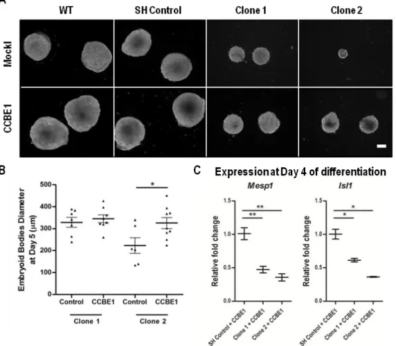
genéticos cardíacos, mostrando que in vitro a sua expressão ocorre aquando da especificação das células para as linhagens cardíacas. Posteriormente gerámos duas linhas estáveis de células estaminais com silenciamento de Ccbe1 para avaliar o seu impacto na cardiogénese. Os resultados demonstram que ao diferenciar estas células em agregados 3D conhecidos como corpos embrióides (nome dado devido à sua semelhança física e funcional com um embrião nos estadios iniciais do desenvolvimento), estas células são incapazes de se especificar em mesoderme cardíaca pois apresentam a expressão de Mesp-1 e Isl-1 reduzida. Em paralelo com estes resultados, foi possível verificar que os corpos embrióides gerados a partir de células estaminais com silenciamento de Ccbe1 apresentam um tamanho muito reduzido. Este defeito é devido não a um aumento da morte celular mas sim a um défice na proliferação das células estaminais silenciadas. Estes defeitos na proliferação estão de acordo com outros estudos efetuados pela nossa equipa, em que fibroblastos embrionários derivados de ratinhos knockout apresentam grandes problemas na proliferação. Adicionalmente, em embriões de galinha foi verificado necessidade de Ccbe1 para a correta proliferação de células precursoras cardíacas para formar o tubo cardíaco. Em conjunto, estes resultados demonstram que CCBE1 tem um papel importante em proliferação. Tais resultados são corroborados por experiências onde foi feita a adição de CCBE1 recombinante ao meio de cultura e se observou a recuperação parcial dos corpos embrióides silenciados. Apesar das dificuldades em produzir quantidades elevadas desta proteína recombinante, os resultados indicam que CCBE1 foi capaz de aumentar a proliferação dos corpos embrióides silenciados. No entanto, as células demonstram-se incapazes de se especificar em mesoderme cardíaca, sugerindo que para além deste papel que Ccbe1 tem em proliferação, o seu papel na cardiogénese é independente deste mecanismo.
Conclui-se assim que Ccbe1 é indispensável para a especificação das células em diferenciação em mesoderme cardíaca. Para vir a ser utilizado no futuro como fator de crescimento em células estaminais em diferenciação, para derivar grandes quantidades de células cardíacas, é necessário desenvolver
xvii ainda mais estudos que permitam ultrapassar as limitações associadas à sua produção e à sua bioatividade.
Paralelamente a estes estudos, uma outra parte do meu trabalho incidiu numa colaboração com uma equipa de bioinformática, na qual nos propusemos a analisar o transcriptoma de diferentes tipos de células progenitoras cardíacas. O objetivo desta análise seria primariamente identificar através de sequenciação RNA novas isoformas de genes envolvidos na cardiogénese, e adicionalmente identificar novos genes não caracterizados com potencial impacto na cardiogénese. Para tal utilizámos a linha de células estaminais duplamente transgénica já referida, da qual isolámos diferentes populações de células progenitoras cardíacas em dias de diferenciação diferentes. Conseguimos analisar o dataset resultante utilizando algumas ferramentas bioinformáticas, que nos permitiu construir uma lista de genes potencialmente envolvidos em cardiogénese ainda não caracterizados. Deste trabalho resultam alguns genes que merecerão um estudo funcional mais detalhado visto estarem claramente expressos nas regiões embrionárias cardiogénicas.
Palavras-chave: cardiogénese; cardiomiócitos; diferenciação de células
estaminais; terapia regenerativa; doenças cardiovasculares; Ccbe1; sequenciação de RNA.
xviii
The identification and use of new growth factors to stimulate the cardiogenic potential of pluripotent cells is a safe and alternative approach to develop cell therapies to address the limited regenerative capacity of the heart.
Collagen and calcium binding EGF domains 1 (Ccbe1) was firstly identified in our laboratory, which encodes for a secreted protein with potential involvement in cardiogenesis. Knockout animal models for this gene and humans with mutations in CCBE1, have lymphangiogenic defects, resulting in the absence of lymphatic vessels. This is in part due to the known described role that CCBE1 has in the processing of the pro lymphangiogenic factor VEGF-C. However, Ccbe1 is also expressed in the embryonic cardiogenic regions of both mouse and chick and in fact, silencing this gene in chick embryos leads to the development of heart defects incompatible with life. Noteworthy, knockout mice show an underdeveloped myocardium. The objective of the present work is to perform a detailed study of the involvement of this gene in the early stages of cardiogenesis.
The results demonstrate that silencing the expression of Ccbe1 or blocking CCBE1 in differentiating stem cells, impairs their specification towards cardiac mesodermal lineages. Additionally, we found that differentiating Ccbe1 KD ESCs have a reduced proliferation rate that leads to smaller EBs. In agreement with this result, when supplementing the differentiating Ccbe1 KD ESCs lines with recombinant CCBE1, we were able to partially rescue the size of the EBs, but the expression of the cardiac mesoderm markers remained downregulated. These data suggest that those defects are independent from each other, but are intimately related to the disruption of Ccbe1, placing CCBE1 as a direct regulator of cell proliferation and cardiac mesoderm specification during ESC differentiation.
Keywords: Cardiogenesis; cardiomyocytes; ESCs diferentiation; cardiovascular
xix
List of contents
Acknowledgements ... xi
Resumo ... xiii
Abstract ... xviii
List of Figures ... xxi
List of abbreviatures, acronyms and symbols ... xxiii
Chapter I General Introduction ... 1
1. Definition and prevalence of cardiovascular disease ... 3
1.1. Limited cardiac regeneration capacity ... 3
2. Current strategies to regenerate the heart ... 3
2.1. Pluripotent and multipotent cell-based therapies in cardiac repair ... 4
2.2. Adult cells-based therapies ... 5
2.3. Cardiac patches and scaffolds in cardiac repair ... 8
2.4. Current challenges and future directions on cardiac tissue repair ... 9
3. Mammalian Heart development: from defined progenitor populations to a 4 chambered organ ... 10
3.1. Endoderm genetic networks defines cardiogenic mesoderm ... 11
3.2. Two defined cardiac progenitor populations ... 13
3.3. ECM in cardiogenesis: collagens and fibronectin ... 15
4. Mouse and human pluripotent stem cell differentiation in vitro recapitulate cardiac differentiation ... 16
5. Identification of genes (splice variants) involved in cardiogenesis: DNA microarrays Vs RNA sequencing ... 20
6. Ccbe1 ... 21
6.1. Protein Structure and Identity ... 21
6.2. Lymphangiogenesis and Hennekan Syndrome ... 21
6.3. Carcinogenesis, proliferation and migration ... 24
6.4. Cardiogenesis ... 26
7. Objectives ... 28
Chapter II Materials and Methods ... 31
2.1. Culture of mouse embryonic fibroblasts (MEFs) ... 33
2.2. Culture of mouse ESCs ... 33
2.3. Differentiation of ESCs ... 33
xx
2.7. cDNA Synthesis ... 36
2.8. Quantitative PCR ... 36
2.9. Production of lentiviral vectors ... 37
2.10. Generation of Ccbe1 knockdown mESCs lines ... 37
2.11. Immunofluorescence in cryosections ... 38
2.12. Immunolabelling ... 38
2.13. Methylene Blue Diffusion Assay ... 39
2.14. Cell Proliferation Assay with Dye eFluor® 670 ... 39
2.15. Production of Recombinant human CCBE1 protein ... 40
2.16. Statistics... 40
RESULTS ... 41
Chapter III Ccbe1 is required for normal cardiac-specification and proliferation in differentiating mouse embryonic stem cells ... 43
3.1. SUMMARY ... 45
3.2. INTRODUCTION ... 46
3.3. RESULTS ... 47
3.4. DISCUSSION ... 60
Chapter IV From Stem Cells to Heart: Identification of novel cardiac genetic players by RNA-seq ... 67
4.1. SUMMARY ... 69
4.2. INTRODUCTION ... 70
4.3. RESULTS ... 71
4.4. DISCUSSION AND CONCLUSION ... 83
Chapter V General Discussion and Future Perspectives ... 87
References ... 95
xxi
List of Figures Chapter I
Figure 1.1 – Schematic representation for the potential uses of cardiovascular
progenitors and cardiomyocytes in cardiac regenerative therapies. ... 6
Figure 1.2 – Cardiac mesoderm formation during gastrulation ... 11 Figure 1.3 – Contribution of the heart fields to the mature tissues of the mature
heart and head. ... 13
Figure 1.4 – Genetic origin of cardiac components ... 14 Figure 1.5 – Growth factors and key transcription factors that regulate fate choices
during early embryonic cardiogenesis and ESCs differentiation ... 17
Figure 1.6 – Schematic representation of the hanging droplet method used to
differentiate ESCs ... 18
Figure 1.7 – Mouse and Human Ccbe1 protein alignment. ... 22 Figure 1.8 - Schematic view of the function of CCBE1 in lymphangiogenesis. ... 23 Figure 1.9 – Ccbe1 expression pattern in cardiogenic regions during mouse
embryogenesis ... 27
Chapter III
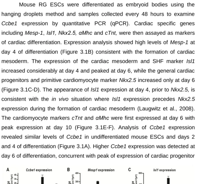
Figure 3.1 - Expression of (A) Ccbe1, (B) Mesp-1, (C) Islet1, (D) Nkx2.5, (E) αMhc,
and (F) cTnt during differentiation of mouse ESCs ... 48
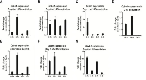
Figure 3.2 - Ccbe1 expression in cardiac progenitors isolated from differentiating
mouse ESCs and embryos at E9.5. ... 49
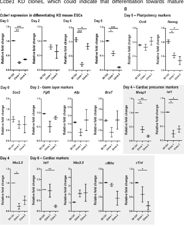
Figure 3.3 - Ccbe1 knockdown leads to reduced cardiac mesoderm formation from
differentiating mouse ESCs... 52
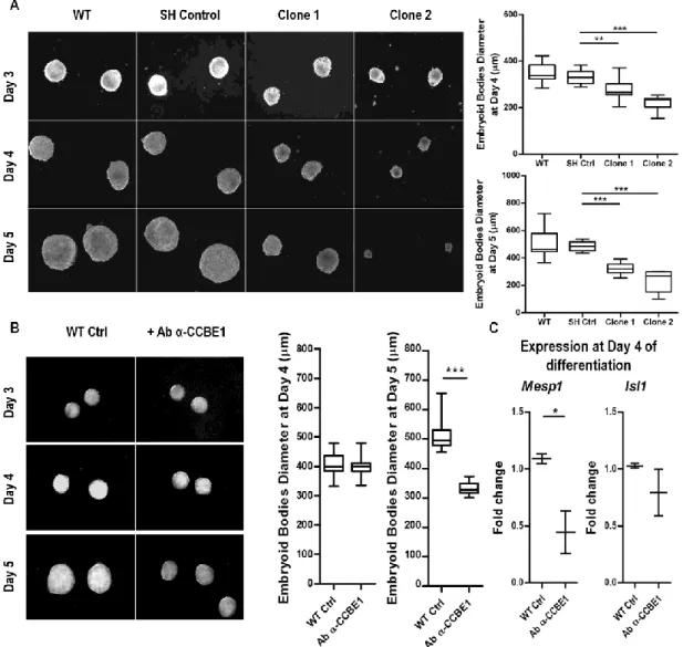
Figure 3.4 - Ccbe1 loss-of-function leads to smaller embryoid bodies. ... 54 Figure 3.5 - Recombinant CCBE1 partially rescues the defects caused by the loss
of Ccbe1. ... 55
Figure 3.6 – Visceral endoderm-like layer is present and cell death is not affected in
the absence of Ccbe1. ... 57
Figure 3.7 - Ccbe1 knockdown decreases the proliferation of differentiating ESCs. ... 59 Figure 3.8 - Defects caused by the absence of Ccbe1 seem unrelated to the role of
CCBE1 in VEGF-C signaling ... 61
Chapter IV
Figure 4.1 – Gate settings used for ESC-derived cardiac progenitor cell sorting. The
xxii
Figure 4.3 – Hierarchical clustering of the expression of all genes ... 74 Figure 4.4 – Boxplot displaying the distribution of the expression values of the
samples from our dataset ... 75
Figure 4.5 – Absolute dsRed and eGFP transcripts count from RNA-seq dataset ... 76 Figure 4.6 – Venn diagram highlighting the number of genes exclusively
up-regulated in the G+R- population at day 4 of differentiation ... 78
Figure 4.7 – Venn diagram highlighting the number of genes exclusively
up-regulated in the G+R- and G+R+ populations at day 6 of differentiation. ... 78
Figure 4.8 – Cardiac Muscle Contraction pathway, which is significantly enriched in
xxiii
List of abbreviatures, acronyms and symbols
#
3D – Three dimensional A
Αfp – Alpha fetoprotein
αMHC – Alpha myosin heavy chain
ao – Aorta
ASC - Adipocyte-derived stem cell B
BMP – Bone morphogenic protein BMMSC – Bone marrow-derived mesenchymal stem cell
C
Ccbe1 – Collagen and calcium-binding
EGF domain-containing protein
CF – Cardiac fibroblast
Cm/s – centimetre per second CNTN2 – Contactin-2
Col - Collagen
CPC – Cardiac progenitor cell CX – Connexin D Da - Dalton dNTP – Deoxynucleotide E E – Embryonic day EB – Embryoid body EC – Endothelial cell ECM – Extracellular matrix EGF – Endothelial growth factor eGFP – Enhanced green fluorescent
protein
EMT – Epithelial-to-mesenchymal transition
EP – Electrophysiological ESC – Embryonic stem cell
F
FACS – Fluorescence-activated cell
sorting
FBS – Fetal bovine serum FGF – Fibroblast growth factor FHF – First heart field
Flk1 – Kinase insert domain receptor
FN – Fibronectin
FOXA2 – Forkhead box protein A2 G
Gata4 - GATA binding protein 4
GO-BP – Gene ontology biological
process database
GO-MF – Gene ontology molecular
function database
H
HCN4 – Potassium/sodium
hyperpolarization-activated cyclic nucleotide-gated channel 4
HFR – Heart forming regions I
Igf2 – Insulin-like growth factor 2 iPSC – Induced pluripotent stem cell
Isl1 - ISL1 transcription factor,
LIM/homeodomain
K
KD – Knockdown
KP – Kegg pathways database L
LA – Left atrium
LEC – Lymphatic endothelial cells LIF - Leukemia inhibitory factor LN – Laminin
LSCV – Left superior caval vein LV – Left ventricle
Lyve1 – Lymphatic vessel endothelial
xxiv
MEF – Mouse embryonic fibroblast
Mef2c – Myocyte enhancer factor 2C
Mesp-1 – Mesoderm posterior protein1
MLC2a/v – Myosin light chain 2a and/or
2v
MSC – Mesenchymal stem cell
MYH – Myosin heavy chain N
NEAA – Non-essential amino acids NF – Neural fold
Nkx2.5 – NK2 homeobox 5
nN/mm2– nanoNewton per squared
millimeter
NPPA – Natriuretic peptide precursor A NRG1 – Neuregulin 1
O
Oct4 - POU class 5 homeobox 1
OFT – Outflow tract P
PBS – Phosphate buffered saline PCR – Polymerase chain reaction PDGF – Platelet-derived growth factor PDGFR – PDGF receptor
PEA – Poly ethyl acrylate PN – Primitive node pt – Pulmonary trunk
PSCs – Pluripotent stem cells
Prox1 – Prospero homeobox 1
PV – Pulmonary vein Q Q – Quadrant qPCR – Quantitative PCR R RA – Right atrium RG – Mouse Nkx2.5-eGFP/SHF-dsRed ESCs
RNA-seq – RNA sequencing rRNA – Ribosomal RNA
S
SAP – Self-assembly peptide Sca1 – Stem cell antigen 1
SCN5A – Sodium channel protein type
5 subunit α
sFrps – Frizzle-like proteins SHF – Second heart field Shh – Sonic hedgehog shRNA – Short hairpin RNA siRNA – Short interfering RNA SMC – Smooth muscle cell
SNP – Single nucleotide polymorphism SOX – SRY-related high-mobility-group
box T T – Brachyury TnC – Troponin C TnT – Troponin T V
VEGF – Vascular endothelial growth
factor
VEGFR – VEGF receptor W
Wnt – Wingless-type MMTV integration
site family, member 1
1
Chapter I
3
1. Definition and prevalence of cardiovascular disease
From its four different chambers to its synchronous electric network, the heart is perfectly engineered to act as an interface to every single different system present in human organism. Due to heart’s complexity, cardiovascular disease can enclose a vast set of cardiac manifestations including inflammatory heart disease, hypertensive heart disease, congenital heart disease and ischemic heart disease. Despite all of these different etiologies cardiovascular disease can have, the ultimate outcome is with no exception very similar – ectopic cardiac function that ultimately leads to scarred and/or dead heart tissue. For example, in ischemic heart disease, coronary insufficiency results in myocardial infarction, and ultimately cardiomyocyte loss.
In Portugal cardiovascular disease is the leading cause of morbidity and mortality in the adult population, being it a proper reflection of that what occurs in developed countries (INE 2013; Jessup and Brozena, 2003).
1.1. Limited cardiac regeneration capacity
As the heart has a very limited regeneration capacity, all injuries caused in heart tissue represent a major medical challenge when it comes to the replacement of the lost tissue. Looking at the major component of the heart the myocardium after an ischemic infarction, contractile myocardial tissue is replaced by non-contractile scar tissue (Cao et al., 2008). Cardiac transplantation has been the standard therapy to overcome a conditioned poorly functioning heart, however is limited by the number of available donors (Jing et al., 2008) and to a series of associated risks such as immunoreactivity, organ rejection and the side effects of immunosuppressive therapies (NHLBI, 2012).
2. Current strategies to regenerate the heart
With the advancement of tissue regeneration technologies on the past two decades, a different light started to be shed on cardiac regeneration, setting in motion the investigation on what could be the real potential of such therapies in restoring lost tissues in damaged hearts. From where we stand now, a lot of progresses have been made in such therapies, as I am going to explain in more detail on the next sections.
4
2.1. Pluripotent and multipotent cell-based therapies in cardiac
repair
The ultimate goal for any cell-based therapy is to regenerate diseased or damaged tissues or cells by the use of autologous, allogenic or xenogenic cells. In the case of the latter two, it would be optimal that these cells lacked immunogenicity in order for the cells to engraft the injured area without triggering an immunological response that could lead to cell rejection and local inflamation. However, life-long lasting immunosupressive therapies often will have to be combined with the use of those cells. On the other hand, autologous cell-based therapies is the ultimate optimal option as this major limitation would be overcome, withdrawing the need to use immunosupressive therapies. Cell-based therapies can comprise diverse delivery strategies, in order to deliver cells into the injured sites or areas, such as systemic intravenous administration or, more specifically, in situ administration, eg. intracoronary administration in myocardial repair strategies (Hastings et al, 2014).
In cardiac repair approaches, it was thought that pluripotent or multipotent stem cells could drive regeneration by differentiating and repopulating the damaged tissue in the heart. Hence, types of cells that preserved to some extent a pluri/multipotent capacity have been so far tested aiming this goal. In fact, there are already excellent reviews about the most various cell types explored in order to develop the most efficient therapy, that are currently on phase I and II clinical trials (Boyle et al., 2006; Sanganalmath and Bolli, 2013; Aguirre et al., 2013; Hastings et al., 2014). Accordingly, a meta-analysis from 50 different clinical studies confirmed that overall local benefit was significant, as ejection fraction increased by 3.96 % for a period of at least 2 years in patients with or without myocardial infarction; while present infarct size was reduced by more than 4% (Jeevanantham et al., 2012).
Most of these cell-based therapies rely on the cardiogenic potential that some cell niches have been identified to preserve in adult mammalians (Kim et al., 2015). Apart from the pluripotent potential of embryonic stem cells (ESCs; discussed in more detail on section 4), other cell niches that have been manipulated aiming towards the same goal include mesenchymal stem cells (MSCs), adipocyte-derived stem cells (ASCs), bone marrow-derived
5 mesenchymal stem cells (BMMSCs) and induced pluripotent stem cells (iPSCs). Although iPSCs have similar differentiation potential as ESCs, one of the major advantages its cell-based therapies offer is that they are patient-specific, meaning that there is a reduced chance of transplant rejection, and are also easy to generate (e.g. with a patient fibroblast sample). In addition, the other cell lines used can only differentiate into more restricted fates as they are multipotent instead of pluripotent like ESCs and iPSCs (Takahashi and Yamanaka, 2006; Gnecchi and Melo, 2009; Nardi and Meireles, 2006; Zuk et al., 2001). However, on the other hand due to their pluripotent state, ESCs and iPSCs have higher tumorigenicity than multipotent or even differentiated cells. Interstingly, the main evidences so far in large mammals and on the ongoing clinical trials have related the benefits of such therapies more likely to a paracrine effect that transplanted cells exert on the surrounding cells rather than to in situ differentiation into new tissue, as initially envisaged (Boyle et al., 2006; Sanganalmath and Bolli, 2013; Aguirre et al., 2013).
2.2. Adult cells-based therapies
Another promising cell population that has been described to have regeneration potential is adult cardiac progenitor cells (CPCs). Despite the heterogeneity, and-nonconsensual origin of CPCs, it has been described that the Sca-1+ compartment of CPCs can contribute, even though at a low rate, to myocardial turnover (extensively reviewed in Valente et al., 2014). Indeed, a clinical trial using cardiospheres-derived Sca1+ cells has shown that these cells contribute to cardiac improvements after myocardial infarction. In this trial it was shown reductions in scar mass (p=0·001), increments in viable heart mass (p=0·01) and regional contractility (p=0·02), and regional systolic wall thickening (p=0.015). However, there were not identified improvements in the left ventricular ejection fraction, the most-expected functional outcome when regenerating the myocardium (Makkar et al., 2012). Therefore there is a need to try to understand at the single cell level, the differences that may exist between the overall Sca1+ CPCs and other Sca1+ stromal cells, such as cardiac fibroblasts (CD90+). It is not clear which cell population, nor to which extent, are
6
Figure 1.1 – Schematic representation for the potential uses of cardiovascular progenitors and cardiomyocytes in cardiac regenerative therapies. Deriving
cardiovascular progenitors from multipotent cells or pluripotent stem cells using growth factors, is a major step for ultimately derive large amounts of cardiomyocytes. On the other hand isolating and purifying adult CPCs populations or cardiovascular progenitors can be transplanted directly into an injured myocardium, or alternatively, be expanded into larger numbers, followed by further differentiation into functional cardiomyocytes. Derived patient-specific cardiomyocytes can be used in various cellular assays, several examples of which are shown, to study and develop therapies for a variety of cardiovascular disorders, including cardiomyopathy, electrophysiological (EP) disorders, and congenital defects. One major goal the production of cardiomyocytes aims is to be used in developing efficient cardiomyocyte transplantation techniques for myocardial regeneration.
7 these different Sca1+ cell populations contributing for cardiac regeneration (Valente et al., 2014). Being confirmed the existence of the multipotent compartment of cells amongst Sca1+ CPCs, it could be speculated that this therapy could also offer the patient-specific benefits, since after patient’s CPCs isolation it would be possible to expand and transplant them into the injured myocardium. Indeed, in Figure 1.1 there is a schematic representation of how isolated cardiovascular progenitors (which can comprehend adult CPC’s) could be used (1) for direct transplantation or (2) cultured and differentiated as a source of cardiomyocytes for further transplantation or patient-specific disease modeling (Davis and Stewart, 2011; Garbern et al., 2013).
In an early 2013 study published in Nature by Senyo and colleagues, it was shown how pre-existing cardiomyocytes could be the major source of new cardiomyocytes found in adult mammals’ hearts, contributing to myocardial turnover. Interestingly, these new cardiomyocytes derived from already existing cardiomyocytes showed to be more abundant in areas adjacent to myocardium injuries, correlating them to a strong contribution for myocardium regeneration (Senyo et al., 2013). As so, exploring this mechanism – transplanting already differentiated cells into the injured areas (e.g. cardiomyocytes) – can be identified as another approach for cardiac cell-based therapies. Such strategy seems a safer alternative than all of the ones considered so far, as with engraftment cells would substitute the exact same cell types lost during an ischemic event, and their capacity to cause tumors in the host organism is rather lower then pluripotent cells. In fact, earlier studies on this approach in rats has proven the technique to be feasible, for the transplanted cardiomyocytes engrafted the host tissue, proliferated and formed cardiac tissue. In addition, transplanted cells were connected to each other by intercalated disks and the newly formed tissue was also more vascularized then the remaining scarred tissue, however their overall arrangement was disorganized when compared to the host cardiac tissue (Li et al., 1996; Sakakibara et al., 2002). Even though this proves it is possible to transplant cardiomyocytes into ischemic injuries, the authors address some concerns with this technique, such as the used cardiomyocytes being from newborn mice and have being rejected after several weeks post-transplantation. Interestingly, in a
8
different study it was indicated that cardiomyocyte transplantation only inhibited the progress of cardiac remodeling in chronic myocardial infarction and did not improve cardiac function significantly (Sakakibara et al., 2002). Nevertheless, to test the viability and efficiency of such approach in humans there would be starting limitations needing to be addressed such as developing protocols that allow a scalable, yet ethical, production of cardiac cells for further transplantation.
One attractive and safe way to achieve this goal could be the use of secreted factors that promote the cardiogenic potential of pluripotent stem cell (both ESCs and iPSCs) or CPCs (Hansson and Lendahl, 2013), but examples in the literature of such factors are still limited (Czyz and Wobus, 2001; Hashimoto and Yuasa, 2013; Khezri et al., 2007; Sato et al., 2006; Takahashi et al., 2003; Zeng et al., 2013). However, combining factors such as hypoxia and bioreactor’s hydrodynamics has shown to be an interesting approach to efficiently maximize the production of cardiomyocytes from iPSCs (Correia et al., 2014). Nonetheless, there is still room for significant improvements in such protocols – on how to alternatively modulate pluripotent or multipotent cell lineages to achive their full cardiogenic potential – as another challenging limitation is related to the non-maturation state these engineered cardiomyocytes present.
2.3. Cardiac patches and scaffolds in cardiac repair
Myocardial infarction leads to ventricular weakening by replacement of the cardiac muscle fibers by non-functional fibrotic scar tissue, leading to ventricular dilation and wall thinning. To avoid this, actual research is also being developed to design cardiac patches that help to improve these defects upon injury. Cardiac patches are three-dimensional scaffolds engineered from natural or synthetic polymers which aim to be engrafted on the site of the injury to help avoiding the progressive impairment of surrounding healthy tissue, and on the other hand also by improving the restoration of the lost functions. By mimicking the extracellular matrix (ECM), cardiac patches can stimulate to a limited extent biological processes such as cell adhesion, proliferation and migration, which intend to drive tissue regeneration. In fact, Holubec et al. reviews how the use
9 of different porcine small intestinal submucosa ECM-derived products in clinical cardiac surgery offers the potential for cellular repopulation and growth in different damaged cardiac tissues (Holubec et al., 2014). However, some approaches of engineering such patches can include the encapsulation of living cells into the polymer mesh, resembling a sheet of living tissue with closer similarity to native myocardial tissue.
To have a clinically relevant effect on damaged hearts, such cardiac patches should have around 1 cm of thickness, be able to generate between 20-50nN/mm2 and also be able to propagate electrical impulses around 25 cm/s (as clearly reviewed and described in Radisic and Christman, 2013). Even though so far the small size of viable cardiac patches for transplantation has been a limitation, Martínez-Ramos and colleagues have recently developed a scalable way to produce injury-size patches which can be grafted into the site of the injured myocardium. While producing scaffolds with similar myocardial physical properties such as elasticity, flexibility and stiffness had been a challenge, this latter group was able to combine a poly (ethyl acrylate) (PEA) scaffold, with self-assembly peptide (SAP) hydrogel RAD16-I and ASCs biohybrid patch that overcomes such limitations (Martínez-Ramos et al., 2014). This clearly shows that synergisms can be created by combining different regenerative strategies. Indeed, in the 6-months follow-up study to their animal models, enhanced systolic and diastolic parameters and also the reduction of the infarct area were identified. Along with the regeneration of the lost myocardium tissue, proper local vascularization of the injured tissue, or even of the engrafted patch, is a requirement for a successful strategy. To meet that need, Ichiara’s team have recently developed a biodegradable surgical patch for high pressure systems that enables the incorporation of endothelial cells (ECs) and smooth muscle cells (SMCs) in the scaffold, therefore opening the way to create vascular grafts (Ichiara et al., 2015).
2.4. Current challenges and future directions on cardiac tissue
repair
As some of the cell-based therapies have shown a significant but yet modest improvement of the cardiac function, primarily related to paracrine effects of the transplanted cells, cell-homing can play an important role in raising the
10
efficiency of such approaches. Taghavi and George review many cell-adhesion markers, growth-factors, chemokines, endothelial nitric oxide synthase and hormones responsible to enhance the homing of transplanted cells to the injured myocardium (Taghavi and George, 2013). Despite such promising alternatives to enhance the efficacy of current cell-based therapies, there seems to be strong suggestions that combining cardiac patches with cell-based therapies can already increase the time window of transplanted cells’ homing and residency on the local of interest. So whether is by increasing the time of exposure of the damaged tissue to the paracrine effects of metabolites produced by the engrafted cells, or whether is by actually easing the homing of more differentiated cell populations (eg. cardiomyocytes and endothelial cells) into the injury site, the way these strategies are evolving offer a very promising fashion in helping the regeneration of the heart.
3. Mammalian Heart development: from defined progenitor
populations to a 4 chambered organ
A better and more detailed understanding of the mechanisms involved in mammalian embryonic cardiogenesis would be beneficial for the development of novel cardiac regenerative therapeutic approaches.
With the growth and development of the embryos, which limits the access to oxygen and nutrients to all the cells, novel embryonic cardiovascular structures are formed to ensure a sufficient supply of nutrients and oxygen to all the cells, and to remove efficiently the cellular waste products. Consequently, around the 3rd week in human embryos and embryonic day (E) 8.5 in mouse embryos, populations of cardiogenic cells start forming the heart to ensure these functions, being the heart the 1st organ to be formed during development (Brade et al., 2013; Carlson, 2014). Due to the conserved similarity in mammalian heart development, mouse cardiogenesis is considered to be a good model for unraveling mechanisms of human heart development. Interestingly, studies in mouse models have determined that specific regions of the embryo are pre-assigned to give rise to specific cardiac structures.
11 During gastrulation, both in mouse and human embryos, precardiac mesodermal cells expressing Brachyury (T) and Mesp1 leave the primitive streak associated with endodermal cells composing the splanchnic mesoderm (Figure 1.2A). Later, these cells migrate anteriorly and adopt a U-shaped disposition – the cardiogenic mesoderm, also called the cardiac crescent (Brade et al., 2013). The endodermal cells that migrate most anteriorly from the
Figure 1.2 – Cardiac mesoderm formation during gastrulation is conserved in human and mice embryos. A) In human, during gastrulation T+ mesodermal cells, Mesp1+ precardiac mesodermal cells and endodermal cells leave the primitive streak
migrating anteriorly and formatting the mesoderm and the endoderm; B) Bmps, released from the newly formed endoderm, signal the formation of a cardiogenic lineage from the mesoderm (red cells), but their influence is limited to the lateral mesoderm because of the release of chordin and noggin from the notochord and Wnt1/3a from the forming neuroectoderm. NF, Neural fold; PN, primitive node. Adapted from Schoenwolf, 2015.
12
primitive streak will form the definitive endoderm which is crucial for cardiac mesodermal cells specification. This process is conserved in mice and human embryos (Lewis and Tam, 2006). During its formation, this structure secretes cardiogenic inductive signals such as bone morphogenic proteins (Bmps), fibroblast growth factor (Fgf), activin, insulin-like growth factor 2 (Igf2) and sonic hedgehog (Shh). These factors contribute to the commitment of mesodermal cells to cardiac fates, and also promote their proliferation and survival (Lewis and Tam, 2006; Schoenwolf, 2015).
3.1. Endoderm genetic networks defines cardiogenic
mesoderm
However all the mesoderm in exposed to these signals, only the cranial part of the lateral mesoderm commits to cardiac fates. In one hand, the lateral specification is related to the inhibitory effects that secreted factors by both the notochord and the neural tube exert on the Bmp signaling (Figure 1.2 B). Chorddin and Noggin are secreted by the notochord, and act by sequestering Bmps, keeping them from binding to their receptors. Wnt1 and Wnt3a are secreted by the neural tube and are antagonizers of the Bmp signaling (Figure 1.2 B; Schoenwolf, 2015). On the other hand, the cranial specification results from the secretion of dickkopf proteins by the cranial definitive endoderm, and from the presence of frizzle-like proteins (sFrps) on those same cells. While sFrps will sequester the Wnt molecules secreted in the cranial mesoderm, dickkopfs molecules will act by binding both to the Wnts and its co-receptors, abrogating their cardiogenic inhibitory signal (Schoenwolf, 2015). Bmp2 signaling will hence act restrictively as an early stimulus to the expression of early cardiogenic transcription factors within the lateral mesoderm, such as Nkx2.5 and Gata4, and its role is also conserved in other vertebrates (Carlson, 2014, Andrée et al., 1998; Schultheiss et al., 1997). For this reason these early mesodermal cardiogenic fields are located bilaterally and later merge to form the cardiac crescent. From this structure two distinct pools of cells can be identified through the expression of unique markers which give rise to specific cardiac structures: the first heart field (FHF) and second heart field (SHF) populations (Kelly et al., 2001; Abu-Issa et al., 2004).
13
3.2. Two defined cardiac progenitor populations
The FHF cardiac progenitors are known to express Nkx2.5 (red cell population in Figure 1.3 and Figure 1.4) soon after the onset of gastrulation under the influence of Bmps secreted by the adjacent endoderm, and are derived from splanchnic mesoderm, which gives rise to the heart tube and subsequently will contribute to the left ventricle and atria (Dehaan 1963, Zaffran et al., 2004; Meilhac et al., 2014; Carlson, 2014). The SHF progenitors are characterized by the expression of Isl1 (green cell population in Figure 1.3 and Figure 1.4),, which together with the Gata transcription factors will drive the expression of a specific SHF enhancer of Mef2c in pharyngeal mesoderm, the embryonic region
Figure 1.4 – Genetic origin of cardiac components. Genetic tracing with a
Mesp1-Cre and Rosa26 conditional reporter shows that almost all cardiac cells in the
14
Figure 1.3 – Contribution of the heart fields to the mature tissues of the mature heart and head. First heart field (red; FHF) and second heart field (SHF; green) and
anterior (pale green/yellow) or posterior (dark green) subdomains of the SHF are shown at different stages of heart and head development. Regions of the heart with a dual origin are shown with colored dots. ao, aorta; LA, left atrium; LSCV, left superior caval vein; LV, left ventricle; OFT, outflow tract; pt, pulmonary trunk; PV, pulmonary vein; RA, right atrium; RSCV, right superior caval vein; RV, right ventricle. Adapted from Meilhac et al., 2014
where this lineage is derived from. Nkx2.5 enhancer in the SHF is then activated by these two transcription factors, leading to its expression in this second pool of embryonic cardiac progenitor cells (Meilhac et al., 2014; Kelly and Evans, 2010). SHF progenitors hence lie medial and slightly caudal to the FHF within the lateral plate mesoderm (Figure 1.3; Schoenwolf, 2015). At E8.0 in mouse and 3rd week in humans, the primordial heart tube is composed mainly by FHF progenitors when the cardiac crescent fuses at midline, after which it starts beating and undergoes rightward looping (Zaffran et al., 2004). Proliferating cells from the SHF start to migrate to the newly-formed heart tube contributing to its elongation and growth at both arterial and venous poles (Figure 1.3). SHF progenitors will give rise to the outflow tract, right ventricle and atria of the developing heart (Buckingham et al. 2005; Kelly et al. 2001). At day 32 in human gestation and E10.5 in mice, the heart presents a well-defined 4 chamber structure, which resembles the form it will have as a mature heart (Brade et al., 2013).
While most of the FHF- and SHF- derived cells are going to mature into cardiomyocytes and compose the myocardium, other cell types found in the heart, i.e. smooth muscle cells (SMCs) and cardiac fibroblasts (CF), will arise
15 from the epicardium. The proepicardial organ is marked by the expression of Tbx18 and Wilm’s tumor protein-1 (Wt1), and is the embryonic structure that gives rise to the epicardium (Figure 1.4). This structure derives from a specialized group of cells within the splanchnic mesoderm during E9.5 in the caudal dorsal mesocardium/septum transversum junction (Meilhac et al., 2014). As the heart looping starts, these cells will start migrating in order to cover all the surface of the myocardium and form the epicardium, and then will undergo epithelial-to-mesenchymal transition (EMT) to enter the myocardium and give rise to CFs and SMCs of the coronary vasculature (Meilhac et al., 2014; Carlson, 2014; Schoenwolf, 2015).
3.3. ECM in cardiogenesis: collagens and fibronectin
The ECM provides structural support for the formation and maintenance of 3 dimensional organs and tissues within an organism. However, the ECM is also a communication net of molecules that allow cells to sense and interpret the environment around them. As a response to those stimuli cells can undergo many cellular processes such as adhesion, migration, proliferation, apoptosis, transformation, and even secrete to that same net additional factors, giving back a response to the surrounding environment. How cells interact with each other and with the surrounding ECM is hence important for the continued understanding of cardiogenesis and cardiac defects (Bowers and Baudino, 2010).
One of the earliest contributions of the ECM to the developing embryonic heart happens before the migration of the mesenchymal cardiac cells to an acellular compartment called the cardiac jelly, located between the myocardium and endocardium of the primitive heart tube. In the mammalian heart the ECM is mostly composed by collagens (Col) of types I, III, IV, VI, fibronectin (FN), laminin (LN) and elastin (Schenke-Layland et al., 2011; Burggren and Keller, 1997). Even though it is not possible yet to understand the role of each one of these single components of cardiac ECM, relevant information is already known on the roles of the FN and Col I, Col IV and LN. In the case of the FN it is known that its loss-of-function in mice leads to severe cardiac malformations (George et al., 1993). More recently, is was described that proliferating niches
16
of Isl1+/Flk1+ cardiac progenitor cells within the right ventricular free wall, the atria and outflow tract of both mouse and human developing hearts, were characterized by a rich Col IV and LN ECM. Indeed, data from the same report strongly suggested that such ECM composition was important to maintain cardiac progenitors in an undifferentiated state, prior to their migration to populate other parts of the heart. Interestingly, while cardiac progenitors migrated from the niche, the surrounding ECM rich in Col I and FN promoted their differentiation towards cardiomyocytes and vascular cells as Isl1 expression was downregulated and cells started to express Troponin C (TnC; Schenke-Layland et al., 2011).
It is getting clear now the determining role of the ECM composition in the developing heart. Now the aim is to try to understand what particular cellular processes do these components regulate and promote in the heart, in order to also exploit such mechanisms in therapeutics (Bowers and Baudino, 2010). In the next section, with the explanation of why are pluripotent stem cells (PSCs) a good model for embryonic cardiogenesis, there will also be addressed some findings that have been made to better understand the role of Col I, Col IV and FN in controlling cellular processes using in vitro models.
4. Mouse and human pluripotent stem cell differentiation in vitro
recapitulate cardiac differentiation
As mentioned earlier, ESCs offer a very promising potential as a source of cells for heart regeneration. In fact, ESCs and iPSCs by being PSCs, when manipulated and differentiated in vitro allow the possibility to recapitulate some of the crucial steps for cardiac specification. Indeed, it is possible to derive cells expressing specific genetic markers of both early- and late- cardiogenesis by differentiating PSCs. However, whether is in vivo or in vitro, the specification of the cardiovascular lineages involves a transition through a sequence of increasingly restricted progenitor cells, proceeding from a pluripotent state to mesoderm and then to cells committed to cardiovascular fates (Figure 1.5; Laflamme and Murry 2011). Culturing and differentiating mouse PSCs as cell aggregates, called embryoid bodies (EBs), has become a routine in many laboratories, since it was proved back in 1985 that spontaneous in vitro
17 differentiation of ESCs could give rise to cells from the 3 embryonic germ-layers (Doetschman et al. 1985). At the same time the differentiation protocol is triggered on PSCs after removal of Leukemia inhibitory factor (LIF) from the culture medium the cells are cultured in hanging drops (Figure 1.6Figure 1.). This technique, which requires the preparation of a cell suspension with a precise cell density, allows the aggregation of the cells with the stimulus of the gravity since these cell suspension drops are cultured in inverted bacterial dishes. After they aggregate, they become EBs, resembling the inner cell mass of early embryos. Simultaneously with this physical change, cells also lose the expression of the pluripotency genes Nanog, Oct4 and Sox2, and start expressing germ-layer specific genes. As the EBs grow in size, they form an outer shell-like layer composed of cells and an enriched collagen IV and laminin
Figure 1.5 – Growth factors and key transcription factors that regulate fate choices during early embryonic cardiogenesis and ESCs differentiation. Growth
factors that regulate fate choices are listed at branch points (green), and key transcription factors and surface markers for each cell state are listed under the cell types (blue). The growth factors are useful for directing the differentiation of ESCs, whereas the markers are useful for purifying cells at defined developmental states. BMPs, bone morphogenetic proteins; CNTN2, contactin-2; CX, connexin; FOXA2, forkhead box protein A2; HCN4, potassium/sodium hyperpolarization-activated cyclic nucleotide-gated channel 4; MESP, mesoderm posterior protein; MLC2a/v, myosin light chain 2a and/or 2v; MYH, myosin heavy chain; NPPA, natriuretic peptide precursor A; NRG1, neuregulin 1; PDGF, platelet-derived growth factor; PDGFR, PDGF receptor; SCN5A, sodium channel protein type 5 subunit α; SOX, SRY-related high-mobility-group box; TBX, T-box transcription factor; VEGF, vascular endothelial growth factor; VEGFR-2, VEGF receptor-2.
18
extracellular matrix (ECM), that can act similar to the endoderm in the embryos by secreting mesoderm inducing morphogens (Li et al., 2001). In fact around day 3, such external layer of cells starts expressing the endoderm-specific genes α-fetoprotein (αfp) and galactosamine epitopes until all the outer shell-like layer is formed by day 4 (Weitzer, 2006). Also during day 3, T which is responsible for inducing mesoderm formation in the embryo starts to be expressed, and is therefore an early mesodermal indicator in differentiating PSCs. After mesoderm specification it is possible to identify transient Mesp-1 and Mesp-2 expression around day 4 in EBs, which are markers of early cardiac commitment. Similarly, in the embryo its expression can be found transiently on the primitive streak prior to migration to the cranial region of the embryo where they become cardiac progenitors (Weitzer 2006; Schoenwolf, 2015). When occurs cardiac mesoderm specification, different early cardiac markers start to be expressed, such as Nkx2.5, Isl1, Gata4 and Tbx18 (reviewed in Meilhac et al., 2014) and such markers can be found to be expressed in differentiating PSCs. After plating the EBs on gelatin-coated culture dishes at day 6, they form cellular structures that within a couple of days start beating and are hence called beating foci. These structures are indicative of cardiac differentiation, and so it is that mature-specific cardiac markers like cardiac Troponin T (TnT) and myosin chains encoding genes (light and heavy chains) start to be expressed. Nonetheless, contrasting with the organized way gastrulation occurs in developing embryos, the way cells commit to cardiac fates in differentiating EBs is rather stochastic, and hence referred to spontaneous differentiation (Weitzer 2006).
Figure 1.6 – Schematic representation of the hanging droplet method used to differentiate ESCs. Undifferentiated ESCs are maintained in culture for 2 passages
prior to differentiation with medium supplemented with LIF. A cell suspension is prepared and cells are put to grow in droplets on an inverted bacterial dish for 2 days, and EBs grow in suspension four additional days. At day 6 of differentiation the formed EBs are plated in to gelatin-coated 6-well plates.
19 Similar to what was described in vivo ColI, ColIV and FN have important roles in promoting the differentiation of PSCs towards cardiac lineages, meaning that the ECM found in differentiating EBs plays also an important role. In fact at day 4 of differentiation, as the EBs commit to early cardiac lineages expressing Mesp1, Isl1 and Flk1, it is shown that the ECM surrounding these cells is rich in ColIV (Schenke-Layland et al., 2011). In addition, culturing undifferentiated ESCs in ColIV substrates was shown to effectively enhance the fold number of Flk1+ cells, while posterior culturing Flk1+ cells in a FN substrate led to expression of αMhc, an early indicator of mature cardiomyocytes. In the one hand, this strongly supports that ColIV is indeed required for the earlier stages of cardiac commitment, proliferation and maintenance of cardiac progenitors in an undifferentiated state. On the other hand, these data supports that once the cells are committed to a cardiac fate, they get sensitive to FN and respond to that stimulus by differentiating into cardiomyocytes (Schenke-Layland et al., 2011). Additionally, inhibiting the synthesis of ColI or blocking its interaction with β1 integrin receptors in differentiating PSCs leads to failure on cardiac lineages commitment and specification, showing it also has an important role on cardiac differentiation and hence is an important cardiac ECM component (Zeng et al., 2013). Of interest, our molecule of study is also a secreted protein and a prospective cardiac ECM component, and was already implied in cellular processes such as cell migration and proliferation in the heart, as it will be explained in detail in section 6. However, its role and function in cardiac differentiation is not well known.
As described before, there are mainly two pools of progenitor cells in the embryo that give rise to the heart - FHF and SHF. These progenitors have only recently been described in mouse ESCs. To do so, these authors generated a transgenic mouse with the red fluorescent protein dsRed under the control of an Isl1-dependent enhancer of the Mef2c gene whose expression is restricted to the SHF. This mouse line was then bred with another transgenic mouse line containing the enhanced green fluorescent protein (eGFP) controlled by the cardiac-specific Nkx2.5 enhancer (Domian et al., 2009). With the expression of these fluorescent markers it was possible to isolate from developing hearts, cell populations expressing one, both or none of these reporters, corresponding to
20
populations of the FHF, SHF and non-cardiac cells as a control. After, blastocysts from these transgenic mice were isolated in order to establish a double transgenic ESC line model, which aimed to become a powerful tool to study the divergent origin of the different cardiac progenitor populations (Domian et al., 2009). By being a good model for embryonic cardiogenesis we chose to work with this double transgenic ESC line as they differentiate into some of the different cardiac populations existing in the developing heart.
5. Identification of genes (splice variants) involved in cardiogenesis: DNA microarrays Vs RNA sequencing
Growth factors or genes that regulate fate choices of differentiating PSCs can in fact offer a powerful tool to boost the production of a desired cell type (known examples in Figure 1.5). Since there are a lot of complex genetic networks interacting during cardiogenesis, the need to identify the factors that could ultimately be manipulated to increase in vivo or in vitro the number of cardiomyocytes or cardiac progenitors for heart regeneration applications, is a field worth exploring. Performing DNA microarrays has been the predominant technique used in the past decade to measure gene expression levels, to identify transcription factors’ binding sites and to genotype single-nucleotide-polymorphisms (SNP), allowing biologists to explore vast amounts of complex digital data. According to this, in our lab was carried out a differential screening using Affymetrix GeneChip system technologies to enable us to identify and study genes expressed and involved in the correct development and differentiation of the vertebrate cardiac progenitor cell lineages. Indeed, this screening led to the identification of more than 700 transcripts differentially expressed in the heart forming regions (HFR), which after bioinformatical analysis and in vivo validation, this number was cut down to a few more than 150. Collagen and calcium-binding EGF domain-containing protein 1 (Ccbe1), our gene of study and interest, was identified among the new genes potentially expressed in the heart precursor cells (Bento et al., 2011). However DNA microarrays have been incredibly useful in a wide variety of applications, they can be particularly problematic for gene families and for genes with multiple splice variants (Bumgarner, 2013).
21 During pre-mRNA maturation to mRNA, alternative splice sites in one gene transcript may give rise to different protein isoforms that differ in their peptide sequence, having consequently different biological and chemical activities. And in fact many genes are known to have several splicing patterns or even thousands (Black, 2003). Now, as the costs of sequencing became cheaper, sequencing is a feasible unbiased approach to measure which nucleic acids are present in a given solution. In addition, it is also independent of our prior knowledge of which nucleic acids may be present and so it can detect closely related gene sequences, novel splice forms or RNA editing that may be missed due to cross hybridization on DNA microarrays (Bumgarner, 2013). RNA sequencing (or simply RNA-seq) is a whole genome transcriptome analysis that allows us to quantify the gene expression in a genome-wide fashion and in a given moment in time. Taking advantage of such technique presents an additional way to identify some of the yet unknown genes or growth factors that regulate cardiogenesis. Nonetheless, at the same time will allow us to identify if some of the already well known cardiac genetic players have different isoforms depending on the nature of the cardiac progenitor population, or if within the same population these change with time.
6. Ccbe1
6.1. Protein Structure and Identity
Ccbe1 encodes a 408 amino acid secreted protein with a calcium-binding EGF-like domain, which has 89% identity with the 406 amino acid human ortholog CCBE1. Even though it is still not possible to determine the correct structure of this protein, it possible to highlight some of the features it has by analyzing its primary amino acid sequence. By aligning mouse and human Ccbe1 sequences (Figure 1.7) and blasting against protein databases, Ccbe1 is shown to have 1 peptide signaling for secretion, 2 collagen domains, 1 calcium binding EGF-like domain, 1 predicted EGF-like domain and 2 glycosylation sites that could be responsible for further protein modifications.
6.2. Lymphangiogenesis and Hennekan Syndrome
During E9.5 in mouse development Ccbe1 is shown to be expressed in tissues surrounding the anterior cardinal vein where Prox1+ lymphatic endothelial cells