UNIVERSIDADE DA BEIRA INTERIOR
Ciências
Production and Purification of DNA G-quadruplex
using pPH600 plasmid
Tiago André Afonso dos Santos
Dissertação para obtenção do Grau de Mestre em
Biotecnologia
(2º ciclo de estudos)
Orientadora: Prof.ª Dr.ª Carla Patrícia Alves Freire Madeira Cruz
Co-orientador: Prof. Dr. João António de Sampaio Rodrigues Queiroz
iii
“Attitude is a little thing that makes a big difference”
Winston Churchill
v
Acknowledgments
First of all, I would like to express my sincere gratitude to my supervisor Dr Carla Cruz and my co-supervisor Professor João Queiroz, for all the guidance, orientation, support and trust that giving me during this year. Your advices and scientific expertise are crucial to the development of this work.
I would also like to express my sincere gratitude to Professor Fani Sousa and Professor Ângela Sousa for the availability, criticism and scientific expertise in all the steps of my work.
My special thanks to Patricia Pereira for his teaching since my first day in the lab. Your friendship, advices, encouragement and scientific expertise are fundamental to the development of this work. Thank you.
I also thank to all the people involved in Health Sciences Research Centre of the University of Beira Interior, especially to the Biotechnology and Biomolecular Sciences group for all their help and to providing me all the resources. I would like to express a special acknowledge to Augusto and Ana Margarida for their friendship, complete availability and support.
To my friends, some of them far but the feeling that giving me was that are always present encourage me and supporting me.
To all of my family, especially to my parents and my brother for all affection, support and care, never doubting me and give me a strength to continue my thrill to success and happiness. I love you.
Finally, i want to thank to my girlfriend, Ana Sofia, for all the patiente, support and love that give me in all moments, always willing to help me in my critical moments and to have a friendly word to encourage me to follow my dreams. I love you.
vii
Resumo
Quando pensamos na estrutura do DNA a imagem que vem imediatamente à nossa mente é a da icónica estrutura em dupla hélice que Watson e Crick descobriram em 1953. No entanto, para além desta estrutura em dupla hélice, o DNA tem capacidade para assumir outras conformações secundárias, não menos relevantes do ponto de vista biológico. Algumas importantes regiões do genoma humano têm um potencial incomum para formar estas estruturas durante o processo de transcrição. O plasmídeo pPH600 tem uma sequência de 604 pb da região de troca Sγ3 da imunoglobulina do ratinho que tem capacidade para formar G-quadruplex durante a transcrição. A predominância de G-loops é maior na topologia superenrolada (sc) do que nas topologias circular aberta (oc) ou linear (ln). O presente trabalho descreve a biosíntese do plasmídeo pPH600 em E. coli DH5α e as estratégias para purificação da isoforma sc do pPH600, diretamente a partir de uma amostra nativa (oc + sc) ou a partir do lisado de E. coli clarificado. As estratégias de purificação são baseadas na cromatografia de afinidade com aminoácidos, aproveitando o reconhecimento biológico com o plasmídeo. Para este propósito dois suportes foram preparados, L-triptofano e L-tirosina, através de imobilização covalente dos aminoácidos à Sepharose CL-6B através do braço espaçador 1,4-butanediol diglicidil éter. O suporte comercial L-arginina-Sepharose 4B foi também utilizado devido à eficiência na purificação da isoforma sc com diferentes plasmídeos utilizando baixas concentrações de sal para ligação e eluição da isoforma sc. Deste modo, foi realizado um
screening inicial com a amostra nativa (oc + sc) de pPH600 para avaliar comportamento dos
três suportes. O suporte que promove a separação das isoformas do pPH600 foi selecionado para purificação da isoforma sc com o lisado de E. coli clarificado. O suporte de L-tirosina foi o que demonstrou resultados proeminentes na separação das duas isoformas, permitindo a recuperação total da isoforma sc de pPH600, através de um gradiente por passos decrescente de 2,25 M para 0 M de (NH4)2SO4 em HEPES 100 mM (pH 7,4) à temperatura de 10ºC.
Subsequentemente, o lisado de E. coli clarificado foi injetado diretamente no suporte de L-tirosina para separar a isoform sc de pPH600 das impurezas internas do hospedeiro (gDNA, RNA, proteínas, endotoxinas e outras conformações de pPH600). A separação foi alcançada com um gradiente por passos decrescente de 2,25 para 1,95 M e posteriormente para 0 M (NH4)2SO4 em
HEPES 100 mM (pH 7,4) à temperatura de 10ºC. O mecanismo de reconhecimento molecular que permite a separação envolve não só as interações hidrofóbicas mas também outras interações não covalentes como, pontes de hidrogénio, empilhamento π-π e van der Waals. Os testes de avaliação do plasmídeo indicam que a isoforma sc de pPH600 resultante do passo de purificação tem um grau de pureza de 98,2%, com um reduzido nível de impurezas. Finalmente, a isoforma sc resultante da purificação foi transcrito e analisado por dicroísmo circular para comprovar a formação do G-loop.
ix
Palavras-chave
G-quadruplex; sc pPH600; cromatografia de afinidade; E. coli; arginina; triptofano; L-tirosina
xi
Resumo Alargado
Nos últimos anos o cancro e as doenças neurodegenerativas têm dominado o interesse dos investigadores devido ao aumento do número de casos diagnosticados e às previsões efetuadas por diversas entidades no sentido do aumento desse número nos próximos anos. Estas doenças têm causado um enorme impacto tanto na saúde como na economia dos países. Até à determinação da composição química e resolução da estrutura da dupla hélice em 1953, pensou-se que o DNA era apenas a molécula que armazenava a informação hereditária. No entanto, com a descoberta da dupla hélice foi possível determinar como a informação genética é transmitida de geração em geração. Muitos estudos foram posteriormente realizados para elucidar possíveis variações da estrutura e a função das mesmas. Várias estruturas secundárias receberam interesse, no entanto os G-quadruplex devido a inúmeros fatores têm recebido grande parte da atenção dos investigadores.
As guaninas têm a capacidade de se organizar e formar estruturas cíclicas através de ligações de hidrogénio, dando origem a G-quartetos. Os G-quartetos através de interações de empilhamento π-π e estabilização com catiões dão origem aos G-quadruplexes. Estas estruturas são polimórficas devido ao elevado número de possibilidades para as formar. Assim, deve ter-se em conta fatores como a quantidade de moléculas envolvidas na formação, ter-sequência dessas moléculas e orientação das cadeias de DNA.
Os G-quadruplex são geralmente encontrados em regiões do genoma com enorme relevância, como são a região de troca da imunoglobulina, os telómeros e regiões regulatórias de oncogenes. Estas estruturas desempenham um papel fundamental em processos celulares essenciais como são a transcrição, a recombinação e a replicação e portanto, têm sido associados a inúmeras doenças.
O processo de transcrição da região de troca da imunoglobulina está diretamente envolvido no desenvolvimento normal das células B e consequentemente na resposta imune. Durante a transcrição formam-se os G-quadruplexes que são o alvo de ligação de diversas proteínas que podem estabilizar ou clivar as estruturas para manter a estabilidade genómica.
Recentemente, vários estudos foram realizados e descreveram a importância do grau de superenrolamento do DNA na formação destas estruturas. A importância deste fenómeno é relacionada não só com o armazenamento da informação genética no núcleo mas, também com a indução de estruturas secundárias devido à energia acumulada durante o superenrolamento do DNA.
Os plasmídeos são moléculas que possuem diferentes topologias: superenrolada, circular aberta e linear e por isso têm diferentes capacidades para formar G-loops. Os G-loops são caracterizados pela formação de um híbrido de RNA-DNA que estabiliza os G-quadruplex na cadeia rica em guaninas. Na literatura está descrito que a percentagem de G-loops formados nos plasmídeos em topologia superenrolada é superior à das outras topologias.
xii
O plasmídeo utilizado foi o pPH600 que contém uma sequência do fragmento da região de troca da imunoglobulina Sγ3 do ratinho e que após transcrição forma G-quadruplexes na cadeia rica em guaninas.
Assim, a necessidade de produzir e purificar a isoforma superenrolada do pPH600 tem grande relevância quando o objetivo é a utilização destas estruturas para estudos in vivo.
A fermentação para amplificação do vetor foi efetuada após transformação das células de E.
coli DH5 com o plasmídeo pPH600. Após fermentação as células forma recolhidas e foi efetuada
a lise das células através de um kit comercial ou através do método de lise alcalina modificado. A etapa cromatográfica foi realizada por cromatografia de afinidade. A cromatografia de afinidade foi a técnica selecionada para purificação de ácidos nucleicos uma vez que permite interações específicas entre o ligando e a biomolécula apresentando vantagens tanto processuais como económicas. Recentemente vários aminoácidos têm sido utilizados como ligandos na cromatografia de afinidade devido às múltiplas interações não covalentes que se podem estabelecer entre as duas unidades básicas, os aminoácidos e os nucleótidos. Neste estudo foi utilizado o suporte comercial, a L-arginina Sefarose e sintetizados dois outros suportes através da imobilização covalente do L-triptofano e da L-tirosina à Sefarose CL-6B. O suporte L-arginina Sefarose tem sido utilizado para purificar diversos plasmídeos com eficiência e por isso foi incorporado neste trabalho. O L-triptofano e a L-tirosina foram selecionados devido às muitas similaridades que apresentam com o suporte de L-histidina, também utilizado recentemente para separar a isoforma superenrolada do plasmídeo de outros contaminantes.
Neste trabalho, pretende-se efetuar um screening inicial da purificação das isoformas do plasmídeo pPH600 com os três suportes e depois selecionar aquele em que melhores resultados forem obtidos para purificação da isoforma superenrolada através de lisado de E. coli. Neste caso, a L-tirosina apresentou os melhores resultados na purificação das isoformas permitindo uma completa separação através de um gradiente decrescente de 2,25 para 0 M de (NH4)2SO4
em HEPES 100 Mm (pH 7.4). A separação completa da isoforma superenrolada de pPH600 das outras impurezas do lisado de E. coli foi obtida através de um gradiente decrescente de 2,25 para 1,95 e finalmente para 0 M de (NH4)2SO4 em HEPES 100 mM (pH 7,4). Após purificação
avaliou-se a qualidade e o nível de impurezas presentes na isoforma superenrolada. Verificou-se que a quantidade de pPH600 superenrolado recuperado foi de aproximadamente 56% e que a pureza da amostra era de aproximadamente 98%. No que diz respeito às impurezas, foram avaliadas através das técnicas definidas pelas agências reguladoras e verificou-se uma diminuição significativa sendo os valores abaixo dos limites aconselhados por estas agências. Por fim, a amostra de pPH600 superenrolado purificada com o suporte de L-tirosina a partir do lisado de E. coli foi sujeita a transcrição e a observação dos G-loops no transcrito foi avaliada por CD.
Verificou-se que o G-quadruplex formado apresentava topologia paralela devido à presença de uma banda negativa a 240 nm e uma banda positiva a 265 nm.
xiii
Abstract
When we think in structure of DNA, the image that comes immediately to mind is the iconic structure in double helix discovered by Watson and Crick in 1953. However, in addition to this structure DNA can assume other secondary structures which are relevant in the biological context. Some important regions of human genome have unusual potential to form this structures upon transcription. The plasmid pPH600 have a sequence of Sγ3 immunoglobulin switch region of murine that are able to form G-quadruplex upon transcription. The G-loops predominance is more evidenced on supercoiled (sc) topology than on relaxed (oc) or linearized (ln) plasmid. The present work describe the biosynthesis of plasmid pPH600 in E. coli DH5α and the strategies for sc pPH600 purification, directly from native sample (oc + sc) and clarified E.
coli lysate. The purification strategies are based on amino acid affinity chromatography taking
advantage of biological recognition to pDNA. For this propose two supports were prepared, L-tryptophan Sepharose and L-tyrosine Sepharose, by covalent immobilisation using 1,4-butanediol diglycidyl ether spacer arm. The commercial support L-arginine Sepharose 4B was also used in the strategy for purifying sc pPH600 since it has already been efficiently applied to separate sc isoforms of different plasmids using mild binding and elution conditions. Therefore, an initial screening with pPH600 native sample (oc + sc) was performed to evaluate the behavior of three supports. The better support in isoform separation was selected to purify sc pPH600 directly from clarified lysate. L-tyrosine support shows the prominent result in separation of two isoforms, allowing the recovery of sc pPH600, through a decreasing stepwise gradient from 2.25 to 0 M (NH4)2SO4 in 100 mM HEPES acid (pH 7.4) with temperature at 10ºC.
Thereafter, the clarified E. coli lysate sample was injected directly onto L-tyrosine support to separate sc pPH600 from internal impurities of E. coli (gDNA, RNA, proteins, endotoxins and other conformations of pPH600). The total separation of sc pPH600 was totally achieved using a stepwise gradient from 2.25, 1.95 and 0 M (NH4)2SO4 in 100 mM HEPES acid (pH 7.4) with
temperature at 10ºC. The underlying mechanism is thought to involve not only hydrophobic but also other non-covalent interactions such as, hydrogen bonds, π-π stacking and van der Waals interactions. Plasmid assessment tests indicated that the sc pPH600 resultant from the purification step presented a purity degree of 98.2%, with an extremely reduced level of impurities.
Finally, the sc pPH600 resultant from purification was transcribed to induce the formation of G-quadruplex and it is confirmed by circular dichroism.
Keywords
G-quadruplex; sc pPH600; Affinity chromatography; E. coli; arginine; tryptophan; L-tyrosine.
xv
Table of contents
CHAPTER I - INTRODUCTION ...1
1.1 - Fundamentals of G4 ... 4
1.1.1 - Formation and structural diversity of G4 ... 4
1.1.2 - Genome locations and functions of G4 ... 7
1.1.3 - Transcription of S regions ... 8
1.1.4 – Circular dichroism of G4 structure ... 10
1.1.5 – Role of G4 in cancer and neurodegenerative diseases ... 11
1.1.5.1 – Aging and cancer ... 11
1.1.5.2 – Neurological diseases ... 12
1.2 Gene therapy and DNA vaccination ... 13
1.2.1 – Gene therapy ... 13 1.2.2 – DNA vaccines ... 15 1.3 - pDNA ... 16 1.3.1 – pDNA specifications ... 17 1.3.2 – pDNA production ... 18 1.3.3 – Downstream processing ... 19 1.3.4 – pDNA purification ... 21
1.3.4.1 – Size exclusion chromatography ... 21
1.3.4.2 – Anion exchange chromatography ... 22
1.3.4.3 – Hydrophobic interaction chromatography ... 22
1.3.4.3 – Affinity chromatography ... 23
CHAPTER II – MATERIALS AND METHODS ... 27
2.1 - Materials ... 27 2.1.1 - pDNA ... 27 2.2.2 – Bacterial strain ... 27 2.2 - Methods ... 28 2.2.1 – pDNA production ... 28 2.2.1.1 – Cell competence ... 28 2.2.1.2 – Transformation ... 29
2.2.1.3 – Plasmid extraction - NZYMiniprep ... 29
2.2.1.4 – pPH600 digestion ... 29
2.2.1.5 – Master cell bank ... 29
2.2.1.6 – Pre inoculum and fermentation ... 30
2.2.2 – Cell lysis ... 31
2.2.2.1 – Plasmid extraction – NZYMaxiprep ... 31
2.2.2.2 – Lysate extraction – Modified alkaline lysis ... 31
2.2.3 – Bead morphology ... 32
2.2.4 – Preparative chromatography ... 32
2.2.4.1 – L-arginine chromatography ... 32
xvi
2.2.4.3 – L-tyrosine chromatography ... 33
2.2.5 – Agarose gel electrophoresis ... 34
2.2.6 – Analytical chromatography ... 34 2.2.7 – Impurities assessment ... 35 2.2.7.1 – gDNA quantification ... 35 2.2.7.2 – Endotoxin evaluation ... 36 2.2.7.3 – Protein analysis ... 36 2.2.8 – Plasmid transcription ... 37
2.2.9 – Circular dichroism spectroscopy ... 37
CHAPTER III – RESULTS AND DISCUSSION ... 39
3.1 – Characterization of plasmid pPH600... 40
3.2 – pPH600 production ... 41
3.3 – pPH600 extraction... 42
3.4 – Characterization of L-tryptophan and L-tyrosine supports ... 44
3.5 – sc pPH600 purification with amino acid based chromatography ... 45
3.5.1 – Pre-purification of sc pPH600 from native sample (oc + sc) ... 45
3.5.1.1 – sc pPH600 pre-purification with L-arginine support ... 45
3.5.1.2 – sc pPH600 pre-purification with L-tryptophan support ... 51
3.5.1.3 – sc pPH600 pre-purification with L-tyrosine support ... 55
3.5.2 – Purification of sc pPH600 from clarified E. coli lysate ... 60
3.6 – sc pPH600 quantification and purity assessment ... 63
3.7 – In vitro transcription and G4 caracterization ... 64
CHAPTER IV – CONCLUSIONS ... 67
CHAPTER V – FUTURE CONSIDERATIONS ... 69
xvii
List of figures
Chapter I – Introduction
Figure 1 - Chemical Structure of DNA and RNA nucleotides ... 1
Figure 2 – G-quartet and G4 structure ... 4
Figure 3 - G4 stoichiometries and folding patterns ... 6
Figure 4 – Diagram of Class Switch Recombination (CSR) from IgM to IgA ... 8
Figure 5 - G-loop formed in a transcribed G-rich region and binding of MutSα ... 9
Figure 6 – Circular dichroism spectra with parallel and antiparallel topologies ... 10
Figure 7 – Compiled data on gene therapy clinical trials ... 14
Figure 8 - Mechanism of DNA vaccines ... 15
Figure 9 – Different conformations of pDNA ... 16
Figure 10 - The three stages of sc pDNA development process ... 18
Figure 11 - Process flow sheet for the purification of sc pDNA ... 19
Chapter II – Materials and methods
Figure 12 – Plasmid pPH600 map ... 28Figure 13 – Standard curve for sc pDNA quantification ... 34
Figure 14 - Standard curve for the quantification of gDNA ... 35
Figure 15 – Standard curve for the quantification of Endotoxin ... 36
Figure 16 - Standard curve for the quantification of proteins ... 36
Chapter III – Results and discussion
Figure 17 – Plasmid pPH600 map and digestions with HindIII and PVUII ... 40Figure 18 – Growth profile of E. coli DH5α harbouring plasmid pPH600 ... 42
Figure 19 – Samples of extraction in 1% agarose gel electrophoresis ... 43
Figure 20 - SEM micrographs ... 44
Figure 21 – Molecular structure of L-arginine coupled to Sepharose 4B through 12 carbon atom spacer arm ... 46
Figure 22 – Chromatographic profile of pPH600 from L-arginine support and respective agarose gel electrophoresis performing a stepwise gradient 10 mM Tris-HCl (pH 8.0) and 500 mM NaCl ... 47
xviii
Figure 24 - Chromatographic profile of pPH600 from L-arginine support and respective
agarose gel electrophoresis performing a stepwise gradient of 110 and 500 mM NaCl in 10 mM Tris-HCl (pH 8.0) ... 50
Figure 25 – Molecular structure of L-tryptophan coupled to Sepharose CL-6B through 12
carbon atom spacer arm ... 51
Figure 26 – Chromatographic profile of pPH600 from L-tryptophan support and respective
agarose gel electrophoresis performing a stepwise gradient of 2.65 and 0 M (NH4)2SO4 in 100 mM HEPES acid (pH 7.4) ... 53
Figure 27 - Main interactions between L-tryptophan and cytidine ... 54 Figure 28 - Chromatographic profile of pVAX-LacZ from L-tryptophan support and
respective agarose gel electrophoresis performing a stepwise gradient of 2.7 and 0 M (NH4)2SO4 in 100 mM HEPES acid (pH 7.4) ... 55
Figure 29 – Molecular structure of L-tyrosine coupled to Sepharose CL-6B through 12
carbon atom spacer arm ... 56
Figure 30 - Main interactions between L-tyrosine and guanine ... 57 Figure 31 - Chromatographic profile of pPH600 isoforms from L-tyrosine support and
respective agarose gel electrophoresis performing a stepwise gradient of 2.25 and 0 M (NH4)2SO4 in 100 mM HEPES acid (pH 7.4) ... 58
Figure 32 - Chromatographic profile showing the optimization of sc pPH600 purification
from clarified E. coli lysate with L-tyrosine support and respective agarose gel electroforesis performing a stepwise gradient of 2.25, 1.6 and 0 M (NH4)2SO4 in 100 mM HEPES acid (pH 7.4) ... 61
Figure 33 - Chromatographic profile showing the purification of sc pPH600 from clarified
E. coli lysate with L-tyrosine support and respective agarose gel
electrophoresis performing a stepwise gradient of 2.25, 1.95 and 0 M (NH4)2SO4 in 100 mM HEPES acid (pH 7.4) ... 62
Figure 34 – RNA G4 formation upon transcription of plasmid pPH600 with agarose gel
xix
List of tables
Chapter I – Introduction
Table 1 - Specifications and recommended assays for assessing pDNA quality ... 17 Table 2 - Different affinity chromatography approaches ... 24
Chapter II – Materials and methods
Table 3 – Summary of binding/elution profiles of plasmid pPH600 isoforms with L-arginine
support in different NaCl conditions ... 48
Table 4 - Summary of binding/elution profiles of plasmid pPH600 isoforms with
L-tryptophan support in different (NH4)2SO4 conditions ... 52
Table 5 - Summary of binding/elution profiles of plasmid pPH600 isoforms with L-tyrosine
support in different (NH4)2SO4 conditions ... 56
Table 6 - Quantitative analysis of the sc pDNA, recovery yield and purity in each peak
recovered from clarified E. coli lysate using L-tyrosine support ... 63
Table 7 - Protein, endotoxin and gDNA assessment from clarified E. coli lysate sample
xxi
List of acronyms
A Adenine
ADA Adenosine deaminase
A-DNA A-form of DNA
AID Activation-induced cytidine deaminase
Aβ Β-amyloid peptides
BCA Bicinchoninic acid
Bcl-2 B cell lymphoma 2
B-DNA B form of DNA
bp Base pairs
C Cytosine
C9ORF72 Chromosome 9 open reading frame 72
CD Circular dichroism
c-Kit V-kit Hardy-Zuckerman 4 feline sarcoma viral oncogene homolog
cm centimeter
c-Myc V-myc avian myelocytomatosis viral oncogene homolog
c-rich region Cytosine rich region
CSR Class switch recombination
DNA Deoxyribonucleic acid
Duplex DNA double strand
E. coli Escherichia coli
EAEMP European Agency for the evaluation of medical products
EDTA Ethylene diamine tetra-acetic acid
EU Endotoxin unit
FDA Food and drug administration
FMR1 Fragile X mental retardation 1
FMRP Fragile X mental retardation protein
G Guanine
g gram
G4 G-quadruplex
gDNA Genomic DNA
g-rich region Guanine rich region
h hours
HEPES acid HEPES (4-(2-hydroxyethyl)-1-piperazineethanesulfonic acid )
hTERT Human telomerase reverse transcriptase
IgA Immunoglobulin A
IgM Immunoglobulin M
xxii
KD Affinity constant
KRAS Kirsten rat sarcoma viral oncogene homolog
L liter
LAL Limulus amebocyte lysate
LB Luria Bertani
ln linear
M Molar
min Minutes
miR1229 Micro RNA 1229
mL milliliter
mM millimolar
mRNA Messenger RNA
MutSα Mismatch repair protein
ng nanogram
nm nanometer
NMR Nuclear magnetic resonance
NOP56 Nucleolar protein 56
oc Open circular
ºC Celsius
OD600 Optical density at 600 nm
PAGE Polyacrylamide gel electrophoresis
PCR Polymerase chain reaction
PDGF-A Platelet-derived growth factor subunit A
pDNA Plasmid DNA
PEG Polyethylene glycol
PNA Peptide nucleic acid
pRb Retinoblastoma protein
premiR-29 Pre micro RNA-29
PRNP Prion protein
RCF Relative centrifugal force
rDNA Ribosomal DNA
RNA Ribonucleic acid
rpm Rotations per minute
S region Switch region
sc supercoiled
SCA36 Spinocerebral ataxia type 36
SCID Severe combined immunodeficiency
SDS Sodium dodecyl sulphate
SEM Scanning electron microscopy
xxiii
T Thymine
TB Terrific broth
TF Transcription factor
Tris Tris(hydroximethyl)methylamine
TSS Transcription start site
U Uracyl
UV Ultraviolet light
V Volts
v/v Volume/volume
VEGF-A Vascular endothelial growth factor A
w/v mass/volume
Z-DNA Z-form of DNA, left handed-DNA
μg microgram
1
Chapter 1
Introduction
Since were discovered in 1869 by Friedrich Meischer, a Swiss Physician, the nucleic acids have been the subject of fields such biology, genetics and biochemistry. Meischer working at University of Tübingen, Germany in chemical composition of leukocytes. In his studies he isolate a hitherto unknown substance from the nuclei of cells. Meischer called the isolated substance
nuclein. Nuclein is a crude extract, and contain many proteins. Subsequently, analysis and
isolation of this substance showed that it had acidic nature and containing phosphorus, and its name was changed to nucleic acid [1, 2]. After that, nucleic acids continues to amaze and intrigue many researchers devoted to its study.
In 1919, Phoebus Levene was the first to suggest that nucleic acids was a linear chain composed by units of a sugar backbone that links to a phosphate groups and nitrogen bases. Levene identified D-ribose and 2-deoxy-D-ribose as the sugars occurring in RNA and DNA, respectively. He also identified the nitrogen bases, purines and pyrimidines. DNA is composed by cytosine (C) and thymine (T) that are pyrimidines and adenine (A) and guanine (G) that are purines. In RNA thymine is replaced by uracyl (U) that is a pyrimidine derivative. He called each unit of phosphate-sugar-base, nucleotide (Fig. 1). Based on this, Levene’s work culminated with the suggestion of a tetranucleotide hypothesis for the structure of nucleic acids, in which nucleotides were always linked in the same order [3].
2
Later in 1940’s decade, Erwin Chargaff and collaborators showed the lack of equality of four bases in most samples of DNA, unlike as required by the tetranucleotide hypothesis for the structure of nucleic acids. He was the first to develop micro-methods such as, paper chromatography and ultraviolet light absorption for accurate analysis of purines and pyrimidines and observed that the amount of adenine is equal to thymine and the amount of guanine is equal to cytosine, but the amount of guanine is not identical to thymine, nor adenine that of cytosine [5]. In this decade many other scientists such as, Avery, MacLeod, McCarthy, Hershey and Chase, contribute with their studies to prove that DNA was the carrier of hereditary information and not the proteins as previously thought.
In 1953, Francis Crick and James Watson discovered double helix of DNA known as B-DNA. This fact, marked a decisive milestone in the history of science in last century. Since then, the B-DNA has usually been regarded as the biologically relevant structure of B-DNA [6].
However, in addition to the normal Watson-Crick duplex, B-form, nucleic acids has potential to form a high number of alternative structural conformations under specific experimental or in
vivo conditions, which are less familiar, although with biological relevance. The negative
supercoiling of DNA can induce sequence-dependent conformational changes that give rise to alternative DNA conformations such as cruciforms, A-DNA, left-handed DNA (Z-DNA), triplexes, four-stranded DNA, known as G-quadruplexes (G4) and others [7].
First evidence that G4 behaved differently from all other nucleobases was first noted in 1910 by Ivar Bang, when he observed that it could spontaneously form a gel. However, only 52 years later, in 1962, the structure has been discovered by Davies and collaborators [8]. The structure has termed G4 because fold within extended guanine-rich regions into four stranded motifs, using an alternative base pairing arrangement [9]. These structures are generated by stacked G-quartets that are composed by four guanine bases in each of which interacts with its neighbours through Hoogsteen hydrogen bonding. These structures are further stabilized by cations (e.g., Na+, K+) that are in the center between the stacks, neutralizing the electrostatic
repulsion of inwardly pointing guanine oxygen’s [10].
Nowadays there is a growing evidence that G4 structures play important roles in various genetic processes [11]. They are often present in biologically regions, such as telomeres, regulatory regions of oncogenes and immunoglobulin switch regions. During last decades, studies indicated that formation of G4 in these regions may play important regulatory roles. Therefore, the polymorphism of G4 could make them valuable molecular targets to study biological processes and for possible therapeutic interventions in cancer and other diseases [12, 13].
The plasmid pPH600 contains a fragment of Sγ3 switch region of murine and upon transcription is able to form G4 structures. The supercoiling show more ability to form G4 as described by Duquette and collaborators [14].
Therefore there is a need to obtain purified sc pPH600 for G4 in vitro assessment. For this propose, affinity chromatography plays a powerful role in separation, identification and purification of sc pDNA. Recently, amino acids such as, L-arginine [15], L-lysine [16], L-histidine [17] and L-methionine [18] have been used as ligands in affinity chromatography to purify sc
3
pDNA from impurities. The principle of technique is based on the natural interactions that occur between amino acids and nucleic acids in the biological environment. Thus, the development of new methods to produce and purify vectors such as, pPH600 that contains a sequence that encodes to G4 became essential for the study.
Based on aforementioned, the main proposes of this work are the development of methods in a typically biotechnological process:
1. Biosynthesis of plasmid pPH600 that contains a sequence that encodes G-quadruplex. 2. Screening of binding/elution conditions of three affinity chromatography supports for
isolation and purification of sc pPH600.
3. Isolation and purification of plasmid sc pPH600 isoform from Escherichia coli (E. coli) clarified lysate using the select support that allowed the desirable sc pPH600 with high yield and purity level.
4. In vitro transcription of sc pPH600 to induce G-quadruplex formation and it detection by circular dichroism (CD).
4
1.1 - Fundamentals of G4
1.1.1 – Formation and structural diversity of G4
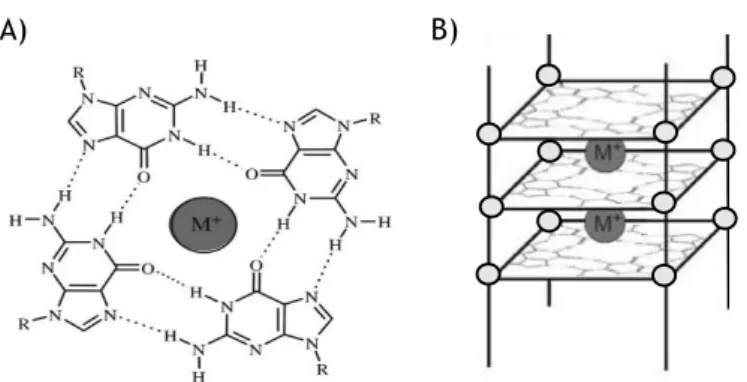
Nucleic acids are flexible molecules that can assume different conformations. Predominantly, DNA in living systems is in double helix structure but in some regions of genome rich in guanines they can exist in an alternative form, known as G4, which can form from DNA or RNA (or other variants, such as PNA) [19, 20]. These structures, as the name suggests, are composed by a set of four guanine nucleotides associated with each other in a cyclic Hogsteen hydrogen-bond (Fig. 2A). These arrays forms G-quartets, which through π-π stacking interactions forms G4 (Fig. 2B). This interactions play an important role in stabilization of the structure [21].
Moreover, the structures are additionally stabilized by monovalent (Rb+, NH
4+, K+, Na+, Cs+, Li+),
divalent (Sr2+, Ca2+, Ba2+, Mg2+), or trivalent (Tb3+, Eu3+) cations, through electrostatically
coordination with the oxygen atoms of the adjacent stacked G-tetrads, neutralizing negative charged formed in central core of G4 (Fig. 2A and 2B) [22]. The order of cation ability to stabilize and/or induce G4 structures is as follow: K+ > NH
4+ > Rb+ > Na+ > Cs+ > Li+ for monovalent
cations [23] and Sr2+ > Ba2+ > Ca2+ > Mg2+ for divalent cations [24]. However, since K+ and Na+
are the main cations in vivo, G4 formation is favoured under physiological conditions. The K+
form is the strongest known coordinating monocation for G4 due to its ionic radius that allow a better coordination with oxygen atoms and higher intracellular concentrations (~140 mM) than Na+ (5-15 mM) [25]. The location of cations between tetrads depends on the nature of the ions:
Na+ ions are observed in a range of geometries, whereas K+ ions are always equidistant between
each tetrad plane [25].
Figure 2 – A) The structure of a G-quartet arrangement with hydrogen Hogsteen bonds and monovalent cation in center of cavity. B) G4 structure. (Adapted from [21]).
5
Supercoiling affects the potential for DNA denaturation and structure formation, and supercoiling local can vary in vivo [14].With the exception of extreme thermophiles in which, DNA is positively supercoiled to protect it from thermal denaturation, supercoiling has a negative sign, which means that the torsional tension reduce the helicity and facilitates strand separation [26].In fact, to induce structural perturbations such as, formation of G4 structures the local unwinding or melting of DNA is necessary and both are generated by DNA supercoiling [27]. Since B-DNA is the minimum energy conformation the transition to G4 is unlikely to occur; however, the free energy generated by supercoiling may be enough to unwinding DNA and generate secondary structures [28]. In plasmids the torsional stress results in a build-up of free energy [29]. In this case, two duplexes wind helically up and down around each other and about a superhelix axis [29]. Since supercoiling affects G4 formation, plasmids can adopt different G4 conformations.
The importance of plasmid topology in G-loop formation was analysed by Duquette and collaborators by comparing formation of G-loops DNA on sc, oc and ln plasmids pPH600 and they observe that formation occurs more frequently in sc pPH600 [14]. More recently, another study performed by Gaynutdinov and collaborators shows that short G-rich peptide nucleic acids (PNA), stabilize G4 formation because have complementarity with C-rich strand, binding with more efficiency to supercoiled plasmids even after linearization of the plasmid [11]. In fact, this approach is very similar to formation of RNA-DNA hybrid; however, instead of using the RNA transcript to stabilize the structure, uses the PNA. Moreover, Lv and collaborators demonstrated that DNA gyrase that is commonly found in prokaryotes and are responsible to the formation of sc in their circular DNA and pDNA can readily drive the formation of G4 under physiological conditions [30]. This arise the importance of obtain pure sc pDNA which is more efficient in G4 formation.
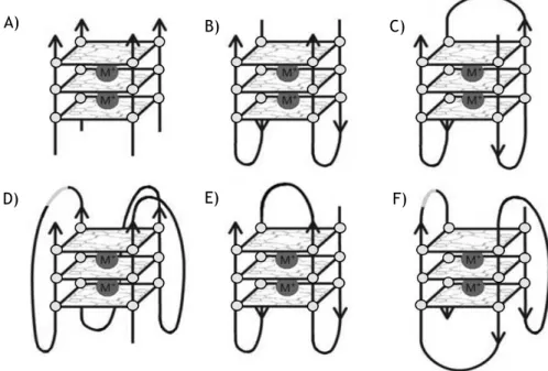
G4 are diverse and polymorphic structures that can be correlated with several features, such as, number of molecules involved in formation of structure, chain orientation of DNA strands, guanine nucleoside glycosidic bonds and loop topology [31].
In the case of number of molecules involved in formation of structure, G4 can be intramolecular (uni-molecular) or intermolecular (bi-molecular or tetra-molecular) [31]. Intramolecular G4 formed by single-stranded are of intensive current research interest due to their potential formation in telomeres and oncogene promoter sequences. Inversely to tetramolecular G4, intramolecular structures are more complex and form quickly exhibiting high polymorphism [31].
6
The sequences with consecutive guanines in uni-molecular and bi-molecular G4 form loops that are positioned on the exterior of core, helping in stabilization of structure. Loops can be diagonal (Fig. 3E), lateral or chain-reversal (also termed propeller) (Fig. 3D), and the presence of a particular loop type in a structure is dependent on the number of G-quartets comprising a G4, on loop length and on sequence [32].
All G4 have four distinct phosphodiester chains and orientation of DNA strands may be parallel (Fig. 3A, 3B, 3C and 3D), anti-parallel (Fig. 3E) or hybrid (Fig. 3F).G4 conformation is influenced by both the DNA sequence and the conditions used to promote folding of G4 such, nature of stabilizing cation. In biological context, the tendency is to form parallel G4 structures in presence of K+, due to its relevance in intracellular media [33].
Figure 3 - G4 stoichiometries and folding patterns. A) Tetramolecular parallel structure; B) Bimolecular antiparallel structure; C) Unimolecular antiparallel structure with alternating parallel strands; D) Unimolecular parallel structure with three double chain reversal loops; E) Unimolecular antiparallel structure with adjacent parallel strands and a diagonal loop; F) Unimolecular mixed structure with three parallel and one antiparallel strands (Adapted from [21]).
All parallel G4 are consistent and have all guanine glycosidic angles in an anti conformation, not displaying significant polymorphisms. In contrast, anti-parallel G4 have both syn and anti guanines, arranged in a way that is particular for a given topology and set of strand orientations, since different topologies have the four strands in different positions relative to each other [34]. A) B) D) C) F) E)
7
1.1.2 - Genome location and functions of G4
Actually, there are powerful tools that allows the prediction of G4-forming sequences in human genome. The sequences that are able to form G4 consists at least four runs of guanines which usually contains at least three guanine residues (e.g., [G≥3NxG≥3NxG≥3]≥4, where N is any nitrogen
base (A; T; G or C)) [10]. If the runs of G≥3 are shorter, much longer or mutated the structural
stability decreases, significantly. This prediction was expressed as an algorithm, called
quadparser. Huppert and Balasubramanian apply quadparser and reveal that human genome
contains approximately 376.000 sequences that have potential to form G4, simultaneously, which are less than would be expectable by chance [35].
Predicted G4 sequences are not located randomly throughout the genome and tend to cluster together in particular important regions of human genome such as, telomeres, regulatory regions of oncogenes, rDNA and immunoglobulin switch region (S region). It is thought that the location of these sequences are a strong evidence of it crucial role in various cellular processes including telomere maintenance, replication, transcription, translation, genetic and epigenetic instability [36].
Telomeres are specialized nucleoproteins that protect extremities of linear chromosomes in order to distinguish them from unwanted double strand breaks. These nucleoproteins are constituted by repetitive sequences of variable lengths characterized by runs of guanines on one strand. Bearing runs of consecutive guanines, 5′ to 3′ telomeric strands from most eukaryotes may potentially fold into G4 [37]. At the G-rich telomere tails, the presence of G4 inhibits extension by telomerase, which is up-regulated in cancer cells, as well as negatively regulating oncogene’s transcription [38].
Aside from telomeres, one of the first region considered for the presence of G4 was gene promoters. Notably, one evidence that G4 have an important function in regulatory processes is your high prevalence in oncogenes and low prevalence in tumor suppressor genes [39]. Based on hallmarks of cancer proposed by Hanahan and Weinberg [40] several oncogenes have been studied, among them are c-Myc, c-Kit and KRAS (self-sufficiency); pRb (insensitivity); Bcl-2 (evasion of apoptosis); VEGF-A (angiogenesis); hTERT (limitless replication); and PDGF-A (metastasis) [41]. Actually, various efforts have been done to design therapeutic targets with high specificity for these regions.
Eukaryotic rDNA is G-rich, rapidly transcribed and contain many sequences that can form G4 in non-template strand. Nucleolin is a very abundant protein in nucleolus and binds with high affinity (KD=1 M) to G4 through two separated domains that recognize G4 as natural binding
targets independent of sequence context. The strong interaction suggest that the presence of nucleolin stabilize G4, prevents renaturation of the duplex and is involved in rDNA transcription, replication, or recombination [42].
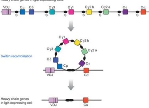
The class switch recombination (CSR) of the immunoglobulin loci is essential to normal B-cell development and immune response and the process occurs in response to antigen stimulation and costimulatory signals. The process allows mature B cells to alter effector portion modifying
8
the mode of antigen clearence without affecting antigen specificity. The mechanism of immunoglobulin isotype switching is regulated, irreversible and alter genomic structure by joins a rearranged and expressed variable region to a new downstream constant region, deleting the DNA between as an excised switch circle (Fig. 4). CSR is targeted to short and repetitive G-rich regions (20-80 bp), called switch regions. S regions are approximately 1–10 kb in length and are located upstream of each of the CH regions that participate in switch recombination: Cµ, Cγ,
Cε, and Cα. There is no S region upstream of Cδ, and RNA processing rather than DNA recombination regulates Cδ expression [43, 44].
Figure 4 – Diagram of Class Switch Recombination from IgM to IgA (CSR). The murine heavy chain locus is shown before and after recombination from µ to α, which results in expression of IgA antibodies. Deleted sequences can be recovered in circular DNA molecules from B cells which have recently completed switch recombination. V, variable region; C, constant region; and S, switch region (Adapted from [43]).
1.1.3 – Transcription of S regions
The process of gene expression begins with transcription in cell nucleus. The transcription is initiated in transcription start site (TSS), which is located immediately downstream of promotor. The region is not transcribed but play a fundamental role in gene transcription. The transcription factors (TFs) binds with specificity to nucleotide sequences in promotor and assist the binding of enzyme that catalyses RNA synthesis, called RNA polymerase [45].
9
The binding of the general TFs on their own produces low levels of transcriptional activity; however, the transcriptional activity is increased or decreased by other TFs that binds specifically to another regions of DNA called enhancers or silencers [45].
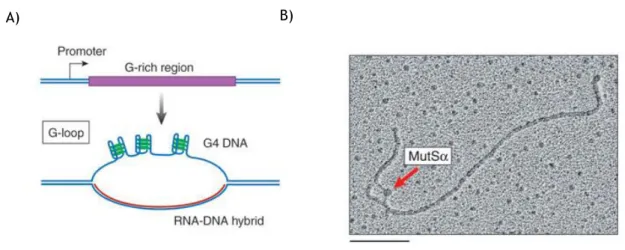
Potential G4 forming sequences were identified in the regions flanking the TSS suggesting and active role in transcription. Recent studies investigated the influence of transcription in G4 formation [46]. G4 can be induced far away from TSS, functioning as silencer or enhancer of transcription. Each S region has a dedicated promoter, and transcription through the S region is necessary to activate recombination and target it to specific S regions [47]. Transcription alters the structure of S region DNA, creating an active molecular partner for recombination. Transcription of G-rich templates such as the S regions, either in vitro or in vivo, causes characteristic large loops to form, which are hundreds of base pairs in length and are called G-loops [48]. G-G-loops contains a stable, cotranscriptional RNA-DNA hybrid on the C-rich template strand and intramolecular G4 DNA interspersed within single-stranded regions on the G-rich strand (Fig. 5A) [48]. These structures are involved in a range of processes, such as regulation in E. coli plasmid replication [48]. The persistence of such hybrids in living cells can result in genomic instability, and conserved factors associated with RNA processing normally prevents the formation of such hybrids. The hybrid formation prevents further transcription, suggesting a regulatory role of these structures [48].
G4 DNA in G-loops is the target for TFs genetically linked to switch recombination. Two switch recombination proteins MutSα (Fig. 5B) and AID binds to G4 structures [46, 49]. The capacity to prevent or induce the formation of G4 with some transcription factors in S region seems to be a fundamental role in immunoglobulin class switch recombination.
Figure 5 - G-loop formed in a transcribed G-rich region and bond of MutSα to G4 structure. (A) Transcription of G-rich region resulted in G-loop and cotranscriptional RNA-DNA hybrid on C-rich region; (B) Bond of MutSα to G4 structure in G-C-rich template (Adapted from [47]) .
10
1.1.4 – Circular dichroism of G4 structure
Circular dichroism (CD) is an excellent method for rapid determination of secondary structure and is widely used in structural studies of biomolecules. The technique takes advantage from the differential absorption of left-handed and right-handed circularly polarized light. The light is polarized by passing through suitable prisms or filters its electric field will oscillate sinusoidal in a single plane. This sinusoidal wave can be visualized as the resultant of two vectors of equal length, rotating in opposite directions, one in clockwise direction and the other in counter clockwise direction. In order to show signal the chromophore should be chiral (optically active) to absorb with different extent the circularly polarized light and to have different indices of refraction for the two waves. The result is the rotation of plane of light and the creation of a new vector, resulting from addition of the two vectors in opposite directions, which traces an ellipse. Generally the results are reported in terms of ellipticity (θ) in degrees [50]. For this reasons the technique is suitable for structural evaluations of biomolecules such as, proteins and nucleic acids.
In particular case of nucleic acids, CD arises from the asymmetric backbone sugars and the helical arrangement of its constituents. The task to determine secondary structures is not easy due to the length of nucleic acids and the empirically determination of structure [51]. Despite these drawbacks CD have advantages over the other methods because is handy, fast, highly sensitive and relative inexpensive, making technique ideal for tracing conformational transitions between nucleic acids arrangements [51]. Moreover, the technique is particularly powerful for monitoring structural changes resulting from changes in environmental conditions such as temperature, ionic strength, and pH [51].
Figure 6 – Circular dichroism spectra with parallel (black line) and anti-parallel (grey line) G4 structure (Adapted from [20]).
11
In recent years the technique has been widely used to determine the topological conformation of G4. The CD spectra of a reference set of DNAs of known G4 structure, with examples of both anti-parallel and parallel types, have been obtained. In general, a peak in CD at 260 nm wavelength and at 240 nm is descriptive of an all-parallel structure, whereas a peak at 295 nm and a trough at 260 nm describes an antiparallel structure (Fig. 6) [20].
1.1.5 - Role of G4 in cancer and neurological diseases
Cancer, aging and neurological diseases represent an enormous source of health and financial burden in worldwide. Together affect hundreds of millions of people in world resulting in increased associated costs. G4 are intrinsically associated to aging process and disease development. The presence in telomeres, promoter regions of oncogenes and the formation of non-B DNA structure in triplex expansion-associated a neurological diseases and cancer are a strong evidence of its importance [52]. In this subsection it will be presented examples inform the literature about genes and proteins involved in these processes.
1.1.5.1 - Aging and cancer
The processes of aging and cancer are closely related and are directly associated to telomere shortening, risk of disease development, decreased of reparation ability and activity of regeneration processes [52]. Telomeres are DNA–protein complexes that cap the ends of linear chromosomes and provide protection against gene erosion at cell divisions, non-homologous end-joining’s and nuclease attacks [53].In this point of view, there seems to be a pathological paradox. On the one hand, telomere shortening accompanies the human aging, suggesting that erosion of chromosomes could be a natural mechanism to prevent the immortal replication of cells that characterizes cancer growth. On the other hand, telomere shortening is observed in many cancer cells that gained an unlimited proliferation potential [54]. Therefore, using telomeres to prevent aging may lead to an increase risk of cancer. This fact is supported by the concept that the incidence of cancer rises exponentially in the final decades of life [55], suggesting that human body try to fight aging and results in cancer. Since, telomeres are constituted by repetitive sequences of variable lengths characterized by runs of guanines on one strand may potentially fold into G4. The stabilization of G4 in telomeres results in telomerase inhibition, telomere shortening, cell growth suppression and induction of apoptosis through disruption of telomere maintenance [55].
In addition to their existence in telomere sequence, G4 are also found in promoter regions of oncogenes close to their TSS’s [55]. As aforementioned there are many oncogenes that are able to form G4 structures and that can be the target for small molecules [55]. A study reported by
12
Hurley and collaborators, shows that the promoter region of c-Myc oncogene controls 80%-90% of the transcriptional activity of this gene [55].
Therefore, as in telomeres the stabilization of G4 results in telomerase inhibition, the stabilization in promotor regions of oncogenes inhibit transcription, supressing oncogenic expression [55].
1.1.5.2 - Neurological diseases
Neurological diseases in general are characterized, among other things, by mitochondrial dysfunction and oxidative stress [56]. The expansion of hexanucleotide GGGGCC repeat region in C9ORF72 gene in few hundred repeats causes frontotemporal dementia and amyotrophic lateral sclerosis [56]. G4 from the C9ORF72 gene may arise from their ability to bind and activate cellular Heme, towards to an oxidative stress [56]. In Alzheimer disease sequestration of Heme group by Aβ peptide, the causal agent of Alzheimer disease, constitute a loss of function for mitochondrial activity but a gain of function in another way by bond of Aβ peptide to Heme group [56]. Therefore, the sequestration of Heme group by G4 suggests that they have an active role in these diseases.
The expansions in FMR1 gene produce pre-mRNA that carry extended regions of G4 structures, abolishing transcription and initiating fragile X syndrome [57]. When the CGG expansion repeats exceed 200 they become hypermethylated, causing transcriptional silencing of the FMR1 gene, resulting in the loss of its encoded protein FMRP, which is a selective RNA-binding protein implicated in regulating dendritic mRNA transport and local protein synthesis at synapses. The expansion of the intronic GGCCTG hexanucleotide repeat in NOP56 causes a unique form of Spinocerebellar ataxia (SCA36), which shows not only ataxia but also motor neuron dysfunction [58]. This characteristic disease phenotype can be explained by the combination of RNA gain of function and miR1292 suppression. In all of cases aforementioned, produced pre-mRNAs appears to sequester essential RNA binding proteins and impar mRNA processing [36].
The PRNP gene encodes the prion protein, which has been implicated in various types of transmissible neurodegenerative spongiform encephalopathies, such as Creutzfeldt-Jakob disease [59]. The expansion of sequence CCCCATGGTGGTGGCTGGGGACAG from 5 to 10-14 repeats in the coding region of this gene cause a dominant form of familial Creutzfeldt-Jakob disease exhibiting early onset and slow progression, which is correlated with misfolding of corresponding prion protein and formation of insoluble protein aggregates in solution [60]. The mechanisms that drive expansions of this G4 repeats are not defined.
13
1.2 - Gene therapy and DNA vaccination
The significant progress of science during the last decades, namely in molecular biology and genetics, increase the understanding of genetic disorders, leading to revolutionary discoveries in techniques that are based on nucleic acids such as, gene therapy and DNA vaccination [61]. The idea of gene therapy has been around for some time, but only with the development of recombinant DNA technology and ability to transfer and express exogenous genes in mammalian cells received attention and progresses are made [62]. In fact, gene therapy is a relatively new paradigm in medicine with enormous therapeutic potential and is defined as the insertion of genetic material directly into cells of a human patient to correcting a genetic disorder or overexpressing proteins that are therapeutically useful [63]. The DNA vaccination is a type of gene therapy that consists in a pDNA build to express a protein of an aggressor agent inducing both humoral and cellular response [64]. In recent years, this techniques received the attention of researchers because the enormous potential to treat and prevent diseases.
1.2.1 - Gene therapy
Gene therapy can be divided into two categories: somatic gene therapy and germ line gene therapy [65]. The difference between the two categories becomes important and legislation allow the use of somatic gene therapy because the genetic change induced not pass to future generations, contrary to germ line therapy [65]. The major advantage of gene therapy is the treatment or elimination of the cause of disease, while most of conventional approaches treat the symptoms [63]. Initially, the idea of using recombinant DNA techniques was to treat or eliminate the cause of diseases that are based only in a single gene, better known as monogenic diseases [66]. In this case, the aim is the replace of expression of a dysfunctional gene by introducing one or more copies of the therapeutic gene in patient [66]. However, monogenic diseases are not the only target of gene therapy and in recent years many forms of cancer, cardiovascular diseases, neurological diseases and other non-treatable infectious diseases became the primary target [65]. The first attempt of gene therapy to treat a disease targeted a form of severe combined immune deficiency (SCID) due to defects in the gene encoding adenosine deaminase (ADA) [67].
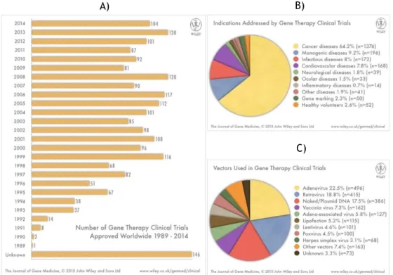
Over the past years, the clinical applications of gene based therapies for treating or preventing various diseases, has been investigated and actually there are 2142 completed or active clinical trials approved (Fig. 7A), in especially to treat or preventing cancer (Fig. 7B).
The efficiency and safety of gene therapy is closely related with different vector systems that have been developed for gene transfer [68].
14
In general, there are two main categories of vectors, viral and non-viral, but all gene therapy applications depend on the fact that the genetic material needs to be delivered across the membrane and ultimately to the nucleus [68].
Viral vectors have better cell targeting properties due to its intrinsic characteristics and at present stage of development generally give the most efficient transfection [68]. The most used viral systems available for gene therapy are adenovirus, retrovirus, lentivirus and adeno-associated virus [68]. For many years it was thought that inactive virus, that have been modified by deleting some areas of their genomes to makes them more safe, would be the ideal vehicle to introduce genetic material in cells. However, a number of widely reported adverse events have focused the attention on associated risks such as, integration into host cell genome, which could possibly cause oncogene activation or tumor-supressor gene inactivation. These events raised safety concerns related to immunogenicity and toxicity [68].
Non-viral vectors based on pDNA give less efficient transfection; however, the safety concerns are clearer and production can be performed in a simply way when compared with the viral ones [69]. This characteristics made it vector very attractive for genetic vaccination and consequently its use in gene therapy arises in recent years (Fig. 7C).
Figure 7 – Compiled data on gene therapy clinical trials. (A) - Clinical trials approved for Gene Therapy 1989 - 2015. The total corresponds to 2142 clinical trials approved. (B) – Diseases addressed and (C) – Vectors used in clinical trials.
(Available at www.wiley.com/legacy/wileychi/genmed/clinical/, May 2015)
A) B)
15
1.2.2 DNA vaccines
Currently, vaccines can be classified into five types: live-attenuated, inactivated microorganisms, subunit vaccines (subunits, polysaccharides and protein conjugates), toxoids and genetic based [70]. However, the use of both live attenuated and inactivated vaccines raises several questions in terms of safety [71]. These limitations need to be surpass using new vaccine platforms that offer broader immunogenicity [71], as in case of DNA vaccines that consist in pDNA build to express an encoded protein following administration in vivo and subsequent transfection of target cells [64]. DNA vaccines have a wide range of economic and technical advantages when compared with conventional ones. DNA vaccines mimics the effects of live-attenuated vaccines because have the capacity to induce both immune and humoral response, through CD4+ and CD8+ T cell responses [72]. The mechanism consists in introduction
of pDNA with a gene of interest in nucleus, where the cell machinery recognizes and induces the transcription of gene with subsequent protein production in cytoplasm [72]. The protein is recognized by specific cells and induce the humoral and immune response (Fig. 8) [72]. Moreover, DNA vaccines can also act as adjuvant of cellular response due to the presence of unmethylated CpG islands that are stimulatory DNA specific sequences [70]. The production cost in large scale is lower than the conventional vaccines, the quality control is much easier, since not need cold chain, because this vaccines are stable at room temperature and can be lyophilized, making distribution easier in remote locals with low economic resources [72]. Until moment, there are four DNA vaccines approved for veterinary use: against West Nile virus in horses; infectious hematopoietic necrosis virus in salmon; malignant melanoma in dog as well as delivery of growth hormone releasing hormone in pigs [72].
16
1.3 - pDNA
The importance of plasmids to molecular biology and their impact in biotechnology industry make them an indispensable molecular tool in life science research [73]. Plasmids are replicative extrachromosomal entities, normally the size of circular double stranded DNA is between 1 kbp and 1000 kbp, meaning that they are very large molecules [74]. Each strand of this molecule is composed by a linear polymer linked by phosphodiester bonds between the hydroxyl group of the 3’ sugar carbon of a nucleotide and the phosphate group of the 5’ sugar carbon of the adjacent nucleotide [75]. The phosphate groups are negatively charged when pH is maintained above 4 [75]. The winding of two anti-parallel DNA strands around each other and around a common axis forms the double helix structure with highly hydrophobic grooves accessible to molecule and solvent [76]. Plasmids contain an origin of replication that is recognized by DNA polymerase allowing growth in bacteria and amplification of large quantities of pDNA for purification [77]. They have also a gene coding for antibiotic resistance and a complete eukaryotic expression cassette composed of the transcribed region domain inserted between an enhancer and terminator. The region is flanking by restriction enzyme sites that allow insert the desired genes. Plasmids can exist in three different conformations (Fig. 9): supercoiled (sc); open circular (oc) and linear (ln) [75]. However, the most relevant isoform in biological context is supercoiled because transfection and gene expression is highly dependent on the degree of superhelicity [78] and can assume different secondary structures of DNA more efficiently [14]. The others conformations are derived from supercoiled by both single strand nick in phosphodiester backbone (oc) and double strand nick in phosphodiester backbone (ln) [76]. The nicking occur by specific nucleases, high temperatures and mechanical stresses [76].
Figure 9 – Different conformations of pDNA: supercoiled (sc); open circular (oc) and linear (ln). (Adapted from [75])
17
1.3.1 – pDNA specifications
Plasmids are the crucial tool in many scientific areas, and large scale processes should be designated to produce a certain amount of plasmid within certain specifications. Thus, there is a need to obtain pure sc pDNA without impurities to allowing efficient analysis and applications. The quality of the plasmid should be assessed by safety, potency and purity, in order to validate the process [75]. For a product which intended to be used in humans, the removal of impurities during the manufacturing process is mandatory to avoid side effects once administered in patients [79]. The impurities for production of recombinant proteins in E. coli is also the same for pDNA production [79]. Therefore, in pDNA production the main impurities are host nucleic acids (gDNA and RNA), proteins and endotoxins).
The regulatory authorities such as, Food and Drug Administration (FDA) and European Agency for the Evaluation of Medical Products (EAEMP), have rigorous guidelines related to the presence of these impurities (Table 1). Recently, a new analytical method have been developed to specifically quantify the amount sc pDNA, oc pDNA and RNA present in pDNA sample [80].
Table 1 - Specifications and recommended assays for assessing pDNA quality. (Adapted from [75])
Requirement Analytical assay Specification
Proteins BCA assay SDS-PAGE Not detectable <0.01 μg/dose RNA Agarose gel electrophoresis Not detectable gDNA Hybridisation blots, PCR, fluorescence <0.05 μg/μg plasmid <0.01 μg/dose Endotoxins Limulus ameobocyte lysate (LAL) assay <5 EU/kg body weight <0.1 EU/μg plasmid Plasmid Homogeneity Agarose gel electrophoresis >90% supercoiled form Potency Cell transfection According to application
18
1.3.2 - pDNA production
The development of processes to achieve the production of large quantities of highly purified supercoiled pDNA have the interest of industry and researchers due to the expansion of gene therapy and DNA vaccination [79].
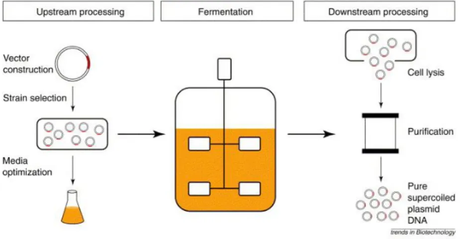
The process development for pDNA production starts on a bench scale with the upstream process, in which the design of vector, choice of host, as well the selection and optimization of fermentation conditions, followed growth of cells and plasmid production and finally, the downstream process, in which the main goals are the cell lysis and purification of sc plasmid DNA (Fig. 10). These three stages of process development (upstream processing, fermentation and downstream processing) are integrated and must not be approached on an individual bases [79].
In general, plasmid production is performed in E.coli, a bacterium with a history of safe use in bio-industry, which growth to an optimal cell concentration, while pDNA is replicated autonomously in intracellular environment [81]. Although other host systems have been used [82]. A single bacterial cell may generate high plasmid number in appropriate conditions. There are two main goals associated with fermentation, the first is maximize the yield of pDNA obtained, while minimizing process that gives rise to impurities such as, gDNA, RNA, proteins and endotoxins, and improves the pDNA stability. In order to achieve this objective should be taking in consideration several parameters such as, the choice of host system and vector, as well the formulation of culture medium. Formulation of culture medium should consider the elemental composition of E.coli, pDNA replication and operative conditions. Finally, the second objective of the fermentation process is maximize the number of copies of sc pDNA in detriment of other isoforms that are usually treated as impurities [83, 84].
Figure 10 - The three stages of sc pDNA development process. The upstream, fermentation and downstream processing. (Adapted from [79])
19
1.3.3 – Downstream Processing
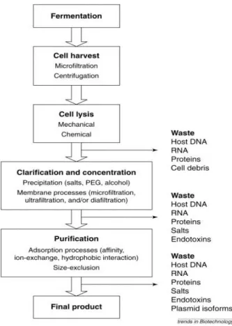
Since pDNA accounts for less than 3% of the total contents of an E. coli cell (depending on plasmid size and copy number) the major obstacles are encountered during downstream process stage (Fig. 11), in which is necessary eliminate other cellular components of the host strain that are usually treated as impurities [83].
After fermentation, the recovery of cells from the broth by microfiltration or centrifugation initiates the downstream processing [79]. Then, cells are ressuspended in appropriate buffer to initiate the cell lysis [79]. The buffer contains ethylene diamine tetra-acetic acid (EDTA) and often carbohydrates such as, glucose and sucrose to facilitate the lysis step and protect plasmid against mechanical stresses [79].
Figure 11 - Process flow sheet for the purification of sc pDNA. Each step of the process is a unit operation that can be improved. The eliminated impurities in each step are also indicated. (Adapted from [79]).
![Figure 1 - Chemical Structure of DNA and RNA nucleotides (Adapted from [4]).](https://thumb-eu.123doks.com/thumbv2/123dok_br/18892315.934111/25.892.300.620.800.1092/figure-chemical-structure-dna-rna-nucleotides-adapted.webp)
![Figure 6 – Circular dichroism spectra with parallel (black line) and anti-parallel (grey line) G4 structure (Adapted from [20])](https://thumb-eu.123doks.com/thumbv2/123dok_br/18892315.934111/34.892.239.612.757.1006/figure-circular-dichroism-spectra-parallel-parallel-structure-adapted.webp)
![Figure 8 - Mechanism of DNA vaccines. Induction of immune and humoral responses. (Adapted from [72])](https://thumb-eu.123doks.com/thumbv2/123dok_br/18892315.934111/39.892.188.752.756.1082/figure-mechanism-vaccines-induction-immune-humoral-responses-adapted.webp)