ARTIGO ORIGINAL
RESUMO
Introdução: O transporte através de sistemas de emergência médica reduz os tempos de tratamento no enfarte agudo do miocárdio com elevação do segmento ST. Os autores estudaram o Registo Nacional de Síndromes Coronários Agudos para avaliar o impacto nacional do transporte através de sistema de emergência médica no tratamento do enfarte agudo do miocárdio com elevação do segmento ST.
Material e Métodos: Foi realizado um estudo retrospetivo, multicêntrico de doentes com enfarte agudo do miocárdio com elevação do segmento ST inseridos no Registo Nacional desde 2010 até 2017. Os doentes foram divididos em Grupo I, representando doentes transportados por viaturas de emergência médica e Grupo II, doentes que chegaram ao Serviço de Urgência por outros meios. Resultados: Do total de 5702 doentes, 25,6% foram transportados por viaturas de emergência médica. Registou-se um aumento no uso de viaturas de emergência médica de 17% nos últimos sete anos. Os sistemas de emergência médica garantiram uma maior taxa de transporte para centros capazes de realizar intervenção coronária percutânea, de bypass do Serviço de Urgência e de fibrinólise no local. O transporte através de viaturas de emergência médica conseguiu uma redução da mediana do atraso para a reperfusão de 59 minutos (p < 0,001). Não houve diferença na mortalidade intra-hospitalar.
Discussão: Nesta amostra nacional, é evidente que os sistemas de emergência médica reduziram significativamente os tempos de reperfusão, associando-se a uma menor incidência de insuficiência cardíaca aguda pós-enfarte. No entanto, esse benefício não resul-tou numa menor mortalidade intra-hospitalar, provavelmente devido ao facto dessa população representar um subgrupo de doentes com doença mais grave e mais comorbilidades.
Conclusão: Os benefícios associados ao uso de sistemas de emergência médica no transporte de doentes com enfarte agudo do miocárdio com elevação do segmento ST não se traduziram numa menor mortalidade intra-hospitalar.
Palavras-chave: Enfarte Agudo do Miocárdio com Elevação do Segmento ST; Factores de Tempo; Intervenção Coronária Percutânea; Reperfusão Miocárdica; Tempo para Tratamento
Impact of Emergency Medical System Transportation
in ST-segment Elevation Myocardial Infarction: A
Nationwide Retrospective Study
Impacto do Transporte por Viatura Médica de Emergência
e Reanimação no Enfarte Agudo do Miocárdio com
Elevação do Segmento ST: Estudo Retrospetivo Nacional
1. Cardiology department. Hospital de São Teotónio. Centro Hospitalar Tondela-Viseu. Viseu. Portugal. 2. National Registry of Acute Coronary Syndromes. Coimbra. Portugal.
Autor correspondente: Júlio Gil Pereira. juliogilpereira@gmail.com
Recebido: 17 de julho de 2018 - Aceite: 28 de outubro de 2019 | Copyright © Ordem dos Médicos 2020
Júlio Gil PEREIRA1, Luís ABREU1, Hugo ANTUNES1, Maria Luísa GONÇALVES1, Bruno MARMELO1,
Davide MOREIRA1, Luís NUNES1, Jorge Oliveira SANTOS1, on the behalf of the Portuguese National Registry of Acute
Coronary Syndromes2
Acta Med Port 2020 Jun;33(6):390-400 ▪ https://doi.org/10.20344/amp.11082 ABSTRACT
Introduction: Emergency medical system transportation has been shown to reduce treatment times in ST-segment elevation myocar-dial infarction. The authors studied the Portuguese National Registry of Acute Coronary Syndromes to determine the nationwide impact of the emergency medical system transportation in the treatment of ST-segment elevation myocardial infarction.
Material and Methods: A multicentric, nationwide, retrospective study of ST-segment elevation myocardial infarction patients inserted in the National Registry from 2010 to 2017 was performed. The patients were divided into: Group I, composed of patients transported by emergency medical system, and Group II, patients arriving to the Emergency department by other means.
Results: Of the 5702 patients studied, 25.9% were transported via emergency medical system. Rates of emergency medical system activation increased by 17% in the last 7 years. The emergency medical system provided a higher rate of transport to a percutaneous coronary intervention capable centre, of Emergency department bypass, of on-site fibrinolysis, and ensured a 59-minute reduction of the median reperfusion time (p < 0.001). There was no difference in in-hospital mortality.
Discussion: In this nationwide cohort, emergency medical system transportation is associated with a reduction in reperfusion times. It provides a higher amount of salvaged myocardium and reduces the incidence of acute heart failure. However, emergency medical system use did not result in lower in-hospital mortality, probably due to confounding factors of higher disease severity and comorbidity. Conclusion: The benefits associated with emergency medical system based transportation of patients with ST-segment elevation myocardial infarction do not translate into lower in-hospital mortality.
Keywords: Myocardial Reperfusion; Percutaneous Coronary Intervention; ST Elevation Myocardial Infarction; Time Factors; Time-to-Treatment
INTRODUCTION
Cardiovascular diseases (CVD), particularly ischemic
heart disease (IHD), is the most common cause of death worldwide.
1,2 In the past four decades, studies have shown
ARTIGO ORIGINAL
by acute myocardial infarction (AMI), particularly with ST-segment elevation myocardial infarction (STEMI).1-4 In
Por-tugal, the same trend was also described in several stud-ies.2,3,5,6 It is undeniable that the best treatment for STEMI is
revascularization as soon as possible, preferably by prima-ry percutaneous coronaprima-ry intervention (P-PCI).1,7-9 Multiple
factors have an impact on the mortality in STEMI patients, such as advanced age, Killip-Kimball class and comorbidi-ties; however, treatment delay is one of the most important factors.1,10 Transportation by emergency medical system
(EMS) vehicles has been proven to have a definite impact in reducing treatment delays and possibly on mortality.1,10-15
The Portuguese National Institute of Medical Emer-gency (INEM) currently employs a special emerEmer-gency and reanimation medical vehicle (VMER). The VMER is a pre-hospital intervention vehicle comprised of a specialized staff, which includes a physician and nurse. It is designed for rapid transportation directly to the patient with the goal of ensuring pre-hospital stabilization and medical care prior to hospital admission. The VMER plays an important role in achieving a pre-hospital diagnosis of STEMI and in deciding the best course of action to employ in each case.
In Portugal, the National Registry of Acute Coronary Syndromes (RNSCA) was established in 2002, under the auspice of the Portuguese Society of Cardiology.16 It is an
observational, multicentric, nationwide prospective study in which each hospital centre participates with data from pa-tients admitted due to acute coronary syndrome (ACS).16
Quality control is assured by each participating centre. The primary goal of the current study is to determine the real-world impact of VMER transportation in patients with STEMI in Portugal. The main objectives include evaluating in-hospital prognosis and determining differences in treat-ment delays. Information from the RNSCA was analysed to determine the actual nationwide effect of EMS transporta-tion. The secondary objectives were: (1) evaluating which patients were more likely to be transported by VMER; (2) characterization of VMER use nationwide and regionally, as well as evolution trends.
MATERIAL AND METHODS Participants
This is a retrospective study of patients in the RNSCA from October 1, 2010 to March 14, 2017. Of the 16 084 pa-tients included in study period, 5702 were selected, based on the diagnosis of admission (only STEMI patients were chosen) and whether information regarding hospital trans-portation was available. Patients suffering STEMI while in hospital facilities and hospital transfers were excluded. The decision on which patients were transported via VMER was made on an individual basis, through an algorithm followed by the INEM professionals who receive the distress calls. The patients were divided into two groups, according to the means of transportation: those who were transported by vehicles with medical and nursing staff (VMER) (Group 1) and those who arrived at the hospital through other means (Group 2), either self-comers or by ambulance
with-out medical or nursing staff (usually firemen). The groups were compared according to clinical, analytical and pro-cedural parameters, collected upon arrival at the hospital. In-hospital complications were determined as well, namely: (1) Re-infarction (recurrence of ischemia-related chest pain with over 20-minutes duration, ECG changes and new el-evation of myocardial biomarkers); (2) Heart failure (HF); (3) Mechanical complication (rupture of the left ventricular free wall, interventricular septum or papillary muscle); (4)
De novo Atrial fibrillation (AFib); (5) Second degree
atrio-ventricular block Mobitz II or of higher grade; (6) Sustained ventricular tachycardia (VT) (over 30 seconds of duration or with hemodynamic instability); (7) Aborted cardiac arrest (successful resuscitation after cardiac arrest of any cause); (8) Ischemic or haemorrhagic stroke during hospitalization; (9) Major haemorrhaging (intracranial bleeding or other bleeding associated with hemodynamic compromise requir-ing intervention accordrequir-ing to the GUSTO criteria or transfu-sion need). The RNSCA has been approved by the Ethics Committee of the Portuguese Society of Cardiology and the National Data Protection Commission (number 3140/2010) and is registered in the platform clinicaltrials.gov (NCT 01642329).
Data analysis
In the statistical analysis, categorical variables were characterized by absolute and relative frequencies and numerical variables by means and standard deviations. Comparative analyses were carried out in relation to demo-graphic variables, treatment timings, therapeutic strategies, specific interventions and general outcome parameters. Comparisons between two groups regarding the categori-cal variables were conducted using the chi-square test, or Fisher’s Exact test. Regarding the continuous variables, t-test was used to compare the means whenever possible, otherwise, the Mann-Whitney U test was used to compare the medians. The chi-square test for trend was considered when evaluating the evolution of VMER use over the years. Predictors of VMER use were determined by adjusting a logistic regression model. The following variables were test-ed as possible prtest-edictors: demographic variables, risk fac-tors, cardiovascular and non-cardiovascular comorbidities, chest pain upon admission. Considering the subsample of patients that use VMER, logistic regression models were also adjusted to determine independent predictors of sev-eral outcomes: HF, shock, ventricular arrhythmias, cardiac arrest during hospitalization and in-hospital mortality. Vari-ables were selected to be included in the model using the Stepwise (Forward) method, together with the Likelihood-Ratio test. For each variable included in the regression model, the adjusted odds ratio and the respective 95% con-fidence interval (CI 95%) were also estimated. Model cali-bration was assessed by Hosmer and Lemeshow test. The quality of the adjustment of the logistic regression models was assessed by determining the area under the receiver operating characteristic curve (AUC). Statistical analyses were conducted using SPSS 19.0®, at a 5% significance
ARTIGO ORIGINAL level for hypothesis-testing. RESULTS
Patients’ Characteristics
A total of 5702 patients were studied, of which 1474 (25.9%) were transported by VMER. Regarding Group 2, 33.0% of patients were transported by an ambulance without a physician and 55.0% arrived at the Emergency department (ED) through their own means. The general characteristics of each group are displayed in Table 1. Table 2 displays the predictors of EMS transportation. Men (p = 0.005), patients with more cardiovascular risk factors and patients with known coronary disease are more frequently transported by EMS. Chest pain characteristics were also predictors of EMS use. Patients who presented with lower blood pressure (p < 0.001) or suffered cardiac arrest (p < 0.001) were more likely to be transported by EMS. There was a significantly lower value of BNP in patients transport-ed via VMER (p < 0.001). VMER registertransport-ed a higher rate of direct admission to a Cath lab and of ED bypass (p < 0.001). VMER also showed a greater likelihood of transport to a PCI-capable centre (p < 0.001).
Regional differences and temporal evolution
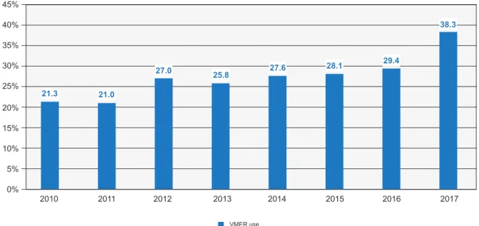
VMER use throughout the regions is displayed in Ta-ble 1. Northern Portugal has significantly less VMER use. On the contrary, almost 60% of patients are transported by EMS in the Southern region and the Islands (Madeira and Azores). In Fig. 1, the temporal evolution of each region is shown. While there was no difference in the Northern region (p = 0.078), the other regions all showed a significant in-crease in EMS use (p < 0.001). This inin-crease translates into a steady and significant rise in the VMER use nationwide (Fig. 2).
Treatment delays
Transportation by EMS vehicles had a significant impact on the time it took to treat patients. The overall timings are displayed in Table 3. EMS consistently reduced almost all measurable timings (p < 0.05), thus effectively reducing both patient-delay and system-delay. The only two timings in which EMS did not have an impact were timings regard-ing fibrinolytic therapy (FT). However, the overall time be-tween symptoms and beginning of FT was significantly re-duced (p = 0.012).
Table 1 – Characterization of the sample (n = 5702) (first section)
STEMI transported by emergency vehicles
(n = 1474)
STEMI transported through other means
(n = 4228) p
Age, years old (mean ± SD) 64 ± 13 64 ± 14 0.674
Male gender, n (%) 77.5 73.8 0.005
Body mass index, kg/m2 (mean ± SD) 27.3 ± 4.5 27.0 ± 4.2 0.034
- Low weight, n (%) 15 (1.1) 22 (0.6) 0.045
- Obesity, n (%) 309 (23.0) 759 (19.9) 0.018
Regions of Portugal
Northern Portugal1, n (%) 79 (5.4) 1416 (33.5) < 0.001
Central Portugal2, n (%) 541 (36.7) 1487 (35.2) 0.189
Southern Portugal and Islands3, n (%) 854 (57.9) 1325 (31.3) < 0.001
Cardiovascular risk factors
Arterial hypertension, n (%) 872 (60.9) 2549 (61.4) 0.710
Diabetes mellitus, n (%) 312 (21.8) 1034 (25.0) 0.016
Dislypidemia, n (%) 769 (55.4) 1994 (50.4) 0.001
Smoking habits, n (%) 568 (38.9) 1498 (35.5) 0.022
Family history of coronary disease, n (%) 108 (8.3) 269 (7.6) 0.454
Previous acute myocardial infarction, n (%) 196 (13.4) 388 (9.2) < 0.001
Previous percutaneous coronary intervention, n (%) 178 (12.2) 311 (7.4) < 0.001
Previous coronary artery bypass graft, n (%) 16 (1.1) 46 (1.1) 0.997
Peripheral artery disease, n (%) 57 (3.9) 119 (2.8) 0.037
Comorbidities
Heart failure, n (%) 27 (1.8) 92 (2.2) 0.433
Chronic obstructive pulmonary disease, n (%) 57 (3.9) 159 (3.8) 0.853
Dementia, n (%) 36 (2.5) 83 (2.0) 0.206
Previous stroke or transient ischemic attack, n (%) 84 (5.7) 273 (6.5) 0.313
ARTIGO ORIGINAL
Table 2 – Predictors of EMS transportation
Predictors Beta OR Confidence interval 95% p
Low Weight 0.881 2.41 1.20 – 4.84 0.013
Obesity 0.191 1.21 1.3 – 1.43 0.024
Female sex -0.238 0.79 0.67 – 0.93 0.005
Previous Angina 0.321 1.38 1.14 – 1.67 0.001
Previous PCI 0.418 1.52 1.21 – 1.90 < 0.001
Chest pain present at admission 0.253 1.29 1.08 – 1.54 0.006
Episodic chest pain 0.083 0.59 0.50 – 0.70 < 0.001
Hosmer-Lemeshow test p - value of 0,365; AUC (CI 95%) of 0.588 (0.570; 0.606)
Table 1 – Characterization of the sample (n = 5702) (final section)
STEMI transported by emergency vehicles
(n = 1474)
STEMI transported through other means
(n = 4228) p
Clinical data at admission
Anterior acute myocardial infarction, n (%) 719 (48.8) 2040 (48.2) 0.710
Inferior acute myocardial infarction, n (%) 746 (50.6) 2142 (50.7) 0.991
New left bundle branch block, n (%) 8 (0.5) 46 (1.1) 0.063
Chest pain, n (%) 1381 (93.7) 3936 (93.1) 0.432
- Pain at rest, n (%) 1270 (95.7) 3725 (95.8) 0.933
- Pain present at admission, n (%) 1156 (87.9) 3328 (85.4) 0.021
- Episodic chest pain, n (%) 315 (24.3) 1307 (34.1) < 0.001
Cardiac arrest, n (%) 26 (1.8) 20 (0.5) < 0.001
Heart rate, bpm (mean ± SD) 77 ± 21 78 ± 20 0.369
Systolic blood pressure, mmHg (mean ± SD) 132 ± 30 135 ± 30 < 0.001
Diastolic blood pressure, mmHg (mean ± SD) 78 ± 18 80 ± 18 < 0.001
Killip-Kimball class I, n (%) 1254 (85.4) 3616 (86.0) 0.617
Killip-Kimball class II, n (%) 112 (7.6) 359 (8.5) 0.280
Killip-Kimball class III, n (%) 41 (2.8) 144 (2.1) 0.121
Killip-Kimball class IV, n (%) 61 (4.2) 144 (3.4) 0.195
Killip-Kimball class II + III + IV, n (%) 214 (14.6) 591 (14.0) 0.617
Laboratory data
Creatinine at admission, mg/dL, median (P25; P75) 0.9 (0.8; 1.1) 0.9 (0.8; 1.1) 0.214
Maximum creatinine, mg/dL, median (P25; P75) 1.0 (0.8; 1.3) 1.0 (0.9; 1.3) 0.26
Glycemia, mg/dL, median (P25; P75) 141 (116; 186) 135 (111; 174) < 0.001
Hemoglobin at admission, g/dL (mean ± SD) 14 ± 1.8 14 ± 1.9 0.491
Hemoglobin, minimum value, g/dL (mean ± SD) 12.8 ± 2.0 12.6 ± 1.9 0.025
Platelets, x 106/L, median (P25; P75) 213 (177; 252) 214 (179; 260) 0.243 BNP, pg/ml, median (P25; P75) 143 (46 ; 363) 195 (76 ; 465) < 0.001 GRACE score - Low risk, n (%) 47 (3.3) 129 (3.3) 0.432 - Intermediate risk, n (%) 354 (25.0) 1046 (26.7) - High risk, n (%) 1017 (71.7) 2739 (70.0) Hospital admission Emergency department, n (%) 354 (24.1%) 2005 (47.6%) < 0.001
Coronary intensive care unit, n (%) 396 (26.9%) 1028 (24.4%) 0.060
Catheterization lab, n (%) 717 (48.7%) 1169 (27.7%) < 0.001
Admission to a non-PCI-capable centre, n (%) 323 (22.3%) 1725 (41.7%) < 0.001
Note: The following Portuguese regions were considered: (1) Northern Portugal, from Oliveira de Azeméis to the north; (2) Central Portugal, from Aveiro and Viseu to Lisbon; (3) Southern Portugal and Islands, from Almada and Barreiro to Faro including Madeira and Azores.
ARTIGO ORIGINAL
Percentage of VMER use
Figure 2 – Evolution of VMER use throughout the years
Chi-square test for Trend p < 0.001
2010 2011 2012 2013 2014 2015 2016 2017 21.3 21.0 27.0 25.8 27.6 28.1 29.4 38.3 VMER use 0% 5% 10% 15% 20% 25% 45% 35% 40% 30%
Therapeutic strategies and other interventions
The multiple therapeutic strategies, results from coro-nary angiography and other interventions are displayed in Table 4. There is a significantly higher use of FT (p = 0.042), particularly pre-hospital fibrinolysis in the VMER group (p < 0.001), in probable relation to expectable delay in the trans-fer to a PCI-capable centre. There was a higher rate of total occlusions of left main coronary artery (p = 0.003), the left anterior descending artery (p = 0.017) and the right coro-nary artery (p = 0.001) in the EMS group. There is more ap-plication of intra-aortic balloon pump (p = 0.012) and of
non-invasive mechanical ventilation (p = 0.006) in Group 2. On the other hand, Group 1 needed significantly more invasive mechanical ventilation (p < 0.001). There are significantly higher values of left ventricular ejection fraction (LVEF) in patients transported by VMER (p < 0.001).
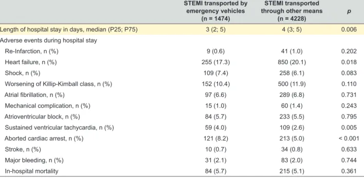
In-hospital mortality and complications
The rate of complications and in-hospital mortality are displayed in Table 5. Regarding in-hospital complications, there is a higher rate of sustained VT (p = 0.005) and aborted cardiac arrest (p < 0.001) in Group 1. On the other
Percentage of VMER use
Figure 1 – Evolution of VMER use throughout the years in the different regions
Chi-square test for Trend: Northern region p = 0.078; Central region p < 0.001 and Southern region and Islands p < 0.001
2010 2011 2012 2013 2014 2015 2016 2017 20.9 19.1 28.4 29.0 27.9 30.8 31.5 33.3 35.5 33.1 38.0 34.3 47.8 46.9 44.3 42.1 10.2 6.7 2.2 4.9 5.6 3.9 4.5 16.7
Northern Portugal Central Portugal Southern Portugal and Islands
0% 10% 20% 30% 40% 50% 60%
ARTIGO ORIGINAL
hand, patients from Group 2 are more likely to develop HF throughout the hospital stay (p = 0.018). When it comes to in-hospital mortality, there are no significant differences be-tween the groups (p = 0.361).
Regarding in-hospital mortality, there is a specific sub-set of patients that are associated with a worse outcome. Women (p < 0.001) and patients with valve disease (p = 0.037) and chronic kidney disease (p = 0.015) are associat-ed with higher mortality. Cardiac arrest (p = 0.008), shock (p < 0.001) and atrial fibrillation (p < 0.001) represent high-risk clinical features that culminate in a worse outcome. Also, the presence of right bundle branch block (RBBB) is associ-ated with higher mortality (p = 0.001), as described in the literature.17 Patients with left ventricular systolic dysfunction
(p < 0.001) and multivessel disease (p = 0.031) are associ-ated with higher in-hospital mortality.
DISCUSSION
Cardiovascular diseases are the most common cause of death in European countries.1-3,6,18 Even though
hospi-talization rates seem to have remained stable in the last few decades, STEMI rates are declining, probably due to
improved coronary risk factor awareness.18 Improvements
in treatment improved CVD prognosis, as shown by the steady reduction in mortality and morbidity.2-4,6,18 Hartley et al2 described an estimated annual percentage change in
IHD mortality in Portugal of -1.74% and -1.47% from 1980 to 2004 and of -8.44% and -8.20% from 2005 to 2009, in male and female patients, respectively. Townsend et al3
also reported that from 2003 to 2013 there was a 40.1% and 42.9% reduction in the age-standardized mortality rate by IHD in Portuguese men and women, respectively. The European Cardiovascular Disease Statistics 2017 report described a reduction in death rates from IHD from 220 and 125 deaths per 100 000 in 1980 to 98 and 49 deaths per 100 000 in 2014, in Portuguese men and women,
respec-tively.6 IHD is also the leading cause of death in both men
and women under 65 years of age,6 which is important for
this population since STEMI tends to affect younger patients more predominantly.1 In Portugal, there is a less steady but
effective decline in premature mortality by IHD, with death rates of 46 and 12 deaths per 100 000 in 1980 compared to 28 and 4 deaths per 100 000 in 2014, in men and women under 65 years of age, respectively.6 This overall reduction
in cardiovascular mortality is certainly related with improve-ments in pharmacological therapy and to an increase in PCI accessibility.19 Pereira et al5 reported a significant increase
in P-PCI from 2002 to 2013, thus promoting mortality and morbidity reduction in STEMI patients. The same group5
also demonstrated the change in the paradigm in STEMI treatment, notably P-PCI overtaking rescue angioplasty as the predominant procedure since 2006.
To truly impact prognosis, the most important part of the STEMI treatment is a timely reperfusion, ideally through P-PCI.1,20 P-PCI is superior to FT in terms of mortality
reduc-tion, reinfarction and stroke, if treatment delay is compara-ble.1,20 Nevertheless, if P-PCI is expected to be carried out
with significant delay, FT is preferable since it can be ini-tiated hastily.8,9 Treatment delay depends on patient-delay
and on system-delay. Patient-delay is difficult to minimize.1
The REACT trial21 employed a series of public education
campaigns in a community to evaluate differences in pa-tient-delay. Luepker et al21 determined that the
interven-tion, while significantly increasing EMS use, time between symptom onset and hospital arrival was not significantly different. In Portugal’s case, the European initiative Stent for Life strategy has been implemented.22 A series of
na-tional campaigns to improve public awareness, as well as organizational measures involving INEM and hospital per-formance, were applied with plans to improve the network of P-PCI centres and thus increasing the number of patients treated by PCI in cases of STEMI.22 The initiative enhanced
Table 3 – Treatment delays to reperfusion in minutes
STEMI transported by emergency vehicles
(n = 1474)
STEMI transported through other means
(n = 4228) p
Time between symptoms and first medical contact 116 (60; 205) 138 (70; 310) < 0.001
Time between first medical contact and admission 72 (45; 105) 80 (43; 148) < 0.001
Time between symptoms and admission 165 (110; 271) 183 (99; 387) 0.002
Time between symptoms and reperfusion 208.5 (150; 308) 267 (180; 435) < 0.001
Time between symptoms and needle 165 (120; 240) 205 (140; 297.5) 0.012
Time between symptoms and balloon 211 (152; 314) 270 (181; 445) < 0.001
Time between first medical contact and reperfusion 93 (49; 139) 120 (75; 190) < 0.001
Time between first medical contact and needle 71 (40; 106) 61.5 (39; 105) 0.994
Time between first medical contact and balloon 94 (50; 141) 125 (79; 195) < 0.001
Time between door and reperfusion 30 (15; 69) 85 (37; 160) < 0.001
Time between door and needle 30 (17; 64.5) 42.5 (20; 84.5) 0.109
Time between door and balloon 29 (15; 69) 88 (40; 165) < 0.001
ARTIGO ORIGINAL
Table 5 – In-hospital mortality and complications
STEMI transported by emergency vehicles
(n = 1474)
STEMI transported through other means
(n = 4228) p
Length of hospital stay in days, median (P25; P75) 3 (2; 5) 4 (3; 5) 0.006
Adverse events during hospital stay
Re-Infarction, n (%) 9 (0.6) 41 (1.0) 0.202
Heart failure, n (%) 255 (17.3) 850 (20.1) 0.018
Shock, n (%) 109 (7.4) 258 (6.1) 0.083
Worsening of Killip-Kimball class, n (%) 152 (10.4) 500 (11.9) 0.110
Atrial fibrillation, n (%) 97 (6.6) 289 (6.8) 0.731
Mechanical complication, n (%) 15 (1.0) 60 (1.4) 0.243
Atrioventricular block, n (%) 84 (5.7) 233 (5.5) 0.795
Sustained ventricular tachycardia, n (%) 59 (4.0) 109 (2.6) 0.005
Aborted cardiac arrest, n (%) 121 (8.2) 213 (5.0) < 0.001
Stroke, n (%) 10 (0.7) 34 (0.8) 0.633
Major bleeding, n (%) 31 (2.1) 83 (2.0) 0.744
In-hospital mortality 84 (5.7) 215 (5.1) 0.361
Table 4 – Therapeutic strategies
STEMI transported by emergency vehicles
(n = 1474)
STEMI transported through other means
(n = 4228) p Type of reperfusion Fibrinolysis, n (%) 104 (8.1) 219 (6.4) 0.042 - pre-hospital fibrinolysis, n (%) 15 (14.4) 9 (4.1) < 0.001 Primary PCI, n (%) 1178 (91.9) 3191 (93.6) 0.042 Angiograhy - Radial access, n (%) 968 (71.4) 2841 (74.1) 0.059
Number of affected vessels
- None, n (%) 28 (2.4) 76 (2.1) 0.527
- One vessel, n (%) 645 (55.9) 1970 (54.8) 0.509
- Two vessels, n (%) 323 (28.0) 1015 (28.2) 0.877
- Three vessels, n (%) 157 (13.6) 532 (14.8) 0.318
Total occlusions
- Left main coronary artery, n (%) 12 (1.0) 12 (0.3) 0.003
- Left anterior descending artery, n (%) 485 (36.9) 1267 (33.2) 0.017
- Circumflex artery, n (%) 139 (11.1) 454 (12.2) 0.305
- Right coronary artery, n (%) 478 (37.1) 1218 (32.2) 0.001
Left ventricular ejection fraction
- LVEF (%) 53 ± 13 49 ± 12 < 0.001 - Normal LVEF (> 50%), n (%) 889 (63.0) 2144 (53.4) < 0.001 - LVEF 40% - 49%, n (%) 309 (21.9) 984 (24.5) 0.050 - LVEF 30% - 39%, n (%) 158 (11.2) 667 (16.6) < 0.001 - LVEF < 30%, n (%) 54 (3.8) 221 (5.5) 0.014 Other Interventions
- Intra-aortic balloon pump, n (%) 8 (0.5) 57 (1.3) 0.012
- Invasive mechanical ventilation, n (%) 78 (5.3) 132 (3.1) < 0.001
- Non-invasive mechanical ventilation, n (%) 21 (1.4) 113 (2.7) 0.006
ARTIGO ORIGINAL
EMS use and shortened revascularization times due to re-duced patient-delay; system-delay remained unchanged however.22 Reducing system-delay is only possible through
organizational measures,1,23 and according to Terkelsen et al10 it can have an impact on mortality. Fordyce et al23
re-cently presented the results of the implementation of the STEMI Systems Accelerator program, which was applied in the United States. They described four key care process-es: (1) prehospital catheterization laboratory activation; (2) single call transfers to PCI capable hospitals for patients presenting to a non-PCI-capable centre; (3) ED bypass for patients referred via EMS; (4) ED bypass for patients trans-ferred from a non-PCI-capable centre.23 EMS played an
important part in employing the implementation of the initia-tive.23 Fordyce et al23 proved that, even though reperfusion
times and system-delay was significantly reduced, mortality remained unchanged. In fact, EMS transportation is cru-cial in reducing system-delay.1,11,15,23 EMS can shorten the
time to reperfusion by arriving faster to the patient location and providing earlier transport to PCI-capable centres or administration of on-site fibrinolysis (O-FT).1,15,24 EMS can
make available advanced cardiac life support personnel in a pre-hospital setting.15 EMS can dispatch STEMI alert to the
nearest PCI-capable centre or even activate catheterization lab in a pre-hospital setting, thus reducing door to balloon times. 11,12,14 However, despite the benefits the EMS seems
to carry, most published studies do not seem to report re-duced mortality.15,23
Regarding our population, men, patients with higher car-diovascular risk (namely dyslipidaemia and smoking habits) and known coronary disease tend to have higher usage of EMS vehicles. There was no significant difference in age. These findings were in some respects similar to what Canto
et al reported.15 Canto et al15 evaluated EMS use in cases
of AMI. When compared to our population, the biggest dif-ferences were: (1) men had 19% lower chance of using EMS; (2) the odds of EMS use were 21% higher with each increasing decade of life; (3) previous stroke or cardiac his-tory tended to have higher EMS use.15 In our population,
pa-tients with extreme weights (low weight or obese papa-tients) were more likely to be transported via VMER, which was not accounted for by Canto et al15 Another important aspect that
affected EMS use was the characteristics of the chest pain. The presence of persistent chest pain was independently associated with EMS use. On the other, paroxysmal chest pain was less likely to use VMER.
Another important fact was the steady rise in VMER use in STEMI patients. The implementation of public awareness initiatives, like the aforementioned Stent for Life, has cer-tainly made an impact in informing the general public. This steady increase of VMER use from 21.3% in 2010 to 38.3% in 2017 (p < 0.001) has been proven to be associated with significant patient- and system-delay reduction.22 Notably,
there is a significant lower rate of EMS transportation in Northern Portugal, a situation that merits the reinforcement of public awareness initiatives in this region to improve overall timings.
The greatest benefits that derive from EMS use are the reduction in reperfusion timings. Studies consistently show an impact of EMS transportation in reducing treatment de-lay.1,10,12-15,23 This study showed that, in Portugal, this is no
exception, VMER transportation ensured a 58-minute re-duction of the median reperfusion time (p < 0.001). VMER reduced the time between symptoms and the first medical contact (FMC), thus enabling a faster STEMI diagnosis. VMER also shortened the time between symptoms and hospital admission, playing a part in ensuring a faster trans-portation to the hospital, while providing advanced life sup-port if necessary. Another imsup-portant aspect is the reduction of the time between hospital admission and beginning of the PCI procedure. EMS transportation is known to reduce Door to Balloon (D2B) times,12,15 as is the case with our
population, and that reduction has been proven to reduce mortality in the study by Nallamothu,25 though that reduction
was not evident in a study by Menees.26 EMS has an
im-portant role in ensuring a field triage as well, thus enabling direct transfer to a PCI-capable centre, which may have an impact on mortality.11,23 In our population, VMER was
as-sociated with a significant lower incidence of admission to a non-PCI-capable centre, demonstrating the importance of field triage and following protocols according to geographi-cal areas of responsibility. However, there is still a high rate of transportation to a non-PCI-capable centre in the VMER group, which is not satisfactory. The authors suspect that this is probably due to two main reasons: (1) misdiagnosis of STEMI and thus transporting to the nearest hospital; (2) impossibility of administering on-site fibrinolysis, in a setting of an expectable delay of over 120 minutes for transporta-tion to a PCI-capable centre, and thus transportatransporta-tion to the nearest hospital being able to initiate FT expeditiously. EMS can also expedite pre-hospital Cath lab activation, reducing D2B times, however the risk of false positive activation is a factor that must be accounted for. This possibility is more frequent in pre-hospital STEMI diagnosis.12,14 Again, in
Por-tugal, VMER ensured a higher rate of ED bypass and of direct admission to the Cath lab, therefore playing an impor-tant role in the reduction of the system-delay.
Perhaps the most unexpected finding was that some tim-ings related with FT were not significantly reduced, namely the time between FMC and FT and between hospital ad-mission and FT. Strikingly enough, the time between FMC and beginning of FT was higher in the EMS group, albeit not significantly. These findings were different from what is described in other studies, which consistently describe shorter times to FT in patients transported by EMS.15,27 The
authors suspect that this might have happened due to pos-sible misdiagnosis of STEMI, inability to administer O-FT, poor coordination of the emergency systems, or simply because the time between FMC with VMER (which occurs in a pre-hospital setting) and FT is understandably higher when compared to in-hospital FMC and FT when O-FT is not available. Fortunately, the overall timing between symp-toms and FT ended up being significantly reduced in Group 1, which reflects the benefits of EMS in arriving faster to
ARTIGO ORIGINAL the patient’s location, thus reducing the time between symp-tom and FMC. VMER enhanced the patients’ chances of
receiving O-FT (14.4% vs 4.1%, p < 0.001). Pre-hospital fibrinolysis has a definite impact on prognosis if adminis-tered early after symptom onset and if transportation to a PCI-ready centre is expected to have a relevant delay, with the 120-minute mark being the most consistent inter-val reported.1,7,27-30 If possible, FT should be administered
in a pre-hospital setting.1,29-31 The presence of an EMS
ve-hicle with medical staff can certainly play a part in increas-ing the rate of pre-hospital fibrinolysis. Studies reveal that O-FT administered by trained staff was associated with a higher adherence to protocols and a decrease in treatment delay and might be associated with lower mortality.24,27 This
finding also raises another question regarding the small number of cases in which FT was administered outside the hospital setting in the absence of an EMS team. Since this is a retrospective and observational study, it is impossible to determine when, where and how these events occurred. It is the authors’ opinion that possibly these cases hap-pened in primary health care centres, where it is possible to administer fibrinolysis with medical supervision and with posterior referral ed ce«ospto PCI-capable centres through transportation means outside the EMS network. These re-sults reveal an issue that must be addressed in order to improve our care, namely the need to reduce the time be-tween symptoms and FT. Organizational measures should be enforced to guarantee that every VMER is capable of administering FT and adequate training should be given to the VMER staff to ensure the administration of FT in the recommended 10-minute interval, if transportation to a PCI-capable centre delay is expected.
Regarding the primary objective of the study, unfortu-nately EMS transport was not associated with mortality re-duction nationwide. This finding is consistent with what is described in other studies. Canto et al15 described higher
crude mortality in the EMS group, which was explained by the fact that the EMS population represented a much sicker cohort. In Portugal, the same applies. The population trans-ported by VMER represents patients of higher risk. There is a higher prevalence of cardiac arrest upon admission and throughout hospitalization, consistent lower blood pressure, higher rate of total occlusion of the left main, left anterior and right coronary arteries, more frequent occurrence of ventric-ular tachycardia and higher usage of invasive mechanical ventilation (which is significantly related to the higher rate of cardiac arrest, QQ test p-value of < 0.001). After individual evaluation of each specific subset of patients regarding KK class and occurrence of cardiac arrest, there was also no significant difference in mortality. The main benefit detected in the VMER group was the significant decrease in devel-opment of HF. This fact is related to the finding of consist-ently higher LVEF and lower BNP levels in Group 1. This is also associated with the higher usage of Intra-aortic balloon pump and non-invasive mechanical ventilation in the treat-ment of HF in group 2 (both procedures were significantly related to HF occurrence, QQ test p -value of < 0.001).
This benefit in LVEF and HF is the result of shorter reperfu-sion times. Longer duration of coronary occlureperfu-sion results in progressive extension of the infarction area and subsequent reduction of the amount of salvageable myocardium.28 The
reduction in overall reperfusion timings attained by VMER resulted in a greater amount of salvaged myocardium and thus led to a higher LVEF and subsequently lower occur-rence of HF. Another interesting aspect would be evaluating if VMER transportation had an impact in long-term progno-sis. According to the literature, left ventricular systolic dys-function is a strong predictor of in-hospital and long-term mortality.1,26,33 Since the shorter reperfusion times found in
the VMER group resulted in higher LVEF, one could expect better long-term results in that group. However, due to re-straints in the RNSCA, it is not possible to evaluate long-term mortality in our population.
Limitations
This study comprises all the limitations that are inher-ent to national registries. First, not all cinher-entres participate in the RNSCA. Each participation is voluntary, so it relies on the accurate description of every ACS case in each centre, which might not include all patients. Since this is a retrospective study, it is impossible to determine the cri-teria used in selecting which patients were transported by VMER. Due to registry restraints, the authors do not have access to each individual centre’s data, meaning it is not possible to evaluate the reasons for each individual treat-ment strategy. This is an observational study; thus, it has all the limitations associated with this method. There was no randomization performed throughout the study and it is impossible to infer causality in some of the parameters that are being analysed. Thus, the results obtained can only be considered indicative.
CONCLUSION
This article reflects the real-world data happening in Portugal. The RNSCA is an invaluable tool in evaluating the current management of ACS in a nationwide setting and can provide useful tools to improve our standard of care. EMS transportation plays a crucial role in the treatment of STEMI. Nationwide, it is evident that EMS is associated with a series of benefits: (1) it ensures transportation for patients with higher cardiovascular risk and with more critical clini-cal presentation; (2) it effectively shortens reperfusion times and enhances the possibility of O-FT when PCI is expected to be delayed; (3) it is associated with a higher amount of salvaged myocardium, as reflected by the higher LVEF, and thus reduces the incidence of HF and possibly enhancing long-term prognosis. Though these benefits were evident, VMER transportation does not impact in-hospital mortality. Probably, this is a result of confounding factors of disease severity and comorbidity in patients that are transported by VMER. Therefore, while this paper was unable to report mortality benefit, a longer follow-up could demonstrate the importance of VMER transportation in long-term prognosis.
ARTIGO ORIGINAL
OBSERVATIONS
A previous version of this work with fewer patients was presented in the Portuguese Congress of Cardiology 2016.
PROTECTION OF HUMANS AND ANIMALS
The authors declare that the procedures were followed according to the regulations established by the Clinical Re-search and Ethics Committee and to the Helsinki Declara-tion of the World Medical AssociaDeclara-tion.
DATA CONFIDENTIALITY
The authors declare having followed the protocols in
use at their working center regarding patients’ data publica-tion.
CONFLICTS OF INTEREST
The authors declare that there are no conflicts of inter-est nor any form of support.
FUNDING SOURCES
This research received no specific grant from any fund-ing agency in the public, commercial, or not-for-profit sec-tors.
REFERENCES
1. Ibanez B, James S, Agewall S, Antunes M, Bucciarelli-Ducci C, Bueno H, et al. 2017 ESC Guidelines for the management of acute myocardial infarction in patients presenting with ST-segment elevation: The Task Force for the management of acute myocardial infarction in patients presenting with ST-segment elevation of the European Society of Cardiology (ESC). Eur Heart J. 2018;39:119-77.
2. Hartley A, Marshall D, Salciccioli J, Sikkel M, Maruthappu M, Shalhoub J. Trends in mortality from ischemic heart disease and cerebrovascular disease in Europe: 1980 to 2009. Circulation. 2016;133:1916-26. 3. Townsend N, Wilson L, Bhatnagar P, Wickramasinghe K, Rayner M,
Nichols M. Cardiovascular disease in Europe: epidemiological update 2016. Eur Heart J. 2016;37:3232-45.
4. Puymirat E, Simon T, Cayla G, Cottin Y, Elbaz M, Coste P, et al. Acute myocardial infarction: changes in patient characteristics, management, and 6-month outcomes over a period of 20 years in the FAST-MI Program (French Registry of Acute ST-Elevation or Non-ST-Elevation Myocardial Infarction) 1995 to 2015. Circulation. 2017;136:1908-19. 5. Pereira H, Campante Teles R, Costa M, Canas da Silva P, da Gama
Ribeiro V, Brandão V, et al. Trends in primary angioplasty in Portugal from 2002 to 2013 according to the Portuguese National Registry of Interventional Cardiology. Rev Port Cardiol. 2016;35:395-404. 6. Wilkins E, Wilson L, Wickramasinghe K, Bhatnagar P, Leal J,
Luengo-Fernandez R, et al. European Cardiovascular Disease Statistics 2017. Brussels: European Heart Network; 2017.
7. Pinto DS, Frederick PD, Chakrabarti AK, Kirtane AJ, Ullman E, Dejam A, et al. Benefit of transferring ST-segment-elevation myocardial infarction patients for percutaneous coronary intervention compared with administration of onsite fibrinolytic declines as delays increase. Circulation. 2011;124:2512-21.
8. Armstrong P, Gershlick A, Goldstein P, Wilcox R, Danays T, Lambert Y, et al. Fibrinolysis or primary PCI in ST-segment elevation myocardial infarction. N Engl J Med. 2013;368:1379-87.
9. Danchin N, Puymirat E, Steg P, Goldstein P, Schiele F, Belle L, et al. Five-year survival in patients with ST-segment-elevation myocardial infarction according to modalities of reperfusion therapy: the French Registry on Acute ST-Elevation and Non-ST-Elevation Myocardial Infarction (FAST-MI) 2005 Cohort. Circulation. 2014;129:1629-36. 10. Terkelsen C, Sørensen J, Maeng M, Jensen L, Tilsted H, Trautner S, et
al. System delay and mortality among patients with STEMI treated with primary percutaneous coronary intervention. JAMA. 2010;304:763-71. 11. Postma S, Kolkman E, Rubinstein SM, Jansma EP, De Luca G,
Suryapranata H, et al. Field triage in the ambulance versus referral via non-percutaneous coronary intervention centre in ST-elevation myocardial infarction patients undergoing primary percutaneous coronary intervention: a systematic review. Eur Heart J Acute Cardiovasc Care. 2017;6:396-403.
12. Stowens J, Sonnad S, Rosenbaum A. Using EMS dispatch to trigger STEMI alerts decreases Door-to-Balloon times. West J Emerg Med. 2015;16:472–80.
13. Scherer TM, Russ S, Jenkins CA, Jones ID, Slovis CM, Cunningham BL, et al. Predictors of ambulance transport in patients with ST-elevation myocardial infarction. Prehosp Disaster Med. 2012;27:226-30. 14. Squire BT, Tamayo-Sarver JH, Rashi P, Koenig W, Niemann JT. Effect
of prehospital cardiac catheterization lab activation on door-to-balloon time, mortality, and false-positive activation. Prehosp Emerg Care. 2014;18:1–8.
15. Canto J, Zalenski R, Ornato J, Rogers W, Kiefe C, Magid D, et al. Use of emergency medical services in acute myocardial infarction and subsequent quality of care: observations from the National Registry of Myocardial Infarction 2. Circulation. 2002;106:3018-23.
16. Santos J, Aguiar C, Gavina C, Azevedo P, Morais J; Registo Nacional de Síndromes Coronárias Agudas da Sociedade. Portuguese Registry of Acute Coronary Syndromes: seven years of activity. Rev Port Cardiol. 2009;28:1465-500.
17. Widimsky P, Rohac F, Stasek J, Kala P, Rokyta R, Kuzmanov B, et al. Primary angioplasty in acute myocardial infarction with right bundle branch block: should new onset right bundle branch block be added to future guidelines as an indication for reperfusion therapy? Eur Heart J. 2012;33:86–95.
18. McManus D, Gore J, Yarzebski J, Spencer F, Lessard D, Goldberg R. Recent trends in the incidence, treatment, and outcomes of patients with STEMI and NSTEMI. Am J Med. 2011;124:40-7.
19. Pereira H, Teles R, Costa M, da Silva P, Ferreira R, da Gama Ribeiro V, et al. Trends in percutaneous coronary intervention from 2004 to 2013 according to the Portuguese National Registry of Interventional Cardiology. Rev Port Cardiol. 2015;34:673-81.
20. Keeley EC, Boura JA, Grines CL. Primary angioplasty versus intravenous thrombolytic therapy for acute myocardial infarction: a quantitative review of 23 randomised trials. Lancet. 2003;361:13–20. 21. Luepker R, Raczynski J, Osganian S, Goldberg R, Finnegan J Jr,
Hedges J, et al. Effect of a community intervention on patient delay and emergency medical service use in acute coronary heart disease: The Rapid Early Action for Coronary Treatment (REACT) Trial. JAMA. 2000;284:60-7.
22. Pereira H, Pinto FJ, Calé R, Pereira E, Marques J, Almeida M, et al. Stent for Life in Portugal: this initiative is here to stay. Rev Port Cardiol. 2014;33:363-70.
23. Fordyce C, Al-Khalidi H, Jollis J, Roettig M, Gu J, Bagai A, et al. Association of rapid care process implementation on reperfusion times across multiple ST-segment elevation myocardial infarction networks. Circ Cardiovasc Interv. 2017;10:e004061.
24. Welsh R, Chang W, Goldstein P, Adgey J, Granger C, Verheugt F, et al. Time to treatment and the impact of a physician on prehospital management of acute ST elevation myocardial infarction: insights from the ASSENT-3 PLUS trial. Heart. 2005;91:1400–6.
25. Nallamothu B, Normand S, Wang Y, Hofer T, Brush J Jr, Messenger J, et al. Relation between door-to-balloon times and mortality after primary percutaneous coronary intervention over time: a retrospective study. Lancet. 2015;385:1114-22.
26. Menees D, Peterson E, Wang Y, Curtis J, Messenger J, Rumsfeld J, et al. Door-to-Balloon time and mortality among patients undergoing primary PCI. N Engl J Med. 2013;369:901-9.
27. Bjorklund E, Stenestrand U, Lindback J, Svensson L, Wallentin L, Lindahl B. Prehospital thrombolysis delivered by paramedics is associated with reduced time delay and mortality in ambulance-transported real-life patients with ST-elevation myocardial infarction. Eur Heart J. 2006;27:1146–52.
28. Boersma E, Maas A, Deckers J, Simoons M. Early thrombolytic treatment in acute myocardial infarction: reappraisal of the golden hour. Lancet 1996;348:771–5.
29. Sinnaeve P, Armstrong P, Gershlick A, Goldstein P, Wilcox R, Lambert Y, et al. ST-segment-elevation myocardial infarction patients randomized
ARTIGO ORIGINAL
to a pharmaco-invasive strategy or primary percutaneous coronary intervention: Strategic Reperfusion Early After Myocardial Infarction (STREAM) 1-year mortality follow-up. Circulation. 2014;130:1139–45. 30. Morrison L, Verbeck P, McDonald A, Sawadsky B, Cook D. Mortality
and prehospital thrombolysis for acute myocardial infarction: a meta-analysis. JAMA. 2000;283:2686–92.
31. Bonnefoy E, Steg P, Boutitie F, Dubien P, Lapostolle F, Roncalli J, et al. Comparison of primary angioplasty and pre-hospital fibrinolysis in acute myocardial infarction (CAPTIM) trial: a 5-year follow-up. Eur Heart J.
2009;30:1598–606.
32. van der Vleuten P, Rasoul S, Huurnink W, van der Horst I, Slart R, Reiffers S, et al. The importance of left ventricular function for long-term outcome after primary percutaneous coronary intervention. BMC Cardiovasc Disord. 2008;8:4.
33. Ng V, Lansky A, Meller S, Witzenbichler B, Guagliumi G, Peruga J, et al. The prognostic importance of left ventricular function in patients with ST-segment elevation myocardial infarction: the HORIZONS-AMI trial. Eur Heart J Acute Cardiovasc Care. 2014;3:67-77.