BJRS
RADIATION SCIENCES
08-01 (2020) 01-18ISSN: 2319-0612 Accepted: 2019-12-04
Bony-based and prostate-based image guidance for
lo-calized prostate cancer radiotherapy.
Fortes
a,bS. S., Rosa
bL. A. R. da
aInstituto Nacional de Câncer José Alencar Gomes da Silva, 20230-130, Praça Cruz Vermelha, 23 – Centro – Rio de
Janeiro – RJ - Brazil
bInstituto de Radioproteção e Dosimetria, 22780-160, Av. Salvador Allende, s/n – Recreio dos Bandeirantes – Rio de
Janeiro – RJ - Brazil [email protected]
ABSTRACT
An important modality for the treatment of prostate cancer is teletherapy. The use of image-guided radiotherapy (IGRT) is a valuable tool in this treatment. This study retrospectively compared how repositioning the patient based on bone structure (B-ISO) and the prostate itself (P-ISO) affected the volumetric dose in the rectum, bladder, and clinical target volume (CTV). Additionally, the normal tissue complication probability (NTCP) for the rectum was computed. We evaluated 155 cone-beam computed tomography (CBCT) from 8 patients. The treatment plans used beam modulation techniques. The planning target volume (PTV) margin adopted in both scenarios was 1 cm. The organs of interest were outlined over each CBCT and then treatment plans were applied so that the absorbed dose could be computed. NTCP values were calculated for the rectum. Analyzing dose-volume metrics published by the Quantitative Analysis of Normal Tissue Effects in the Clinic (QUANTEC), there was no significant difference between the two repositioning strategies for the rectum and bladder. There was a slight degradation in CTV coverage for the B-ISO strategy, but still with adequate coverage. Analysis of the uniform equivalent dose (EUD) and NTCP for the rectum showed little sensitivity to the strategy used. The present study showed that the use of CBCT in radiotherapy for prostate cancer treatment did not significantly improve volumetric doses for the rectum, bladder, and CTV, as well as NTCP for the rectum.
1. INTRODUCTION
Prostate cancer is estimated to be the most common in men in Brazil, excluding non-melanoma skin cancer [1]. Teletherapy is an important treatment modality for localized prostate cancer. The radiotherapy technique for prostate irradiation has changed significantly over the years [2]. The use of beam intensity-modulated radiotherapy (IMRT), a technique capable of distributing high doses of radiation to the target volume with a highly conformal dose distribution, allowed the scaling of the prescribed prostate dose [3]. Many researchers [5, 6] have shown that IMRT reduces treatment side effects and improves survival rates when compared to the 3D conformal radiotherapy technique (3DCRT). However, additional caution is required when distributing high doses of radiation to the prostate, as it may vary its position and shape during both intrafraction and interfraction treatment [6].
The use of image-guided radiotherapy (IGRT) allows positional adjustments for geometric deviations. IGRT represents an advanced tool that can contribute to the success of radiotherapy [7]. Cone-Beam Computed Tomography (CBCT) is an in-room CT imaging modality that allows the prostate gland to be identified. It also allows evaluating anatomical variations of other structures such as rectum and bladder. Different IGRT modalities and image frequency require different PTV margins [8].
At our institution, the imaging protocol for the conventional size fraction (2 Gy per day) is a pair of kilovolt planar images in the first three days and, from the second week, weekly images. This study retrospectively compared how bone-based positional adjustment (B-ISO) and prostate-based adjustment (P-ISO) affected volumetric dose to the prostate, rectum, and bladder. The probabilities of toxicity to the rectum were also compared.
2. MATERIALS AND METHODS
2.1. Patients
A total of 8 patients with confirmed prostate adenocarcinoma were studied retrospectively. The patients underwent external RT for localized prostate treatment without pelvic lymph node involvement.
2.2. Simulation and treatment planning
A Brilliance Big Bore CT scanner (Philips Healthcare, Bothell, WA) was used to acquire the simulation images. All scans were performed with the patient in the supine position, head to the gantry, pillow under the head, using knee support, and arms positioned over the chest, holding a ring. 2 mm slices were used. Laser markings projected onto the patient’s skin were identified with radiopaque markers on tomographic images. These were used as the origin in the planning TC.
In all cases, patients were instructed to keep the rectum empty and the bladder full for both the acquisition of planning CT and for each of the treatment fractions. Patients were also instructed to follow a diet to prevent or reduce diarrhea during treatment.
Using the Eclipse v.13.6 planning system (Varian Medical Systems, Palo Alto, CA), the volumes of interest were contoured and treatment plans and dose distribution calculations were performed. CTV, rectum, and bladder volumes were delineated according to the guideline RTOG [9]. The PTV margin was 10 mm, isotropic. All patients were irradiated with a dose of 74 Gy, distributed in 37 fractions of 2 Gy per fraction. Treatment plans used beam energy of 6 MV (5 patients) and 10 MV (3 patients). 7 cases were planned with IMRT, and 1 with VMAT. IMRT plans used 5 or 7 radiation beams.
Accepted treatment plans in all cases had CTV covered by 100% of the prescribed dose. The acceptance criteria were the dose-volume metrics published by QUANTEC [10]. The accepted criteria for rectum are V75Gy <15%, V70Gy <20%, V65Gy <25%, V60Gy <35%, V50Gy <50%. If these metrics are not violated, the probability of late rectal toxicity grade ≥ 2 is less than 15%. For the bladder, the accepted metrics are V80Gy <15%, V75Gy <25%, V70Gy <35%, V65Gy <50%.
2.3. CBCT and estimated volumetric doses
Treatments were performed on a Trilogy linear accelerator (Varian Medical Systems, Palo Alto, CA). The patients were initially positioned by aligning the lasers at the reference markings made on the skin. CBCT was then acquired. Images were taken using the pelvis mode, 125 kV, 80 mAs, 13 ms, and full scan with half-fan bowtie filter. The planning CT images and the daily CBCT were rigidly registered manually using translation only for the B-ISO and P-ISO strategies. All contours were made using the anatomy of the day. The organs analyzed here, CTV, rectum, and bladder, were contoured over each CBCT image. The rectum and bladder volumes were analyzed and compared with those of the planning CT. A total of 155 CBCTs were performed (an average of approximately 19 for each patient).
In the B-ISO strategy, a pair of anterior and lateral planar images were compared with digitally reconstructed radiography (DRR) images. Using bone match, the necessary correction was introduced and the isocenter was defined. In the P-ISO strategy, the prostate was used for registration between the planning TC and the CBCT.
For each CBCT, using the isocenters defined by the B-ISO and P-ISO repositioning strategies, the original plan was used to calculate the dose distribution, considering the anatomy of the day. The values of monitor units (MU), beam weight, and gantry angle were kept the same as those used for the planning CT. For each patient and each repositioning strategy, daily doses in the volumes of interest were calculated. Additionally, daily and cumulative dose-volume histograms (DVH) were calculated for each patient and both scenarios. DVH data were exported to a spreadsheet, where dose and volume information was extracted for analysis and comparison with planning CT.
2.4. Estimation of Normal Tissue Complication Probability (NTCP)
The dose-response model used to calculate the probability of occurrence of given toxicity was the Lyman-Kutcher-Burman (LKB) model described in detail by Lyman et. al. [11]. The equation that describes the model is as follows:
where EUD is the uniform equivalent dose, which represents the dose which, if evenly distributed throughout the organ volume, would produce the same effect as a given heterogeneous dose distribution as specified by DVH. The EUD is calculated by the equation [12]:
(2)
The function that describes the calculation of NTCP is described by:
(3), with x = (D – D50(V)) / (mD50(V)), D is the dose for the volume fraction V, D50(V)=D50V-n.
Lyman's model has four parameters: V, n, m, and D50. V is the volume fraction with a dose D; n
determine tissue volume dependence on tissue and takes into account differences in tissue architecture; m is the parameter that controls the slope of the dose-response curve, and D50 is the
dose point whose probability of complication is 50%. The endpoint analyzed was rectal bleeding (G2). The parameters used were those published by Gulliford et al. [13], m = 0.16, n = 0.18, and D50 = 68.9 Gy.
2.5. Statistical analysis of data
To analyze the statistical significance of comparisons between the samples of the two groups studied, the two-tailed paired Wilcoxon test was used. This is a nonparametric analysis method using rank. This test is indicated when the distribution of a variable is not normal [14].
3. RESULTS
3.1. Characteristics of the studied strategies
Table 1 summarizes the characteristics of the B-ISO and P-ISO strategies, showing the main similarities and differences.
Table 1: Summary of the characteristics of B-ISO and P-ISO strategies.
Adopted Strategy
B-ISO P-ISO
Patients (n) 8 8
PTV margin 1 cm, isotropically 1 cm, isotropically
Patient prepare Fill the bladder and keep the rectum empty. Fill the bladder and keep the rectum empty. Prescribed dose 74 Gy 74 Gy
Technique IMRT/RA IMRT/RA
Image protocol in the first 3 days and then Orthogonal planar images 1 x / week.
CBCT in various fractions with online correction.
3.2. Cumulative dose on CTV
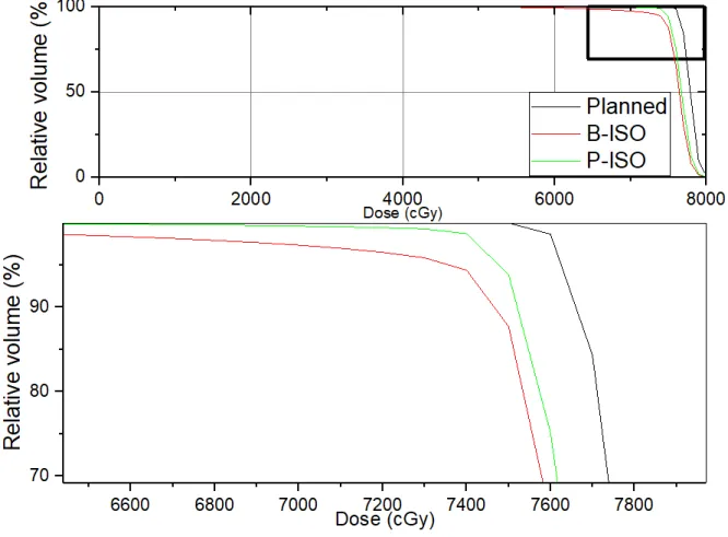
Figure 1 shows the planned CTV DVHs as well as the cumulative dose resulting from the B-ISO and P-B-ISO repositioning strategies. The planning margin used in all cases was 1 cm, isotropic. The curves presented refer to the case where the largest difference in the volume of CTV covered by the prescription dose was found considering the two strategies studied. The V100% was 100% for the planned DVH, 98.7% for the P-ISO strategy, and 94.4% for B-ISO. The volume of CTV encompassed by 95% of the prescribed dose was 100%, 99.6%, and 97.2% for planned, P-ISO, and B-ISO, respectively.
Figure 1: Dose-volume histogram of planned CTV (gray) and accumulated dose using B-ISO (red) and P-ISO (green) repositioning strategies. The planning margin adopted was 1 cm, isotropic. The
inferior graph is a zoom of superior graph.
3.3. CTV, rectum, and bladder dosimetric data
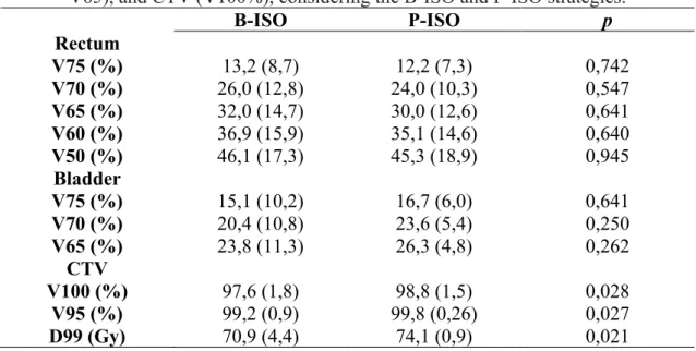
Table 2 shows summarize CTV, rectum, and bladder dosimetric information, considering the B-ISO and P-B-ISO strategies. The dose-volume metrics V75, V70, V65, V60, and V50 for the rectum are presented. These metrics represent the percentage volume of the rectum receiving 75, 70, 65, 60, and 50 Gy. In parentheses is expressed the standard deviation of the mean. Similarly, for the bladder, the metrics V75, V70, and V50 were analyzed. For both organs at risk (OAR), for all metrics analyzed, no significant difference was observed between the B-ISO and P-ISO strategies. Table 2 also shows the percentage volume of CTV covered by 100% and 95% of the prescribed
dose (V100% and V95%). A decrease in the volume of CTV irradiated by the prescription dose for the
B-ISO strategy was observed when compared to P-ISO (97.6% for B-ISO and 98.8% for P-ISO, p = 0.028). Similar behavior was observed for V95% (99.2% and 99.8%, p = 0.027). The dose encompassing 99% of the target volume (D99) was also systematically lower for the B-ISO strategy (70.9 Gy B-ISO and 74.1 Gy P-ISO, p = 0.021). CTV coverage in the B-ISO strategy was negatively affected with statistical significance for the three metrics analyzed. However, it still maintained dosimetrically adequate requirements for CTV.
Table 2: Volume dose metrics for the rectum (V75, V70, V65, V60, and V50), bladder (V75, V70, V65), and CTV (V100%), considering the B-ISO and P-ISO strategies.
B-ISO P-ISO p Rectum V75 (%) 13,2 (8,7) 12,2 (7,3) 0,742 V70 (%) 26,0 (12,8) 24,0 (10,3) 0,547 V65 (%) 32,0 (14,7) 30,0 (12,6) 0,641 V60 (%) 36,9 (15,9) 35,1 (14,6) 0,640 V50 (%) 46,1 (17,3) 45,3 (18,9) 0,945 Bladder V75 (%) 15,1 (10,2) 16,7 (6,0) 0,641 V70 (%) 20,4 (10,8) 23,6 (5,4) 0,250 V65 (%) 23,8 (11,3) 26,3 (4,8) 0,262 CTV V100 (%) 97,6 (1,8) 98,8 (1,5) 0,028 V95 (%) 99,2 (0,9) 99,8 (0,26) 0,027 D99 (Gy) 70,9 (4,4) 74,1 (0,9) 0,021
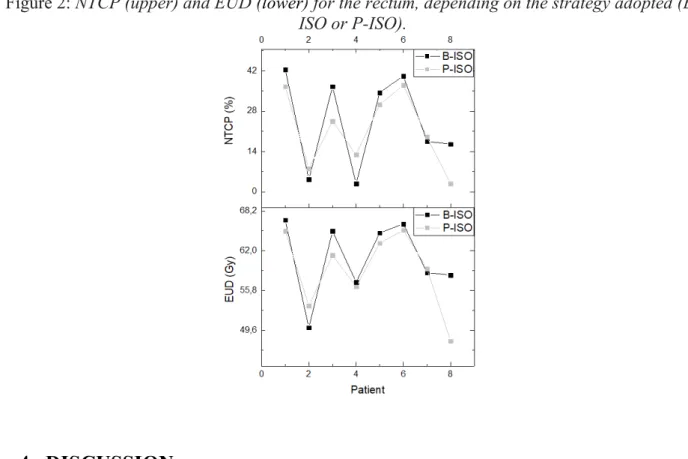
Figure 2 shows for each patient studied, graphically, the values of the equivalent uniform dose (EUD) and NTCP for the rectum as a function of the adopted repositioning strategy. As observed in the analysis of dose-volume metrics (Table 1), except for patient 8, the trend observed for both EUD (60.9 Gy for B-ISO group and 59.0 Gy for P-ISO) and NTCP (24.3% for B-ISO and 21.4% for P-ISO) showed little sensitivity in relation to the adopted strategy, showing no significant difference between both strategies (p = 0.148 for EUD and p = 0.313 for NTCP). ). For all patients, the metrics evaluated in Figure 2 were computed based on the DVHs resulting from the accumulated rectum dose. EUD and NTCP bladder analyses were not included because in all patients the probability of complications tended to zero.
Figure 2: NTCP (upper) and EUD (lower) for the rectum, depending on the strategy adopted (B-ISO or P-(B-ISO).
4. DISCUSSION
Before distributing the prescribed dose in a fraction of radiotherapy treatment it is important to ensure proper setup. Setup errors can cause relevant deviations in dose delivered to target and organs at risk. In prostate cancer treatments, the target may shift relative to pelvic bone references. This may render positioning based on bone parameters inadequate [15]. The use of CBCT provides a three-dimensional reconstruction with better soft tissue contrast than seen in planar images [16].
Many studies report dosimetric superiority in prostate CBCT based IGRT over bone markers based IGRT [15, 17, 18]. Results of this study showed no dosimetric difference for rectum and bladder when CBCT was used. The rectum volumes receiving doses of 75, 70, 65, 60 and 50 Gy, shown in table 2, were systematically smaller for the P-ISO group. For the bladder, volumes receiving 75, 70, and 65 Gy were higher when CBCT was used compared to B-ISO. But without statistical significance in both cases. Degradation of dose distributed at CTV was observed for the group in which the B-ISO strategy was used for the V100%, V95%, and D99% metrics. The latter
had the most pronounced effect (p = 0.021). Zucca et al. used MV-CBCT to analyze 13 patients undergoing prostate radiotherapy and concluded that soft tissue-based positioning is more appropriate than bone references for verifying and correcting setup errors immediately before treatment [18]. Nakamura et al. retrospectively analyzed 96 patients, observed a significantly lower incidence of gastrointestinal toxicity in the prostate-based group compared to the bone-based group without sacrificing tumor control [3]. They used a 1-3 mm lower PTV margin for the group in which CBCT was used. In a study involving two institutions, Chung and colleagues analyzed 25 patients treated with IMRT [17]. 10 patients were treated without IGRT, with 1 cm PTV margins, 0.5 cm posterior, and 15 patients treated with IGRT (CBCT) and implanted fiducials, with 2-3 mm PTV margin. IGRT significantly reduced rectal and bladder doses, with no significant difference in target volume coverage.
On the other hand, Zhong et al. retrospectively compared 65 patients treated with IMRT and IGRT with a group of 62 patients treated with IMRT [2]. The adopted PTV margins were equal. Similar to the results of the present study, they reported a lower rectal volume receiving doses of at least 40 Gy and 70 Gy in the IGRT-treated group, but without statistical significance. There was no significant better biochemical control in 5 years for either group. In a retrospective study of 30 patients and 270 CBCTs, Hirose and colleagues concluded that among six correction strategies analyzed, only the simple use of tattooing (non-correction) was significantly worse [19]. They reported that the use of bone correction still ensures the planned dose distribution in prostate cancer IMRT. The study evaluated two scenarios: 8 mm PTV margin, 5 mm posteriorly, and 5 mm isotropically margin. Variations in dose-volume metrics showed a consistent trend for both scenarios. Recently, in a phase 3 randomized controlled trial, Tondel et al. used questionnaires for endpoint evaluation and compared a group treated with weekly orthogonal images (n = 129) with a PTV margin of 15 mm and one group with daily CBCT (n = 128), with a PTV margin of 7 mm [20]. They concluded that daily CBCT with reduced margins showed no advantage over side effects reported by patients at the end of treatment.
Many studies have shown a lower incidence of risk organ toxicity, or probability of occurrence (NTCP), when using CBCT for patient repositioning. However, many of them used lower PTV margins when compared to the bone setup correction scenario [3, 17, 18]. Some researchers who
compared bone and prostate-based repositioning strategies using the same PTV margin did not see significant superiority in either strategy [2, 19]. Even with reduced PTV margins, Tondel and colleagues did not observe superiority in the daily CBCT group [20]. Additionally, the present study, using the same PTV margin (1 cm) in both scenarios, did not prove the superiority of one strategy over another. Even observing a slight degradation in CTV coverage, in both scenarios, the dose distribution in this volume can be considered dosimetrically adequate, as discussed by Mzenda et al. in their review [21]. Even for the B-ISO strategy, 99% of the CTV was covered by 70.9 Gy (> 95% of the prescribed dose) and the volume receiving 95% of the dose averaged 99.2%.
The results of the present study can be considered surprising. Estimating adequate PTV margin is a challenge in radiotherapy. Especially in the treatment of the prostate, inter and intra-fractional movements may cause adverse effects [22]. Dosimetric advantages over OARs using IGRT are only enhanced if PTV margins are reduced [2].
Some studies have shown clinical superiority of IGRT use in prostate RT in terms of tumor toxicity and control [7, 23]. In addition to the adopted PTV margins, other factors may contribute to CBCT not being superior to the bone-based strategy. The frequency of images is related to the benefit of IGRT. Kupelian et al. showed that, among eight imaging protocols, the use of daily imaging was what enabled the greatest reduction in PTV margins [24]. The residual error may be substantial in the treatment fractions without IGRT in a daily imaging scheme. However, a higher frequency impacts the increase in the dose absorbed by the patient [25] and increased costs and resources needed [26]. Additionally, Zelefsky et al. showed a greater benefit in tumor control when the prescribed dose was scaled for low risk (≥ 75.6 Gy) and high risk (≥ 81 Gy) patients [27]. Therefore, a potential benefit of IGRT over tumor control can be minimized if prescribed doses are not scalated.
This study retrospectively analyzed 155 CBCTs from 8 patients (mean approximately 19 CBCTs per patient) undergoing radiotherapy to treat localized prostate cancer at a dose of 74 Gy in 37 sessions. The doses distributed in CTV, rectum, and bladder resulting from the correction of bone parameter setup errors (B-ISO) and the prostate gland (P-ISO) were compared. Dosimetric variations in the studied organs were not sensitive to the strategy used. A 1 cm PTV margin
circumferentially was used in both groups. Limiting factors of this study may be considered (a) the number of patients studied. Volumetric doses in the rectum were systematically lower when P-ISO was adopted. Perhaps a larger n could produce a statistically significant difference in one or more metrics analyzed. (b) The rigid registration method between the planning CT and CBCT was used. Song et. al. reported that the use of deformable fusion has only a moderate influence on the RT dosimetry of prostate cancer [28]. (c) Intrafraction variations were not considered in this study. Many studies have focused on investigating the intrafraction movement of the prostate [29, 30]. (d) CBCT use was not daily. This study had the merit of performing CBCT in many treatment fractions (mean of 19 per patient), while some studies used lower frequencies of imaging [31, 32]. The higher the frequency of IGRT, the fewer assumptions are required, as the more accurate estimate of the distributed dose is.
The reduction of residual error after adopting a setup error correction strategy has been investigated in radiotherapy [19]. This is particularly challenging in the case of prostate cancer treatment, as it can move inter and intrafraction, and suffer interfraction deformations [8]. Several strategies have been investigated as fiducial marker implants [15], real-time location transponder implants [33], mechanical limitation of prostate movement through the use of rectal balloon [34], transabdominal ultrasound before each fraction [35], real-time transperineal ultrasound [36], MV-CBCT [18] or KV-MV-CBCT [37]. Prostate movements and anatomical variations may occur from patient to patient, and even in an individual patient [8]. Strategies to reduce margins on prostate moving targets should be considered as work in progress.
5. CONCLUSION
By analyzing 155 CBCTs from 8 radiotherapy treated prostate cancer patients using an isotropic 1 cm PTV margin, our study observed rectal volumes receiving slightly lower specific doses when CBCT was used. Opposite behavior was observed for the bladder. Slight degradation of the dose distributed at CTV was observed for group B-ISO, but meeting dosimetric requirements for CTV. For PTV margins of this magnitude, prostate-based repositioning did not significantly improve rectal, bladder, and CTV dose when compared to the bone-based repositioning strategy.
REFERENCES
[1] BORGES, k. A. M., SCHILITHZ, F. O. C., LIMA, F. C. S., FERREIRA, J. D., DE MORAIS, L. A., SANTOS, M. O., REBELO, M. S., COSTA, R. M. O. Estimativa 2018: incidência do câncer no Brasil. Instituto Nacional de Câncer José de Alencar
Gomes da Silva, Coordenação de Prevenção e Vigilância, 2017.
[2] ZHONG, Q., GAO, H., LI, G., XIU, X., WU, Q., LI, M., XU, Y. Significance of image guidance to clinical outcomes for localized prostate cancer. BioMed Research Int, v.
14, 2014.
[3] NAKAMURA, K., MIZOWAKI, T., INOKUCHI, H., IKEDA, I., INOUE, T., KAMBA, T., OGAWA, O., HIRAOKA, M. Decreased acute toxicities of intensity‑modulated radi-ation therapy for localized prostate cancer with prostate‑based versus bone‑based image guidance. Int J Clin Oncol, v. 23, p. 158-164, 2018.
[4] ZIETMAN, A.L., DESILVIO, M.L., SLATER, J.D., ROSSI, C. J. Jr., MILLER, D. W., ADAMS, J. A., SHIPLEY, W. U. Comparison of conventional-dose vs. high-dose con-formal radiation therapy in clinically localized adenocarcinoma of the prostate: a ran-domized controlled trial. JAMA, v. 294, p.1233–1239, 2005.
[5] ZELEFSKY, M. J., CHAN H., HUNT M., YAMADA, Y., SHIPPY, A. M., AMOLS, H. Long-term outcome of high dose intensity-modulated radiation therapy for patients with clinically localized prostate cancer. J Urol, v. 176, p. 1415–1419, 2006.
[6] LANGEN, K.M., WILLOUGHBY, T.R., MEEKS, S.L., SANTHANAN, A., CUN-NINGHAN, A., LEVINE, L., KUPELIAN, P. A. Observations on real-time prostate gland motion using electromagnetic tracking. Int J Radiat Oncol Biol Phys, v. 71, p.
1084–1090, 2008.
[7] BUJOLD, A., CRAIG, T., JAFFRAY, D, DAWSON, L.A. Image-Guided Radiotherapy: Has It Influenced Patient Outcomes? Semin Radiat Oncol, v. 22, p. 50-61, 2012.
[8] KUPELIAN, P.A., TWYLA, R., WILLOUGHBY, M.S., KLEIN, E. A., MAHADEVAN, A. Impact of image guidance on outcomes after external beam radiotherapy for localized prostate cancer. Int. J. Radiation Oncology Biol Phys, v. 70, n. 4, p. 1146–1150, 2008.
[9] GAY, H.A., BARTHOLD, H.J., O’MEARA, E., BOSCH, W. R., EL NAQA, I., AL-LOZI, R., ROSENTHAL, R. A., LAWTON, C., LEE, W. R., SANDLER, H., ZIET-MAN, A., MYERSON, R., DAWSON, L. A., WILLETT, C., CACHNICH, L. A., JHINGRAN, A., PORTELANCE, S., RYU, J., SMALL, W. Jr., GAFFNEY, D., VISWANATHAN, A. N., MICHALSKI, J. M. Pelvic normal tissue contouring guide-lines for radiation therapy: a Radiation Therapy Oncology Group consensus panel atlas.
Int. J. Radiat Oncol Biol Phys, v. 83, n. 3, p. 353-362, 2012.
[10] MARKS, L. B., YORKE, E. B., JACKSON, A., TEN HAKEN, R. K., CONSTINE, L. S., EISBRUCH, A., BENTZEN, S. M., NAM, J., DEASY, J. O. Use of Normal Tissue Complication Probability Models in the Clinic. Int J Radiat Oncol Biol Phys, v. 76,
supl. 3, p. S10-S19, 2010.
[11] LYMAN, J. T., WOLBARST, A. B. Optimization of radiation therapy, IV: A dose-volume histogram reduction algorithm. Int J Radiat Oncol BioI Phys, v. 17, n. 2, p.
433-436, 1989.
[12] MOHAN, R., MAGERAS, G.S., BALDWIN, B., BREWSTER, L. J., KUTCHER, G. J., LEIBEL, S., BURMAN, C. M., LING, C. C., FUKS, Z. Clinically relevant optimization of 3-D conformal treatments. Med Phys, v. 19, n. 4, p. 933–944, 1992.
[13] GULLIFORD, S. L., PARTRIDGE, M., SYDES, M., WEBB, S., EVANS, P. M., DEARNALEY, D. P. Parameters for the Lyman Kutcher Burman (LKB) model of nor-mal tissue complication probability (NTCP) for specific rectal complications observed in clinical practice. Radiother Oncol, v. 102, p. 347-351, 2012.
[14] NAHM, F. S. Nonparametric statistical tests for the continuous data: the basic concept and the practical use. Korean J of Anesth, v. 69, n. 1, p. 8-14, 2016.
[15] SCHALLENKAMP, J. M., HERMAN, M. G., KRUSE, J. J., PISANSKY, T. M. Prostate position relative to pelvic bony anatomy based on intraprostatic gold markers and elec-tronic portal imaging. Int J Radiat Oncol Biol Phys, v. 63, p. 800-811, 2005.
[16] FIORINO, C., FOPPIANO, F., FRANZONE, P., BROGGI, S., CASTELLONE, P., MARCENARO, M., CALANDRINO, R., SANGUINETI, G. Rectal and bladder motion during conformal radiotherapy after radical prostatectomy. Radiother Oncol, v. 2005, n.
74, p. 187-195, 2006.
[17] CHUNG, H. T., XIA, P., XAN, L. W., PARK-SOMERS, E., ROACH, M. 3rd. Does im-age-guided radiotherapy improve toxicity profile in whole pelvic-treated high-risk pros-tate cancer? Comparison between IG-IMRT and IMRT. Int. J. Radiation Oncology Bi-ol Phys, v. 73, n. 1, p. 53–60, 2009.
[18] ZUCCA, S., CARAU, B., SOLLA, I., GARIBALDI, E., FARACE, P., LAY, G., MELEDDU, G., GABRIELE, P. Prostate image-guided radiotherapy by megavolt cone-beam CT. Strahlenther Onkol, v. 187, p. 473-478, 2011.
[19] HIROSE, Y., NAKAMURA, M., TOMITA, T., KITSUDA, K., NOTOGAWA, T., NIKI, K., NAKAMURA, K., ISHIGAKI, T. Evaluation of different set-up error corrections on dose-volume metrics in prostate IMRT using CBCT images. J Radiat Research, v. 55,
p. 966-975, 2014.
[20] TONDEL, H., LUND, J. A., LYDERSEN, S., WANDERAS, A. D., AKSNESSAEDER, B., JENSEN, C. A., KAASA, S., SOLBERG, A. Radiotherapy for prostate cancer – Does daily image guidance with tighter margins improve patient-reported outcomes compared to weekly orthogonal verified irradiation? Results from a randomized con-trolled trial. Radioth Oncol, v. 126, p. 229-235, 2018.
[21] MZENDA, B., HOSSEINI-ASHRAFI, M. E., PALMER, A., HODGSON, D. F. Deter-mination of targeting volumes in radiotherapy and the implications of technological ad-vances: a literature review. J Radioth Pract, v. 8, p. 41-51, 2009.
[22] KUPELIAN, P.A., LANGEN, K.M., ZEIDAN, O.A., MEEKS, Z. L., WILLOUGHBY, T. R., WAGNER, T. H., JESWANE, R., RUCHALA, K. J., HAIMERL, J. OLIVEIRA,
G. H. Daily variations in delivered doses in patients treated with radiotherapy for local-ized prostate cancer. International Journal of Radiation Oncology Biology Physics, v.
66, n. 3, p. 876–882, 2006.
[23] ZELEFSKY, M. J., KOLLMEIER, M., COX, B., FIDALEO, A., SPERLING, D., PEI, X., CARVER, B., COLEMAN, J., LOVELOCK, M., HUNT, M. Improved clinical out-comes with high-dose image-guided radiotherapy compared with non-IGRT for the treatment of clinically localized prostate cancer. International Journal of Radiation Oncology Biology Physics, v. 84, n. 1, p. 125–129, 2012.
[24] KUPELIAN, P.A., LANGEN, K.M., WILLOUGHBY, T.R., ZEIDAN, O. A., MEEKS S. L. Image-guided radiotherapy for localized prostate cancer: treating a moving target.
Semin Radiat Oncol, v. 18, p. 58-66, 2018.
[25] WALTER, C., BODA-HEGGEMANN, J., WERTZ, H., LOEB, I., RAHN, A., LOHR, F., WENZ, F. Phantom and in-vivo measurements of dose exposure by image-guided ra-diotherapy (IGRT): MV portal images vs. kV portal images vs. cone-beam CT. Radio-therapy and Oncology, v. 85, n. 3, p. 418–423, 2007.
[26] PERRIER, L., MORELLE, M., POMMIER, P., LAGRANGE, J. L., LAPLANCHE, A., DUDOUET, P., SUPIOT, S., CHAUVET, B., NGUYEN, T. D., CREHANGE, G., BECKENDORF, V., PENE, F., MURACCIOLI, X., BACHAUD, J. M., LE PRISÉ, E., DE CREVOISIER, R. Cost of prostate image-guided radiation therapy: results of a ran-domized trial. Radiotherapy and Oncology, v. 106, n. 1, p. 50–58, 2013.
[27] ZELEFSKY, M.J., PEI, X., CHOU, J.F., SCHECHTER, M., KOLLMEIER, M., COX, B., YAMADA, Y., FIDALEO, A., SPERLING, D., HAPPERSETT, L., ZHANG, Z. Dose escalation for prostate cancer radiotherapy: predictors of long-term biochemical tumor control and distant metastases-free survival outcomes. European Urology, v. 60,
n. 6, p. 1133–1139, 2011.
[28] SONG, W.Y., WONG, E., BAUMAN, G.S., BATTISTA, J. J., VAN DYK, J. Dosimetric evaluation of daily rigid and nonrigid geometric correction strategies during on-line
im-age-guided radiation therapy (IGRT) of prostate cancer. Med Phys, v. 34, p. 352–65,
2007.
[29] BITTNER, N., BUTLER, W. M., REED, J. L., MURRAY, B. C., KURKO, B. S., WALLNER, K. E., MERRICK, G. S. Electromagnetic tracking of intrafraction prostate displacement in patients externally immobilized in the prone position. Int J Radiat On-col Biol Phys, v. 77, p. 490–5, 2010.
[30] XIE, Y., DJAJAPUTRA, D., KING, C. R., HOSSAIN, S., MA, L., XING, L. Intrafrac-tional motion of the prostate during hypofractionated radiotherapy. Int J Radiat Oncol Biol Phys, v. 72, p. 236–46, 2008.
[31] HUANG, T.C., CHOU, K.T., YANG, S.N., CHANG, C. K. LIANG, J. A., ZHANG, G. Fractionated changes in prostate cancer radiotherapy using cone-beam computed tomog-raphy. Medical Dosimetry, v.40, p. 222-225, 2015.
[32] LI, W., VASSIL, A., GODLEY, A., MOSSOLI, L. M., SHANG, Q., XIA, P. Using daily diagnostic quality images to validate planning margins for prostate interfraction varia-tions. J Appl Clin Med Phys, v. 17, n. 3, p. 61-74, 2016.
[33] KUPELIAN, P., WILLOUGHBY, T., MAHADEVAN, A., DJEMIL, T., WEINSTEIN, G., JANI, S., ENKE, C., SOLBERG, T., FLORES, N., LIU, D., BEYER, D., LEVINE, L. Multi-institutional clinical experience with the Calypso System in localization and continuous, real-time monitoring of the prostate gland during external radiotherapy. In-ternational Journal of Radiation Oncology Biology Physics, v. 67, n. 4, p. 1088–
1098, 2007.
[34] VAN LIN, E. N. J. T., VAN DER VIGHT, L. P., WITJES, J. A. The effect of an endorec-tal balloon and off-line correction on the interfraction systematic and random prostate position variations: a comparative study. International Journal of Radiation Oncology Biology Physics, v. 61, n. 1, p. 278–288, 2005.
[35] SCARBROUGH, T. J., GOLDEN, N. M., TING, J. Y., FULLER, C. D., WONG, A., KUPELIAN, P. A., TOMAS, C. R. Jr. Comparison of ultrasound and implanted seed marker prostate localization methods: Implications for image-guided radiotherapy.
In-ternational Journal of Radiation Oncology Biology Physics, v. 65, n. 2, p. 378–387,
2006.
[36] BAKER, M. e BEHRENS, C.F. Prostate displacement during transabdominal ultrasound image-guided radiotherapy assessed by real-time four-dimensional transperineal moni-toring. Acta Oncol, v. 54, n. 9, p. 1508-1514, 2015.
[37] NIJKAMP, J., POS, F. J., NUVER, T. T., DE JONG, R., REMEIJER, P., SONKE, J. J., LEBESQUE, J. V. Adaptive radiotherapy for prostate cancer using kilovoltage cone-beam computed tomography: first clinical results. International Journal of Radiation Oncology Biology Physics, v. 70, n. 1, p. 75–82, 2008.