656
Rev Soc Bras Med Trop 49(5):656-659, September-October, 2016 doi:10.1590/0037-8682-0145-2016
Case Report
Corresponding author: Dr. K. Jagadish Kumar.
e-mail: [email protected]
Received 28 April 2016
Accepted 12 July 2016
INTRODUCTION
Dengue infection is a disease entity that can have different clinical presentations and often demonstrates an unpredictable clinical progression and outcome. There have been increasing reports of dengue fever (DF) and dengue hemorrhagic fever (DHF) with atypical manifestations due to involvement of liver, kidneys, heart, or nervous system (expanded dengue syndrome)(1).These atypical manifestations may be potentially
serious and may result in increased rates of morbidity and mortality. Therefore, clinicians should be aware of these atypical manifestations. Acute pancreatitis is a rare complication of DF(2).
We report a case of acute pancreatitis complicating DHF; this is a very infrequently reported complication.
CASE REPORT
A 10-year-old girl, known with autoimmune hemolytic anemia, presented with a one-day history of fever, vomiting, and body aches. On examination, her vital signs were stable. Abdominal examination revealed a 2cm hepatomegaly and a palpable spleen. Examination of other systems was unremarkable. The investigations and the course of her illness are depicted in Table 1 and Figure 1. She developed persistent vomiting with abdominal pain. A diagnosis was made of acute pancreatitis complicating DHF. She was managed conservatively and was discharged in a stable condition after 19 days of hospitalization.
Acute pancreatitis complicating
dengue hemorrhagic fever
Kalenahalli Jagadish Kumar
[1], Anitha Chandrashekar
[1], Chetak Kadabasal Basavaraja
[1]and Halasahalli Chowdegowda Krishna Kumar
[1][1]. Department of Pediatrics, Jagadguru Sri Shivarathreeswara Medical College, Jagadguru Sri Shivarathreeswara University, Mysore, India.
Abstract
Dengue infection can have spectrum of manifestations, often with an unpredictable clinical progression and outcome. There have been increasing reports of atypical manifestations. Abdominal pain or tenderness and persistent vomiting (warning signs) are present in the majority of cases with severe dengue prior to clinical deterioration. We report a 10-year-old child who presented with fever, persistent vomiting, and abdominal pain. A diagnosis of acute pancreatitis was made. This is a very infrequently reported complication of dengue hemorrhagic fever.
Keywords: Dengue hemorrhagic fever. Acute pancreatitis. Persistent vomiting.
DISCUSSION
In DF, abdominal and gastrointestinal symptoms are common(3) (4). Presentation with an acute abdomen in DF
may pose a diagnostic dilemma and is a challenge for the treating clinician. In DHF, up to 40% of patients may present with abdominal pain(3).Abdominal pain or tenderness and
persistent vomiting are classiied as warning signs(1). These
symptoms (abdominal pain and vomiting) have been noted in the majority of patients with severe dengue infection prior to clinical deterioration(5).Hence,there is a need for close
monitoring of children with DF who display such warning signs. In a retrospective review of 8,559 patients with DF, 67% had abdominal and gastrointestinal symptoms. The most common symptom was nausea (52%), followed by abdominal pain (36%), and vomiting (29%)(4). In dengue infection, the causes
of abdominal pain include hepatitis, pancreatitis, acalculous cholecystitis, and peptic ulcer disease(3).Acute pancreatitis is a
rare complication of DF(2). In a study of DF by Khanna et al.,
the various causes of abdominal pain were reported to include acute hepatitis [n = 20 (36.4%)], acalculous cholecystitis [n = 9 (16.4%)], acute pancreatitis [n = 8 (14.5%)], appendicitis [n = 3 (5.5%)], spontaneous bacterial peritonitis [n = 2 (3.6%)], enteritis [n = 8 (14.5%)], peptic ulcer disease [n = 2 (3.6%)], and gastric erosions [n = 3 (5.5%)](3).
Acute pancreatitis in children is associated with signiicant
morbidity and mortality(6). It was reported that of 589 cases of
acute pancreatitis in children, viral infections accounted for 10%(6). In a study by Setiawan et al., 29% (43/148) of children
657
Kumar KJ et al.
Acute pancreatitis in dengue hemorrhagic fever
TABLE 1
Investigation chart.
Hb TLC Platelets Serum AST ALT Amylase Lipase Chest x-ray
(g/dL) (× 109/L) (× 109/L) bilirubin (mg/dL) [U/L] [U/L] [U/L] [U/L] indings Abdominal sonography indings
Admission 6.1 11.11 276 Total=8.8 63 45 - - Normal
N=91% Direct=0.8
Day 3 11.1 147.47 - - - Hepatosplenomegaly, 5-mm-thick gall bladder wall
- Lakhs edema
Day 7 9.7 - 66 - 43737 318 284 421 Right upper and Pancreas bulky, enlarged 24mm in body region,
middle lobe probe tenderness present. Ascites, bilateral pleural consolidation. Right effusion, hepatosplenomegaly, 5mm thick gall
pleural effusion bladder wall edema
Day 8 10.3 3.29 38 Total=2.38 200 183 - - -
N=41%, Direct=1.3
L=52%
Day 9 10.9 3.5 146 - - - Right upper and Pancreas bulky, enlarged 24mm in body region,
N=43%, middle lobe probe tenderness present. Ascites and bilateral
L=45% consolidation. Right pleural effusion, hepatosplenomegaly. Gall bladder
pleural effusion wall edema.
Day 13 10.3 - 209 Total=3.4 55 118 195 292 Normal Pancreas normal, mild ascites, mild pleural effusion
Direct=0.4
Blood culture and urine culture: sterile; dengue serology: NS1 antigen positive, IgM positive, IgG negative; Peripheral smear for malarial parasite, Widal test, Weil-Felix test, Typhidot-M, and HIV test: all negative; serology for hepatotrophic viruses (hepatitis A, B, C, E): negative. On day 7, echocardiogram: normal. Arterial blood gas: pH 7.30, pCO2 32mmHg, pO2 52.8mmHg, HCO3 15.5mmol/L; kidney function tests (urea, creatinine, Na+, K+): normal on days 1, 3, 7 and 8. Total proteins and albumin: normal On day 8: PT=16.9 (control=14.2) seconds; APTT=33 (control=29) seconds; INR=1.24.
658
Rev Soc Bras Med Trop 49(5):656-659, September-October, 2016
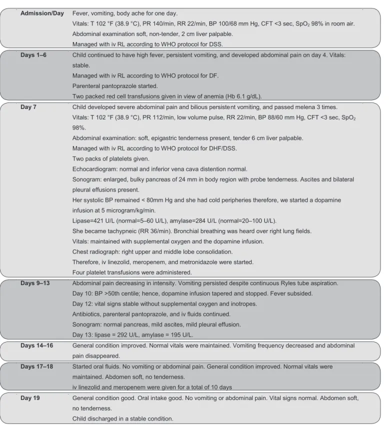
FIGURE 1. Sequence of events after admission
T: temperature; PR: pulse rate; RR: respiratory rate; BP: blood pressure; CFT: capillary illing time; SpO2: oxygen saturation; iv: intravenous; RL: Ringer’s lactate; WHO: World Health
Organization; DSS: Dengue shock syndrome; DF: Dengue fever; Hb: hemoglobin; DHF: Dengue hemorrhagic fever.
Admission/ Day Fever, vomiting, body ache for one day.
Vitals: T 102 °F (38.9 °C), PR 140/min, RR 22/min, BP 100/68 mm Hg, CFT <3 sec, SpO2 98% in room air.
Abdominal examination soft, non-tender, 2 cm liver palpable.
Managed with iv RL according to WHO protocol for DSS.
Days 1–6 Child continued to have high fever, persistent vomiting, and developed abdominal pain on day 4. Vitals:
stable.
Managed with iv RL according to WHO protocol for DF.
Parenteral pantoprazole started.
Two packed red cell transfusions given in view of anemia (Hb 6.1 g/dL).
Day 7 Child developed severe abdominal pain and bilious persistent vomiting, and passed melena 3 times.
Vitals: T 102 °F (38.9 °C), PR 112/min, low volume pulse, RR 22/min, BP 88/60 mm Hg, CFT <3 sec, SpO2
98%.
Abdominal examination: soft, epigastric tenderness present, tender 6 cm liver palpable.
Managed with iv RL according to WHO protocol for DHF/DSS.
Two packs of platelets given.
Echocardiogram: normal and inferior vena cava distention normal.
Sonogram: enlarged, bulky pancreas of 24 mm in body region with probe tenderness. Ascites and bilateral
pleural effusions present.
Her systolic BP remained < 80mm Hg and she had cold peripheries therefore, we started a dopamine
infusion at 5 microgram/kg/min.
Lipase=421 U/L (normal=5–60 U/L), amylase=284 U/L (normal=20–100 U/L).
She became tachypneic (RR 36/min). Bronchial breathing was heard over right lung fields.
Vitals: maintained with supplemental oxygen and the dopamine infusion.
Chest radiograph: right upper and middle lobe consolidation.
Therefore, iv linezolid, meropenem, and metronidazole were started.
Four platelet transfusions were administered.
Days 9–13 Abdominal pain decreasing in intensity. Vomiting persisted despite continuous Ryles tube aspiration.
Day 10: BP >50th centile; hence, dopamine infusion tapered and stopped. Fever subsided.
Day 12: vital signs stable without supplemental oxygen and inotropes.
Antibiotics, parenteral pantoprazole, and iv fluids continued.
Sonogram: normal pancreas, mild ascites, mild pleural effusion.
Day 13: lipase = 292 U/L, amylase = 195 U/L.
Days 14–16 General condition improved. Normal vitals were maintained. Vomiting frequency decreased and abdominal
pain disappeared.
Days 17–18 Started oral fluids. No vomiting or abdominal pain. General condition improved. Normal vitals were
maintained. Abdomen soft, no tenderness.
iv linezolid and meropenem were given for a total of 10 days
Day 19 General condition good. Oral intake good. No vomiting or abdominal pain. Vital signs normal. Abdomen soft,
no tenderness.
659
in 6 (75%) of 8 patients with mild DHF and in 10 (83%) of 12 patients with severe DHF. All children with mild DHF had a normal-sized pancreas and in all 10 severe cases children with increased serum levels of amylase and lipase had an enlarged pancreas(7).
In a previous study, 14 out of 328 cases of DHF/dengue shock syndrome (DSS) had an acute abdomen; causes included
acute cholecystitis (n = 10), nonspeciic peritonitis (n = 3), and
acute appendicitis (n = 1); none had acute pancreatitis(8).In a
study conducted in Pakistan, however, 43 (12%) out of 357 patients with DF had an acute abdomen and three (0.8%) had acute pancreatitis. All three patients with acute pancreatitis developed acute respiratory distress syndrome, and two died(9).In our patient, DHF was diagnosed according to the
World Health Organization’s (WHO) criteria. She presented with fever, a bleeding tendency, thrombocytopenia (platelets <100 × 109/L), and ascites, and her serology results were
positive for nonstructural protein 1 (NS1) antigen and dengue immunoglobulin M (IgM) antibodies. She had continuous persistent vomiting that started one day prior to admission and stopped three days before discharge. Her initial abdominal sonogram and liver enzymes levels were normal. Given that the child’s epigastric pain and associated persistent vomiting were not relieved by parenteral pantoprazole, and that she was hypotensive, we suspected acute pancreatitis and ordered serum amylase and lipase testing, and a repeat abdominal ultrasound. We diagnosed acute pancreatitis in view of clinical symptoms (abdominal pain and vomiting), hypotension, enlargement of the pancreas on ultrasound examination without features of hepatobiliary disorders, and increased serum amylase and lipase levels. We started antibiotics empirically for pneumonia and acute pancreatitis. Serum amylase and lipase levels decreased after one week. In DHF, the involvement of the pancreas may be due to direct viral invasion, secondary to host immune reactivity, or due to hypotension(2).
Like septic shock, acute pancreatitis can be fatal(10). As such,
DHF/DSS can also cause mortality if not treated. Therefore, acute pancreatitis as a complication of DHF is dangerous, and clinicians should know when to suspect in patients with DF. Acute pancreatitis as a complication of DHF may be underdiagnosed due to lack of awareness(10). Hence, clinicians
might not request serum amylase or lipase investigation, despite abdominal pain and vomiting. Lee et al. compared 14 patients
with hyperlipasemia (one with additional hyperamylasemia) and 57 without hyperlipasemia/hyperamylasemia among 71 DHF patients who presented with abdominal pain. They found that three patients in the hyperlipasemia group had pancreatitis, all of whom had enzyme elevation > 3 times the of normal(11).
In acute pancreatitis, serum amylase usually rises within a few hours of the onset of symptoms and return to normal values
within 3-5 days. However, because of sensitivity, speciicity,
and positive and negative predictive value limitations, serum amylase alone cannot reliably be used to diagnose acute
pancreatitis; the more speciic serum lipase test is preferred.
Serum lipase remains increased for a longer period than amylase after disease presentation(12). Abdominal pain and vomiting are
common in DF, especially in severe DF. Even though important
common causes include acute gastritis, hepatitis, and acalculous cholecystitis, acute pancreatitis should be kept in mind as one of the causes. Simple investigations like serum lipase, amylase (levels more than 3 times the upper limit of normal), and abdominal ultrasound will establish the diagnosis.In a patient with dengue illness who has abdominal pain, it is probably
justiied to estimate and monitor serum lipase and amylase levels
and to perform serial abdominal sonography.
To conclude, clinicians should be alert when there are warning symptoms (abdominal pain and persistent vomiting) in patients with DF, and should order testing of serum lipase and amylase levels along with abdominal sonography. Even though acute pancreatitis is a rare complication, early diagnosis and prompt treatment is necessary to prevent morbidity and mortality.
Conlicts of Interest
The authors declare that there is no conlict of interest.
REFERENCES
1. World Health Organization Regional Ofice for South-East Asia.
Comprehensive Guidelines for Prevention and Control of Dengue and Dengue Haemorrhagic Fever. Revised and expanded edition. World Health Organization: 2011. 196p.
2. Wijekoon CN, Wijekoon PW. Dengue hemorrhagic fever presenting with acute pancreatitis. Southeast Asian J Trop Med Public Health 2010; 41:864-866.
3. Khanna S, Vij JC, Kumar A, Singal D, Tandon R. Etiology of abdominal pain in dengue fever. Dengue Bull 2005; 29:85-88.
4. Ramos-De La Medina A, Remes-Troche JM, González-Medina MF, Anitúa-Valdovinos MdelM, Cerón T, Zamudio C, et al. Abdominal and gastrointestinal symptoms of dengue fever. Analysis of a cohort of 8559 patients. Gastroenterol Hepatol 2011; 34:243-247.
5. Ong A, Sandar M, Chen MI, Sin LY. Fatal dengue hemorrhagic fever in adults during a dengue epidemic in Singapore. Int J Infect Dis 2007; 11:263-267.
6. Benila M, Weizman Z. Acute pancreatitis in childhood. analysis
of literature data. J Clin Gastroenterol 2003; 37:169-172.
7. Setiawan MW, Samsi TK, Wulur H, Sugianto D, Pool TN. Epigastric pain and sonographic assessment of the pancreas in dengue hemorrhagic fever. J Clin Ultrasound 1998; 26:257-259.
8. Khor BS, Liu JW, Lee IK, Yang KD. Dengue hemorrhagic fever patients with acute abdomen: clinical experience of 14 cases. Am J Trop Med Hyg 2006; 74:901-904.
9. Shamim M. Frequency, pattern and management of acute abdomen in dengue fever in Karachi, Pakistan. Asian J Surg 2010; 33:107-113.
10. Simadibrata M. Acute pancreatitis in dengue hemorrhagic fever. Acta Med Indones 2012; 44:57-61.
11. Lee IK, Khor BS, Kee KM, Yang KD, Liu JW. Hyperlipasemia/ pancreatitis in adults with dengue haemorrhagic fever. Pancreas 2007; 35:381-382.
12. Tenner S, Baillie J, DeWitt J, Vege SS. Management of acute pcreatitis. Am J Gastroenterol 2013; 108:1400-1415.