Prevalence of mesiodens in orthodontic patients with deciduous and
mixed dentition and its association with other dental anomalies
Tulio Silva Lara1, Melissa Lancia2, Omar Gabriel da Silva Filho3, Daniela Gamba Garib4, Terumi Okada Ozawa5
How to cite this article: Lara TS, Lancia M, Silva Filho OG, Garib DG, Oza-wa TO. Prevalence of mesiodens in orthodontic patients with deciduous and mixed dentition and its association with other dental anomalies. Dental Press J Orthod. 2013 Nov-Dec;18(6):93-9.
Submitted: September 10, 2011 - Revised and accepted: October 24, 2011
» The authors report no commercial, proprietary or inancial interest in the prod-ucts or companies described in this article.
Contact address: Tulio Silva Lara
Hospital de Reabilitação de Anomalias Craniofaciais / Setor de Ortodontia Rua Silvio Marchione, 3-20 – CEP: 17.012-900 – Bauru/SP — Brazil. E-mail: [email protected]
» Patients displayed in this article previously approved the use of their facial and in-traoral photographs.
1 PhD in Orthodontics, UNESP.
2 Masters student in Rehabilitation Sciences, Hospital for Rehabilitation of
Craniofacial Anomalies/ University of São Paulo (USP).
3 MSc in Orthodontics, UNESP. 4 Full professor in Orthodontics, FOB-USP. 5 PhD in Orthodontics, UNESP.
Objective:To determine the prevalence of mesiodens in deciduous and mixed dentitions and its association with
other dental anomalies. Material and Methods: Panoramic radiographs of 1,995 orthodontic patients were analyzed retrospectively, obtaining a final sample of 30 patients with mesiodens. The following aspects were analyzed: gender ; number of mesiodens; proportion between erupted and non-erupted mesiodens; initial position of the supernumerary tooth; related complications; treatment plan accomplished; and associated dental anomalies. The frequency of dental anomalies in the sample was compared to reference values for the general population using the chi-square test (c2),
with a significance level set at 5%. Results: The prevalence of mesiodens was 1.5% more common among males (1.5:1). Most of the mesiodens were non-erupted (75%) and in a vertical position, facing the oral cavity. Extraction of the mesiodens was the most common treatment. The main complications associated with mesiodens were: delayed eruption of permanent incisors (34.28%) and midline diastema (28.57%). From all the dental anomalies analyzed, only the prevalence of maxillary lateral incisor agenesis was higher in comparison to the general population. Conclu-sion: There was a low prevalence of mesiodens (1.5%) in deciduous and mixed dentition and the condition was not associated with other dental anomalies, except for the maxillary lateral incisor agenesis.
Keywords:Supernumerary tooth. Child. Prevalence.
Objetivo:determinar a prevalência de mesiodens nos estágios de dentição decídua e mista, e verificar sua associação com outras anomalias dentárias. Métodos: radiografias panorâmicas de 1.995 pacientes ortodônticos foram analisadas retrospectivamente, obtendo-se uma amostra de 30 pacientes com o mesiodens. Os seguintes aspectos foram analisados: distribuição entre os sexos, número de mesiodens; se irrompido ou não irrompido; posição; complicações; tratamento instituído, e anomalias dentárias associadas. A frequência de anomalias dentárias na amostra estudada foi comparada a valores de referência para a população em geral por meio do teste qui-quadrado (χ2), com um nível de significância de 5% (p < 0,05). Resultados: a prevalência de mesiodens foi de 1,5%, sendo mais comum no sexo masculino (1,5:1). A maior parte dos mesiodens estavam não irrompidos (75%) e numa posição vertical, voltada para a cavidade bucal. O tratamen-to mais empregado foi a exodontia. As principais complicações associadas ao mesiodens foram o atraso na erupção dos incisivos permanentes (34,28%) e diastema mediano (28,57%). Pacientes com mesiodens não apresentaram prevalência aumentada de microdontia, agenesia de dentes permanentes ou outros supranumerários. De todas as anomalias analisadas, apenas a prevalência de agenesia de incisivo lateral superior mostrou-se aumentada em comparação à população em geral. Conclusão: o mesiodens foi encontrado em uma prevalência baixa (1,5%) nas dentições decídua e mista, e não apre-sentou associação com outras anomalias dentárias, com exceção da agenesia de incisivo lateral superior.
INTRODUCTION
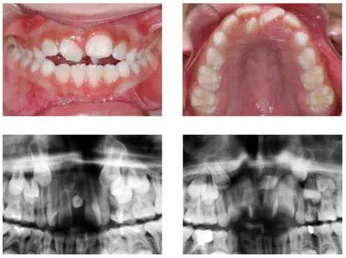
The term mesiodens refers to supernumerary teeth located in the pre-maxilla region, precisely be-tween the maxillary central incisors (Figs. 1 and 2). Mesiodens is the most frequent type of supernumer-ary tooth.1,2 The prevalence of mesiodens reported in the literature varies from 0.15 to 7.8% (Table 1), with a higher prevalence in males, with a proportion of 2:1.3-10 Although it has not been precisely established, its etiology seems to be related to genetic factors, giv-en the records of family recurrgiv-ence.4,11-13 A dominant autosomal trait has been suggested, with incomplete penetrance in some generations13 and x chromo-some linked inheritance due to the higher prevalence among males.
The mesiodens is often unique3,4,5,7,14 and anoma-lous in size and shape,11 but may vary in morphol-ogy from a small rudimentary conical shape4,7,8,9,14,18 to a complex form with several tubercles. It rarely erupts spontaneously,8,10,14,17 which only occurs in situations in which the mesiodens faces the oral cav-ity. Most often, the mesiodens is inverted, with the crown positioned towards the nasal cavity and the root apex facing the oral cavity.3,8,9 The presence of mesiodens can lead to local irregularities of which the most common are: delayed eruption or impaction of adjacent teeth, displacement or rotation of adjacent
teeth, development of dentigerous cysts, resorption of adjacent roots, crowding, midline diastema or maxil-lary incisors root dilaceration.1,3,4,5,7-11,14,17
Studies have suggested a genetic and hereditary background in the etiology of dental anomalies of number, size and position. Such evidence comes from investigations carried out with families, monozygotic twins and the observation of associations in the oc-currence of certain anomalies.11,19,20 Tooth agenesis is oten associated with other dental anomalies, such as microdontia, ectopia and delayed dental develop-ment.19,20,21 Peck20 has recently denominated the asso-ciation of these occurrences as dental anomaly patterns (DAP), as a single mutant gene may be responsible for more than one morphological or functional trait.
Two previous studies veriied the association be-tween supernumerary teeth in general and other dental anomalies, including tooth agenesis, microdontia and ectopic eruption.19,21 The indings revealed that the fre-quency of supernumerary teeth was not higher in pa-tients with hypoplastic dental anomalies.19,21 However, no previous study has investigated the exclusive associa-tion of mesiodens with other dental anomalies.
Thus, the aim of the present study was to verify the prevalence of mesiodens in children with decid-uous and mixed dentitions and its association with other dental anomalies.
Figure 1 - Mesiodens erupted in the oral cavity.
Figure 2 - Radiographic image showing
MATERIAL AND METHODS
The orthodontic records of 1,995 patients with de-ciduous and mixed dentition, taken from the archives of the Prois Preventive and Interceptive Orthodontics Course (Bauru, SP, Brazil) were retrospectively lyzed. Panoramic and periapical radiographs were ana-lyzed by a single examiner. The inclusion criteria were: patients aged between 4 and 13 years old; with decidu-ous or mixed dentition; presence of at least one super-numerary tooth in the midsagittal region of the maxilla. The exclusion criteria were: presence of craniofacial anomalies; presence of syndromes; history of tooth ex-traction and incomplete documentation. Ater the ini-tial analysis, a sample of 30 patients with mesiodens and a mean age of 8 years and 3 months was obtained.
The following aspects were analyzed in the orth-odontic records of the sample: 1) sex; 2) number of mesiodens; 3) proportion between erupted and non-erupted mesiodens; 4) initial position of the super-numerary tooth; 5) related complications; 6) treat-ment planning accomplished; and 7) associated dental anomalies. The associated dental anomalies
investigated included agenesis of permanent teeth, microdontia, ectopic eruption of permanent maxil-lary first molars, tooth transpositions, palatally dis-placed canines (PDC), distoangulation of mandibular second premolar, infraocclusion of deciduous molars, delayed tooth development and supernumerary teeth (in addition to mesiodens).
Diagnosis of palatally displaced maxillary canines followed the radiographic parameters suggested by Lindauer et al22 confirmed by the interpretation of periapical radiographs by the tube shift method of object localization using two projections with sig-nificantly different x-ray tube angulations. Consider-ing the findConsider-ings of Ericson and Kurol23 in which the attempt to determine the eruption path of maxillary canines radiographically is generally of little value in children under 10 years old, those subjects whose only diagnostic records were from an age under 10 years were excluded from the sample when evaluating pala-tally displaced canines. Diagnosis of distoangulation of mandibular second premolars followed the crite-ria described by Shalish et al24 using the lower edge
Author Number of subjects
analyzed Age range Method Origin of the sample
Prevalence of mesiodens
Montenegro et al1 36,057 5 to 56y Analysis of patient’s ile Unidad Ambulatoria de
Cirurgía Bucal (Spain) 0.15%
Gündüz et al14 23,000 4 to 14y Radiographic Ondokuz Mayis
University (Turkey) 0.3%
Buenviaje and Rapp15 2,439 2 to 20y Radiographic University of Pittsburgh
(USA) 0.4%
Järvinen and Lehtinen16 1,141 3 to 4y Clinical
University of Kuopio/ Public Health Centre
(Finland)
0.4%
Tyrologou et al9 11,500 3 to 15y Clinical and radiographic
Department of Paediatric
Dentistry – Institute for Postgraduate Education
(Sweden)
0.8%
Hurlen and Humerfelt17 63,029 9 m to 80y Clinical and radiographic University of Oslo
(Norway) 1.4%
Salcido-García et al18 2,241 2 to 55y Radiographic Facultad de Odontología
UNAM (Mexico) 1.6%
Kaller6 3,523 4 to 18y Clinical and radiographic
Los Barrios Community Clinic – Hispanic area of
Dallas (USA)
2.2%
Huang et al5 543 2.5 to 7y Clinical and radiographic Chang Gung Memorial
Hospital (China) 7.8%
Present study 1,995 4 to 13y Radiographic Universidade de
São Paulo (Brazil) 1.5%
of the mandible as a base line. The maxillary lateral incisor was considered as presenting microdontia when the maximal mesiodistal crown diameter was smaller than that of the opposing mandibular lateral incisor in the same patient.19 This category also in-cluded conical maxillary lateral incisors.
The prevalence of dental anomalies in the sample was compared with reference values for the general population by means of the chi-square test (c2), with the significance level set at 5% (P < 0.05). The odds ratio (OR) was calculated with 95% of confidence intervals to measure the strength of associations be-tween supernumerary mesiodens and the presence of other dental anomalies.
RESULTS
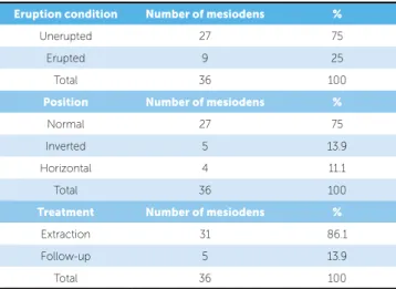
The prevalence rate of mesiodens in the sample cor-responded to 1.5% with a male-female ratio of 1.5:1 (Tables 1 and 2). Among the afected patients, 80% had only one mesiodens and 20% had two mesiodens (Ta-ble 3). Three out of four mesiodens were unerupted and in an upright position (facing the oral cavity), and more than 80% had extraction indication (Table 5). The most common complications related to mesiodens included delayed eruption of maxillary central incisors and mid-line diastema (Table 5). No association was found be-tween mesiodens and other dental anomalies, except for maxillary lateral incisor agenesis (Tables 6).
DISCUSSION
This study retrospectively assessed the complete orthodontic records of 1,995 patients who presented deciduous or mixed dentition at orthodontic treat-ment onset. A total of 36 mesiodens were diagnosed in 30 patients (average of 1.2 mesiodens per patient), corresponding to 1.5% of the overall sample. This prevalence was similar to that described in studies by Hurlen and Humerfelt17 (1.4%) and Salcido-García et al18 (1.6%), and is very close to the mean frequen-cy observed for the prevalence values compiled in Table 1 (1.67%).
With regard to sex distribution, mesiodens was more common among males, with a male to fe-male proportion of 1.5:1 (Table 2), which corrob-orates previous reports.5-10,14 A retrospective study carried out in India,8 on a sample of 30 patients with mesiodens, found the same male to female
Table 2 - Gender distribution of the sample comprised of children with
me-siodens.
Sex Number of individuals %
Male 18 60
Female 12 40
Total 30 100
Table 3 - Number of mesiodens per patient in the sample.
Number of mesiodens Number of individuals %
1 24 80
2 6 20
Total 30 100
Table 4 - Eruption condition, position and treatment planning for mesiodens
in the sample.
Eruption condition Number of mesiodens %
Unerupted 27 75
Erupted 9 25
Total 36 100
Position Number of mesiodens %
Normal 27 75
Inverted 5 13.9
Horizontal 4 11.1
Total 36 100
Treatment Number of mesiodens %
Extraction 31 86.1
Follow-up 5 13.9
Total 36 100
Table 5 - Complications associated with mesiodens in the sample.
Complications Number of cases (%)
Delayed eruption of maxillary
permanent incisors 12 (34.28%)
Midline diastema 10 (28.57%)
Rotation or axial inclination of erupted
permanent incisors 6 (17.14%)
Resorption of teeth adjacent to mesiodens 1 (2.85%)
Root anomaly 2 (5.57%)
None (asymptomatic) 4 (11.42%)
Total 35 (100%)
Dental Anomaly
Prevalence rate in the sample
n Reference
values n c² P Odds Ratio
Confidence interval
Tooth agenesis
(excluding third molars)
10.00% 3/30 5.00%
Grahnen27 53/1064 1.51 0.219 2.12 0.62 7.21
Mandibular second premolar
agenesis
3.33% 1/30 3.00%
Polder et al25 1479/48274 0.01 0.932 1.09 0.15 8.01
Maxillary second
premolar agenesis 3.33% 1/30
1.50%
Polder et al25 722/48274 0.69 0.407 2.27 0.31 16.70
Maxillary lateral
incisor agenesis 10.00% 3/30
1.90%
Le Bot and Salmon28
109/5738 10.28 0.001 5.74 1.71 19.20
Small maxillary
lateral incisor 3.33% 1/30
4.70%
Baccetti21 47/1000 0.12 0.726 0.70 0.09 5.24
Mandibular
second premolar distoangulation
3.33% 1/30
0.19%
Matteson et al29
52/26264 14.64 <0.001 17.38 2.32 129.99
Palatally displaced
canines (PDC)
3.33% 1/30
1.70% Dachi and
Howell30
25/1450 0.44 0.507 1.97 0.26 15.00
Supernumerary
teeth 10.00% 3/30
3.90%
Baccetti21 39/1000 2.77 0.096 2.74 0.80 9.41
Table 6 - Prevalence of dental anomalies in the sample compared with reference values.
A single mesiodens was found in 80% of the sample, whereas the remaining 20% presented two mesiodens (Table 3). A very similar proportion was described in a clinical and radiographic study involving 90 patients and 113 mesiodens,10 in which the majority of patients (78%) had a single mesiodens while the remaining pa-tients had two. Kim and Lee7 described a similar ten-dency with 75% of patients exhibiting only one me-siodens and 25% exhibiting two meme-siodens. A patient with three mesiodens was described in a retrospective study involving Japanese children.3
Among the 36 mesiodens in the sample, 27 (75%) were unerupted while nine (25%) had erupted in the oral cavity (Table 4). Studies are unanimous in dem-onstrating that the majority of mesiodens remains impacted6,8-10,14,18 and are often discovered only in routine radiographs requested for other reasons.9 Mesiodens may also be identified in radiographs re-quested for follow-up of maxillary incisors trauma or due to delayed eruption of maxillary permanent incisors. These factors are the most common causes of mesiodens diagnosis.9
cooperation during this stage.4,9 Thus, the clinical and radiographic follow-up can also be indicated in cases in which the mesiodens is not causing a mal-occlusion or will not interfere in the orthodontic treatment.9,14 In the present study, 86.1% of the me-siodens were surgically removed right after diagno-sis, whereas the remaining 13.5% were followed-up radiographically due to the absence of any interfer-ence in the dental development (Table 4).
The main complications associated with me-siodens were: delayed eruption of permanent incisors (34.28%), midline diastema (28.57%) and rotation or axial inclination of permanent incisors (17.14%). Other local disorders were also associated with the presence of mesiodens as shown in Table 5. Similar findings have been previously reported.7,9,14
Regarding the presence of other dental anomalies associated with mesiodens, 22 patients (73.3% of the sample) did not exhibit any associated dental anomaly. The other eight patients (26.7%) exhibited 12 associated dental anomalies including microdontia of maxillary lateral incisors, other supernumerary teeth, delayed development of second premolars, distoangulation of mandibular second premolars and tooth agenesis (Table 6).
The statistical analyses revealed that patients with mesiodens did not have an increased prevalence of permanent tooth agenesis in general or microdontia of the maxillary lateral incisors (Table 6). However, the prevalence of agenesis of maxillary permanent lateral incisors was approximately five times high-er in the sample when compared with the genhigh-eral population. This uncommon association between a supernumerary mesiodens and the agenesis of
max-illary lateral incisor has been described in a case re-port previously published and the interpretation of the authors regarding this association was a possible transposition between a malformed lateral incisor and the central incisor.26
One patient (3.3%) in the sample exhibited dis-tal angulation of the mandibular second premolars. Due to the small prevalence of this anomaly in the general population (0.19%), this result was con-sidered statistically significant (Table 6). However, considering the small size of the sample, this result could have randomly occurred and should be inter-preted with caution. Larger samples are needed to confirm such association.
Ectopic eruption of maxillary canines was diag-nosed in one patient. However, since the patient was eight years old and the diagnosis of ectopia in pan-oramic radiographs is more reliable among patients over the age of 10,23 this anomaly was not considered.
CONCLUSION
1. The prevalence of mesiodens in the deciduous and mixed dentition corresponded to 1.5%, with a male to female proportion of 1.5:1. 2. Mesiodens was associated with local
disor-ders, such as maxillary incisor rotation, delayed eruption or impaction of maxillary incisors, midline diastema, permanent incisor root re-sorption and dilaceration.
3. Mesiodens was associated with other dental anomalies in 26.7% of the sample.
1. Montenegro PF, Castellón EV, Aytés LB, Escoda CG. Retrospective study of 145 supernumerary teeth. Med Oral Patol Oral Cir Bucal. 2006;11(4):339-44.
2. Stafne EC. Supernumerary teeth. Dental Cosmos. 1932;74:653-9. 3. Asaumi JI, Shibata Y, Yanagi Y, Hisatomi M, Matsuzaki H,
Konouchi H, et al. Radiographic examination of mesiodens and their associated complications. Dentomaxillofac Radiol. 2004;33(2):125-7. 4. Ersin NK, Candan U, Alpoz AR, Akay C. Mesiodens in primary, mixed and
permanent dentitions: a clinical and radiographic study. J Clin Pediatr Dent. 2004;28(4):295-8.
5. Huang WH, Tsai TP, Su HL. Mesiodens in the primary dentition stage: a radiographic study. ASDC J Dent Child. 1992;59(3):186-9.
6. Kaller LC. Prevalence of mesiodens in a pediatric Hispanic population. ASDC J Dent Child. 1998;55(2):137-8.
7. Kim SG, Lee SH. Mesiodens: a clinical and radiographic study. J Dent Child. 2003;70(1):58-60.
8. Roychoudhury A, Gupta Y, Parkash H. Mesiodens: a retrospective study of ifth teeth. J Indian Soc Pedod Prev Dent. 2000;18(4):144-6.
9. Tyrologou S, Koch G, Kurol J. Location, complications and treatment of mesiodentes – a retrospective study in children. Swed Dent J. 2005;29(1):1-9.
10. Von Arx T. Anterior maxillary supernumerary teeth: a clinical and radiographic study. Aust Dent J. 1992;37(3):189-95.
11. Gallas MM, Garcia A. Retention of permanent incisors by mesiodens: a family afair. Br Dent J. 2000;188(2):63-4.
12. Marya CM, Kumar BR. Familial occurrence of mesiodens with unusual indings: case reports. Quintessence Int. 1998;29(1):49-51.
13. Sedano HO, Gorlin RJ. Familial occurrence of mesiodens. Oral Surg Oral Med Oral Pathol. 1969;27(3):360-1.
14. Gündüz K, Celenk P, Zengin Z, Sümer P. Mesiodens: a radiographic study in children. J Oral Sci. 2008;50(3):287-91.
15. Buenviaje TM, Raap R. Dental anomalies in children: a clinical and radiographic survey. ASDC J Dent Child. 1984;51(1):42-6.
16. Järvinen S, Lehtinen L. Supernumerary and congenitally missing primary teeth in Finnish children. An epidemiologic study. Acta Odontol Scand. 1981;39(2):83-6.
17. Hurlen B, Humerfelt D. Characteristics of premaxillary hyperdontia. A radiographic study. Acta Odontol Scand. 1985;43(2):75-81. REFERENCES
18. Salcido-García JF, Ledesma-Montes C, Hernández-Flores F, Pérez D, Garcés-Ortíz M. Frequency of supernumerary teeth in Mexican population. Med Oral Patol Oral Cir Bucal. 2004;9(5):407-9, 403-6. 19. Garib DG, Peck S, Gomes SC. Increased occurrence of dental
anomalies associated with second-premolar agenesis. Angle Orthod. 2009;79(3):436-41.
20. Peck S. Dental anomaly patterns (DAP): a new way to look at malocclusion. Angle Orthod. 2009;79(5):1015-6.
21. Baccetti T. A controlled study of associated dental anomalies. Angle Orthod. 1998;68(3):267-74.
22. Lindauer SJ, Rubenstein LK, Hang WM, Andersen WC, Isaacson RJ. Canine impaction identiied early with panoramic radiographs. J Am Dent Assoc. 1992;123(3):91-2, 95-7.
23. Ericson S, Kurol J. Radiographic assessment of maxillary canine eruption in children with clinical signs of eruption disturbance. Eur J Orthod. 1986;8(3):133-40.
24. Shalish M, Peck S, Wasserstein A, Peck L. Malposition of unerupted mandibular second premolar associated with agenesis of its antimere. Am J Orthod Dentofacial Orthop. 2002;121(1):53-6.
25. Polder BJ, Van’t Hof MA, Van der Linden FP, Kuijpers-Jagtman AM. A meta-analysis of the prevalence of dental agenesis of permanent teeth. Community Dent Oral Epidemiol. 2004;32(3):217-26.
26. Segura JJ, Jiménez-Rubio A. Concomitant hipohyperdontia: simultaneous occurrence of a mesiodens and agenesis of a maxillary lateral incisor. Oral Surg Oral Med Oral Pathol Oral Radiol Endod. 1998;86(4):473-5.
27. Grahnen H. Hypodontia in the permanent dentition. A clinical and genetical investigation. Odontol Revy. 1956;7:1-100.
28. Bot PL, Salmon D. Congenital defects of the upper lateral incisors (ULI): condition and measurements of the other teeth, measurements of the superior arch, head and face. Am J Phys Anthropol. 1977;46(2):231-43. 29. Matteson SR, Kantor ML, Proit WR. Extreme distal migration of the
mandibular second bicuspid. A variant of eruption. Angle Orthod. 1982;52(1):11-8.