Therapeutic Effects of Topical Netrin-4
Inhibits Corneal Neovascularization in
Alkali-Burn Rats
Yun Han1,2‡, Yi Shao3‡, Tingting Liu1,2, Yang-Luowa Qu1,2, Wei Li1,2‡, Zuguo Liu1,2‡
*
1Eye Institute of Xiamen University, Xiamen, Fujian, China,2Fujian Provincial Key Laboratory of Ophthalmology and Visual Science, Xiamen, Fujian, China,3Department of Ophthalmology, the First Affiliated Hospital of Nanchang University, Nanchang, Jiangxi, China
‡YH and YS contributed equally to this work. ZL and WL also contributed equally to this work.
*zuguoliu@xmu.edu.cn
Abstract
Netrins are secreted molecules involved in axon guidance and angiogenesis. However, the role of netrins in the vasculature remains unclear. Netrin-4 and netrin-1 have been found to be either pro- or antiangiogenic factors. Previously, we found that netrin-1 acts as an anti-angiogenic factor in rats by inhibiting alkali burn-induced corneal neovascularization. Here, we further investigate the effects of netrin-4, another member of the same netrin family, on neovascularizationin vitroandin vivo. We found that netrin-4 functions similarly as netrin-1 in angiogenesis.In vitroangiogenesis assay shows that netrin-4 affected human umbilical vein endothelial cell (HUVEC) tube formation, viability and proliferation, apoptosis, migra-tion, and invasion in a dose-dependent manner. Netrin-4 was topically appliedin vivoto al-kali-burned rat corneas on day 0 (immediately after injury) and/or day 10 post-injury. Netrin-4 subsequently suppressed and reversed corneal neovascularization. Netrin-Netrin-4 inhibited corneal epithelial and stromal cell apoptosis, inhibited vascular endothelial growth factor (VEGF), but promoted pigment epithelium-derived factor (PEDF) expression, decreased NK-KB p65 expression, and inhibits neutrophil and macrophage infiltration. These results indicate that netrin-4 shed new light on its potential roles in treatmenting for angiogenic dis-eases that affect the ocular surface, as well as other tissues.
Introduction
Netrins are laminin-like secreted molecules that are involved in axon guidance, angiogenesis, and the formation of blood vessel networks [1–4]. The netrin system consists of at least five li-gands (netrin-1, -2, -4, -G1a, and -G1b) and six receptors (neogenin, DCC, Unc5A, -B, -C, and -D) [5]. In nervous system, netrins act as bifunctional cues for angiogenesis in axonal guid-ance. Recently, both netrin-1 and netrin-4 were implicated in angiogenesis [6–11], but the role of netrins in vasculature remains unclear.
a11111
OPEN ACCESS
Citation:Han Y, Shao Y, Liu T, Qu Y-L, Li W, Liu Z (2015) Therapeutic Effects of Topical Netrin-4 Inhibits Corneal Neovascularization in Alkali-Burn Rats. PLoS ONE 10(4): e0122951. doi:10.1371/journal. pone.0122951
Academic Editor:Jing Chen, Children's Hospital Boston, UNITED STATES
Received:September 22, 2014
Accepted:February 16, 2015
Published:April 8, 2015
Copyright:© 2015 Han et al. This is an open access article distributed under the terms of theCreative Commons Attribution License, which permits unrestricted use, distribution, and reproduction in any medium, provided the original author and source are credited.
Data Availability Statement:All relevant data are within the paper and its Supporting Information files.
Conflicting results have been reported regarding the roles of netrin-1 and netrin-4 in angio-gensis in previous studies. On one hand, some studies report the anti-angiogenic effects of netrin-4 on VEGF-stimulated endothelial cells [9], human microvascular endothelial and human pancreatic carcinoma cells via the inhibition of Akt and JNK1/2 phosphorylation [10]. Moreover, netrin-4 is anti-angiogenic and inhibits endothelial cell function by binding to neo-genin and recruiting Unc5B [9]. Additionally, netrin-4 can also cause thein vivofilopodial re-traction of the endothelial cell [12]. Netrin-1 has also been shown to inhibit postnatal
angiogenic sprouting and neovascularization by activating the Unc5B receptor [13]. In addi-tion, netrin-1 or netrin-4 overexpression in tumor cells delays tumor angiogenesis in various animal models [9,13,14]. On the other hand, studies also demonstrated that netrin-4 acts as a pro-angiogenic factor [8,15–17]. It has been reported that netrins stimulate cell proliferation and migration in primary endothelial cell (EC) cultures [11,17] and vascular smooth muscle cells (VSMCs) [18]. These pro-angiogenic effects are independent from cognate netrin receptor expression in EC [11,17], and neogenin is the only receptor involved in netrin signaling in VSMCs [18]. The same group also reported that netrin-4 induces lymphangiogenesis [16]. In addition, netrin-4 has been shown to enhance angiogenesis after cerebral ischemia [8]. As a re-sult the exact role of netrin-4 in angiogenesis remains uncertain. Netrin-4 has been detected in mouse cornea, and the deletion of netrin-4 in Ntn4 homozygous null (Ntn4−/−) mice did not disrupt Bowman’s membrane (BM) in the cornea [19], suggesting that netrin-4 is not a struc-tural element of ocular BM. Instead, netrin-4 might function in regulating epithelial cell prolif-eration or migration.
In this study, we first used human umbilical vein endothelial cells (HUVECs) as a model of vascular endothelial cells to assess the effects of netrin-4in vitro. Next, we used a well-estab-lished corneal neovascularization model—the alkali-burn model—to assess the therapeutic ef-fects of netrin-4 on corneal angiogenesis. Our results suggest the crucial role of netrin-4 in the regulation of corneal angiogenesis.
Materials and Methods
Cell culture
HUVECs were purchased from Promo Cell (Heidelberg, Germany) and cultured in endothelial cell basal medium 2 (EBM2) containing low serum (2% FCS) and endothelial cell growth sup-plement (Promo Cell). Endothelial cells were used after 2–6 passages. Because the complete medium contains a large amount of netrin-4, and because endothelial cells produce and secrete high levels of netrin-4 in normal culture media [15], the endothelial cells were serum-deprived for 16 hours before thein vitroexperiments and the effects of netrin-4 were analyzed in the ab-sence of serum. Plates were observed under an inverted microscope to determine confluence and morphology.
Cell counting kit-8 (CCK-8) assay
The respective HUVEC cell suspensions (100μL containing 5×10³cells/well) were dispensed
in triplicate into 96-well plates and incubated for 24 hours. Netrin-4 was added to the medium at a concentration of 5000 ng/mL. After 72 hours, 10μL Cell CCK-8 solution (Dojindo,
Kuma-moto, Japan) was added to the wells, and the plates were incubated for 1 hour. Absorbance (i.e., optical density [OD]) was read at 450 nm using a universal microplate reader (Bio-Tek, Winooski, VT, USA), and a graph of OD at 450 nm against concentration was plotted. Each mark represents the mean of the collected readings, and the procedure was repeated at least three times. Within 4 hours, OD at 570 nm was determined using a microplate reader (ELX800, BIO-TEK Corporation, USA).
YH decision to publish and preparation the manuscript. 3. This work was also supported by grants from the international major project in National Natural Science Foundation of China (NSFC No. 81330022), the National Natural Science Foundation of China NSFC No. 81270978), the National Fund Committee on both sides of the Taiwan Straits joint project (No.U1205025). The funders had roles in purchasing reagent, data collection and analysis and decision to publish the manuscript.
In vitro tube formation assay
HUVECs were serum-starved in EBM2 medium (0.1% FBS without growth factor) (Lonza, USA) for 12 hours, and HUVECs were seeded to a density of 10,000 cells/well on growth fac-tor-depleted Matrigel (BD Biosciences, NSW, Australia) in 24-well plates. Netrin-4 (100, 500, 1000, or 5000 ng/mL) or the PBS (control supernatant) was added, and the results were quanti-fied 6 hours later. Microscopic fields containing the tube structures that formed in the gel were photographed at low magnification (10×). At least five fields in each well were examined. Be-fore they were photographed, cells were fixed with 4% paraformaldehyde. Tube length was quantified using Image J software.
Flow cytometric cell-cycle analysis
HUVECs were grown under the above-described conditions in 35-mm dishes until confluent. Netrin-4 was added to the medium at a concentration of 100, 500, 1000, or 5000 ng/mL. Incu-bation was continued for 72 hours at 37°C. Cells were removed using 0.05% trypsin in PBS buffer, pelleted (centrifugation at 300gfor 5 minutes), washed twice in 1% BSA in PBS, resus-pended in 1% BSA in PBS, and fixed in 70% cold ethanol. A flow cytometer (FACScan; BD Bio-sciences, Franklin Lakes, NJ, USA) was used to acquire all data.
Cell apoptosis analysis
Duplicate HUVECs were plated onto 60-mm culture dishes at a density of 2×10⁵. When the cells achieved 70–80% confluence, HUVECs were treated with 100 ng/mL, 500 ng/mL, 1000 ng/mL, or 5000 ng/mL netrin-4. Incubation was continued for 72 hours at 37°C. Apoptosis was analyzed using an Apoptosis and Necrosis Assay Kit with Hoechst 33342 and PI staining (Beyotime Institute of Biotechnology, Shanghai, China). Briefly, the cultured HUVECs were trypsinized, washed twice with PBS, and then suspended in 100μL cell-staining buffer. Then,
5μL Hoechst 33342 and 5μL of 10μg/mL PI was added, and the cells were incubated at 4°C or
the cells were kept on ice for 20 minutes in the dark. After incubation, apoptotic cells were im-mediately analyzed using flow cytometry.
Wound closure assay for assessing migration
HUVEC migration was assessed using the wound-healing assay as previously described [20, 21]. HUVECs were seeded (1×10⁵cells/well) in duplicate to 1% gelatin-coated 24-well plates (Corning, Schiphol, Netherlands). Cells were grown until confluence, and a scratch wound was applied in two perpendicular directions using a sterile pipet tip (200-μL yellow tip), thereby
creating linear, cross-stripe scrapes that were 2 mm apart. Monolayers were washed with PBS to remove floating cells, the experimental medium (containing 100, 500, 1000, or 5000 ng/mL netrin-4) was added, and the cells were incubated for an additional 24 hours. Cell migration to where the scrapes were introduced was photographed at different time points using an inverted microscope.
Cell invasion assay
HUVEC invasion across Matrigel was evaluated in the invasion chambers. Cell inserts (8-mm pore size, 6.5-mm diameter; Corning) were coated with 15–25 mL Matrigel and placed in 24-well plates. HUVECs were plated at a density of 2×10⁵cells/well in the upper chamber, while either 0 or 800μL netrin-4 (100, 500, 1000, or 5000 ng/mL) was added to the lower
bud. Inserts were then fixed in 4% formalin for 10 minutes at room temperature and stained with 40, 6-diamidino-2-phenylindole (DAPI). Cells were observed under a fluorescent micro-scope. Cells that migrated to the lower surfaces were counted at 40× magnification in five predetermined fields.
Alkali-burned rat corneas and treatment
Wistar rats (180–220 g; 2 months old; male; n = 30) were purchased from Shanghai Shilaike Laboratory Animal Co, Ltd., Shanghai, China. Animal experiments were carefully performed in accordance with the guidelines of the Association for Research in Vision and Ophthalmolo-gy (ARVO) Statement for the Use of Animals in Ophthalmic and Vision Research, and all ani-mal experimental procedures were approved by the Experimental Aniani-mal Committee of Xiamen University (approval ID: XMUMC2013-02-1). Animals were housed in a temperature, humidity, and light controlled room. Food and water were available ad libitum. All rats were confirmed to be free of ocular diseases before experimentation. Alkali burns were applied to the rat corneas as previously reported [7,22,23]. The procedures of corneal alkali burn were briefly as follows: The rats were anesthetized with an intraperitoneal injection of 40 mg/kg pen-tobarbital and received topically administration with a drop of tetracaine. A round filter paper (3.5 mm in diameter) soaked with 1 N NaOH was placed on the center of the corneal surface for 30 sec to induce alkali burn. The ocular surface was then rinsed with 10 mL PBS.
After applying the alkali burn injury, animals were randomly divided into four equally sized groups (7 rats per group). Rats in two groups received the topical administration of PBS (10μ
L was administered four times per day, for 14 or 24 consecutive days), and the rats in the other two groups received the topical administration of recombinant mouse netrin-4 (R&D Systems, Minneapolis, MN, USA) using a pipette (10μL was administered four times per day at a
con-centration of 5000 ng/mL in PBS, for 14 or 24 consecutive days). Treatments were adminis-tered for 14 consecutive days, all eyes were observed on day 1, 4, 7 and 14 with slit lamp microscopy as described below for evaluation of corneal NV and inflammation and measure-ment of corneal epithelium damage. The rats were sacrificed with an overdose of pentobarbital sodium on postoperative day14, eye balls were removed, and the corneas were dissected and stored at -80°C until histological examination or protein extraction. In the fourth group, treat-ment started at day 10 after injury and were continued for another 14 days in order to observe the regressive effects of netrin-4 on corneal neovascularization. All eyes were observed on day 10, 14, 17, 20 and 24 with slit lamp microscopy as described below for evaluation of corneal NV and inflammation. The rats were sacrificed with an overdose of pentobarbital sodium on postoperative day 24, eye balls were removed, and the corneas were dissected and stored at -80°C until histological examination or protein extraction.
For the histology examination, the corneas were embedded in OCT, cross-sectioned, Hema-toxylin and Eosin staining (H&E staining) was conducted as described below. For the Western blot analysis, the whole cornea tissue including limbal (about 0.5 mm width) area was carefully dissected and Western blotting was conducted as described below.
Slit-lamp microscopic observation
by calculating the wedge-shaped area (S) of the vessel growth using the following formula:
S¼C=123:1416 ½r2
ðr IÞ2
where S is the area, C is time, I is the radius to the border of the vessel, and r is the radius of the cornea [24]. The inflammatory index was analyzed according to various parameters, including ciliary hyperemia, central corneal edema, and peripheral corneal edema, as previously de-scribed [25].
Rat cornea samples and HUVEC cell culture supernatant collection and
Netrin-4 ELISA
The entire corneal tissue was carefully dissected and extracted with cold PBS (PH7.4), homoge-nized by grinder, centrifugation 20min at the speed of 3000 r.p.m., remove supernatant. HUVEC cell culture supernatant detects secretory component, collect sue a sterile container, centrifugation 20min at the speed of 3000 r.p.m. We analyzed the rat cornea and HUVEC su-pernatant netrin-4 levels using a rat netrin-4 ELISA kit (Hui Ying Biotechnology Co., Ltd., Shanghai, China) according to the manufacturer’s protocol.
Histology Analysis
Rat cornea samples were fixed overnight in 4% paraformaldehyde (PFA) in PBS, followed by dehydration in a series of ascending alcohol and paraffin embedding. Deparaffinized sections (5μm) were stained with hematoxylin and eosin (H&E).
Apoptosis Detection Assay
To detect apoptosis of the corneal cells, the corneas were embedded in OCT, cross-sectioned, and subjected to terminal deoxynucleotidyl transferase-mediated nick end labeling (TUNEL) staining (Dead End Fluorometric TUNEL system; Promega, Shanghai, China), according to the manufacturer’s protocol. Cellular nuclei were stained with 4-6-Diamidino-2-phenylindole (DAPI), and apoptotic cells were examined under a laser confocal microscope (Fluoview 1000, Olympus, Tokyo, Japan). The cellular nuclei and apoptotic cells were counted in three sections from each sample.
Western blot assay
To determine protein expression levels in the cornea, the entire corneal tissue was carefully dis-sected and extracted using cold RIPA buffer and proteinase inhibitor cocktail (Merck, Darm-stadt, Germany). Equal amounts of proteins were extracted from the lysates and subjected to electrophoresis on 10% sodium dodecyl sulfate-polyacrylamide gels, then electrophoretically transferred to polyvinyl difluoride (PVDF) membrane. After 30 minutes of blocking in 2% BSA, the blots were incubated with primary antibodies against netrin-4 (1:200; Santa Cruz, USA), vascular epidermal growth factor (VEGF; 1:200; Santa Cruz, USA), PEDF (1:200; Santa Cruz, USA), and NF-KB p65 (1:200; Santa Cruz, USA).β-actin (1:10,000; Bio-Rad, Hercules, CA, USA) was used as the loading control. After three washes with Tris-buffered saline with 0.05% Tween 20 for 10 minutes each, the membranes were incubated with horseradish peroxi-dase (HRP)-conjugated secondary antibodies and Ig G (Bio-Rad) and rabbit anti-goat antibod-ies (1:5000; Dako, Shanghai, China) for 1 hour at room temperature. The results were
Statistical Analysis
Summary data are reported as the mean ± SD. The inflammatory index and the area and length of the corneal neovascularization were analyzed using ANOVA followed by Bonferroni post hoc comparison. Group means were analyzed using the studentttest, andp<0.05 is consid-ered statistically significant. Statistical analyses were conducted using GraphPad Prism for Windows (version 5.00; GraphPad Software Inc., USA).
Results
Netrin-4 concentrations in rat cornea and HUVEC
The levels of netrin-4 in endothelial cells and cornea before and after alkali-burn were first de-termined by using ELISA, IHC and Western blot. The level of netrin-4 in HUVEC is about 123pg/mL (Fig 1C), and it is about 72pg/mL in normal rat cornea (Fig 1C), which could not be detected 7 days after alkali-burn (Fig 1C). Meanwhile, we performed immunohistochemical staining and found that netrin-4 (Fig 1A) was expressed in both normal corneal epithelium and stroma. Strong staining was detected in the membrane of epithelial cells. But there wasn’t netrin-4 expression 7 days after alkali-burn (Fig 1B). Western blot results showed that netrin-4 expressed in normal rat cornea, after alkali-burn treatment there was no expression on day 1and 3. After 7days, there was weak netrin-4 expression in rat cornea. Then netrin-4 expression increased slowly at day 14 and 21(Fig 1D).
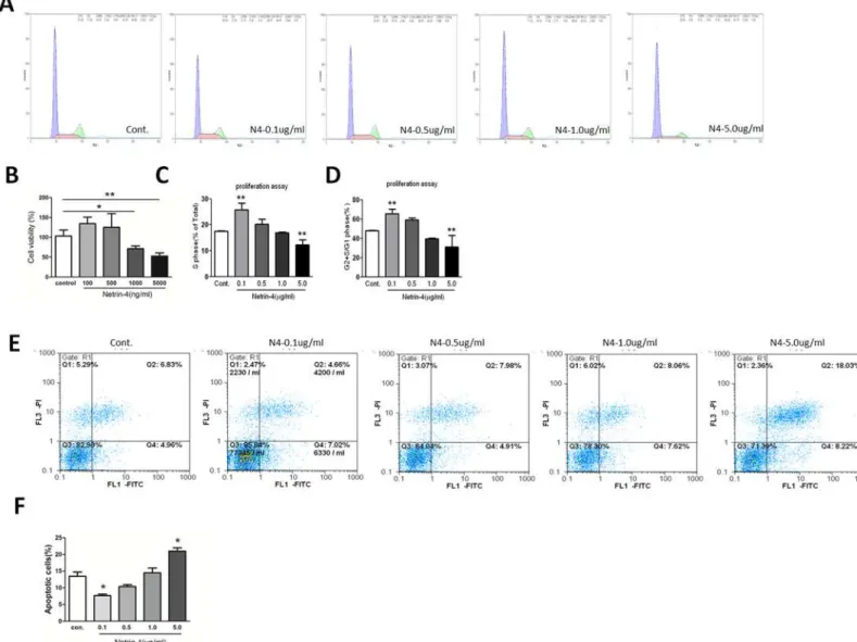
Netrin-4 decreases HUVECs viability in a dose-dependent manner by
regulating HUVEC proliferation and apoptosis
We first measured the cell viability with CCK-8 assays in the presence of different concentra-tions of netrin-4 (100, 500, 1000, 5000 ng/mL), which reduced HUVEC viability at 1000 and 5000 ng/mL compared with the control (Fig 2B). The effects of netrin-4 on cell cycle were as-sessed using flow cytometry. After treating for 72 hours, cells treated with 100, 500, 1000, and 5000 ng/mL netrin-4, 5000ng/mL netrin-4 decreases the percentage of cells in S phase as well as the ratio of cells in G2+S phase compared with G1 phase. However, the opposite effects were observed at the concentration of 100 ng/mL (Fig2A,2Cand2D). In addtion, HUVECs were starved overnight and then treated with 100, 500, 1000, and 5000 ng/mL netrin-4, and apopto-sis was analyzed using flow cytometry. Representative dot plots of annexin V/PI staining are shown (Fig 2E). The lower left quadrant shows the vital population, the lower right quadrant shows the apoptotic population (annexin V+/PI-), and the upper right quadrant shows the late apoptotic/necrotic population (annexin V+/PI+). The total number of apoptotic HUVECs in-creased after treatment with 5000 ng/mL netrin-4, and accordingly the total percentage of apo-ptotic cells increased from 12.12 ± 0.27% to 20.45 ± 0.51% at the high dosage (Fig 2E). The results demonstrate that netrin-4 promotes HUVECs apoptosis at the concentration of 5000 ng/mL but decreases HUVEC apoptosis (Fig 2F).
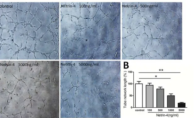
Netrin-4 inhibited HUVECs tube formation
Netrin-4 effects on HUVEC migration and invasion
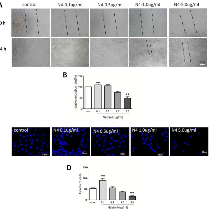
After verifying the effects of netrin-4 on cell proliferation, we assessed its effects on HUVEC migration using a scratch-wound model. After the scratch wound was created, the wounds were allowed to heal with or without netrin-4. The control wounds closed within approximate-ly 24 hours, while the group treated with 5000 ng/mL netrin-4 did not heal within 24 hours. There was still 980μm blank in the netrin-4-treated group at 24 h. After 24 hours treatment,
the migration rates were 42.6 and 26.1μm/hour in the control and netrin-4–treated groups,
re-spectively (Fig4Aand4B). Based on the results of the transwell invasion assays, the 5000 ng/ mL netrin-4–treated group demonstrated lower invasive abilities than the controls, but 100ng/ mL netrin-4 increases HUVEC invasive abilities (Fig4Cand4D). These results suggest that netrin-4 exhibits a dose-dependent effects on HUVEC proliferation, migration, apoptosis, and invasion. At 5000ng/mL, netrin-4 can inhibit HUVEC proliferation, migration, invasion, but increase apoptosis. However, at lower dosage 100ng/mL, the role of netrin-4 is opposite. Fig 1. Levels of netrin-4 in endothelial cells, and in cornea before and after alkali-burn were detected by ELISA, IHC and Western blot.Netrin-4 expression in normal (A) and after alkali-burn rat cornea (B) by immunohistochemical staining. Netrin-4 concentrations in rat cornea and HUVEC (C). Netrin-4 concentration in normal rat cornea and different time after alkali-burn treatment.
Netrin-4 prevents corneal neovascularization in alkali-burned rat
corneas
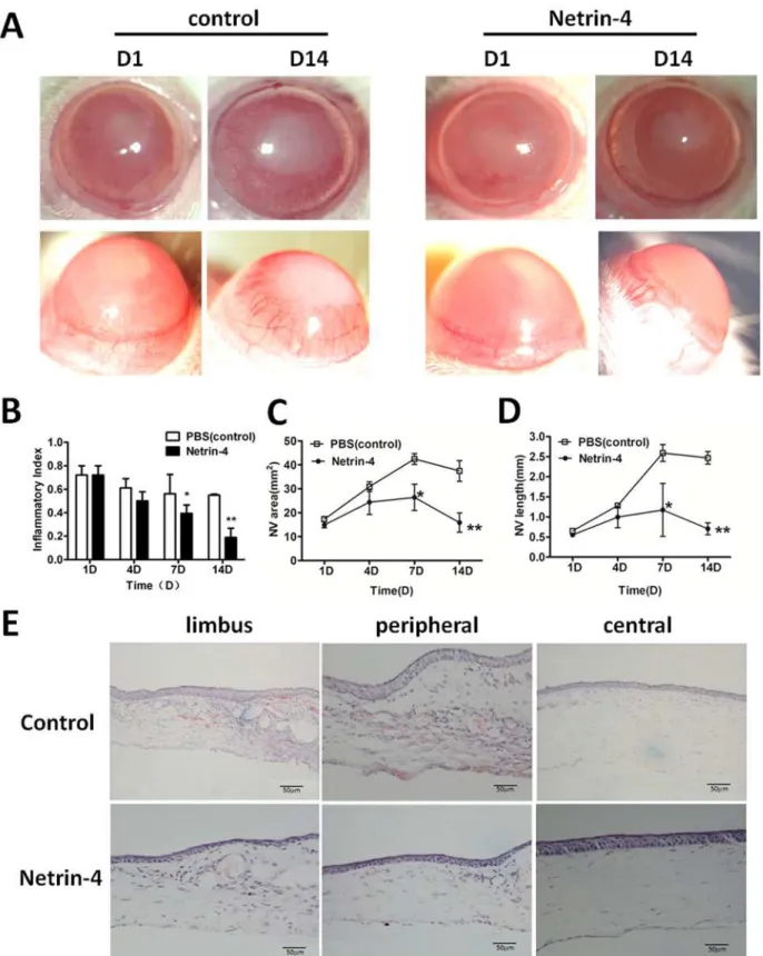
Alkali burn has been shown to cause severe neovascularization in the cornea [27]. We evaluat-ed the effect of netrin-4 on corneal neovascularization, and we usevaluat-ed the alkali burn model to compare vessel formation between injured corneas that received netrin-4 treatment and con-trols. The onset of peripheral neovascularization occurred on day 1 after the alkali burns were applied to both groups (Fig 5A). The inflammatory index showed slight decrease from day 1 to Fig 2. Cell viability, HUVEC proliferation and apoptosis on different doses of netrin-4 was detected by using the CCK-8 assay and flow cytometry.
(A) Cultured HUVECs were serum-starved for 24 h followed by 72 h culture in serum free media with or without dosage netrin-4 protein. The cells were then harvested, stained with propidium iodide (PI), and analyzed by flow cytometry. The cell proliferation rate was expressed as the percentage of cells in the S phase and as the G2 + S/G1 ratio (**p<0.01). (B) Cell viability detected by CCK-8. (C) S phase percentage of cells. (D) G2 + S/G1 ratio. (E) Apoptosis assay by flow cytometry, analysis with annexin V-FITC / PI double staining. The numbers in the upper left, upper right, lower left and lower right quadrants represent the percentage of necrotic cells (annexin V positive, PI positive), advanced apoptotic cells (annexin V negative, PI positive), viable cells (annexin V negative, PI negative) and early apoptotic cells (annexin V positive, PI negative), respectively. The values represent the mean percentages of apoptotic cells in different groups (*p<0.05). (F) Quantitative results of the apoptotic cells. Each value represents the mean±SD, n = 3.*p<0.05;**p<0.01; compared with the control group.
day 14 in PBS group, while there was dramatic reduction in the netrin-4 group, and there was significant difference between the two groups at day 7 and day14 (Fig 5B).
In the control group, there was an ingrowth of new blood vessels toward the peripheral cor-neas on day 4, which reached the central corcor-neas on day 7, and new blood vessels were detected on day 14 (Fig5Cand5D). In contrast, corneas treated with netrin-4 demonstrated only a mild increase in new blood vessels, which was maintained at low levels throughout the study period (Fig 5A). The new blood vessel areas and lengths were much lower than control corneas on days 7 and 14 (Fig5Cand5D). Hematoxylin-eosin (H&E) staining showed persistent, new blood vessel formation in the anterior part of the corneal stroma in the control group on day 14 after applying the alkali burns, which was clearly indicated by the remaining red blood cells in the blood vessels. However, the netrin-4–treated group developed only a few blood vessels in the limbus and none in the peripheral or central corneas (Fig 5E).
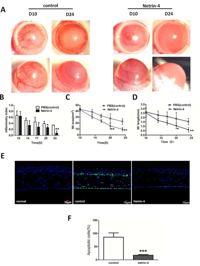
Netrin-4 promotes the regression of corneal neovascularization and
inhibits apoptosis in alkali-burned rat corneas
To determine if netrin-4 induces the regression of well-formed corneal neovascularization, netrin-4 treatment was started on day 10 after applying alkali burns. In the rat alkali-burn model, corneal neovascularization peaked 10 days after injury (Fig 6C). Thereafter, gradual blood vessel regression was noted in the control group (Fig 6C). By 24 days post injury, the blood vessels regressed from the central cornea to the peripheral cornea in the control group. Fig 3. Effect of netrin-4 on HUVECs tube formation.Tube formation was assessed in tubulogenesis assay in vitro model, HUVECs were serum-starved with EBM2 medium (0.1% FBS, no growth factor) (Lonza) for 12 hours. HUVECs (1.5×10⁴) were then cultured in a 24-well plate (Gibco) coated with 150 mL Matrigel Basement Membrane Matrix GFR (BD Biosciences). Different concentrations of netrin-4 (100, 500, 1000, and 5000 ng/mL) or PBS (control) were added, and the results were quantified 6 h later using Image J. (B) Representative images out of three independent experiments are shown.*p<0.05;**
p<0.01. Each experiment was performed three times and representative pictures are shown. Data are expressed as mean±SD.
On the other hand, the rapid regression of corneal neovascularization was noticed when the eyes were treated with netrin-4, and almost all of the blood vessels regressed to the limbal area 24 days after treatment (Fig 6A). The inflammatory index showed gradual decrease in the PBS group, while it was further decreased in the netrin-4 group at days 17, 20, and 24 (Fig 3B). The Fig 4. Effect of netrin-4 on HUVECs migration and invasion.HUVEC were treated during 24 h in medium without serum, in the presence of EBM2 alone or EBM2 with different concentrations of netrin-4. (A)Pictures were taken at 0 h and 24 h after insert removing. (B) Results are expressed as percentage of wound closure (percentage closure) (mean±SD). (C) Netrin-4 affects HUVEC migration in a transwell assay. HUVEC cells were allowed to migrate for 24 h at 37°C in a 5% CO2humidified incubator. After 24 h, the cells that migrated across the membrane were stained with DAPI and the fluorescence intensity was measured. (D)The % invasion is compared to the fluorescence intensity of migrated cells in the absence of netrin-4 (**p<0.01). Each experiment was performed three times and a representative picture of each condition is shown. Data are expressed as mean±SD.
area of corneal neovascularization (Fig 6C) and length (Fig 6D) on day 24 were significantly lower in the netrin-4 group in comparison with the control group. Meanwhile, to determine the effect of netrin-4 on alkali-burn induced apoptosis, we performed TUNEL assay on corneas treated with PBS or netrin-4 post alkali injury. As expected, there were no apoptotic cells pres-ent in normal rat corneas (Fig 6E). After alkali-burn, there were still many apoptotic cells in the basal epithelia, stromal, and endothelia at day 7. However, when the corneas were treated with netrin-4, there were much fewer apoptotic cells compared with the PBS control, and the majority of the apoptotic cells resided in the endothelia (Fig 6E). Statistical analysis showed a significant difference of apoptotic cells between the two groups (Fig 6F)
Netrin-4 affects the expression of VEGF, PEDF, NF-KB p65, PMN and
ED1 (CD-68) in alkali-burned rat corneas
It is well established that corneal neovascularization is tightly regulated by a dynamic, natural equilibrium between local proangiogenic and antiangiogenic molecules [28–31]. Among them, VEGF and PEDF are considered to play major roles. To investigate the mechanism hownetrin-4 inhibits and reverses corneal neovascularization after alkali burns application, we used West-ern blot analysis to assess VEGF and PEDF protein levels. Our results show that VEGF is ex-pressed at low levels in normal rat corneas, but there was a dramatic increase in the control group on day 14 after alkali burns. In the later stage, there was a decrease in VEGF expression on day 24. After netrin-4 treatment, VEGF expression was lower than that of the control group on days 14 and 24 (Fig7Aand7B). PEDF was expressed in the normal corneas and dramatical-ly decreased on days 14 and 24 after alkali burns. However, PEDF was restored in the netrin-4 treatment group, although its expression was still lower than that observed in the normal cor-neas (Fig7Aand7D). To further illustrate the function of netrin-4 in corneal inflammation, we conducted western blot NF-KB p65 and immunostaining of PMN and ED1(CD-68) anti-bodies to detect inflammation, neutrophil and macrophage infiltration after alkali burns. In the normal rat cornea, NK-KB p65 expressed lower than after alkali-burn. However, netrin-4 sig-nificantly decreased NK-KB p65 expression at day 1, 4, 7 (Fig 7E). Meanwhile, we detected netrin-4 inhibits neutrophil and macrophage infiltration by immunostaining (Fig 7F).
Discussion
Netrins have been shown to play various important roles in central biological processes includ-ing cell guidance, adhesion, differentiation, and survival as well as in angiogenesis. Netrin-1 and netrin-4 are the most extensively studied members of the netrin family [32]. We previously reported that netrin-1 acts as an anti-angiogenic factor by inhibiting alkali-burn–induced cor-nea neovascularization in rats [7]. In this study, we investigated the effects of netrin-4, which is a member of the same protein family, onin vitroandin vivocornea neovascularization in rats. The level of netrin-4 in normal rat cornea is about 72 pg/ml. Meanwhile, I also detected the concentration of netrin-4 7days after alkali-burn, which cannot be detected. We applied 5000 area) in the control group increased from day 1 to day 7, and there was a mild decrease on day 14 after the alkali burns. In contrast, corneas treated with netrin-4 showed only a mild increase in NV area on day 7. There was a significant difference between the two groups on day 7 and day 14 (**p<0.01). (D) The average new blood vessel length (NV length) increased from day 1 to day 7 and decreased on day 14 in the PBS group, while the NV length continued to be very short in the netrin-4 treatment group. There were significant differences between the two groups on day 7 and day 14 (**p<0.01). (E) H&E staining showed prominent new blood vessel formation from the limbal areas to the central corneas in the control group on day 14, which was well-indicated by the red blood cells that remained in the blood vessels. However, the netrin-4 treatment group only showed a few blood vessels in the limbal areas and none in the peripheral and central corneas.
ng/mL netrin-4 seems quite far from physiological concentrations. But after the tear dilution and incomplete absorption, we detected the concentration of netrin-4 at14 days after alkali-burn and netrin-4 treatment is about 64 pg/ml, and 24 days is about 72 pg/ml. They almost re-covered to the normal level. We, and others, have analyzed thein vitroeffects of netrin-4 on cells [9,18] and exogenous netrin-4 [16,19]. However, this is the first study to evaluate the ef-fects of netrin-4 on cornea neovascularization. Netrin-4 has been detected in mouse cornea, and the deletion of netrin-4 in Ntn4 homozygous null (Ntn4−/−) mice did not disrupt Bow-man’s membrane (BM) in the cornea[19], suggesting that netrin-4 is not a structural element of ocular BM. Instead, netrin-4 might function in other ways by regulating epithelial cell prolif-eration or migration.
Netrin-4 is richly deposited in vascular BM. Vascular endothelial cells are most likely the cellular source of netrin-4, as vascular endothelial cells are reported as a source of netrin-4 (ac-cording toin situhybridization [33] and microarray analyses [34]). We also determined how netrin-4 affects HUVECs, and we investigated the effects of netrin-4 on tube formation, prolif-eration, apoptosis, migration, and invasion in HUVECs. The wound-healing process that takes place in cellular monolayers involves complex processes, including migration, and prolifera-tion. Different doses of netrin-4 alter tube formation and the rates of adhesion and/or migra-tion of HUVECs. In our study, we found that 0.1μg/mL netrin-4 promotes HUVEC survival,
proliferation, migration and invasion, but inhibits apoptosis and demonstrates pro-angiogenic effectsin vitro. These findings are consistent with recent studies illustrating that netrin-4 stim-ulates the migration of both human microvascular endothelial cells (HMVECs) and HUVECs in a dose-dependent mannerin vitro[17] and netrin-4 induces thein vitroproliferation, migra-tion, adhesion, tube formamigra-tion, and survival of human lymphatic dermal human microvascular endothelial cells (HMVEC-dLys) at physiological doses (i.e., 500 ng/mL) [16]. In addition, 5000 ng/mL netrin-4 inhibited HUVEC survival (Fig 2B), proliferation (Fig 2C), migration (Fig 4A) and invasion (Fig 4C). Netrin-4 also induced apoptosis (Fig 2E) and demonstrated anti-angiogenicin vitro, which is consistent with a published report that concluded that netrin-4 (25μg/mL) inhibits HMVEC proliferation and migration [10]; furthermore, netrin-4 has
anti-adhesive properties in endothelial cells previously [17]. Based on these data, we conclude that netrin-4 either acts as a pro- or anti-angiogenic factorin vitro, depending on dosage, con-sistent with netrin-4 regulates glioblastoma cell proliferation and migration in a concentration-dependent manner (0, 50, 200, 3000 ng/mL) [35].
Alkali burns produce substantial angiogenesis in the cornea. We found that 5000 ng/mL netrin-4 reduced the corneal inflammatory index (Figs5Band6B) and inhibited (Fig5A,5C and5D) and reversed (Fig6A,6Cand6D) neovascularization in alkali-burned corneas. This study shows that the potent anti-angiogenic effects of netrin-4 could be used to treat corneal al-kali burns. Netrin-4, when applied at different stages of the alal-kali burn, not only inhibited (Fig 5) but also reversed (Fig 6) corneal neovascularization. This is the first time that netrin-4 has been shown to attenuate corneal neovascularization in a corneal model of alkali burn; this is also consistent with the findings that netrin-4 significantly reduces pathological angiogenesis in Matrigel (6μg/mL netrin-4) and laser-induced choroidal neovascularization models (1μg/
mL netrin-4) [9] and netrin-4 overexpression delays tumor angiogenesis and growth. These dramatic decrease of NV area in the netrin-4 treatment group on day 20 and day 24, and there was a significant difference between the two groups (*p<0.05 and**p<0.01). (D) The average NV length was continuously reduced from day 10 to day 24 in both groups, and the length was shorter in the netrin-4 treatment group on days 17, 20, and 24 than in the other group (*p<0.05 and**p<0.01). (E) Netrin-4 reduced alkali burn-induced apoptosis of corneal cells. (F) A statistical analysis of the apoptotic cells on day 7 between the two groups showed significant difference (***p<0.001).
netrin-4–involved inhibitory effects are associated with decreased tumor cell proliferation and increased tumor cell apoptosis [9,14]. Netrin-4 most likely negatively regulates proliferation in the cornea during normal corneal development and maintenance [19].
In the early stages of alkali burns, the majority of the central corneal epithelial cells went into apoptosis. However, netrin-4 significantly reduced the number of apoptotic cells, not only in the epithelia, but also among keratocytes and endothelia cells (Fig 6E). Several studies showed that netrins regulate apoptosis via their dependence receptors [36] and downstream signaling pathways involving v-Akt murine thymoma viral oncogene homologue, extracellular signal-regulated protein kinase and apoptosis signal-regulationg kinase 1[5]. Neogenin and UNC5 dependence receptors induce apoptosis in the absence of netrin ligand and inhibit apo-ptosis when netrin is bound [37].
As shown before, traumatic disorders can disrupt the balance between angiogenic and anti-angiogenic factors in the cornea and tilt the balance toward angiogenesis [38]. Our results dem-onstrate that netrin-4 can restore balance between VEGF and PEDF that is disrupted by alkali burns. In our study, VEGF was downregulated after netrin-4 treatment, supporting the notion that netrin-4 may affect corneal neovascularization by inhibiting macrophage infiltration. In-terestingly, PEDF expression was restored in netrin-4–treated corneas. PEDF is a potent anti-angiogenic factor found in retinoblastoma cells, retinal pigment epithelia, iris, and cornea [39]. PEDF promotes endothelial cell apoptosis and also inhibits endothelial cell migration and tube formation [40]. The effect of netrin-4 on the expression of VEGF and PEDF may be a represen-tation of the mechanisms that underlie the inhibitory effect of netrin-4 on corneal neovascular-ization. Future studies are needed to explore the mechanisms by which netrin-4 regulates PEDF expression. Macrophages act in concert with neutrophils to phagocytose debris and in-vading pathogenic microorganisms and are a source of growth factors that promote resolution of inflammation as well as cell migration and proliferation for wound healing. On the other hand, neutrophils release into the injured tissue oxidative, hydrolytic, and pore-forming mole-cules which can damage host cells; macrophages also secrete abundant inflammatory cytokines, chemokines, and angiogenic factors which contribute to angiogenesis and scar formation of the wounded tissue. Therefore, exaggerated or constant influx and presence of neutrophil and macrophage is detrimental [41,42]. In our study, neutrophil and macrophage infiltration was significantly reduced approximately 1 week post injury, which may have a major impact on the resolution of corneal inflammation after alkali burns.
In summary, this study demonstrates that exogenous netrin-4 inhibits HUVEC tube forma-tion, proliferaforma-tion, migraforma-tion, and invasion, but promotes HUVEC apoptosis. In addiforma-tion, netrin-4 inhibits and reverses neovascularization in alkali-burned corneas. Corneal neovascu-larization is a major cause of corneal blindness and a risk factor for rejection following allograft corneal transplantation. The multifunctional features of netrin-4 may shed new light on possi-ble treatments for angiogenic diseases that affect the ocular surface, as well as other tissues. However, the underlying molecular mechanism is largely unknown. A futher study using siRNA silencing, inhibitor or mutant mice could be used in the future.
corneas and was dramatically decreased on days 14 and 24 after alkali burns, whereas it was restored after netrin-4 treatment. Densitometry of protein expression compared withβ-actin showed significant differences between the control group and the netrin-4 treatment group in (B) VEGF and (C) PEDF on days 14 and 24 (**p<0.01). (E) NK-KB p65 expression detected by western blot. (F) PMN and ED1(CD68) immunostaining after alkali burn day 5, 7.
Supporting Information
S1 Fig. Effect of different doses of netrin-4 on NV area and inflammatory index induced by alkali-burn rat cornea.(A) 5000 ng/mL netrin-4 inhibited CNV area significantly compared with other groups on day 14 (p<0.01). (B) On the 7d, inflammatory index reduced signifi-cantly by 5000 ng/mL netrin-4 (p<0.01).
(TIF)
Acknowledgments
We thank Zhirong Lin, Hui He and He Wang for technical support in the experiments, thank Yi Liao revised the manuscript.
Author Contributions
Conceived and designed the experiments: YH YS WL ZL. Performed the experiments: YH YS TL YLQ. Analyzed the data: YH YS. Contributed reagents/materials/analysis tools: WL ZL. Wrote the paper: YH YS WL ZL. Obtained permission for use of cell line: YH YS.
References
1. Eichmann A, Makinen T, Alitalo K. Neural guidance molecules regulate vascular remodeling and vessel navigation. Genes Dev, 2005; 19: 1013–1021. PMID:15879551
2. Eichmann A, Noble F L, Autiero M, Carmeliet P. Guidance of vascular and neural network formation. Curr Opin Neurobiol, 2005; 15: 108–115. PMID:15721752
3. Freitas C, Larrivee B, Eichmann A. Netrins and UNC5 receptors in angiogenesis. Angiogenesis, 2008; 11: 23–29. doi:10.1007/s10456-008-9096-2PMID:18266062
4. Klagsbrun M, Eichmann A. A role for axon guidance receptors and ligands in blood vessel development and tumor angiogenesis. Cytokine Growth Factor Rev 2005; 16: 535–548. PMID:15979925
5. Cirulli V, Yebra M. Netrins: beyond the brain. Nat Rev Mol Cell Biol, 2007; 8: 296–306. PMID: 17356579
6. Castets M, Coissieux MM, Delloye-Bourgeois C, Bernard L, Delcros JG, Bernet A, et al. Inhibition of en-dothelial cell apoptosis by netrin-1 during angiogenesis. Dev Cell, 2009; 16: 614–620. doi:10.1016/j. devcel.2009.02.006PMID:19386270
7. Han Y, Shao Y, Lin Z, Quyang L, Wang H, Zhou Y, et al. Netrin-1 Simultaneously Suppresses Corneal Inflammation and Neovascularization. Invest Ophthalmol Vis Sci, 2012; 53: 1285–1295. doi:10.1167/ iovs.11-8722PMID:22323486
8. Hoang S, Liauw J, Choi M, Guzman R, Steinberg G. Netrin-4 enhances angiogenesis and neurologic outcome after cerebral ischemia. J Cereb Blood Flow Metab, 2009; 29: 385–397. doi:10.1038/jcbfm. 2008.128PMID:18985053
9. Lejmi E, Leconte L, Pedron-Mazoyer S, Ropert S, Raoul W, Lavalette S, et al. Netrin-4 inhibits angio-genesis via binding to neogenin and recruitment of Unc5B. Proc Natl Acad Sci USA, 2008; 105: 12491–12496. doi:10.1073/pnas.0804008105PMID:18719102
10. Nacht M, Martin T S, Byrne A, Klinger KW, Teicher BA, Madden SL. Netrin-4 regulates angiogenic re-sponses and tumor cell growth. Exp Cell Res, 2009; 315: 784–794. doi:10.1016/j.yexcr.2008.11.018 PMID:19094984
11. Nguyen A, Cai H. Netrin-1 induces angiogenesis via a DCCdependent ERK1/2-eNOS feed-forward mechanism. Proc Natl Acad Sci U S A, 2006; 103: 6530–6535. PMID:16611730
12. Lu X, le Noble F, Yuan L, Jiang Q, de Lafarge B, Sugiyama D, et al. The netrin receptor UNC5B medi-ates guidance events controlling morphogenesis of the vascular system. Nature, 2004; 432: 179–186. PMID:15510105
13. Larrivee B, Freitas C, Trombe M, Lv X, Delafarge B, Yuan L, et al. Activation of the UNC5B receptor by Netrin-1 inhibits sprouting angiogenesis. Genes Dev, 2007; 21: 2433–47. PMID:17908930
15. Lambert E, Coissieux M-M, Laudet V, Mehlen P. Netrin-4 Acts as a Pro-angiogenic Factor during Zeb-rafish Development. Biochemistry and Molecular Biology, 2012; 287: 3987–3999.
16. Larrieu-Lahargue F, Welm A, Thomas K, Li D. Netrin-4 induces lymphangiogenesis in vivo. Blood, 2010; 115: 5418–5426. doi:10.1182/blood-2009-11-252338PMID:20407033
17. Wilson B, Ii M, Park K, Suli A, Sorensen LK, Larrieu-Lahargue F, et al. Netrins promote developmental and therapeutic angiogenesis. Science, 2006; 313: 640–644. PMID:16809490
18. Park KW, Crouse D, Lee M, Karnik SK, Sorensen LK, Murphy KJ, et al. The axonal attractant netrin-1 is an angiogenic factor. Proc Natl Acad Sci U S A, 2004; 101: 16210–16215. PMID:15520390
19. Li Y N, Pinzón-Duarte G, Dattilo M, Claudepierre T, Koch M, Brunken WJ. The expression and function of netrin-4 in murine ocular tissues. Experimental Eye Research, 2012; 96: 24–35. doi:10.1016/j.exer. 2012.01.007PMID:22281059
20. Bijman M, Amerongen GN, Laurens N, Hinsbergh VW, Boven E. Microtubule-targeting agents inhibit angiogenesis at subtoxic concentrations, a process associated with inhibition of Rac1 and Cdc42 activi-ty and changes in the endothelial cytoskeleton. Mol Cancer Ther 2006; 5: 2348–2357. PMID:
16985069
21. Bijnsdorp I, Vrijland K, Vroling L, Fukushima M, Peters GJ. Increased migration by stimulation of thymi-dine phosphorylase in endothelial cells of different origin. Nucleosides Nucleotides Nucleic Acids, 2010 b; 29: 482–487. doi:10.1080/15257771003730201PMID:20544542
22. Planck SR, Rich LF, Ansel JC, Huang XN, Rosenbaum JT. Trauma and alkali burns induce distinct pat-terns of cytokine gene expression in the rat cornea. Ocul Immunol Inflamm, 1997; 5: 95–100. PMID: 9234373
23. Liu X, Lin Z, Zhou T, Zong R, He H, Liu Z, et al. Anti-angiogenic and anti-inflammatory effects of SERPI-NA3K on corneal injury. PLoS One, 2011; 6: e16712. doi:10.1371/journal.pone.0016712PMID: 21304961
24. D’Amato R, Loughnan M, Flynn E, Folkman J. Thalidomide is an inhibitor of angiogenesis. Proc Natl Acad Sci U S A, 1994; 91: 4082–4085. PMID:7513432
25. Laria C, Alio J, Ruiz-Moreno J. Combined non-steroidal therapy in experimental corneal injury. Ophthal-mic Res, 1997; 29: 145–153. PMID:9211467
26. Arnaoutova I, Kleinman H. In vitro angiogenesis: endothelial cell tube formation on gelled basement membrane extract. Nat Protoc, 2010; 5: 628–635. doi:10.1038/nprot.2010.6PMID:20224563
27. Yamada J, Dana M, Sotozono C, Kinoshita S. Local suppression of IL-1 by receptor antagonist in the rat model of corneal alkali injury. Exp Eye Res, 2003; 76: 161–167. PMID:12565803
28. Amano S, Rohan R, Kuroki M, Tolentino M, Adamis A. Requirement for vascular endothelial growth fac-tor in wound- and inflammation-related corneal neovascularizatio. Invest Ophthalmol Vis Sci, 1998; 39: 18–22. PMID:9430540
29. Ambati BK, Nozaki M, Singh N, Takeda A, Jani PD, Suthar T, et al. Corneal avascularity is due to solu-ble VEGF receptor-1. Nature, 2006; 443: 993–997. PMID:17051153
30. Mwaikambo B, Sennlaub F, Ong H, Chemtob S, Hardy P. Activation of CD36 inhibits and induces re-gression of inflammatory corneal neovascularization. Invest Ophthalmol Vis Sci, 2006; 47: 4356–4364. PMID:17003426
31. Stuart P, Pan F, Plambeck S, Ferguson T. FasL-Fas interactions regulate neovascularization in the cor-nea. Invest Ophthalmol Vis Sci, 2003; 44: 93–98. PMID:12506060
32. Lai Wing Sun K, Correia JP, Kennedy TE. Netrins: versatile extracellular cues with diverse functions. Development, 2011; 138: 2153–69. doi:10.1242/dev.044529PMID:21558366
33. Koch M, Murrell J, Hunter D, Olson P, Jin W, Keene D, et al. A novel member of the netrin family, β-netrin, shareshomology with theβchain of laminin: identification, expression, and functionalcharac-terization. AJ Cell Biol 2000; 151: 221–234. PMID:11038171
34. Ho M, Yang E, Matcuk G, Deng D, Sampas N, Tsalenko A, et al. Quertermous T. Identification of endo-thelial cell genes by combined database mining and microarray analysis. Physiol. Genomics, 2003; 13: 249–262. PMID:12644598
35. Hu Y, Ylivinkka I, Chen P, Li L, Hautaniemi S, Nyman TA, et al. Netrin-4 promotes glioblastoma cell pro-liferation through integrin beta4 signaling. Neoplasia, 2012; 14: 219–27. PMID:22496621
36. Forcet C, Ye X, Granger L, Corset V, Shin H, Bredesen DE. The dependence receptor DCC (deleted in colorectal cancer) defines an alternative mechanism for caspase activation. Proc Natl Acad Sci U S A, 2001; 98: 3416–21. PMID:11248093
38. Chang J, Gabison E, Kato T, Azar D. Corneal neovascularization. Curr Opin Ophthalmol, 2001; 12: 242–249. PMID:11507336
39. Ortego J, Escribano J, Becerra S, Coca-Prados M. Gene expression of the neurotrophic pigment epi-thelium-derived factor in the human ciliary epithelium. Synthesis and secretion into the aqueous humor. Invest Ophthalmol Vis Sci, 1996; 37: 2759–2767. PMID:8977492
40. Becerra S. Focus on molecules: pigment epithelium-derived factor (PEDF). Exp Eye Res, 2006; 82: 739–740. PMID:16364294
41. Martin P, Leibovich SJ. Inflammatory cells during wound repair: the good, the bad and the ugly. Trends Cell Biol, 2005; 15: 599–607. PMID:16202600