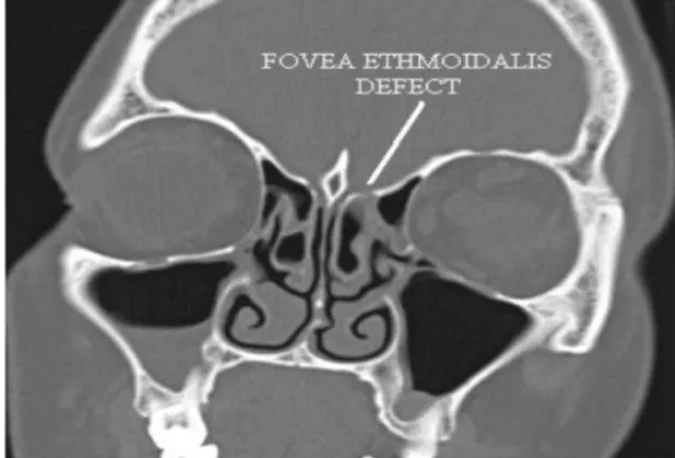
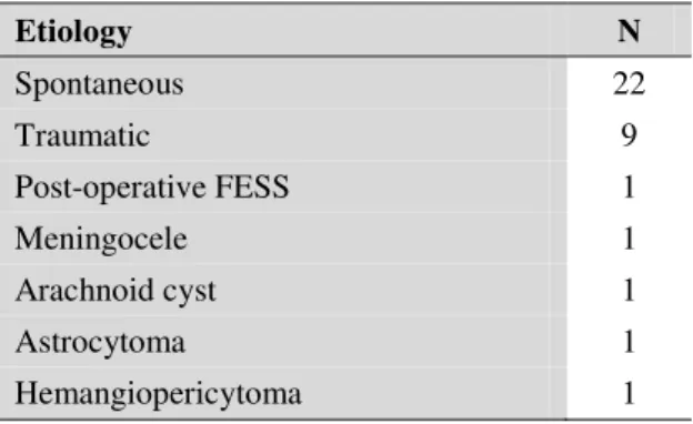
Endoscopic Repair of CSF Rhinorrhea: An Institutional Experience
Texto
Imagem
Documentos relacionados
The aims of this study were to evaluate the integrity of brain white matter and basal ganglions with magnetic reso- nance imaging (MRI) and computed tomography (CT) in the acute
The diagnosis of mature cystic ter- atoma by computed tomography (CT) and magnetic resonance imaging (MRI) is fairly straightforward as such modalities are more fat sensitive.. At
The purpose of this study is to assess anomalies found in structural neuroimaging exams (brain computed tomography (CT) and magnetic resonance imaging (MRI)) in the initial
However, the appearance of this cyst on cone beam computed tomography (CBCT) and magnetic resonance imaging (MRI) have received relatively little attention.. CBCT and
Objective: To evaluate the feasibility of quantifying visceral adipose tissue (VAT) on computed tomography (CT) and magnetic reso- nance imaging (MRI) scans, using freeware, as well
A new autopsy technique consists of the internal examination of death bodies using computed tomography (CT) and magnetic resonance imaging (MRI), without opening
Computerized tomography (CT) and magnetic resonance imaging (MRI) (Fig. 1) revealed a large posterior paravertebral lesion of soft tissue mass, with invasion of the epidural space
A computed tomography (CT) scan ( Figure 1 ) and magnetic resonance imaging (MRI) scan of the chest ( Figure 2 ) revealed the presence of a nodular lesion in the right pulmonary
