Iranian Journal of Basic Medical Sciences
www.mums.ac.ir/basic_medical/en/index
Human T Lymphotropic Virus Type I (HTLV-I) Oncogenesis: Molecular
Aspects of Virus and Host Interactions in Pathogenesis of Adult T cell
Leukemia/Lymphoma (ATL)
Sanaz Ahmadi Ghezeldasht
1, Abbas Shirdel
2, Mohammad Ali Assarehzadegan
3, Tahereh
Hassannia
4, Hosian Rahimi
2, Rahele Miri
1, S. A. Rahim Rezaee *
51 Research Centre for HIV/AIDS, HTLV and Viral Hepatitis, Iranian Academic Centre for Education, Culture & Research (ACECR), Mashhad Branch, Mash-had, Iran
2 Inflammation and Inflammatory diseases research Centre, Medical School, Mashhad University of Medical Science, Mashhad, Iran. 3 Department of Immunology, Faculty of Medicine, Ahvaz Jundishapur University of Medical Sciences, Ahvaz, Iran
4 Internal Medicine Dept, Medical School, Arak University of Medical Sciences, Arak- Iran 5 Immunology Research Centre, Mashhad University of Medical Sciences, Mashhad, Iran
* Corresponding author: Rezaee S. AR, Immunology Research Centre, Immunology Dept. Qaem Hospital, Mashhad University of Medical Sciences, Mash-had, Iran. Tel:+98-511 8436626; E-mail: [email protected]
© 2013 mums.ac.ir All rights reserved.
This is an Open Access article distributed under the terms of the Creative Commons Attribution License (http://creativecommons.org/licenses/by/3.0), which per-mits unrestricted use, distribution, and reproduction in any medium, provided the original work is properly cited.
A R T I C L E I N F O
A B S T R A C T
Keywords:
Adult T Cell Leukemia/Lymphoma
HTLV-I
Oncoviruses
Oncogenecity
Article type:
Review article
Please cite this paper as:
Ahmadi Ghezeldasht S, Shirdel A, Assarehzadegan MA, Hassannia T, Rahimi H, Miri R, Rezaee S. AR. Human T Lymphotropic Virus
type I (HTLV-I) oncogenesis: molecular aspects of virus and host interactions in pathogenesis of Adult T cell leukemia/lymphoma
(ATL). Iran J Basic Med Sci: 2013;16:179-95.
The study of tumor viruses paves the way for understanding the mechanisms of virus
pathogenesis, including those involved in establishing infection and dissemination in
the host tumor affecting immune-compromised patients. The processes ranging from
viral infection to progressing malignancy are slow and usually insufficient for
establish-ment of transformed cells that develop cancer in only a minority of infected subjects.
Therefore, viral infection is usually not the only cause of cancer, and further
environ-mental and host factors, may be implicated.
HTLV-I, in particular, is considered as an oncovirus cause of lymphoproliferative
dis-ease such as adult T cell leukemia/lymphoma (ATL) and disturbs the immune responses
which results in HTLV-I associated meylopathy/tropical spastic parapresis (HAM/TSP).
HTLV-I infection causes ATL in a small proportion of infected subjects (2-5%) following a
prolonged incubation period (15-30 years) despite a strong adaptive immune response
against the virus.
Overall, these conditions offer a prospect to study the molecular basis of tumorgenicity
in mammalian cells. In this review, the oncogencity of HTLV-I is being considered as an
oncovirus in context of ATL.
Introduction
Human tumor viruses
Oncogenecity refers to viruses that may cause cancers.
Generally, the viruses that associated with malignancies
are known as tumorviruses. Some researchers prefer to
consider human tumor viruses as a distinct group of
vi-ruses, however, the known tumorviruses such as
Hepati-tis B and C viruses, Epstein-Barr virus (EBV), human
her-pesvirus 8 (HHV-8), Human papillomavirus (HPV), and
Human T lymphotropic virus type I (HTLV-I) are various
Article history:
riers (15), whereas small percentage of infected
individu-als develops the neoplastic disease; adult T-cell leukemia
(ATL) and the inflammatory condition HTLV-I- associated
myelopathy/tropical spastic paraparesis (HAM/TSP) (16).
Only 5% of HTLV-I infected people develop HAM/TSP (17).
HTLV-I biology and pathogenesis
Genome structure and organization
The HTLV-I is an enveloped dimeric positive- sense
sin-gle-stranded RNA virus, and like other retroviruses, the
linear genome encodes the structural and enzymes such
as gag, env, and pol, (Figure 1) (18). Moreover, it contains
a unique region at the 3’ end, referred to the pX region,
which encodes regulatory proteins, such as Tax, HBZ (for
HTLV-I bZIP factor) and Rex. The biology of regulatory
proteins is explained in the following sections.
in case of families, genomes and life cycles. The
aforemen-tioned viruses are associated with malignancies such as
hepatocellular carcinoma (HCC), Burkitt’s lymphoma,
nasopharyngeal carcinoma (NPC), Kaposi’s sarcoma (KS),
Multicentric Castleman’s Disease (MCD) and Adult T cell
leukemia/lymphoma (ATL) (Table 1).
HBV is a double-stranded DNA virus of the
Hepadnaviri-dae family which infects 350 million people worldwide.
Hepatitis B can lead to liver diseases ranging from the
acute hepatitis to chronic hepatitis, cirrhosis and
hepato-cellular carcinoma (HCC) (1-2). HCV is a positive-stranded
RNA virus which belongs to the Flaviviridae family. It is
estimated that approximately 180 million people are
infected with hepatitis C virus worldwide (3- 4) The
Ep-stein–Barr virus (EBV), also called human herpesvirus 4
(HHV-4), is the virus of herpes family, and is one of the
most common viruses in humans. It is best known as the
cause of infectious mononucleosis (glandular fever). It is
also associated with particular forms of cancer, such as
Hodgkin’s lymphoma, Burkitt’s lymphoma,
nasopharyn-geal carcinoma, and central nervous system lymphomas
associated with HIV (5-7).
Kaposi’s sarcoma-associated herpesvirus (KSHV) is the
most recently discovered human herpesvirus. It is the
ae-tiologic agent of Kaposi’s sarcoma (KS), a tumor which
af-fects more frequently on patients diagnosed with AIDS
that does not receive any treatment. KSHV is also a
proba-ble cause of two lymphoproliferative diseases: multicentric
Castleman’s disease and primary effusion lymphoma (8).
Human T-cell lymphotropic virus type I (HTLV-I) was the
first discovered human retrovirus (9) and it has been
esti-mated that HTLV-I infects 10-20 million people worldwide
(10). This virus is endemic in several regions of the world,
such as southwestern Japan, the Caribbean basin, Central
Africa, South America, the Melanesian Islands and the
Middle East (11- 12). The prevalence of HTLV-I infection in
Iran (Mashhad) is estimated to be 2-3% of the entire
popu-lation and 0.7% among blood donors (12-14). The majority
of HTLV-I-infected individuals remain asymptomatic
car-Figure 1.Genome organization of HTLV-I
The full length of HTLV-I mRNA encodes the gag protein (p55) which is then cleaved by the viral protease making the matrix (p19), capsid (p24), and nucleocapsid (p15) proteins. A sequence, from the 3 end of gag to the 5 end of pol, encodes the protease which results from ribosomal frame-shifting. The HTLV-I long-terminal repeat (LTR) at both 5’ and 3’ ends of the genome contains the viral promoter and regulatory elements. In ad-dition, the viral mRNA encodes the pol protein, single spliced mRNA en-codes the env protein and a double spliced mRNA enen-codes the Tax and Rex regulatory proteins (19, 20).
GAG
POL ENV
X-I PRO
X-IV X-II
LTR LTR
PR
P RT IN
Cleavage by PR C
C 5/
5/ 5/
Antisense
ORF AAAAAAA3/ Genomic RNA
Ribosomal Frameshifts
SU TM AAAAenv RNAAAA3/
tax
rex AAAAAAA3
/
Tax/rex RNA Aiternative
initiation C
HBZ
HBZ-SI
Antisense transcription X-III
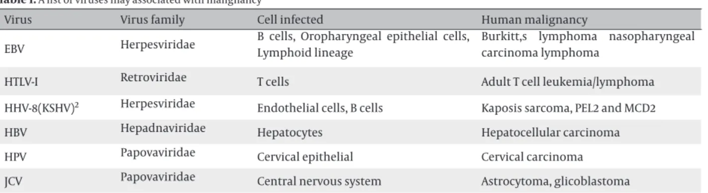
Table 1. A list of viruses may associated with malignancy
Virus
Virus family
Cell infected
Human malignancy
EBV
Herpesviridae
B cells, Oropharyngeal epithelial cells,
Lymphoid lineage
Burkitt,s lymphoma nasopharyngeal
carcinoma lymphoma
HTLV-I
Retroviridae
T cells
Adult T cell leukemia/lymphoma
HHV-8(KSHV)
2Herpesviridae
Endothelial cells, B cells
Kaposis sarcoma, PEL2 and MCD2
HBV
Hepadnaviridae
Hepatocytes
Hepatocellular carcinoma
HPV
Papovaviridae
Cervical epithelial
Cervical carcinoma
JCV
Papovaviridae
Central nervous system
Astrocytoma, glicoblastoma
1-These tumors appear in immunosuppressed patients bear copies of EBV genomes.
2- KSHV: Kaposis sarcoma associated herpesvirus, PEL: Primary effusion Lymphoma, MCD: Multicentral Castleman’s disease
Tax protein
The HTLV-I trans-activator (Tax) is a 40-kDa protein
con-taining 353 amino acid residues located within the U3
region of the LTR (21-23). An atypical nuclear localization
sequence spans the first 48 amino acids and an
amino-terminal domain that interacts with the cellular
tran-scription factors, cyclic AMP-responsive element-binding
protein (CREB) and serum-response factor (SRF) (24-27).
Interactions with these factors are responsible for
HTLV-I LTR-trans-activation, as well as the activation of certain
mitogenic genes during cellular transformation.
Tax interacts with CREB and the p300/CBP co-activator
family on three 21 bp-repeats in the HTLV-I LTR and is
reported to stabilize the formation of CREB/ATF-dimers
bound to the DNA (28-29). The kinase-inducible domain
like domain in Tax may mediate many of the interactions
including the expression of many cellular genes
deregu-lated by viral trans-activator. In fact, the competition for
employing nuclear of p300/CBP may prepare
Tax-depen-dent repression of some transcription factors, such as
p53 and c-Myb (30-31). Altogether, tax ax induces or
re-presses the expression of a large variety of cellular genes
as well as regulating viral gene expression (32-33).
There-fore, emerging evidence suggests that Tax serves as the
primary oncogenic mediator of HTLV-I (34). Tax induces
cell immortalization and transformation in vitro (35-39)
as well as tumor formation in transgenic mice (40).
Rex
Rex is a 27-kDa phosphoprotein encoded by ORF III that
localizes the nucleolus of infected cells (41-42). Rex, p27,
plays a pivotal role in viral replication and the
regula-tion of viral structural genes by funcregula-tioning as a
post-transcriptional regulator that increases the expression of
singly spliced and unspliced viral mRNAs (env, gag, and
pol, respectively) (43).
Rex is a specific RNA binding protein that binds to a
cis-acting Rex responsive element (RxRE), a highly stable
structure located within the R region of the viral LTR
(44-45). Rex-mediated regulation is required to balance the
spliced and unspliced mRNAs which is necessary for the
production of infectious virus. However, the detailed
mechanism(s) of Rex regulation is still highly
controver-sial and not fully understood (46).
p12I
ORF I encodes a 12-kDa protein (p12I). Although, p12I
ex-pression is difficult to be displayed n HTLV-I- infected cells,
some evidences have suggested its importance (20). While
p12I does not seem necessary for HTLV-I replication in
vi-tro (47-48), deletion of the acceptor splice site for the p12I
mRNA results in diminishing viral infectivity in vitro (49).
This viral protein is found to have weak oncogenic
activ-ity, shares amino acid similarities with other viral
oncop-roteins (50), and binds to the IL-2-receptor (IL-2R) (51). p12I
is necessary for the infection of primary lymphocytes in
vitro (52). Therefore, p12I may have an important role in
the activation of host cells at the early stages of infection
where interaction of the protein with host cell signal will
contribute to the host cell activation, thus affecting more
viral infection.
Two variants of the p12I protein have been
demonstrat-ed: one has a Lysine at position 88 which is found in
HTLV-I strains from TSP-HAM patients; the second has an
Argi-nine at position 88 which is found in HTLV-I strains from
all ATL patients and healthy carriers (53).
It has been suggested that p12I has significant roles
during the early stage of HTLV-I infection establishment.
Furthermore, the ability of p12I for binding to the MHC
I heavy chain and making it susceptible for degradation
may decrease the viral peptide MHC I- complexes on the
infected cell surface and protect them from lysis by
cyto-lytic T lymphocytes (CTLs) (54).
Transmission of HTLV-I
HTLV-I can infect some cell types including T cells, B
cells, and synovial cells. Many of the studies
demon-strated that the transmission of HTLV-I should occur by
healthy infected cells and is very inefficient by free virion
(REF). This is because HTLV-I transmits naturally by the
cell-to-cell manner. Whilst an HTLV-I-infected cell
attach-es to uninfected cells, the HTLV-I-infected cells form
“viro-logical synapses” with uninfected cells (55). The receptor
for HTLV-I is glucose transporter type 1 (GLUT1) (56). Its
expression on T lymphocytes is enhanced by mitogens or
transforming growth factor (TGF-β) (57), which has been
shown to increase the infectivity of HTLV-I.
HTLV-I-infected cells in the human body are
transmit-ted via three major routes: (1) mother-to-infant
transmis-sion (mainly breast-feeding), (2) sexual transmistransmis-sion, (3)
parenteral transmission. It is worth noting that fresh
fro-zen plasma from seropositive donors does not transmit
HTLV-I (58). Moreover, as living cells can be eliminated
by freezing and thawing, therefore, feeding the mother’s
frozen breast milk to infants will not increase the risk of
viral transmission (59).
Clonal proliferation of HTLV-I-infected cells
HTLV-I clonal cells are more heterogeneous and less
sta-ble during sero-positivity than in long-term carriers (62).
Several factors may effect on this phenomenon including
the host defense pressure by cellular immunity (63), the
viral factors, particularly oncogenic proteins, and the
sites of the integration.
Oncogenic function of HTLV-I proteins
Immortalization / transformation of T cells
Many studies have demonstrated that HTLV-I genes (e.g.
tax and HBZ), can induce various cellular dysfunctions,
genetic and epigenetic alterations, and the host
im-mune system may be involved in the leukemogenesis of
ATL (64). However, there are no clear determinants that
differentiate the subject develop ATL from those who
re-main asymptomatic.
Immortalization of lymphocytes infected by HTLV-I
re-quires Tax, however, Tax-immortalized cells, but
nontran-formed cells are still IL-2 dependent. Therefore, infected
cells could not pass the G1 check point without
exoge-nous growth factors such as IL-2. In addition the
inacti-vation of tumor suppressor gene p53 contributes to the
accumulation of genetic mutations (65). Alternatively,
alterations and errors are accumulated progressively by
several viral proteins in the host genome during the
la-tent period, finally leading to the onset of ATL (66).
CD4+-CD8+ cells, and immature CD4--CD8- cells from
bone marrow can be transformed by HTLV-I. Transformed
cells not only display an activated phenotype but also in
some cases retain T-cell functional properties. Among
the functional activities of HTLV-I, transformed cells have
reported to be able to induce suppressor-cell activity (67)
and retain antigen-specific responses and cytotoxic
func-tion (68).
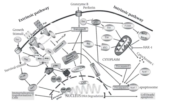
HTLV-I and apoptosis
The programmed cell death is one of the major
effec-tors mechanisms in NK cell and CTLs for killing the
vi-rus infected and malignant cells. The effect of HTLV-I
infection on programmed cell death is less clear. Tax has
been shown to repress Bax gene-expression as is shown
in figure 3 (69). Since Bax promotes apoptosis by
inhibit-ing Bcl-2, this may imply a molecular mechanism for the
resistance of HTLV-I-infected T cell lines to apoptosis
in-ducing stimuli (70). However, several reports have shown
that HTLV-I-infected T cells can be induced to undergo
apoptosis (69, 71). Other reports have suggested that Tax
may induce apoptosis too.
Activation of NF-κB by immune stimuli extrinsic and
intrinsic pathways, which are based on degradation of
IkB or processing of p100, respectively. The canonical
pathway is stimulated by diverse cellular stimuli, such as
antigens and cytokines, and is dependent on the trimeric
IKK complex as well as certain upstream kinases, such as
MEKK3 and PKCψ. The noncanonical pathway responds
to a subset of TNF family members, including BAFF and
CD40L, and requires NIK and its downstream kinase IKKα
but not IKKβ or IKKγ. Tax activates both NF-κB pathways
by physically targeting two different IKK complexes, both
requiring the adaptor protein IKKγ. Formation of the
noncanonical Tax/ IKK complex requires the interaction
of Tax with both IKKγand p100.
Therefore, manipulation of both the intrinsic and
ex-trinsic pathways is very important for dissemination of
virus to evade from NK cell and CTLs antiviral activities.
The precise mechanism that HTLV-I inhibits apoptosis is
unclear, but it is assumed that Tax may have the central
role by suppressing p53 and BAX and stimulation of
sur-vival signals via PI3K and NF-κB pathways.
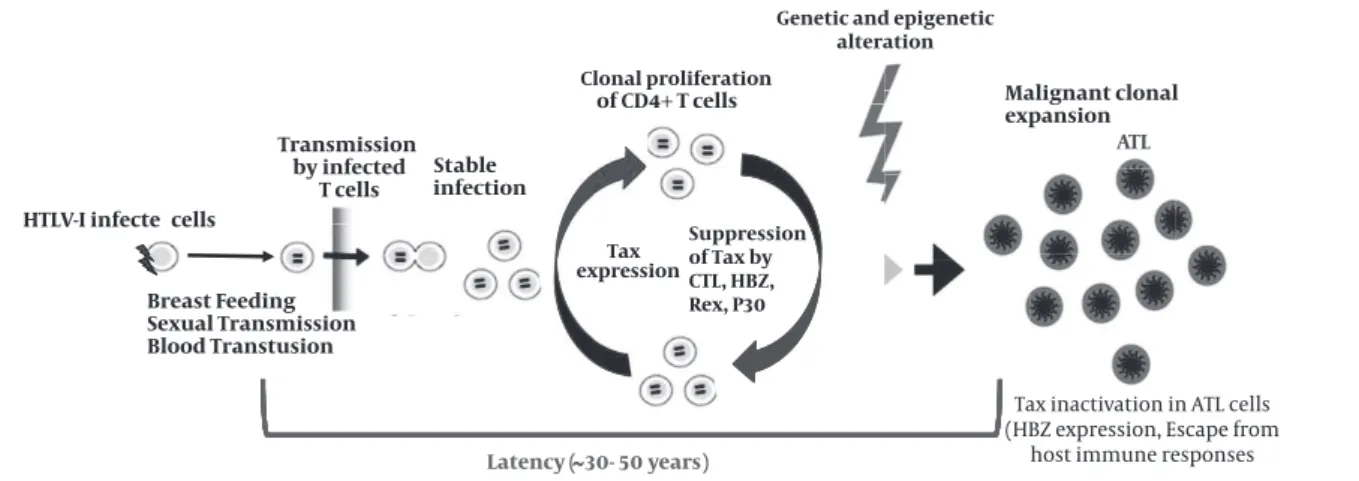
Figure 2.HTLV-I transmission, infection, virus-host interactions, and onset of ATL
Cell-to-cell spreading is the main rout of HTLV-I transmission. Tax mainly promotes T cell proliferation of HTLV-I infected cells. However, other HTLV-I proteins can enhance this process. Cytolytic T cells (CTLs) can prevent the proliferation by killing of Tax expressing targets cells. The expression of Tax is inactivated by several mechanisms, suggesting that Tax is not necessary in ATL stage and HBZ may be more effective. Idea from (65) with modification.
HTLV-I infecte cells
ding Transmission
by infected T cells
t
Latency (
able infection
Tax
x
expression
of CD4+ T cells
30- 50 years)
ATL
Tax inactivation in ATL cells (HBZ expression, Escape from
host immune responses
Breast Feeding
St
Clonal proliferation
Genetic and epigenetic alteration
Sexual Transmission Blood Transtusion
Malignant clonal expansion
Proviral load
Recently, HTLV-I proviral load levels have been evaluated
as important predictors of development of ATL and HAM/
TSP. Many studies show that HTLV-I proviral load are
sig-nificantly higher in ATL and HAM/TSP compared to HTLV-I
healthy carriers (72-75), (76-77).
Proviruses of HTLV-I are clonally integrated in ATL
pa-tients, but randomly integrated in HAM/TSP patients. The
efficiency of HTLV-I replication is shown by the
propor-tion of PBMCs that carry HTLV-I provirus (78). Moreover,
the amount of HTLV-I proviral DNA in-patients with HAM/
TSP is 3-5 folds and in patients with ATL is 5-15 folds higher
than I carriers (61, 79-80). The integration of
HTLV-I in PBMC of HAM/TSP patients is usually polyclonal (78).
Non clonal integration has been observed in HTLV-I
carri-ers of non-HAM/TSP family (81). Taken together, these
re-sults suggest that increased HTLV-I proviral DNA load and
the presence of IgM antibody and high titres of IgG and
IgA antibodies to HTLV-I proteins appear to distinguish
HAM/TSP patients from HTLV-I carriers. Furthermore,
high titres of HTLV-I antibody and proviral load among
HTLV-I carriers maybe useful predictive markers for
de-velopment of HAM/TSP (75).
Previous studies of infected human subjects suggest
that high proviral load is associated with increased
ten-dency to develop HTLV-I-associated HAM/TSP, while ATL is
associated with extremely high levels of provirus (76-77,
82-83) (Akbarin
et al
, unpublished data). Counting HTLV-I
infected cells in healthy carriers and ATL patients (84)
de-termine the susceptibility of HTLV-I associated diseases
(85-86) the influence of cytokines (87-88, 90). A high
HTLV-I proviral load is currently considered as one of the main
indicators for the progression to ATL. In a prospective
study in Japan, 14 participants of asymptomatic HTLV-I
carriers progressed to ATL, all of whose baseline
provi-ral load levels were high (range, 4.17 to 28.58 copies/100
PBMCs). Therefore, the author suggested that those with
a high proviral load level (
∼
> 4-7 copies/100 PBMCs) are
included in a high-risk group for developing ATL.
Statisti-cally analyses confirmed that a higher proviral load level
was a strong factor in the development of ATL (91).
Pro-viral load measurement by Tax expression is also
valuable to monitor viral activity in HTLV-I associated
diseases in ATL patients and it is useful for monitoring of
patients following administration of medications such
as interferon-α and determination of the efficiency of
medication (92-93).
Figure 3.Schematic representation of pathways used by Tax to prevent programmed cell death
With modification from Rezaee et al, 2006, to fit for HTLV-I. Apoptosis is one of the main mechanisms of immune system for killing virus infected or malignant cells. The extrinsic pathway of apoptosis (left side) is triggered by the binding of ligands to death-inducing membrane proteins. This signal-ing event leads to the recruitment of Fas-associated death receptor (FADD) which in turn activates caspase 8. Consequently, caspase 8 activates ‘effector’ caspases (caspases 3, 6 and 7) and releasing caspase-activated DNase (CAD) which in turn lead to cell death. During conditions of cellular stress, such as DNA damage and growth-factor deprivation, the intrinsic apoptosis pathway (the pathway that is activated most frequently by the tumor suppressor protein p53) is activated (right side). The intrinsic apoptotic pathway converges on the disruption of mitochondrial membranes and consequently, after some molecular activation events, results in releasing CAD. The balance of the pro- and anti-apoptotic bcl-2 family proteins determines whether apoptosis proceeds. There is overlap between the intrinsic and extrinsic apoptotic pathways, as caspase 8 can cleave and activate the bcl-2 family protein. Therefore, manipulation of both the intrinsic and extrinsic pathways is very important for dissemination of virus to evade from NK cell and CTLs antiviral activities. The precise mechanism that HTLV-I inhibits apoptosis is unclear, but it is assumed that Tax may have the central role by suppressing p53 and BAX and stimulation of survival signals via PI3K and NF-κB pathways.
Extrinsic pathway
Granzyme B Perforin
Growth Stimuli
HAX -1
Survival stimuli
Cytochrome c
apoptosome
Intrinsic pathway
CYTOPLASM
Immortalization/ transformation T
Tax
HTLV-I transcription by Tax
Appropriate HTLV-Ireplication and viral gene
expres-sion are associated with sufficient expresexpres-sion of viral
oncoprotein Tax. The mechanism by which Tax activates
viral transcription is well known. For viral transcription,
Tax interacts with CREB and recruits the co activator CREB
binding protein (CBP), forming a nucleoprotein complex
on the three viral cyclic AMP-responsive elements (CREs)
in the HTLV-Ipromoter. Each three 21 bp repeats contains
a cAMP response element (CRE) core. In the presence of
Tax, gene expression driven by multiple copies of the 21-
bp repeat element can increase up to 100-fold or higher.
Cellular basic domain-leucine zipper (bZip),
transcrip-tion factors-CREB, ATF-1, the 21-bp repeats, and Tax form
stable ternary complexes (94,101). In these complexes, Tax
binds the bZIP domains of CREB/ATF-1 (28, 102-104) which
binds to the DNA of the G/C-rich sequences achieving the
DNA sequence of LTR trans-activation. (94, 99, 105-106).
The importance of this segment of viral DNA is well
es-tablished, however, some studies have failed to identify
an interaction between Tax and the DNA. In the context
of these complexes, Tax recruits more transcriptional
co-activators, CREB binding protein (CBP)/p300 and some
other transcription factors for an appropriate gene
acti-vation (29, 94, 106-107).
Activation of NF-κB pathway by tax
NF-κB/Rel family of transcription factors are suppressed
by I-κB proteins in cytoplasm, (108). During activation of
NF-κB by extracellular stimuli such as interleukin-1(109),
tumor necrosis factor-α (TNF-α), bacterial
lipopolysaccha-ride (LPS), or by HTLV-I Tax, I-κBα and I-κBβ become serine
phosphorylated by I-κB kinase (IKK). Then these
inhibito-ry proteins are marked for rapid degradation in
protea-some complex (Figure 3). Activation of NF-κB pathway is
very important in proliferative disorders.
The non-canonical pathway of NF-κB activation is
im-portant for B-cell proliferation and lymphoid
organogen-esis and is activated in response to some inducers such as
lymphotoxin β and B cell activating factor (108). Different
physiological inducers of NF-κB activate either the
canon-ical or non-canoncanon-ical pathway, whereas, Tax can activate
both. Activation of I-κB kinase (IKK) by Tax is due to a
di-rect interaction between Tax and IKKγ (110-113). Tax binds
directly to the 201-250 amino acid residues in IKKγ
(110-114). Recent data have indicated that via a tripartite
inter-action, Tax, protein phosphatase 2A (PP2A) and IKKγ form
a stable ternary complex. In this context, PP2A activity is
inhibited or diminished (115). These results suggest that
PP2A is a negative regulator of activated, phospho-IKK,
and PP2A inhibition by IKKγ-bound Tax which maintains
IKK in a phosphorylated and active state, causing
consti-tutive phosphorylation and degradation of I-κB, nuclear
translocation of NF-κB/Rel, and potent activation of genes
under NF-κB/Rel control (Figure 3).
The classical pathway is mediated by the NF-κB-inducing
kinase (NIK) and IKKα, and is independent of IKKγ (116).
Phosphorylation of p100 targets p100 for ubiquitination
and processing by activated NIK-IKKα complex (116-117).
Tax-mediated p100 processing, however, requires both
IKKα and IKKγ (112, 116, 118). Tax appears to alter p100’s
con-formation by direct binding to two short amino-terminal
helices (αA and αβ) in p100 (119) and at the same time
ac-tivate IKKγ/IKKα to facilitate p100 phosphorylation and
processing (112, 116, 119). Because the non-canonical
path-way is silent in T lymphocytes, its aberrant activation by
Tax in T-cells may play an important role in Tax-mediated
T-cell activation and transformation. On the other hand,
cell transfection experiments support the importance of
Tax as an intracellular NF-κB inducer, it should be noted
that cells from ATL patients have elevated NF-κB activity
even when Tax-expression is ultimately shutdown. This
finding suggests that Tax may be used to initiate but may
not need to maintain NF-κB activation (120). In fact,
Higu-chi
et al
. (121) have proposed that CD30 serves a role in Tax
independent activation of NF-κB. CD30 is a member of the
TNF receptor superfamily and interestingly is a marker of
malignancy in Hodgkin’s lymphoma (122). Finally, NF- κB
activation by Tax over regulates many cellular genes
in-cluding those of IL-2 receptor α chain, GM-CSF,
stimula-tory surface receptors OX40, IL-13, IL-15, ICAM1, and
anti-apoptotic proteins (111, 123-124). Role of these proteins in
proinflammatory response and lymphocyte survival are
obvious and are likely to be critical to the development of
HAM/TSP and ATL.
HBZ
The complementary strand of HTLV-I proviral genome
generates distinct 2.6 kb and 2.9 kb transcripts, driven by
promoter in the 3’ LTR (125). Subsequently, a novel viral
protein encoded by these transcripts was identified (126).
This protein, designated HBZ (for HTLV-I bZIP factor),
con-tains nuclear localizing motifs (127), and a N-terminal
transcriptional activation domain and a leucine zipper
motif in its C terminus (128) (Figure 1). C-terminal leucine
zipper (LZ) region deletion of HBZ in an infectious
provi-ral clone of HTLV-I had no effect on the ability of the virus
to replicate and immortalize lymphocytes for growth in
culture. Eliminating HBZ expression results in significant
reductions in proviral load and attenuated antibody
re-sponse against the viral proteins in rabbit model (129).
CREB-2 and preventing recruitment of CREB-CREB-2 to the TRE and
cyclic AMP response element sites in a dose dependent
manner (130), providing evidence for HBZ’s role as a
po-tential negative regulator of Tax-mediated viral
transacti-vation is important.
Other cellular transcriptional factors appear to be the
target of HBZ functions. HBZ interacts with Jun-D and
stimulates its transcriptional activity (131) while
sup-pressing transactivation by c-Jun (132). HBZ represses AP-1
transcriptional factor activity by impairing both the
DNA-binding ability and the stability of c-Jun protein (133).
These studies provide evidence for possible alteration of
cellular processes by HBZ following HTLV-I infection.
ATL
ATL is the most common outcome of infection with
HTLV-I, and in areas of endemicity, it occurs in 2-5% of
HTLV-I-infected individuals (134). It was reported that the
median age of onset of ATL is 56 years (135), suggesting a
long period of latent infection (134). It has led to the
as-sumption that most, if not all, cases of ATL develop in
in-dividuals infected with HTLV-I since birth (136).
ATL can be presented in one of several forms (which may
be stages in the natural history of ATL). Although there
are significant geographic variations in the contribution
of these forms in the initial presentation, the most
com-mon presentation is acute leukemia.
The most benign detectable form of ATL is an
asymp-tomatic pre-leukemic phase which is usually diagnosed
incidentally when the examination of a peripheral blood
smear reveals abnormal lymphocytes with
characteris-tics of lobulated nuclei (flower cells, Figure 2) (137).
Smoldering ATL is the most benign symptomatic form
of disease and is characterized by cutaneous but not
vis-ceral lesions, a normal peripheral blood leukocyte count,
and a few circulating leukemic cells. The development of
chronic ATL is marked by visceral involvement and is
evi-denced by lymphadenopathy, hepatosplenomegaly, and
peripheral blood leukocytosis. However, both of the
rela-tively benign ATL can progress to acute ATL.
The median life expectancy of individuals with acute
ATL is 11 months. Patients with acute ATL have cutaneous
and visceral involvement, peripheral blood leukocytosis,
elevated levels of lactate dehydrogenase and bilirubin,
and often hypercalcemia. ATL is associated with severe
immunosuppression as evidenced by the
susceptibil-ity of these patients to various opportunistic infections,
including Pneumocystis carinii pneumonia,
cryptococ-cal meningitis, candidal esophagitis, and disseminated
cytomegalovirus infection (138). Occasional long-term
survival has been described, but the disease is usually
unresponsive to conventional chemotherapy. Recent
re-ports of immunotherapeutic trials allow for a degree of
optimism (139).
ATL is generally resistant to the chemotherapy and
car-ries a dismal prognosis particularly for the acute and
lymphoma subtypes. Promising results were obtained
re-garding the combination of zidovudine and
interferon-alpha. Chronic ATL has a relatively better outcome, but
poor long-term survival is noted when patients are
man-aged by a watchful-waiting policy or by chemotherapy.
In ATL cell lines, arsenic trioxide shuts off constitutive
NF-kappa B activation and potentiates interferon-alpha
apoptotic effects through proteasomal degradation of
Tax. Clinically, arsenic/interferon therapy exhibits some
efficacy in refractory aggressive ATL patients. The
afore-mentioned data were leading us to investigate the
effi-cacy and safety of the combination therapy of arsenic,
interferon-alpha, and zidovudine in 10 newly-diagnosed
chronic ATL patients. An impressive 100% response rate
was observed including 7 complete remissions, 2
com-plete remissions but with more than 5% circulating
atypi-cal lymphocytes, and 1 partial response. In conclusion,
treatment of chronic ATL with arsenic, interferon-alpha,
and zidovudine is feasible. Overall, the clinical trial
data strengthen the concept of oncogene-targeted
can-cer therapy (139) and a long-term follow up will clarify
whether this will end to disease cure.
HTLV-I proteins in ATL development
The pX region of the HTLV-I genome encodes a number
of nonstructural proteins, including Tax(140), Rex (141),
and the accessory proteins encoded by the open reading
frames I (p12I and p27I) and II (p13II and p30II) (Figure 1).
The most notable viral regulatory protein is Tax (34,140,
142,).
Although the basis of cellular transformation by Tax is
not fully understood (143-144); (145), Some studies have
demonstrated that Tax is the major transforming protein
of HTLV-I, However, no common integration site is found
among patients, integration of the proviral genome into
host cell DNA is monoclonal in transformed cells,
suggest-ing that integration occurs prior to the transformation.
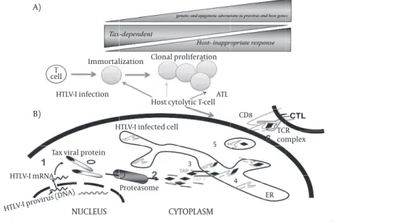
chang-es enable ATL cells to chang-escape from the host immune
sys-tem because tax is the most immune-dominant protein
(Figure 1 and 5). This finding reveals a duality in the Tax
protein: its expression induces proliferation and inhibits
apoptosis of HTLV-I-infected cells, and evokes the host’s
immune response including cytotoxic T cells to kill
virus-infected cells. Therefore, after dissemination of virus and
during latency, loosing tax protein is a pivotal mechanism
for HTLV-I to escape from host immune system.
Although Tax lacks a cellular homologue (149), it
func-tions like an oncogene, inducing leukemogenesis by
causing aberrant transactivation of cellular growth
regu-latory genes. A large group of cellular genes involved in
T-cell growth are activated by Tax including interleukin
2 2), the high-affinity subunit of the IL-2 receptor
(IL-2Ra), IL-3, IL-4, granulocyte–macrophage
colony-stimu-lating factor, proliferating cell nuclear antigen, TGF-β,
vimentin, proenkephalin, egr-1, egr-2, fos, c-myc, bcl-xL,
and Jun (150).
The mechanisms by which Tax modulates viral and host
cell transcription factors toward transformation are of
considerable interest. Altogether, the studies suggest
that Tax may initiate a cascade of events leading toward
transformation that, at later stages, may not require or
must be eliminated due to immunogenicity for the host
immune response (Figure 4).
Activation of the PI3 kinase pathway and ATL
Some studies have reported constitutive activation of
the phosphoinositide 3-kinase (PI3K) pathway as a
com-mon characteristic of HTLV-I- and Tax-transformed cells
(Figure 2). Treatment of IL-2-independent HTLV-I
trans-formed T-cells with inhibitors of PI3 kinase leads to a
p27KIP1- dependent cell cycle arrest (151).
Somatic changes in ATL cells
Mutation of p53, and deletion of p16 have been reported
in ATL with a poor prognosis (152).Therefore; these
genet-ic changes are believed to be associated with the disease
progression. A transcriptional profile of ATL cells by DNA
chip analysis identified aberrantly transcribed genes
(153). Among them, the expression of tumor suppressor
in lung cancer 1 gene is overregulated in ATL cells (154).
However, its ectopic expression is associated with
leuke-mogenesis possibly due to conferring an adhesive
pheno-type to ATL cells.
Epigenetic changes are recognized as mechanisms
implicated in oncogenesis as well as genetic changes.
Since genetic changes in specific genes in ATL cells have
not been identified except for p53 and p16, and there is
no consistent chromosomal change, it is possible that
epigenetic changes such as DNA methylation (155) play
an important role in leukemogenesis by inhibiting the
transcription of tumor suppressor genes or inducing
ab-errant expression of oncogenes. On the other hand, EGR3
gene has been demonstrated to be hypermethylated in
ATL cells (156). EGR3 is a transcriptional factor, which is
essential for transcription of the FasL gene (157). Normal
activated T lymphocytes express FasL as well as Fas
anti-gen. Apoptosis induced by autocrine mechanisms is
des-Figure 4.A schematic illustration of HTLV-I oncogenesis, HTLV-I infection, host CTLs responses and onset of ATL
Tax-mediated immortalization of HTLV-I-infected T cells toward leukemogenesis by genetic and epigenetic changes in provirus and host genes. However, HTLV-I gene expression also can be recognized by T cells result in activation of host immune system which eliminates Tax-expressing target cells. There-fore, the host immune system pressure in such circumstances is in favor of proliferation of Tax negative HTLV-I infected cells.
CYTOPLASM
NUCLEUS
A)
B)
Immortalization Clonal proliferation
HTLV-I infection T
cell
HTLV-I infected cell
Tax viral protein
HTLV-I mRNA
Proteasome
Host cytolytic T-cellATL
5 CD8
TCR complex
ER 3
4
ignated activation-induced cell death and controls the
immune response (158). Although ATL cells express Fas
antigen, they do not produce FasL, thereby enable ATL
cells to escape from programmed cell death. Thus,
epi-genetic changes of the host cell may play a pivotal role in
oncogenesis of ATL.
Tax protein and cell cycle deregulation
Several lines of evidence indicate that p40tax is the
on-cogene responsible for viral lymphocyte-transforming
and leukemogenic properties (38). Mechanistically,
sev-eral biochemical features of the protein can cooperate to
transform, in which transcriptional stimulation of
cellu-lar signal transducers, cytokines (159) and anti-apoptotic
effectors exist. Tax has capacity to stimulate aneuploidy
and interfere with DNA repair (160), which could
indi-rectly support malignant progression. A major
mecha-nistic explanation for the mitogenic and immortalizing
effects of the Tax oncoprotein is provided by its ability to
stimulate the G1- to S-phase transition in T-cells (161-163).
In mammalian cells, G1-progression is controlled by
the sequential activation of several cyclins and
cyclin-dependent kinases (CDKs), starting with CDK4, CDK6
and CDK2. Tax activates CDK4, CDK6 and CDK2 leading to
phosphorylation of retinoblastoma (Rb) tumor
suppres-sor proteins and liberation of the transcription factor E2F
(161, 164). Moreover, Tax may induce Rb degradation (165)
and increase cellular E2F synthesis (166-167). Several
indi-rect effects of Tax and features of HTLV-infected cells may
support the impact of Tax on CDK.
In general, Tax shortens the length of G1 and accelerates
entry into S phase (150, 168). In Tax-expressing cells and
ATL cells in culture, Tax stimulates cyclin/cdk activity;
re-sulting in hyperphosphorylated Rb. Tax also increases E2F
transcription and protein levels (164,167,169).The
cumula-tive effect of hyperphosphorylated Rb and increased E2F
transcription cause the availability of more active E2F,
which facilitates the entry of Tax-expressing cells into S
phase. Tax also increases the levels and activity of several
cyclin/cdk partners, which are important in G1
progres-sion and S phase entry.
Cyclin D2 expression is also overregulated by
interleu-kin-2 receptor (IL2-R) signals (170- 171). Tax may
cooper-ate with interleukin-2 (IL-2) signaling either indirectly,
through stimulating the expression of IL-2Rα or directly,
by activating the cyclin D2 promoter (172- 173). Tax also
represses the function of distinct tumor suppressor
pro-teins which interfere with G1- to S-phase transition. (174).
The interaction of the cyclin D/CDK components provides
a major explanation for the G1-phase stimulating effects
of Tax. Direct association with Tax enhances CDK4 activity.
This increased kinase activity in the presence of Tax may
be explained by intensified association of CDK4 and its
positive cyclin regulatory subunit and resistance of the
complex to inhibition by p21CIP1(175) .
The 40 N-terminal amino acids of Tax are sufficient to
bind cyclin D2 and CDK4. Within CDK4 a N- and a
C-termi-nal domain are relevant for Tax binding. Taken together,
these findings suggest that Tax stimulates G1- to S-phase
transition by supporting the association of CDK4 and
cy-clin D2. Furthermore, these studies support the conclusion
that CDK4 activity is stimulated through conformational
changes of the enzyme directly mediated by Tax (176).
Cell cycles checkpoints are activate in DNA damage. Tax
disrupts the DNA damage-induced G1/S checkpoint
(con-trolled by cyclin E/cdk 2), in part by inactivation of p53.
Normally when DNA damage is detected during G1, p53
activates the cdk inhibitor p21waf1, which in turn, binds
and inactivates cdk2. Although p53 is inactivated,
Tax-expressing cells have higher levels of p21waf1 because Tax
transactivates the p21waf1 promoter (177-178); (164). Loss
of p53 function prevents proper G1/S arrest, p53-mediated
apoptosis, and DNA repair, all of which contribute to
cellu-lar transformation. A series of studies have demonstrated
that in HTLVI-infected cells, Tax has the ability to abrogate
the transactivating function of p53 (Figure 2).
Interesting-ly, whereas p53 mRNA levels remain unaffected in these
cells, p53 protein levels are elevated, implying that Tax
re-presses p53 function by increasing protein stability and/
or through post-translational modifications (179).There
are conflicting opinions on the mechanism by which Tax
mediates the loss of p53 function in vivo. Although Tax
does not bind to p53, it alters its subcellular localization,
or disrupts its DNA-binding activity (71, 180-181).
However, more studies should be conducted to reach
more reasonable mechanisms to understand how HTLV-I
can pass the cell cycle checkpoints and push the cell
to-ward mitosis and proliferation leading to cell
transfor-mation.
still require IL-2 to proliferate do not have a constitutively
activated JAK-STAT pathway (187-188).
ATL cells secrete an increased amount of TGF-β because
of Tax transactivation of the TGF-β promoter (189-190);
however, the cells are resistant to growth inhibition by
TGF-β (191). This resistance occurs due to the numerous
ef-fects of Tax on the TGF-β pathway.
Although Tax enhances TGF-β production, infected cells
are resistant to TGF- β growth inhibition because Tax
dis-rupts the TGF- β signaling pathway and interferes with
the function of growth inhibiting proteins induced by
TGF-β.
An additional disruptive effect of Tax emanates from its
impact on the ability of the cell-cycle machinery to
regu-late DNA replication and cell division (142,192-194).
Despite the fact that nearly 70% of all cancers
demon-strate aneuploidy (including both ATL and
HTLV-I-in-fected cells), only rarely have genetic defects which are
identified in mitotic checkpoint genes, implying that
other events must alter function of the mitotic spindle
checkpoint (MSC) (195-196). Research on both DNA repair
and MSC are vital to understand HTLV-I-mediated cellular
transformation and are highly prior for future studies.
Activation of NF-κB by HTLV-I
NF-κB activation by immune stimuli
The NF-κB proteins are normally sequestered in the
cyto-plasm by physical interaction with a family of inhibitory
proteins, including IkBa, IkBb, and related proteins (197).
The NF-κB precursor proteins, p105 and p100, contain
IkB-like sequences in their C-terminal portion and also
function as NF-κB inhibitors (198). Thus, the processing of
these precursor proteins serves to both generate mature
NF-κB subunits and disrupt their IkB-like function.
The latent NF-κB complexes can be activated by diverse
immune stimuli, such as antigens, cytokines, and
micro-bial components, which target two alternative NFkB
sig-naling pathways: the canonical and noncanonical
path-ways (108, 199).
Aberrant activation of NF-κB by HTLV-I
Under normal conditions, the signals mediating NF-κB
activation in T cells as well as most other cell types are
transient and stimulate predominantly the canonical
NF-κB pathway. Such a signaling mechanism ensures the
rap-id, but short-lived, nuclear expression of NF-κB members
that are required for temporal proliferation and survival
of antigen-stimulated T cells. This mechanism is achieved
through different levels of regulation. First, following
stimulation by an antigen, both the T cell receptor (TCR)
and its proximal signaling molecules are downregulated,
thus preventing persistent signaling through the cell
sur-face receptor (200). Second, the NF-κB signaling pathway
involves a negative feedback mechanism, whereby the
activated NF-κB induces the expression and de novo
syn-thesis of the inhibitory protein IkBa (201-202). The newly
synthesized IkBa is able to enter the nucleus and stop
the function of NF-κB. Despite its tight control in normal
T cells, NF-κB is constitutively activated in both
HTLV-I-transformed T-cell lines and freshly isolated ATL cells
(186, 203-205). The persistent activation of NF-κB by
HTLV-I appears to be mediated through a mechanism that does
not involve the TCR or its proximal signaling molecules,
such as Src and Syk families of protein tyrosine kinases
(PTKs). In fact, a characteristic of HTLV-I-transformed T
cells is the loss of TCR and upstream PTKs (183, 206-207,).
It is generally believed that the viral Tax protein serves as
an intracellular NF-κB inducer that acts by bypassing the
TCR-proximal signaling factors.
A main point of Tax-stimulated NF-κB activation is the
marked induction of nfkb2 gene product, p52, as well as
the canonical NF-κB members (208-209) (Figure 3). In
nor-mal T cells, p52 exists largely as its precursor, p100, even
when the cells are activated by T-cell mitogens (209).
When human T cells are infected with HTLV-I, p100
under-goes active processing, a phenotype that is also detected
in a large panel of HTLV-I-infected T-cell lines (209) and
leukemic cell-derived ATL cell lines (210).
Over the past decades, significant progress has been
achieved through understanding the mechanism of
NF-κB activation by Tax. Indeed, Tax binds to several NF-NF-κB
members, including RelA, p50, and p52 (26, 211). Tax also
interacts with members of the IkB family, such as IkBa,
and the NF-κB precursor proteins p105 and p100 (208,
212). While such virus/host interactions may contribute
to the activation of NF-κB by Tax, it is, however, clear that
Tax cannot directly activate NF-κB via physical
interac-tions with NF-κB or IkB members. Strong evidence
sug-gests the requirement of the cellular protein kinase IKK
in Tax mediated NF-κB activation.
Some studies revealed that Tax induces the degradation
of both IkBa and another IkB member, IkBb (213-214). Since
Tax has no kinase activity, these findings argued for the
activation of a cellular IKK by Tax. More direct evidence
for the involvement of a cellular kinase in Tax-mediated
NF-κB activation came from the finding that Tax induces
IkBa phosphorylation at two regulatory serines such
as serine-32 and serine-36 (215), which also serves as the
sites of IkBa phosphorylation induced by cellular stimuli
(216). Constitutive IKK activity was detected in both
HTLV-I-infected and Tax-transfected cells. An essential role for
IKK in Tax-mediated NF-κB activation was subsequently
confirmed by genetic studies using both non-lymphoid
and T-cell systems (217).
phosphoryla-tion of the RelA subunit of NF-κB, a modificaphosphoryla-tion that is
re-quired for the transactivation function of NFkB. Although
ΙΚ
K
β
is essential for Tax-induced nuclear translocation of
the canonical NF-κB (218),
ΙΚ
K
α
plays an important role
in Tax-induced phosphorylation of RelA. Thus, Tax targets
different axes of the NF-κB signaling network by
stimulat-ing different IKK components.
Implications of NF-κB in HTLV-I-induced T-cell
transfor-mation
Through activation of NF-κB, Tax induces the expression
of various NF-κB target genes that promote cell growth
and survival as well as suppressing the expression of
target genes of p53 involved in DNA repair and cell cycle
checkpoint regulation.
Activation of NF-κB is also essential for Tax-induced
IL-2-independent T-cell growth (219). Loss of viral gene
ex-pression, particularly Tax in the late stages of ATL, has
been shown by studies which represented the expression
of the viral proteins and was barely detectable in the
pe-ripheral blood lymphocytes freshly isolated from ATL
pa-tients (220, 221).
This could partly be explained by the epigenetic
chang-es like hypermethylation (Koiwa
et al
2002) or defect (147)
in the 5’ LTR of the HTLV-I provirus. In addition, nonsense
or missense mutations of the tax gene were reported in
certain ATL cases (222).
The frequent lack of detectable viral gene expression in
ATL cells is thought to be a result of the host immune
sur-veillance. Tax is essential for immortalization of infected
T cells (35, 223); however, it is also known as a major target
of cytolitic T-lymphocyte mediated immunity (224-225)
for eliminating HTLV-I infected cells. This process may
facilitate selective outgrowth of cells that lost viral gene
expression and in turn acquired Tax-independent growth
advantages through alterations of host gene expression
(Figure 5).
Conclusion
Investigating virus and host interaction lead us to a
better understanding of molecular mechanisms of viral
protein and host cellular response in pathogenesis of
in-fection or dissemination of virus. In case of HTLV-I, such
cellular and viral protein interactions pave the way for
discovery of new classes of cellular modulators, which
may induce cell cycle deregulation and disrupting host
immune responses toward malignancy and
autoimmu-nity.
Conducting molecular research for better
understand-ing of molecular behavior of HTLV-I can assist the
re-searcher to find selective therapeutic agents for
malig-nancy and autoimmunity. With regard to the oncogenesis
of Tax and its impact on survival signaling pathways such
as NFkB and PI3K-Akt pathways, new therapeutic
oppor-tunities for ATL has been presented in two collaborative
studies (139, 226). Moreover, new therapeutic approach
is in progress in our group on HAM/TSP patients based on
the result of an IFN-α study (227) and with regard to
epi-genetic studies (Moshfegh,
et al
,).
Based on the molecular interactions between HTLV-I
and host, there are more hopes that these activities have
evolved under selective pressure to become highly
spe-cific for malignancy (ATL) and Autoimmunity (HAM/TSP).
Acknowledgments
The authors wish to thank Dr Ghayour Karimiani, the
University of Manchester, for proof reading of this
manu-script and his valuable comments. Professor Reza Farid
Hosseini kindly provided very helpful comments. This
manuscript is dedicated to the memory of Professor
Has-san Baradaran, a friend, mentor and colleague.
References
1. Chevaliez S, Pawlotsky JM. Diagnosis and management of chron-ic viral hepatitis: antigens, antibodies and viral genomes. Best Pract Res Clin Gastroenterol 2008; 22:1031-1048.
2. Moradi A, Khodabakhshi B, Sadeghipour M, Besharat S, Tabarraei A. Concurrent infections of hepatitis C and HIV in hepatitis B pa-tients in the north-east of Iran. Trop Doct 2011; 41:129-131. 3. Ahmad B, Ali S, Ali I, Azam S, Bashir S. Response rates of standard
interferon therapy in chronic HCV patients of Khyber Pakh-tunkhwa (KPK). Virol J 2012; 9:18.
4. Pagano JS, Blaser M, Buendia MA, Damania B, Khalili K, Raab-Traub N, et al. Infectious agents and cancer: criteria for a causal relation. Semin Cancer Biol 2004; 14:453-471.
5. Hirase C, Maeda Y, Yamaguchi T, Miyatake J, Kanamaru A. mTOR inhibition and adult T-cell leukemia. Leuk Lymphoma 2009; 50:645-647.
6. Maeda E, Akahane M, Kiryu S, Kato N, Yoshikawa T, Hayashi N, et al. Spectrum of Epstein-Barr virus-related diseases: a pictorial re-view. Jpn J Radiol 2009; 27:4-19.
7. Shimamoto Y, Nomura S, Ishii K, Shimizu M, Ozaki Y, Ito T, et al. Adult T-cell leukemia after immunosuppressive therapy for sys-temic lupus erythematosus. Int J Hematol 2009 ; 89:128-129. 8. Rezaee SA, Cunningham C, Davison AJ, Blackbourn DJ. Kaposi’s
sarcoma-associated herpesvirus immune modulation: an over-view. J Gen Virol 2006; 87:1781-1804.
9. Verdonck K GE, Van Dooren S, Vandamme AM,Vanham G, Gotuz-zo E. Human T-lymphotropic virus 1: recent knowledge about an ancient infection. Lancet Infect Dis 2007; 7:266-281.
10. Trevino A, Aguilera A, Caballero E, Benito R, Parra P, Eiros JM, et al. Trends in the prevalence and distribution of HTLV-I and HTLV-II infections in Spain. Virol J 2012; 9:71.
11. Yamashiro T, Kamiya H, Miyara T, Gibo S, Ogawa K, Akamine T, et al. CT scans of the chest in carriers of human T-cell lymphotropic virus type 1: presence of interstitial pneumonia. Acad Radiol 2012; 19:952-957.
12. Naderi M, Paryan M, Azadmanesh K, Rafatpanah H, Rezvan H, Mirab Samiee S. Design and development of a quantitative real time PCR assay for monitoring of HTLV-I provirus in whole blood. J Clin Virol 2012; 53:302-307.
14. Rafatpanah H, Hedayati-Moghaddam MR, Fathimoghadam F, Bidkhori HR, Shamsian SK, Ahmadi S, Rezaee SA, et al. High preva-lence of HTLV-I infection in Mashhad, Northeast Iran: a popula-tion-based seroepidemiology survey. J Clin Virol 2011; 52:172-176. 15. Saito M, Bangham CR. Immunopathogenesis of human T-cell
leukemia virus type-1-associated myelopathy/tropical spas-tic paraparesis: recent perspectives. Leuk Res Treatment 2012; 2012:259045.
16. Abdelbary NH, Abdullah HM, Matsuzaki T, Hayashi D, Tanaka Y, Takashima H, et al. Reduced Tim-3 expression on human T-lym-photropic virus type I (HTLV-I) Tax-specific cytotoxic T lympho-cytes in HTLV-I infection. J Infect Dis 2011; 203:948-959. 17. Rafatpanah H, Farid R, Golanbar G, Jabbari Azad F. HTLV-I
Infec-tion: virus structure, immune response to the virus and genetic association studies in HTLV-I-infected individuals. Iran J Allergy Asthma Immunol 2006; 5:153-166.
18. Franchini G, Tartaglia J, Markham P, Benson J, Fullen J, Wills M,
et al. Highly attenuated HTLV type Ienv poxvirus vaccines induce protection against a cell-associated HTLV type I challenge in rab-bits. AIDS Res Hum Retroviruses 1995; 11:307-313.
19. Ciminale V, Pavlakis GN, Derse D, Cunningham CP, Felber BK. Complex splicing in the human T-cell leukemia virus (HTLV) fam-ily of retroviruses: novel mRNAs and proteins produced by HTLV type I. J Virol 1992; 66:1737-1745.
20. Koralnik IJ, Gessain A, Klotman ME, Lo Monico A, Berneman ZN, Franchini G. Protein isoforms encoded by the pX region of hu-man T-cell leukemia/lymphotropic virus type I. Proc Natl Acad Sci U S A 1992; 89:8813-8817.
21. Beimling P, Moelling K. Direct interaction of CREB protein with 21 bp Tax-response elements of HTLV-ILTR. Oncogene 1992; 7:257-262.
22. Zhao LJ, Giam CZ. Human T-cell lymphotropic virus type I (I) transcriptional activator, Tax, enhances CREB binding to HTLV-I 21-base-pair repeats by protein-protein interaction. Proc Natl Acad Sci U S A 1992; 89:7070-7074.
23. Paca-Uccaralertkun S, Zhao LJ, Adya N, Cross JV, Cullen BR, Boros IM, et al. In vitro selection of DNA elements highly responsive to the human T-cell lymphotropic virus type I transcriptional acti-vator, Tax. Mol Cell Biol 1994; 14:456-462.
24. Adya N, Giam CZ. Distinct regions in human T-cell lymphotropic virus type I tax mediate interactions with activator protein CREB and basal transcription factors. J Virol 1995 ; 69:1834-1841. 25. Smith MR, Greene WC. Characterization of a novel nuclear
local-ization signal in the HTLV-I tax transactivator protein. Virology 1992; 187:316-320.
26. Suzuki T, Hirai H, Fujisawa J, Fujita T, Yoshida M. A trans-activator Tax of human T-cell leukemia virus type 1 binds to NF-kappa B p50 and serum response factor (SRF) and associates with en-hancer DNAs of the NF-kappa B site and CArG box. Oncogene 1993; 8:2391-2397.
27. Goren I, Semmes OJ, Jeang KT, Moelling K. The amino terminus of Tax is required for interaction with the cyclic AMP response element binding protein. J Virol 1995; 69:5806-5811.
28. Perini G, Wagner S, Green MR. Recognition of bZIP proteins by the human T-cell leukaemia virus transactivator Tax. Nature 1995; 376:602-605.
29. Harrod R, Tang Y, Nicot C, Lu HS, Vassilev A, Nakatani Y, et al. An ex-posed KID-like domain in human T-cell lymphotropic virus type 1 Tax is responsible for the recruitment of coactivators CBP/p300. Mol Cell Biol 1998; 18:5052-5061.
30. Kaida A, Ariumi Y, Ueda Y, Lin JY, Hijikata M, Ikawa S, et al. Func-tional impairment of p73 and p51, the p53-related proteins, by the human T-cell leukemia virus type 1 Tax oncoprotein. Onco-gene 2000; 19:827-830.
31. Colgin MA, Nyborg JK. The human T-cell leukemia virus type 1 oncoprotein Tax inhibits the transcriptional activity of c-Myb through competition for the CREB binding protein. J Virol 1998; 72:9396-9399.
32. Sun SC, Maggirwar SB, Harhaj EW, Uhlik M. Binding of c-Rel to STAT5 target sequences in HTLV-I-transformed T cells. Oncogene
1999; 18:1401-1409.
33. Ng PW, Iha H, Iwanaga Y, Bittner M, Chen Y, Jiang Y, et al. Genome-wide expression changes induced by HTLV-I Tax: evidence for MLK-3 mixed lineage kinase involvement in Tax-mediated NF-kappaB activation. Oncogene 2001; 20:4484-4496.
34. Franchini G, Fukumoto R, Fullen JR. cell control by human T-cell leukemia/lymphoma virus type 1. Int J Hematol 2003; 78:280-296.
35. Grassmann R, Dengler C, Muller-Fleckenstein I, Fleckenstein B, McGuire K, Dokhelar MC. Transformation to continuous growth of primary human T lymphocytes by human T-cell leukemia vi-rus type I X-region genes transduced by a Herpesvivi-rus saimiri vector. Proc Natl Acad Sci U S A 1989; 86:3351-3355.
36. Pozzatti R, Vogel J, Jay G. The human T-lymphotropic virus type I tax gene can cooperate with the ras oncogene to induce neoplas-tic transformation of cells. Mol Cell Biol 1990; 10:413-417. 37. Tanaka A, Takahashi C, Yamaoka S, Nosaka T, Maki M, Hatanaka M.
Oncogenic transformation by the tax gene of human T-cell leu-kemia virus type I in vitro. Proc Natl Acad Sci U S A 1990; 87:1071-1075.
38. Grassmann R, Berchtold S, Radant I, Alt M, Fleckenstein B, Sodro-ski JG, et al. Role of human T-cell leukemia virus type 1 X region proteins in immortalization of primary human lymphocytes in culture. J Virol 1992; 66:4570-4575.
39. Akagi T, Shimotohno K. Proliferative response of Tax1-transduced primary human T cells to anti-CD3 antibody stimulation by an interleukin-2-independent pathway. J Virol 1993 ; 67:1211-1217. 40. Grossman WJ, Kimata JT, Wong FH, Zutter M, Ley TJ, Ratner L.
Development of leukemia in mice transgenic for the tax gene of human T-cell leukemia virus type I. Proc Natl Acad Sci U S A 1995; 92:1057-1061.
41. Nagashima K, Yoshida M, Seiki M. A single species of pX mRNA of human T-cell leukemia virus type I encodes trans-activator p40x and two other phosphoproteins. J Virol 1986; 60:394-399. 42. Inoue Y, Kuroda N, Shiraki H, Sato H, Maeda Y. Neutralizing
activ-ity of human antibodies against the structural protein of human T-cell lymphotropic virus type I. Int J Cancer 1992; 52:877-880. 43. Hidaka T, Nakahata S, Hatakeyama K, Hamasaki M, Yamashita
K, Kohno T, et al. Down-regulation of TCF8 is involved in the leukemogenesis of adult T-cell leukemia/lymphoma. Blood 2008;112:383-393.
44. Ballaun C, Farrington GK, Dobrovnik M, Rusche J, Hauber J, Bohn-lein E. Functional analysis of human T-cell leukemia virus type I rex-response element: direct RNA binding of Rex protein cor-relates with in vivo activity. J Virol 1991; 65:4408-4413.
45. Yoshida M, Osame M, Usuku K, Matsumoto M, Igata A. Viruses de-tected in HTLV-I-associated myelopathy and adult T-cell leukae-mia are identical on DNA blotting. Lancet 1987; 1:1085-1086. 46. Hanly SM, Rimsky LT, Malim MH, Kim JH, Hauber J, Duc Dodon M,
et al. Comparative analysis of the HTLV-I Rex and HIV-1 Rev trans-regulatory proteins and their RNA response elements. Genes Dev 1989; 3:1534-1544.
47. Derse D, Mikovits J, Ruscetti F. X-I and X-II open reading frames of HTLV-I are not required for virus replication or for immortaliza-tion of primary T-cells in vitro. Virology 1997; 237:123-128. 48. Robek MD, Wong FH, Ratner L. Human T-cell leukemia virus type
1 pX-I and pX-II open reading frames are dispensable for the immortalization of primary lymphocytes. J Virol 1998; 72:4458-4462.
49. Collins ND, Newbound GC, Albrecht B, Beard JL, Ratner L, Lair-more MD. Selective ablation of human T-cell lymphotropic virus type 1 p12I reduces viral infectivity in vivo. Blood 1998; 91:4701-4707.
50. Franchini G, Mulloy JC, Koralnik IJ, Lo Monico A, Sparkowski JJ, An-dresson T, et al. The human T-cell leukemia/lymphotropic virus type I p12I protein cooperates with the E5 oncoprotein of bovine papillomavirus in cell transformation and binds the 16-kilodal-ton subunit of the vacuolar H+ ATPase. J Virol 1993; 67:7701-7704. 51. Mulloy JC, Crownley RW, Fullen J, Leonard WJ, Franchini G. The