ISSN 0100-879X
BIOMEDICAL SCIENCES
AND
CLINICAL INVESTIGATION
www.bjournal.com.br
www.bjournal.com.br
Volume 43 (7) 600-697 July 2010
Institutional Sponsors
The Brazilian Journal of Medical and Biological Research is partially financed by
Hotsite of proteomics metabolomics developped by:
Braz J Med Biol Res, July 2010, Volume 43(7) 681-686
Effect of carotid and aortic baroreceptors on cardiopulmonary reflex:
the role of autonomic function
Effect of carotid and aortic baroreceptors on
cardiopulmonary reflex: the role of
autonomic function
T.L. Fernandes
1*, A.C. Piratello
1*, V.
Farah
2, P.
Fiorino
2, E.D. Moreira
1,
M.C. Irigoyen
1and E.M. Krieger
11Unidade de Hipertensão, Instituto do Coração, Faculdade de Medicina,
Universidade de São Paulo, São Paulo, SP, Brasil
2Centro de Ciências Biológicas e da Saúde,
Universidade Presbiteriana Mackenzie, São Paulo, SP, Brasil
Abstract
We determined the sympathetic and parasympathetic control of heart rate (HR) and the sensitivity of the cardiopulmonary recep-tors after selective carotid and aortic denervation. We also investigated the participation of the autonomic nervous system in the
Bezold-Jarish reflex after selective removal of aortic and carotid baroreceptors. Male Wistar rats (220-270 g) were divided into
three groups: control (CG, N = 8), aortic denervation (AG, N = 5) and carotid denervation (CAG, N = 9). AG animals presented increased arterial pressure (12%) and HR (11%) compared with CG, while CAG animals presented a reduction in arterial pres-sure (16%) and unchanged HR compared with CG. The sequential blockade of autonomic effects by atropine and propranolol indicated a reduction in vagal function in CAG (a 50 and 62% reduction in vagal effect and tonus, respectively) while AG showed
an increase of more than 100% in sympathetic control of HR. The Bezold-Jarish reflex was evaluated using serotonin, which induced increased bradycardia and hypotension in AG and CAG, suggesting that the sensitivity of the cardiopulmonary reflex
is augmented after selective denervation. Atropine administration abolished the bradycardic responses induced by serotonin in all groups; however, the hypotensive response was still increased in AG. Although the responses after atropine were lower than
the responses before the drug, indicating a reduction in vagal outflow after selective denervation, our data suggest that both
denervation procedures are associated with an increase in sympathetic modulation of the vessels, indicating that thesensitivity
of the cardiopulmonary receptors was modulated by baroreceptor fibers.
Key words: Carotid denervation; Aortic denervation; Bezold-Jarish reflex
Introduction
The physiological role of arterial baroreceptors in the maintenance of circulatory homeostasis has been well es-tablished. The surgical deafferentation of these barorecep-tors as described by Krieger (1) leads to a marked increase in
arterial pressure variability (APV), providing an experimental
model of the complete absence of baroreceptor function,
since the arterial baroreceptor reflex plays an important role in preventing short-term wide fluctuations (2-4). However, there is a different degree of baroreflex impairment in the
presence of cardiovascular diseases such as hypertension, myocardial infarction and heart failure, causing a reduction
of baroreflex sensitivity associated with increased APV (5,6). Indeed, the reduction of baroreflex sensitivity has
been demonstrated to be a significant predictor of cardiac
mortality (7,8).
The reflex control of the cardiovascular system also
depends on the activation of cardiopulmonary receptors (9).
The Bezold-Jarish reflex is one of the cardiac reflexes that contribute to cardiovascular control. This reflex is charac -terized by an increase in parasympathetic efferent activity associated with inhibition of sympathetic efferent activity resulting in hypotension and bradycardia. The chemical stimulation of cardiopulmonary receptors with serotonin is
an effective method for inducing the Bezold-Jarish reflex in animals (10,11). The sensitivity of the Bezold-Jarish reflex
evaluated with serotonin was increased after sinoaortic
Correspondence: M.C. Irigoyen, Unidade de Hipertensão, INCOR, FM, USP, Av. Dr. Enéas de Carvalho Aguiar, 44, 05403-000
São Paulo, SP, Brasil. Fax: +55-11-3069-5048. E-mail: [email protected]
*These authors contributed equally to this study.
682 T.L. Fernandes et al.
denervation (SAD), suggesting that there is an important functional role in the maintenance of cardiovascular homeo-stasis by the cardiopulmonary receptors in the absence of the baroreceptors (12).
The cardiovascular effects observed in complete (nonselective) SAD depend on the net effect of eliminating
excitatory and inhibitory influences (chemoreceptor and
baroreceptor elimination, respectively). However, there is no information about the separate effects of each of the two sets of baroreceptors.
Thus, in the present study, we tested the hypothesis that only carotid or aortic denervation can selectively modulate the autonomic control of the circulation and the sensitivity of the cardiopulmonary receptors. Moreover, since there is some evidence that alterations in the characteristics of
baroreflex sensitivity reflect alterations in autonomic con -trol of the cardiovascular system (6), we also determined the participation of the autonomic nervous system in the
Bezold-Jarish reflex after selective removal of aortic and
carotid baroreceptors.
Material and Methods
Animals
The experiments were performed on male Wistar rats
weighing 220-270 g, housed in cages with free access to water and food, and maintained in a room with a constant temperature (23°C) on a 12-h light/dark cycle. All surgical procedures and protocols used were in accordance with
the Guidelines for Ethical Care of Experimental Animals
and were approved by the Institutional Animals Care and Use Committee. The rats were randomly divided into three groups: carotid denervation (CAG, N = 9), aortic denervation (AG, N = 5), and control (CG, N = 8).
For the surgical procedures the animals were anes-thetized intraperitoneally (ip) with a mixture of 80 mg/kg
ketamine (Parke-Davis, Brazil) and 12 mg/kg xylazine
(Bayer, Brazil). The control group was submitted to sham operation.
Baroreceptor denervation
The SAD method described by Krieger (1) was used, ex
-cept that the aortic fibers of the barore-ceptors were pre -served in the carotid denervation. A midline neck incision wasmade, and sternocleidomastoid muscles were reflected laterally,exposing the neurovascular sheath. The carotid
bifurcation was exposed, the carotid fibers were interrupted
bilaterally and the carotidbody was resected.
For aortic denervation, a midline neck incision was made
and the aortic fibers of the baroreceptors that travel along
the sympathetic trunk or as isolated nerves were sectioned,
keeping the carotid fibers and carotid body intact. Another contingent of baroreceptor aortic fibers located along the
inferior laryngeal nerve was interrupted when sectioning the superior laryngeal nerve.
Catheterization and arterial pressure recording
One day after carotid and aortic denervation, arterial and venous catheters were placed in the right femoral artery and vein for direct measurements of arterial pressure (AP)
and for drug administration. The catheters were exteriorized
through the back of the neck.
The AP signals were recorded continuously in conscious, freely moving rats. The arterial catheter was connected to a transducer (Blood Pressure XDCR, Kent© Scientific,
USA), and AP signals were recorded over a 20-min period by a microcomputer equipped with an analogy-to-digital converter board (CODAS, 2000 Hz sampling frequency, Dataq Instruments, Inc., USA). The recorded data were analyzed on a beat-to-beat basis to quantify changes in mean arterial pressure (MAP) and heart rate (HR).
AP and HR variability was evaluated on the basis of the standard deviation.
Gasometry
One day after carotid and aortic denervation, a small amount of blood (0.2 mL) was collected from the femoral artery and analyzed with an ABL-5 instrument (Radiometer, Copenhagen, Denmark) to determine pCO2 andpO2.
Bezold-Jarish reflex test
The responses to stimulation of chemosensitive
cardio-pulmonary receptors (Bezold-Jarish reflex) were determi -nate in conscious rats. After recording 20 min of resting AP
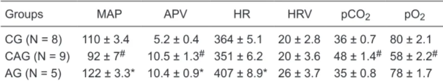
Table 1. Mean arterial pressure (MAP), arterial pressure variability (APV), heart rate (HR), heart rate variability (HRV), and pCO2 and pO2 concentrations in the control
group (CG), carotid group (CAG) and aortic group (AG).
Groups MAP APV HR HRV pCO2 pO2
CG (N = 8) 110 ± 3.4 5.2 ± 0.4 364 ± 5.1 20 ± 2.8 36 ± 0.7 80 ± 2.1 CAG (N = 9) 92 ± 7# 10.5 ± 1.3# 351 ± 6.2 20 ± 3.6 48 ± 1.4# 58 ± 2.2#
AG (N = 5) 122 ± 3.3* 10.4 ± 0.9* 407 ± 8.9* 26 ± 3.7 35 ± 0.8 78 ± 1.7
#P ≤ 0.001 for the CAG group versus the CG group; *P ≤ 0.001 for the AG group versus
and HR, the animals were successively injected with 2, 4, and 8 µg/kg serotonin (5-HT; Sigma, USA) while MAP and HR were recorded. For data analysis, control and peak changes of MAP and HR after each dose were analyzed with a microcomputer (IBM-AT/CODAS, Brazil). Injections were not repeated until the recorded parameters had returned to pre-injection levels. The tests were repeated before and after pharmacological blockade of the parasympathetic nervous system.
Pharmacological blockade
Pharmacological blockade was performed in the same animals on 2 consecutive days.
On the 1st day of the study, immediately after the resting HR was recorded, 4 mg/kg atropine (Sigma), a cholinergic receptor antagonist, was injected intravenously (iv). After atropine injection, AP and HR were recorded for 15 min
to permit the antagonists to exert their full effects and the
Bezold-Jarish test was then repeated. Propranolol (4 mg/ kg; Sigma), a beta-adrenergic receptor antagonist, was administered iv after the Bezold-Jarish test, and the AP and HR responses were recorded for 5 min. The intrinsic heart rate (IHR) was measured after simultaneous blockade with propranolol and atropine. On the 2nd day of the study,
propranolol was administered first to obtain the inverse
sequence of blockade.
The vagal effect was the difference between the maxi -mum HR after atropine injection and the control HR, on the 1st day. The sympathetic effect was the difference between the control HR and minimum HR after propranolol injec-tion on the 2nd day. The vagal tone was calculated as the difference between the IHR and the HR after propranolol injection on the 2nd day. The sympathetic tone was the difference between the HR after atropine injection and the IHR, on the 1st day.
Statistical analysis for the comparison of MAP, APV, HR, HR variability, pCO2, pO2, IHR, vagal, and sympathic
effects was performed by one-way ANOVA followed by
the Newman-Keuls test. The responses of the reflex after
successive injections of serotonin were also compared by two-way ANOVA followed by the Newman-Keuls test. The results are reported as means ± SEM.
Results
Aortic denervation increased MAP while carotid denerva-tion decreased it. The APV of CAG and AG was increased compared with CG (P = 0.001 and P = 0.0005, respectively). The HR of the AG was increased when compared with CG (P = 0.001). CAG rats presented a reduction of pO2 (P =
0.0001) accompanied by an increase of pCO2 (P = 0.0001)
compared with CG animals (Table 1).
The IHR obtained after pharmacological blockage was lower in CAG than in CG (344 ± 8.0 vs 401 ± 6.9 bpm; P = 0.0003), and the vagal effect was decreased in AG (P = 0.009) and CAG (P = 0.03) compared with CG (AG = 50 ± 11.2, CAG = 58 ± 8.3 vs CG = 114 ± 13 bpm). The sympathetic effect was higher in AG compared with CG (83 ± 2.1 vs 41 ± 5 bpm; P = 0.0002; Figure 1A). Vagal and sympathetic tonus was lower in CAG when compared with CG (P = 0.04), as shown in Figure 1B.
As expected, serotonin induced significant bradycardia
and hypotension. The bradycardic response was higher in AG than CG (275 ± 15 vs 163 ± 17 bpm, respectively; P = 0.001; Figure 2B).
The hypotensive response to serotonin (decreased MAP) was increased in AG (P = 0.0001) and CAG (P = 0.0008) compared with CG (AG = 46 ± 5 and CAG = 36 ± 4 vs CG = 19 ± 2 mmHg, respectively; Figure 2A).
The hypotensive response after atropine administration was higher in AG (P = 0.0001) than in CG (AG = 11 ± 2 vs
CG = 4.5 ± 0.7 mmHg) and the bradycardic response did not differ between groups (Figure 2A,B).
684 T.L. Fernandes et al.
Discussion
Clinical and experimental studies have demonstrated the
role of cardiopulmonary receptors in normal and pathophysi-ological conditions, showing the importance of these re-ceptors in cardiovascular control (9,11,13-16). In fact, the
Bezold-Jarish reflex obtained by serotonin administration has been evaluated in several experimental studies and
was found to be impaired in spontaneously hypertensive rats (17) and in renal hypertensive rats (18). In contrast, this
reflex is enhanced in hypertensive rats by inhibition of nitric oxide synthase (19) and after acute myocardial infarction
(16) and is still preserved in diabetic rats (11). These
stud-ies have confirmed the idea that the neural reflex control of circulation depends not only on baroreflex input but also on cardiopulmonary reflexes (9,13,14).
The interplay between arterial and cardiopulmonary
baroreflexes has been demonstrated in several experi -mental studies (10,12,17,20). Tonic inhibition of both
ca-rotid chemoreflex and arterial baroreflex is produced by
cardiopulmonary vagal afferent activity (21). However,
the role of the cardiopulmonary reflex in the control of the circulation in the absence of baroreflex control after SAD
is not completely understood. In dogs, it was suggested that cardiopulmonary afferents could be counteracting the increase in sympathetic activity after removal of arterial baroreceptors (22,23). In addition, Chianca Júnior and
Machado (12) showed a significant increase in the sensi
-tivity of the Bezold-Jarish reflex in both acute and chronic
sinoaortic denervated rats. However, there is no information about the separate participation of the aortic baroreceptors or of the carotid baroreceptors in Bezold-Jarish sensitivity. Thus, in the present study, we evaluated the sensitivity of
the Bezold-Jarish reflex after acute (24 h) selective aortic
or carotid denervation.
In the present study, we have confirmed that the animals
submitted to aortic denervation had increased MAP and HR while animals submitted to carotid denervation had a reduction in MAP with no changes in HR, as previously demonstrated (24-30). Moreover, APV was higher in both groups of animals submitted to denervation when compared
with intact controls.Studies have shown that sinoaortic den-ervated rats had hypertension, tachycardia, and increased APV in the acute phase, whereas in the chronic phase HR and AP returned to normal and APV remained increased (4). Regarding the arterial blood gases, CAG animals
showed significantly reduced arterial pO2 and significantly
increased pCO2. Huckstorf et al. (31) and Franchini and
Krieger (30) obtained similar data when comparing carotid body-denervated and sham-operated control rats.
The stimulation of the cardiopulmonary receptors by the injection of serotonin usually induces bradycardic and hypotensive responses (Bezold-Jarish effect) (10-12). In
the present experiment, these responses were increased
in AG compared with CG, suggesting that the sensitivity
of the cardiopulmonary reflex is augmented in AG. Para -sympathetic blockade with atropine caused a marked reduction in the bradycardic and hypotensive response
induced by serotonin in all groups, confirming data from
Chianca Júnior and Machado (32), which showed that the
hypotensive response induced by the Bezold-Jarish reflex
was due to both sympathetic withdrawal and decreased cardiac output produced by vagal-induced bradycardia. However, the hypotensive response was still augmented in AG when compared with CG. The higher hypotensive response in AG after atropine blockade suggests an intense withdrawal of peripheral sympathetic activity in this group
after serotonin stimulation. Indeed, this finding is consistent
with studies that demonstrated an increase in sympathetic activity in animals with aortic denervation (33), as well as a reduced baroreceptor function of carotid baroreceptors, as previously described in rats (34).
Our data indicate that atropine administration abolished serotonin-induced bradycardia responses while hypotensive responses were maintained. Although the responses after atropine were lower than the responses before administra-tion of the drug, the blockade indicated that both denervaadministra-tion procedures are associated with an increase in sympathetic modulation of the vessels. Indeed, after atropine block-ade all groups showed reduced hypotensive responses, although the AG response was more marked than that of
the other groups. This finding suggests an overactivity of
the sympathetic system in this group. On the other hand, hypotension in CG and CAG animals was predominantly
due to parasympathetic influence on the heart.
The chemoreflex induced by activation from peripheral
chemoreceptors located in the aortic and carotid bodies
has been extensively studied as one of the neural controls
of blood pressure regulation (35-37). These
chemosensi-tive cells are excited by decreases in pO2 or increases in
pCO2 or pH that will stimulate the terminal innervations of
the carotid sinus nerve (38). Our data showed a marked reduction in IHR associated with a decrease in vagal func-tion after selective carotid denervafunc-tion. These results could
be explained at least in part by the hypoxia observed after
carotid denervation, since these animals showed a decrease in pO2 and pH simultaneously with an increase in pCO2.
The relationship between hypoxia and the autonomic
nervous system has been demonstrated in several animal studies, which have evaluated the cardiovascular autonomic
responses to hypoxia. In fact, recently, Sugimura et al. (39),
using spectral analysis methods, have demonstrated that
acute progressive hypoxia inhibits cardiac parasympathetic
activity in hypertensive rats. Therefore, a possible mecha-nism for the alterations observed in cardiac parasympathetic
control in CAG could be related to hypoxia itself (40).
In conclusion, our data showed that both carotid and aortic baroreceptors selectively modulated the sensitivity of the cardiopulmonary receptors. This modulation was associated with the effects of chemo- and baroreceptor
fiber activities coming from the carotid region or with tonic
inhibition of sympathetic activity depending on aortic nerve input to the central nervous system.
Acknowledgments
Research supported by FAPESP (#99/09959-0).
References
1. Krieger EM. Neurogenic hypertension in the rat. Circ Res
1964; 15: 511-521.
2. Cowley AW Jr, Liard JF, Guyton AC. Role of baroreceptor
reflex in daily control of arterial blood pressure and other
variables in dogs. Circ Res 1973; 32: 564-576.
3. Jacob HJ, Brown DM, Bunker RK, Daly MJ, Dzau VJ, Good-man A, et al. A genetic linkage map of the laboratory rat,
Rattus norvegicus. Nat Genet 1995; 9: 63-69.
4. Soares PP, Porto CS, Abdalla FM, De La Fuente RN, Moreira ED, Krieger EM, et al. Effects of rat sinoaortic denervation
on the vagal responsiveness and expression of muscarinic
acetylcholine receptors. J Cardiovasc Pharmacol 2006; 47: 331-336.
5. Floras JS, Jones JV, Hassan MO, Sleight P. Effects of acute
and chronic beta-adrenoceptor blockade on baroreflex sen -sitivity in humans. J Auton Nerv Syst 1988; 25: 87-94. 6. Eckberg DL, Sleight P. Human baroreflex in health and dis
-ease. Oxford: Clarendon Press; 1992.
7. La Rovere MT, Bigger JT Jr, Marcus FI, Mortara A, Schwartz
PJ. Baroreflex sensitivity and heart-rate variability in predic -tion of total cardiac mortality after myocardial infarc-tion.
ATRAMI (Autonomic Tone and Reflexes After Myocardial
Infarction) Investigators. Lancet 1998; 351: 478-484. 8. Schwartz PJ, La Rovere MT. ATRAMI: a mark in the quest
for the prognostic value of autonomic markers. Autonomic
Tone and Reflexes After Myocardial Infarction. Eur Heart J
1998; 19: 1593-1595.
9. Zanchetti A, Mancia G. Cardiovascular reflexes and hyper -tension. Hypertension 1991; 18: III-13-III-21.
10. Thoren P. Role of cardiac vagal C-fibers in cardiovascular
control. Rev Physiol Biochem Pharmacol 1979; 86: 1-94. 11. Oliveira VL, Moreira ED, Farah VD, Consolim-Colombo F,
Krieger EM, Irigoyen MC. Cardiopulmonary reflex impair
-ment in experi-mental diabetes in rats. Hypertension 1999; 34: 813-817.
12. Chianca Júnior DA, Machado BH. The sensitivity of the
Bezold-Jarisch reflex is increased in rats with sinoaortic
deafferentation. Braz J Med Biol Res 1994; 27: 775-781.
13. Mark AL. The Bezold-Jarisch reflex revisited: clinical implica
-tions of inhibitory reflexes originating in the heart. J Am Coll Cardiol 1983; 1: 90-102.
14. Mark AL, Kerber RE. Augmentation of cardiopulmonary
baroreflex control of forearm vascular resistance in border -line hypertension. Hypertension 1982; 4: 39-46.
15. Meyrelles SS, Cabral AM, Vasquez EC. Impairment of the
Bezold-Jarisch reflex in conscious rats with myocardial hy -pertrophy. Braz J Med Biol Res 1994; 27: 1065-1069. 16. Lacerda JE, Consolim-Colombo FM, Moreira ED, Ida F, Silva
GJ, Irigoyen MC, et al. Influence of cardiopulmonary reflex
on the sympathetic activity during myocardial infarction.
Auton Neurosci 2007; 133: 128-135.
17. Verberne AJ, Guyenet PG. Medullary pathway of the
Bezold-Jarisch reflex in the rat. Am J Physiol 1992; 263: R1195-R1202.
18. Thames MD, Johnson LN. Impaired cardiopulmonary
baroreflex control of renal nerves in renal hypertension. Circ Res 1985; 57: 741-747.
19. Araujo MT, Cabral AM, Vasquez EC. Exaggerated Bezold-Jarisch reflex in the hypertension induced by inhibition of nitric oxide synthesis. Braz J Med Biol Res 1995; 28: 1009-1012.
20. Bishop VS, Hasser EM. Arterial and cardiopulmonary
re-flexes in the regulation of the neurohumoral drive to the
circulation. Fed Proc 1985; 44: 2377-2381.
21. Koike H, Mark AL, Heistad DD, Schmid PG. Influence of
cardiopulmonary vagal afferent activity on carotid
chemore-ceptor and barorechemore-ceptor reflexes in the dog. Circ Res 1975; 37: 422-429.
22. Persson P, Ehmke H, Kirchheim H, Seller H. Effect of sino-aortic denervation in comparison to cardiopulmonary deaf-ferentiation on long-term blood pressure in conscious dogs.
Pflugers Arch 1988; 411: 160-166.
686 T.L. Fernandes et al.
dogs. Acta Physiol Scand 1991; 142: 221-228.
24. Bedran de Castro MT, Moreira ED, Krieger EM. Reflex and
central components of carotid occlusion in conscious rats: effect of lesion of the medial forebrain bundle. Hypertension
1986; 8: 47-51.
25. Krieger EM. The acute phase of neurogenic hypertension in the rat. Experientia 1970; 26: 628-629.
26. Fink GD, Bryan WJ, Mann M, Osborn J, Werber A. Con-tinuous blood pressure measurement in rats with aortic baroreceptor deafferentation. Am J Physiol 1981; 241: H268-H272.
27. Patel KP, Ciriello J, Kline RL. Noradrenergic mechanisms in brain and peripheral organs after aortic nerve transection.
Am J Physiol 1981; 240: H481-H486.
28. Zhang TX, Ciriello J. Effect of paraventricular nucleus lesions on arterial pressure and heart rate after aortic baroreceptor denervation in the rat. Brain Res 1985; 341: 101-109. 29. Zhang TX, Ciriello J. Kainic acid lesions of paraventricular
nucleus neurons reverse the elevated arterial pressure after aortic baroreceptor denervation in the rat. Brain Res 1985; 358: 334-338.
30. Franchini KG, Krieger EM. Carotid chemoreceptors influ -ence arterial pressure in intact and aortic-denervated rats.
Am J Physiol 1992; 262: R677-R683.
31. Huckstorf C, Behm R, Habeck JO, Ruckborn K, Franz U. Blood pressure, heart rate and arterial blood gas reactions
to acute hypoxia in carotid body denervated spontaneously
hypertensive rats. Biomed Biochim Acta 1987; 46: 925-931.
32. Chianca DA Jr, Machado BH. Microinjection of NMDA an-tagonist into the NTS of conscious rats blocks the
Bezold-Jarisch reflex. Brain Res 1996; 718: 185-188.
33. Irigoyen MC, Moreira ED, Ida F, Pires M, Cestari IA, Krieger EM. Changes of renal sympathetic activity in acute and chronic conscious sinoaortic denervated rats. Hypertension
1995; 26: 1111-1116.
34. Franchini KG, Krieger EM. Neurogenic hypertension in the rat. In: Ganten D, de Jong W (Editors), Handbook of
hyper-tension: experimental and genetic models of hypertension. Amsterdam: Elsevier Science Publishers B.V.; 1994. p 482-500.
35. Marshall JM. Analysis of cardiovascular responses evoked following changes in peripheral chemoreceptor activity in the rat. J Physiol 1987; 394: 393-414.
36. Hayward LF, Felder RB. Peripheral chemoreceptor inputs to the parabrachial nucleus of the rat. Am J Physiol 1995; 268: R707-R714.
37. Vardhan A, Kachroo A, Sapru HN. Excitatory amino acid
receptors in the nucleus tractus solitarius mediate the re-sponses to the stimulation of cardio-pulmonary vagal
affer-ent C fiber endings. Brain Res 1993; 618: 23-31.
38. Gonzalez C, Almaraz L, Obeso A, Rigual R. Oxygen and acid
chemoreception in the carotid body chemoreceptors. Trends Neurosci 1992; 15: 146-153.
39. Sugimura M, Hirose Y, Hanamoto H, Okada K, Boku A,
Morimoto Y, et al. Influence of acute progressive hypoxia
on cardiovascular variability in conscious spontaneously hypertensive rats. Auton Neurosci 2008; 141: 94-103. 40. Ziegler MG, Nelesen RA, Mills PJ, Ancoli-Israel S, Clausen
JL, Watkins L, et al. The effect of hypoxia on baroreflexes
and pressor sensitivity in sleep apnea and hypertension.