Effects of propofol on damage of rat
intestinal epithelial cells induced by heat
stress and lipopolysaccharides
J. Tang
1*, Y. Jiang
1*, Y. Tang
2, B. Chen
2, X. Sun
3, L. Su
2and Z. Liu
21Department of Anesthesia, Nanfang Hospital, Southern Medical University, Guangzhou, China 2Department of Intensive Care Unit, Guangzhou General Hospital of Guangzhou Military Command, Guangzhou, China 3Laboratory of Traditional Chinese Medicine Syndrome, School of Traditional Chinese Medicine,
Southern Medical University, Guangzhou, China
Abstract
Gut-derived endotoxin and pathogenic bacteria have been proposed as important causative factors of morbidity and death during heat stroke. However, it is still unclear what kind of damage is induced by heat stress. In this study, the rat intestinal epithelial cell line (IEC-6) was treated with heat stress or a combination of heat stress and lipopolysaccharide (LPS). In addition, propofol, which plays an important role in anti-inflammation and organ protection, was applied to study its effects on cellular viability and apoptosis. Heat stress, LPS, or heat stress combined with LPS stimulation can all cause intestinal epithelial cell damage, including early apoptosis and subsequent necrosis. However, propofol can alleviate injuries caused by heat stress, LPS, or the combination of heat stress and LPS. Interestingly, propofol can only mitigate LPS-induced intestinal epithelial cell apoptosis, and has no protective role in heat-stress-induced apoptosis. This study developed a model that can mimic the intestinal heat stress environment. It demonstrates the effects on intestinal epithelial cell damage, and indicated that propofol could be used as a therapeutic drug for the treatment of heat-stress-induced intestinal injuries.
Key words: Propofol; Heat stress; Lipopolysaccharides; Apoptosis; Intestinal epithelial cells
Introduction
Heat stroke is a life-threatening condition in which the core body temperature rises rapidly to 406C or higher and the body’s heat-regulating mechanism breaks down. Moreover, heat stroke causes abnormalities in the central nervous system such as delirium, convulsions, and coma after exposure to a high ambient temperature (classic or non-exertional heat stroke) or strenuous exercise (exer-tional heat stroke). Current knowledge on heat stroke suggests that the pathophysiological consequence of heat stroke may not be due to an immediate effect of exposure to heat but may be a result of the systemic inflammatory response that follows thermal injury. Despite adequate lowering of the body temperature and intensive care support, heat stroke can often lead to multiple organ dysfunction syndrome (MODS) and death in these patients (1-3).
The gut contains a large number of the bacteria and endotoxins or lipopolysaccharides (LPS) in the body.
Gut-derived endotoxin and pathogenic bacteria have been proposed as important causative factors of morbidity and death during clinically relevant stresses such as heat stroke (4,5), sepsis (6), burn injury (7,8), and ischemia-reperfusion injury (9), as well as in critically ill patients (10). A number of studies on pathological examination of the small intestine reveal that lesions in this organ are common, and endotoxemia is present during heat stroke (11-15).
Intestinal epithelial cells are key components of the intestinal mucosal barrier. Damages to these cells will increase the permeability of the intestinal mucosal barrier, leading to increased gut-derived bacterial endotoxin translocation. In the process of heat stress or severe heat stroke, intestinal epithelial cells are attacked by environmental heat. The cells are also stimulated by intestinal bacteria and bacterial LPS. However, the type of damage induced by these stimuli on intestinal epithelial
Correspondence: Z. Liu, Department of Intensive Care Unit, Guangzhou General Hospital of Guangzhou Military Command, Guangzhou, China. Fax: ++86-20-3665-5909. E-mail: serpentking@sina.com
*These authors contributed equally to this study.
cells is still unclear. The first objective of the present study was to detect damage characteristics of intestinal epithelial cells under the combined stimuli of heat stress and LPS.
Propofol is an intravenous general anesthetic drug and is widely used in the induction and maintenance of anesthesia and sedation in intensive care units and for patients who need mechanical ventilation. Recent studies have shown that, in addition to the anesthetic effects, propofol also plays an important role in anti-inflammation and organ protection. For example, propofol can reduce the production of proinflammatory cytokines, alter the production of nitric oxide (16), and inhibit neutrophil function, leading to alleviation of the inflammatory response (17). Propofol can also regulate platelet aggregation (18,19) and activate endogenous organ protection against hypoxic and ischemic injury (20). Our previous studies suggest that the intestinal inflammatory reaction plays an important role in intestinal injury during animal heat stroke (15). In view of the anti-inflammatory and organ-protective functions of propofol, the second purpose of this study was to detect the probable protective role of propofol in heat-stress-induced intestinal epithelial cell injury.
In this study, we developed an in vitro micro-environment for heat-stressed intestinal epithelial cells and investigated the injuries caused by heat stress and LPS. We also studied the effects of propofol on cellular injuries. These results would help to further understand the mechanisms of endotoxin translocation caused by intestinal barrier dysfunction during intestinal heat stress and provide potential therapy.
Material and Methods
Treatment of cells with heat stress, LPS, and propofol
Rat IEC-6 cells (provided by Southern Medical University, China) were cultured in Dulbecco’s modified Eagle’s medium (DMEM) containing 5% fetal bovine serum. Cells were passaged using ethylenediaminetetraacetic acid (EDTA)-trypsin to reach 80-90% confluence. After two passages, the cells were divided into the following groups: control, heat stress (HS), LPS (Sigma, USA) treatment, propofol (Sigma) treatment (P), HS++LPS, HS++P, LPS++P, and HS++LPS++P.
For heat stress, the medium of cells at 70% confluence was replaced with serum-free DMEM and incubated at 436C for 1 h. Subsequently, cells were incubated under normal conditions (376C and 5% CO2)
for 23 h. For LPS stimulation, cells were cultured in serum-free DMEM supplemented with 1mg/mL LPS for
24 h under normal conditions. For HS++LPS, 1mg/mL
LPS was added to the cells, followed by 1 h of heat stress at 436C and another 23 h of incubation under normal conditions. For propofol treatment, cells at 70% con-fluence were grown in serum-free DMEM supplemented
with 50mM propofol for 24 h under normal conditions. For
HS++P, cells were treated with 50mM propofol followed
by 1 h of heat stress at 436C and 23 h of incubation under normal conditions. For LPS++P, cells were treated with 1mg/mL LPS and 50mM propofol and incubated for 24 h
under normal conditions. For HS++LPS++P, cells were treated with 1mg/mL LPS and 50mM propofol and
incubated at 436C for 1 h followed by 23 h of incubation under normal conditions.
Detection of cell viability
The MTT [3-(4,5-dimethylthiazol-2-yl)-2,5-diphenylte-trazolium bromide] method was used to detect cell viability after different treatments. After removal of the medium, 150mL MTT (0.5 mg/L) was added to the cells and then
incubated for 4 h. Subsequently, the supernatant was removed and 150mL dimethyl sulfoxide added, with
shaking for 5 min, until the crystals were fully dissolved. Absorbance (A) at 490 nm was measured for each well, and cell viability was calculated using the following equation: cell viability=Atreatment/Acontrol.
Detection of apoptosis
Double staining with annexin V-fluorescein isothyo-cyanate (FITC) and propidium iodide (PI) was used to detect apoptosis. The cells in each group were washed with phosphate-buffered saline twice and detached with EDTA-free trypsin. Subsequently, cells were resuspended in 500mL binding buffer and stained with 1mL annexin
V-FITC and 1mL PI. Apoptosis was detected using flow
cytometry within 1 h in order to avoid false results. Apoptosis was calculated using the following equation: apoptosis rate=number of apoptotic cells/(number of apoptotic cells++number of living cells)6100%.
Statistical analysis
All data were analyzed using the SPSS 16.0 software (IBM, USA). Quantitative data were reported as means±SD. All comparisons were conducted after homogeneity of variance (P.0.05) was confirmed. Comparison between each group was performed using single factor analysis of variance (one-way ANOVA). Multiple comparisons among different groups were performed using a least-significant difference test or the Student-Newman-Keuls method. The Welch method was used for data with heterogeneity of variance, and the Dunnett T3 test was used for multiple comparisons. P,0.05 was considered to be statistically significant.
Results
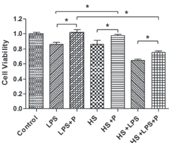
Effect of heat stress and LPS on cell viability
(P,0.05). There was no significant difference between the LPS and HS groups (P.0.05). These results indicated that either heat stress or LPS can cause a certain degree of damage in IEC-6 cells and a combination of these two treatments would produce a synergistic effect (Figure 1).
Effect of propofol on heat stress and combination of heat stress and LPS-induced reduction of cell viability
There was no significant difference between the P group and the control group (P=0.373) in cell viability. However, cell viability in the combination group was significantly higher than that in the HS group (P=0.021). These results indicate that propofol does not affect cell growth, but plays a protective role in heat-stress-induced damage. Moreover, cell viability in the LPS++P group was significantly higher than that in the LPS group (P,0.001). Cell viability in the HS++LPS++P group was significantly higher than that in the HS++LPS group (P,0.001). These results indicate that propofol can protect cell damage caused by the combination of heat stress and LPS stimulation (Figure 2).
Effect of heat stress and LPS on apoptosis
There were significant differences in apoptotic rates among the control, HS, LPS, and LPS++HS groups (P,0.001). Apoptotic rate in the LPS, HS, and HS++LPS groups was significantly higher than that in the control group (P,0.001). Apoptotic rate in the HS++LPS group was also higher than that of the LPS or HS groups (P,0.05), However, there was no significant difference between the LPS and HS groups. These results indicated that heat stress or LPS could increase apoptosis of IEC cells and the combination of these two treatments could produce a synergistic effect (Figure 3).
Effect of propofol on heat stress and combination of heat stress and LPS-induced apoptosis
Propofol treatment did not affect apoptotic rate. In addition, the apoptotic rate in the HS++P group was not significantly different from that in the HS group (P.0.05). These results indicated that propofol treatment may have no significant effect on heat-stress-induced apoptosis. Apoptotic rate in the LPS++P group was significantly lower than that in the LPS group (P=0.047). In addition, the
Figure 1. Effects of heat stress (HS) and lipopolysaccharide (LPS) on the cell viability of IEC-6 cells. Data are reported as means±SD, n=6. *P,0.05vscontrol, and#P
,0.05vsHS or LPS (one-way ANOVA).
Figure 2.Effects of propofol (P) on the cell viability of IEC-6 cells after heat stress (HS), lipopolysaccharide (LPS), or a combina-tion of HS and LPS treatment. The MTT [3-(4,5-dimethylthiazol-2-yl)-2,5-diphenyltetrazolium bromide] method was used for eva-luation. Data are reported as means±SD, n=6. *P,0.05, Student-Newman-Keuls test.
Figure 3. Effects of heat stress (HS) and lipopolysaccharide (LPS) on the apoptosis ratio of IEC-6 cells. Data are reported as means±SD, n=6. *P,0.01vscontrol,#P
apoptotic rate in the HS++LPS++P group was significantly lower than that in the HS++LPS group (P=0.011). These results indicate that treatment with propofol can reduce apoptosis caused by LPS or a combination of LPS and heat stress (Figure 4).
Discussion
The mechanism in heat-stress-induced multiple organ failure is not fully understood. The presence of endotoxemia and intestinal bleeding suggest that a change in intestinal epithelial permeability may play an important role in this process (21). Moseley et al. (22) suggested that most of the tight junctions in intestinal epithelial cells could open at 38.36C; and, when the temperature exceeded 41.56C, all the tight junctions would be opened, resulting in increased permeability to macromolecules. Early studies on heat stroke in animal models also showed that leakage of microorganism endotoxins from the intestine was increased after thermal damage in the intestinal mucosa (23). Lambert et al. (12) utilized the fluorescent molecule FITC-dextran to study the intestinal permeability of rats in different heat stress conditions, and histological analysis showed that the extent of the damage and permeability of the intestinal epithelia were associated with the degree of heat stress.
It could be predicted that, under heat-stress condi-tions, endotoxins, bacteria, and antibodies in the intestine were constantly translocated into the blood and lymph, which resulted in the release of various inflammatory
mediators and aggravation of the systemic inflammatory response syndrome (SIRS). Occurrence of SIRS further increased mucosal barrier damage, eventually leading to MODS (24). However, direct experimental evidence of epithelial cell damage in heat stress is not available.
In this study, we mimicked the micro-environment of intestinal epithelial cells under severe heat stress conditions and determined the cell viability and apoptotic rate. As predicted, heat stress or LPS stimulation could induce damage to intestinal epithelial cells (including early apoptosis and subsequent necrosis). In addition, a synergistic effect was produced when heat stress and LPS stimulation were combined. These results indicate that the unique environment during the process of intestinal heat stroke can induce multiple types of cell damage mediated by different molecules and signaling pathways. Our previous studies had shown that intestinal epithelial cells secreted significantly higher IL-8 under the condition of heat stress or LPS stimulation (25). However, further studies are needed to determine whether other cytokines or molecules are involved in this process.
Propofol plays an important role in anti-inflammation and organ protection. Thus, we determined the effect of propofol on heat-stress-induced cell injuries. Our results showed that propofol could alleviate the injuries caused by heat stress, LPS, or a combination of heat stress and LPS. There is additional evidence supporting the protec-tive role of propofol in intestinal injury. Liu et al. (26) found that propofol attenuates intestinal ischemia-reperfusion (I/R)-induced mucosal injury in an animal model. The response may be attributable to the antioxidant properties of propofol and the effects of inhibiting over-production of nitric oxide and in decreasing ET-1 levels (26). They also found that propofol pretreatment attenuates I/R-induced intestinal epithelial apoptosis (27). Sun et al. (28) found that propofol could inhibit traumatic brain injury that promoted a significant increment in plasma endotoxin, the L/M ratio, and intestinal levels of NF-kappa B, TNF-alpha, and IL-6 in animal models. Vasileiou et al. (29) also found that anesthesia with propofol alleviated intestinal injury and efficiently prevented lipid oxidation following intestinal ischemia-reperfusion in Wistar rats. Yagmurdur’s (30) results suggest that propofol as an anesthetic agent may prevent bacterial translocation by scavenging reactive oxygen species and inhibiting lipid peroxidation in an animal model of burn injury, and they also found that propofol could offer protection against apoptosis of enterocytes (31).
Interestingly, propofol can only inhibit LPS-induced intestinal apoptosis, but has no protective role in heat-stress-induced apoptosis. We speculated that the organ protective effect of propofol was mainly associated with anti-inflammation and reduction of oxidative stress injury, which are similar to LPS-induced cell damage and quite different from heat-stress-induced physical damage. In
Niederlechner’s (32) study on glutamine’s cytoprotective mechanism in heat-stressed intestinal epithelial cells, epidermal growth factor receptor and its associated signal transduction pathway molecules such as activated extra-cellular regulated protein kinases 1/2 and decreased p38 mitogen-activated protein kinases signaling may play an important role, and provide a basis for future study. In conclusion, this study developed a model that can mimic the intestinal heat stress environment, demonstrated its effects on intestinal epithelial cell damage, and indicated
that propofol could be used as a therapeutic drug for the treatment of heat-stress-induced intestinal injuries.
Acknowledgments
Research supported by grants from National Natural Science Foundation of China (#81071529,#81101467) and Guangdong Province Natural Science Foundation of China (#10151001002000001, #10451001002005047,
#S2012010008120).
References
1. Bouchama A, Knochel JP. Heat stroke.N Engl J Med2002; 346: 1978-1988, doi: 10.1056/NEJMra011089.
2. al-Mashhadani SA, Gader AG, al Harthi SS, Kangav D, Shaheen FA, Bogus F. The coagulopathy of heat stroke: alterations in coagulation and fibrinolysis in heat stroke patients during the pilgrimage (Haj) to Makkah. Blood Coagul Fibrinolysis 1994; 5: 731-736, doi: 10.1097/ 00001721-199410000-00009.
3. Dematte JE, O’Mara K, Buescher J, Whitney CG, Forsythe S, McNamee T, et al. Near-fatal heat stroke during the 1995 heat wave in Chicago.Ann Intern Med1998; 129: 173-181, doi: 10.7326/0003-4819-129-3-199808010-00001. 4. Gathiram P, Wells MT, Brock-Utne JG, Gaffin SL.
Antilipopolysaccharide improves survival in primates sub-jected to heat stroke.Circ Shock1987; 23: 157-164. 5. Gathiram P, Wells MT, Brock-Utne JG, Gaffin SL.
Prophylactic corticosteroid increases survival in experimen-tal heat stroke in primates.Aviat Space Environ Med1988; 59: 352-355.
6. Turnbull IR, Javadi P, Buchman TG, Hotchkiss RS, Karl IE, Coopersmith CM. Antibiotics improve survival in sepsis independent of injury severity but do not change mortality in mice with markedly elevated interleukin 6 levels.Shock2004; 21: 121-125, doi: 10.1097/01.shk.0000108399.56565.e7. 7. Gianotti L, Braga M, Vaiani R, Almondo F, Di Carlo V.
Experimental gut-derived endotoxaemia and bacteraemia are reduced by systemic administration of monoclonal anti-LPS antibodies. Burns 1996; 22: 120-124, doi: 10.1016/ 0305-4179(95)00100-X.
8. Sittig K, Deitch EA. Effect of bacteremia on mortality after thermal injury. Arch Surg 1988; 123: 1367-1370, doi: 10.1001/archsurg.1988.01400350081012.
9. Gathiram P, Wells MT, Brock-Utne JG, Wessels BC, Gaffin SL. Oral administered nonabsorbable antibiotics prevent endotoxemia in primates following intestinal ischemia.J Surg Res1988; 45: 187-193, doi: 10.1016/0022-4804(88)90064-9. 10. Marshall JC. The International Sepsis Forum’s controver-sies in sepsis: how will sepsis be treated in 2051?Crit Care 2002; 6: 465-467, doi: 10.1186/cc1539.
11. Hall DM, Buettner GR, Oberley LW, Xu L, Matthes RD, Gisolfi CV. Mechanisms of circulatory and intestinal barrier dysfunction during whole body hyperthermia.Am J Physiol Heart Circ Physiol2001; 280: H509-H521.
12. Lambert GP, Gisolfi CV, Berg DJ, Moseley PL, Oberley LW, Kregel KC. Selected contribution: hyperthermia-induced intestinal permeability and the role of oxidative and
nitrosative stress.J Appl Physiol2002; 92: 1750-1761. 13. Bouchama A, Roberts G, Al Mohanna F, El-Sayed R, Lach
B, Chollet-Martin S, et al. Inflammatory, hemostatic, and clinical changes in a baboon experimental model for heatstroke. J Appl Physiol 2005; 98: 697-705, doi: 10.1152/japplphysiol.00461.2004.
14. Liu Z, Liu JH, Liu Y, Tang Y, Meng F, Sun X, et al. Proteomic analysis and identification of intestinal FBP as a predictor of gut dysfunction during heatstroke in mice.J Surg Res2012; 173: 332-340, doi: 10.1016/j.jss.2010.09.043.
15. Liu Z, Sun X, Tang J, Tang Y, Tong H, Wen Q, et al. Intestinal inflammation and tissue injury in response to heat stress and cooling treatment in mice.Mol Med Rep2011; 4: 437-443, doi: 10.3892/mmr.2011.435.
16. Gonzalez-Correa JA, Cruz-Andreotti E, Arrebola MM, Lopez-Villodres JA, Jodar M, De La Cruz JP. Effects of propofol on the leukocyte nitric oxide pathway:in vitroand ex vivostudies in surgical patients.Naunyn Schmiedebergs Arch Pharmacol2008; 376: 331-339, doi: 10.1007/s00210-007-0220-4.
17. Tang J, Chen X, Tu W, Guo Y, Zhao Z, Xue Q, et al. Propofol inhibits the activation of p38 through up-regulating the expression of annexin A1 to exert its anti-inflammation effect.PLoS One2011; 6: e27890, doi: 10.1371/journal.-pone.0027890.
18. Aoki H, Mizobe T, Nozuchi S, Hiramatsu N.In vivoandin vitrostudies of the inhibitory effect of propofol on human platelet aggregation. Anesthesiology 1998; 88: 362-370, doi: 10.1097/00000542-199802000-00015.
19. Tang J, Sun Y, Wu WK, Zhong T, Liu Y, Xiao J, et al. Propofol lowers serum PF4 level and partially corrects hypercoagulo-pathy in endotoxemic rats.Biochim Biophys Acta2010; 1804: 1895-1901, doi: 10.1016/j.bbapap.2010.06.018.
20. Yamada H, Kawano T, Tanaka K, Yasui S, Mawatari K, Takahashi A, et al. Effects of intracellular MgADP and acidification on the inhibition of cardiac sarcolemmal ATP-sensitive potassium channels by propofol.J Anesth2007; 21: 472-479, doi: 10.1007/s00540-007-0551-9.
21. Lambert GP. Role of gastrointestinal permeability in exer-tional heatstroke.Exerc Sport Sci Rev2004; 32: 185-190, doi: 10.1097/00003677-200410000-00011.
22. Moseley PL, Gapen C, Wallen ES, Walter ME, Peterson MW. Thermal stress induces epithelial permeability.Am J Physiol1994; 267: C425-C434.
heat-stressed primates.Aviat Space Environ Med1987; 58: 1071-1074.
24. Zhou DY. Bacterial and endotoxin translocation: prevention and treatment.Chin J Gastro2003; 8: 3-5.
25. Tang LQ, Yuan FF, Liu YW, Xu QL, Su L, Liu ZF. Effects of lipopolysaccharide combined with heat stress on the expression and secretion of cytokines released from human intestinal epithelial cells SW480.Infect Inflam Repair2012; 13: 6-9.
26. Liu KX, Rinne T, He W, Wang F, Xia Z. Propofol attenuates intestinal mucosa injury induced by intestinal ischemia-reperfusion in the rat.Can J Anaesth2007; 54: 366-374, doi: 10.1007/BF03022659.
27. Liu KX, Chen SQ, Huang WQ, Li YS, Irwin MG, Xia Z. Propofol pretreatment reduces ceramide production and attenuates intestinal mucosal apoptosis induced by intest-inal ischemia/reperfusion in rats.Anesth Analg2008; 107: 1884-1891, doi: 10.1213/ane.0b013e3181884bbf.
28. Sun J, Wang L, Shen J, Wang Z, Qian Y. Effect of propofol on mucous permeability and inflammatory mediators
expression in the intestine following traumatic brain injury in rats. Cytokine 2007; 40: 151-156, doi: 10.1016/ j.cyto.2007.09.003.
29. Vasileiou I, Kalimeris K, Nomikos T, Xanthopoulou MN, Perrea D, Agrogiannis G, et al. Propofol prevents lung injury following intestinal ischemia-reperfusion.J Surg Res2012; 172: 146-152, doi: 10.1016/j.jss.2010.07.034.
30. Yagmurdur H, Akca G, Aksoy M, Arslan M, Baltaci B, Dikmen B. The effects of ketamine and propofol on bacterial transloca-tion in rats after burn injury.Acta Anaesthesiol Scand2005; 49: 177-182, doi: 10.1111/j.1399-6576.2004.00560.x.