CASE SELECTION AND OUTCOME OF RADICAL PERINEAL
PROSTATECTOMY IN LOCALIZED PROSTATE CANCER
JEFFREY M. HOLZBEIERLEIN
(1), PETER LANGENSTROER
(2), H.J. PORTER II
(1),
J. BRANTLEY THRASHER
(1)Section of Urology, University of Kansas Medical Center (1), Kansas City, Kansas,USA,
and Division of Urology, Medical College of Wisconsin (2), Milwaukee, Wisconsin, USA
ABSTRACT
Radical prostatectomy continues to play a central role in the management of localized pros-tate cancer. The majority of patients diagnosed with prospros-tate cancer will undergo radical prospros-tatec- prostatec-tomy. A decrease in the morbidity of this surgical procedure has been accomplished through an im-proved understanding of pelvic anatomy and a greater understanding of the natural history of prostate cancer. Recently, minimally invasive techniques have been applied to radical prostatectomy (laparoscopic prostatectomy) in order to further decrease the morbidity of this operation. What re-mains to be determined is whether this approach confers the same long term surgical outcomes as the open approach. One method which offers known long term outcomes coupled with decreased morbid-ity is the radical perineal prostatectomy. The purpose of this paper is to review the criteria for patient selection as well as outcomes of the radical perineal prostatectomy.
Key words: prostatic neoplasms; prostatectomy; perineal; outcomes; surgical technique
Int Braz J Urol. 2003; 29: 291-9
INTRODUCTION
Perineal prostatectomy is the oldest means of prostate resection and has its origins from the perineal lithotomy which was first described in 400 BC (1). In 25 AD, Celsus developed a curved perineal incision which would eventually become the basis for the incision used in the perineal prostatectomy today (1). Covillard is credited with performing the first removal of a portion of the prostate during re-moval of a bladder stone through the perineum in 1639, although he and other surgeons, at the time, used a median incision in the perineum rather than the curved incision described by Celsus (2). Through-out the 18th and 19th centuries, several surgeons
re-ported the removal of portions of the prostate similar to Covillard; however, the first planned prostate enucleation through a median perineal incision was performed by Guthrie in 1834 (2). This subsequently led to the use of the median perineal incision for the
removal of prostatic carcinoma. In 1866, Kuchler was the first to suggest that the entire prostate could be removed using this approach, but it was Billroth, in 1867, who first described the perineal prostatectomy for the treatment of prostate cancer in a professional journal (3).
Figure 1 – Dr. Young’s original depiction of prostatic tractor used for enucelation of the hypertrophied lobes of the prostate
Figure 2 – The Young retractor which is still used today for the
seminal vesicles, and that the cancer was usually con-tained within Denovillier’s fascia (4). During this same time period, Dr. Halsted was performing the radical mastectomy for the treatment of breast can-cer. Together they developed a radical operation to remove the prostate, the fascia of Denovillier, the seminal vesicles, ampullae of the vasa, and the vesi-cal neck with a portion of the trigone, and thus per-formed the first “radical” perineal prostatectomy in 1904 (5). This radical perineal prostatectomy has re-mained virtually unchanged in regards to technique since it was first described by Dr. Young.
Minor modifications of Young’s original pro-cedure have been made in order to reduce the mor-bidity of the operation. First, after the development of urinary calculi on the silk sutures used for the vesicourethral anastomosis, Dr. Young began using chromic catgut rather than silk (5). Next, Dr. Hans Wildbolz described a technique to preserve the tis-sue surrounding the external urinary sphincter to re-duce the incidence of urinary incontinence (1). Also, prior to 1928, gauze pads were routinely packed into the perineal wound with a portion of the pad exposed for later removal. In 1928, Gibson recommended that these pads be omitted during closure. This modifica-tion significantly decreased wound problems as well as fistula formation (6). Another significant contri-bution was introduced by Dr. Elmer Belt in 1939. Dr. Belt described a new approach to the prostate through the perineum between the longitudinal fibers of the rectum and the circular fibers of the external anal sphincter (7). This approach dramatically decreased blood loss. However, Dr Belt also recommended leav-ing behind the apex of the prostate to achieve better urinary control, and opening the anterior layer of Denonvillier’s fascia during the dissection. Dr. Young considered these last 2 changes in violation of the principals of cancer surgery and discouraged their use in radical perineal prostatectomy (RPP) (5).
In 1945, the development of the retropubic approach for the removal of the benign prostate would soon lead to the use of the radical retropubic pros-tatectomy for the treatment of prostate cancer (8,9). However, the procedure was soon abandoned due to the adoption of radiation therapy for prostate cancer, as it was thought to have less morbidity. Through the
1960’s and early 1970’s, literature began to accumu-late on the morbidity associated with radiation, but it continued to play a significant role in the treatment of prostate cancer due to the significant morbidity, especially blood loss, associated with radical pros-tatectomy. Finally, in 1979 Reiner & Walsh reported early meticulous ligation of the dorsal vein during the radical retropubic approach which greatly de-creased the blood loss associated with the procedure (10). In addition, Walsh et al., after performing de-tailed anatomical dissections in the male pelvis, pub-lished the first description of the nerve-sparing radi-cal retropubic prostatectomy leading to wide accep-tance of this procedure for the treatment of prostate cancer (11).
In recent years there has been renewed inter-est in the radical perineal prostatectomy technique for a number of reasons. First, the research of Weldon & Tavel in the late 1980’s demonstrated that nerve-sparing techniques could be also be applied to the perineal approach (12). Second, with predictive mod-els such as the Partin tables and the Kattan nomo-gram, patients at low risk for pelvic lymph node me-tastases can be identified, thus allowing for the safe exclusion of a pelvic lymph node dissection (13). Fi-nally, with the advent of minimally invasive tech-niques and a focus on decreasing the morbidity of radical prostatectomy, perineal prostatectomy has had resurgence. In addition, as opposed to laparoscopy, the perineal prostatectomy has long-term data on out-comes available (14).
PATIENT SELECTION
retropubic approach (15). The only significant com-plication particular to learning the perineal approach was that of rectal injury. However, all of these rectal injuries were closed primarily at the time of RPP and resulted in no long term sequelae. This study contra-dicts the commonly held belief that the perineal ap-proach is more difficult to teach and learn.
EXTENT OF DISEASE
Any form of prostatectomy, whether it is laparoscopic, radical retropubic, or radical perineal is curative only if all of the cancer can be removed during the procedure. In the RPP approach it is im-perative that patients have organ confined disease in order for the procedure to be curative. This includes patients with clinical stages T1b, T1c, or T2 disease diagnosed by digital rectal examination. Furthermore, using predictive models such as the Kattan nomogram may help exclude patients who are at high risk for extra-capsular disease (13). For example, a patient who has a clinical stage T1c cancer, but a PSA of 12 and Gleason score of 9 has a high chance of extra-capsular disease and may be best served by an alter-native form of treatment (13).
As patients who undergo RPP do not routinely have pelvic lymph nodes sampled, patients at high risk for nodal metastases are typically not candidates for this approach. Some surgeons have combined laparoscopic pelvic lymph node dissections with RPP for patients at greater risk for lymph node metastases. The drawback of this is of course the increased op-erative time as well as the expertise required to per-form laparoscopic lymph node dissection. As men-tioned previously, with the predictive models avail-able, patients with a low probability of lymph node metastases can be selected (16). Furthermore with the stage migration that has been seen in prostate cancer since the introduction of PSA, patients can be accu-rately selected to undergo RPP with the exclusion of a pelvic lymph node dissection (17).
PATIENT CHARACTERISTICS
There are practical considerations in regards to the patients who may or may not be candidates for
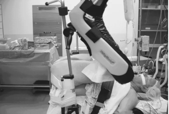
RPP. Patient size is one such consideration. Typically, obese patients have less subcutaneous fat on the perineum as compared to the lower abdominal area making RPP a better approach than the retropubic approach. However, if the patient is morbidly obese then the positioning required for RPP may pose a problem. Patients are placed in an exaggerated litho-tomy position in order to place the perineum in a po-sition which is essentially parallel to the floor (Fig-ure-4). In morbidly obese patients this may increase the ventilatory pressures to > 40 cm of H20 resulting in poor oxygenation and inability to perform the pro-cedure. A simple office test that demonstrates the patient’s ability to tolerate the exaggerated lithotomy position from a respiratory standpoint involves hav-ing the patient lie supine on the exam table and brhav-ing his knees to his chest. If the patient is able to tolerate this test, then he will likely tolerate the positioning required for RPP.
If the patient’s body habitus is such that the base of the prostate gland is not palpable on digital rectal examination this may make dissection during RPP very difficult due to the depth of the wound. Also, if the patient has a narrow distance between his ischial tuberosities such that the prostate gland is wider than this distance then perineal removal of the prostate is very difficult. As a general rule, prostate glands greater than 100 g are difficult to remove through the perineal approach. If this approach is to be used in large prostates, one many consider down-sizing of the prostate with an LH-RH agonist prior to prostatectomy. Other patient characteristics that may exclude them from the perineal approach are hip anky-losis, patients who have had lower extremity ampu-tations, and patients with hip prostheses. These are relative contraindications and should be individual-ized to each patient.
ADVANTAGES OF RPP
laparoscopic approach, as the perineal dissection is through virgin tissue. Furthermore, in patients who have had prior pelvic irradiation for their prostate cancer and undergo prostatectomy (salvage prostate-ctomy) the perineal approach has tended to be tech-nically advantageous as compared to the retropubic approach.
OUTCOMES
To date there has been no direct comparison of laparoscopic prostatectomy versus radical perineal prostatectomy. Most of the comparisons have been between perineal prostatectomy and the radical ret-ropubic approach, although there are only a few stud-ies which can be found directly comparing these ap-proaches. One of the first published reports directly comparing retropubic versus perineal prostatectomy was from Boxer et al. in 1977 (18). In this study of
329 patients, Boxer et al. examined several variables including mortality due to the procedure, overall sur-vival rates, incontinence, and long term complications. The authors found no significant differences between the two groups in the variables examined except for an increased blood loss of 700 ml in the retropubic group versus the perineal group. This study was a poor comparison for efficacy as many patients in the study had received estrogen therapy either pre or post operatively. In addition, only 20% of the patients had undergone pelvic lymphadenectomies leading to stag-ing inaccuracies and difficulties in comparstag-ing the true cancer control rates of the 2 techniques.
A more contemporary series is that by Frazier et al. who compared 122 patients who underwent RPP versus 51 patients who underwent radical retropubic prostatectomy (RRP) (19). Variables examined were operative times, blood loss, hospital stay, short and long-term complications (including incontinence and
impotence), length of catheter drainage, weight of the specimen, and disease extent. For the purposes of operative time, only those patients who underwent a pelvic lymphadenectomy in conjunction with RPP were included. The authors concluded that there were no statistically significant differences between the 2 groups in terms of positive margin rates, short-term or long-term complications, and urethral or bladder neck involvement. Seventeen of the 22 patients (77.3%) in the RPP group who underwent nerve spar-ing procedures were potent after surgery. Unfortu-nately, no data on the potency rates in the RRP group were available making a direct comparison impos-sible in this study. Again, the only significant differ-ences seen were in the estimated blood loss and trans-fusion requirements with both being significantly greater for the RRP group. Criticisms of the study include the lack of potency data in the RRP group, and the failure to match patients in the 2 groups by preoperative data. Furthermore, all RPP’s were formed by 1 surgeon while 3 different surgeons per-formed the RRP’s.
A smaller study by Haab et al. compared 71 patients who underwent either RRP (36 patients) or RPP (35 patients) for clinically localized cancer of the prostate (20). In this study, patients were matched by their preoperative data including PSA. Similar variables to the Frazier study were examined, includ-ing: operative time, number of blood transfusions, peri-operative complications, sexual and urinary func-tion, positive margin rates, and specimen weights. The only significant differences noted were in the trans-fusion requirements (100% RRP vs. 54% RPP) and anastomotic strictures (2 RRP and 0 RPP). The inci-dence of rectal injuries and wound infections was the same between the groups as was the incidence of positive margins, biochemical recurrence rates, and continence. The conclusions were that the 2 proce-dures provide similar disease control outcomes but with significantly less blood loss in the RPP group. This study brings to light one of the major criticisms of any study comparing RRP with RPP, which is the lack of a pelvic lymph node dissection in the RPP patients making true disease control outcomes diffi-cult to measure due to staging inaccuracies. However, with predictive nomograms patients can be accurately
selected in which the risk of node positivity is mini-mal. Therefore, this criticism should not preclude a meaningful and accurate comparison of the 2 proce-dures such as was performed in this study.
These trials indicated that margin positivity and biochemical failure rates are equivalent between the 2 procedures. However, a more recent article by Boccon-Gibod et al. compared the incidence of posi-tive surgical margins in patients undergoing RRP ver-sus RPP (21). Ninety-four patients (48 RRP and 46 RPP) with clinically localized prostate cancer were retrospectively reviewed. The patients were stratified according to clinical stage, extra-capsular extension with and without positive margins, and iatrogenic positive margins (incision into the prostate). The au-thors reported a 56% incidence of positive margins in the perineal group versus 61% in the retropubic group. Biochemical recurrence rates at a mean fol-low-up of 25 months were the same for each group (33%). What was surprising in this study was the in-cidence of positive margin rates in patients with pT2 tumors which was significantly higher in the RPP group (43% versus 29%, p < 0.05). In addition, the incidence of iatrogenic margins was dramatically higher in the RPP group (90%) versus the RRP group (37%) (p < 0.05). Their conclusions were that RRP is a better approach for the treatment of prostate can-cer, as it affords a lower likelihood of capsular inci-sion. Problems with these conclusions are that de-spite the reported incidence of positive margins bio-chemical recurrence rates were the same. Further-more, the RPP’s in this study were not performed by surgeons experienced in this technique. In other stud-ies utilizing data from surgeons with significant ex-perience in the RPP technique, positive margin rates and iatrogenic positive margin rates are similar to those reported for RRP (20).
statisti-cally significant differences between the groups for PSA failures, margin positivity, or organ confined rates. The only significant differences shown were a higher blood loss in the RRP group (p < 0.001) and a higher rectal injury rate in the RPP group (p < 0.03). There was no difference in the rates of incontinence, impotency, bladder neck contractures, or post-opera-tive complications. All of the aforementioned stud-ies are reviewed in Table-1.
COMPLICATIONS
Rectal injuries have been shown to occur more frequently in RPP than in RRP (22). Although,
Study
Boxer et al. 1977
Frazier et al. 1992
Haab et al. 1994
Boccon-Gibbod et al. 1998
Uniformed Services Urology Research Group 2001
Patient Population
329 patients
(265 RPP vs. 64 RRP)
173 patients
(122 RPP vs. 59 RRP)
71 patients
(35 RPP vs. 36 RRP)
94 patients
(48 RPP vs. 46 RRP)
1,698 patients
(316 RPP vs. 1,382 RRP)
Significant Findings
Increased blood loss
(average 700 ml) in RRP group
Increased blood loss (average for RPP = 565 ml vs. 2000 ml for RRP)
Increased blood loss in RRP group vs. RPP group.
Increased anastomotic strictures in RRP (2) vs. RPP (0).
Increased incidence of positive surgical margins for patients with pT2 disease in RPP group. Increased capsular incision in RPP group.
Increased blood loss in RRP group.
Increased incidence of rectal injuries in RPP group.
Study Problems
Patients received estrogen pre or post operatively
Only 20% had pelvic lym-phadenectomy
All RPP’s performed by 1 sur-geon vs. 3 sursur-geons for RRP Patients not matched on pre-operative data
Lack of pelvic lymph node dissections lead to staging in-accuracies
Lack r experienced RPP sur-geons
Biochemical recurrence rates the same despite more posi-tive margins
Retrospective
RPP – radical perineal prostatectomy; RRP – radical retropubic prostatectomy
Table 1 – Comparison studies.
su-tures in a Lembert fashion for the second layer.The surgical field is then copiously irrigated with 1 L of antibiotic irrigation and then two-finger anal dilation is performed to reduce sphincter tone. Broad spec-trum antibiotics are given for 48 hours and a low resi-due diet encouraged for 5 days post-operatively.
Fecal soilage after radical prostatectomy is a particular complication that was not reported until relatively recently. In 1998, Bishoff et al. reported a significant rate of fecal incontinence in patients after prostatectomy (24). Patients were mailed a question-naire asking about both fecal and urinary inconti-nence. From these questionnaires, 3, 9, 3, and 16 per-cent reported daily, weekly, monthly, or less than monthly fecal incontinence respectively after RPP. This was less although still present in the RRP group who reported rates of 2, 5, 3, and 8 percent, daily, weekly, monthly, or less than monthly fecal inconti-nence. This experience is different from the authors’ experience as well as the experience of other experi-enced surgeons employing radical perineal prostate-ctomy. Also, this study did not employ a validated quality of life questionnaire for prostate cancer, once again calling in to question the validity of the data. We are currently reviewing data from a nationwide database to determine the incidence of bowel bother and bowel dysfunction after RRP and RPP.
A unique morbidity to RPP is lower extrem-ity neuropraxia. The etiology is presumed to be to
undue pressure on the sural nerve due to positioning. Price et al. reported that 43 of 111 patients (38.7%) undergoing RPP experienced some degree of lower extremity neuropraxia (25).
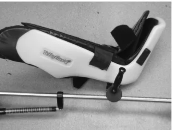
Fortunately, these cases of neuropraxia were of short duration (2-3 days) and resolved in all cases. We also experienced this problem at our institution until recently when we began using the Yellofins Stir-rups™ (Figure-5) and subsequently we have not seen this complication again. This is due to the fact that the stirrups support the entire leg from the calf down to the foot in a boot like support. This minimizes any pressure on the fibular head and ankle which prevents the neuropraxia.
CONCLUSION
Radical perineal prostatectomy is an example of a surgical technique which has stood the test of time. With only a few technical modifications since its original description, it offers outcomes similar to radical retropubic prostatectomy, the standard ap-proach for the treatment of localized prostate cancer. Its advantages include decreased pain, blood loss, and convalescence, the same arguments currently being made in favor of laparoscopic prostatectomy. In ad-dition, it is the optimal approach for obese patients, patients with prior pelvic surgery, or patients with prior pelvic radiation. As shown in this paper, proper patient selection is critical to the success of the pro-cedure and the minimization of complications. Fur-thermore, a detailed understanding of the perineal anatomy combined with surgeon experience make RPP is necessary for success, but for the experienced surgeon RPP is an attractive option for the selected patient with localized prostate cancer.
REFERENCES
1. Weyrauch HM: Surgery of the prostate. Philadelphia, WB Saunders Co. 1959; pp 172-230.
2. Gouley JWS: Some points in the surgery of the hyper-trophied prostate. Trans of Am Surg Assoc. 1885; 3: 163-92.
3. Billroth T: Carcinoma der Prostata. Chir Erfahrungen, Zurich, 1860-67. Archiv Fur Klinische Chirurgie. Bd X, S. 548, 1869.
4. Young HH: Hugh Young: A surgeon’s autobiography. New York, Harcourt, Brace and co. 1940; pp 104-34. 5. Young HH: The cure of cancer of the prostate by radi-cal perineal prostatectomy (prostate-seminal vesiculec-tomy): History, literature and statistics of Young’s op-eration. J Urol. 1945; 53: 188-256.
6. Gibson TE: Improvements in perineal prostatectomy. Surg Gynecol Obstet. 1928; 47: 531-9.
7. Belt E: Radical perineal prostatectomy in early carci-noma of the prostate. J Urol. 1942; 48: 287-97. 8. Soutar HS: Complete removal of the prostate. Brit Med
J. 1947; 1: 917-8.
9. Memmelaar J: Total prostatovesiculectomy - retropu-bic approach. J Urol. 1949; 62: 340-8.
10. Reiner WB, Walsh PC: An anatomical approach to the surgical management of the dorsal vein and Santorini’s plexus during radical retropubic prostatectomy: The apical dissection. J Urol. 1987; 138: 543-50. 11. Walsh PC, Lepor H, Eggleston JD: Radical
prostatec-tomy with preservation of sexual function: Anatomi-cal and pathologiAnatomi-cal considerations. Prostate 1983; 4: 473-85.
12. Weldon VE, Tavel FR: Potency sparing radical perineal prostatectomy: Anatomy, surgical technique, and initial results. J Urol. 1988; 140: 559-62. 13. Partin AW, Kattan MV, Subong E,Walsh PC, Wojno
KJ, Oesterling JE et al.: Combination of prostate-spe-cific antigen, clinical stage, and Gleason score to pre-dict pathological stage of localized prostate cancer. A multi-institutional update. JAMA 1997; 277: 1445-51. 14. Paulson DF: Impact of radical prostatectomy in the management of clinically localized disease. J Urol. 1994; 152: 1826-30.
15. Mokulis J, Thompson I: Radical prostatectomy: is the perineal approach more difficult to learn? J Urol. 1997; 157: 230-2.
16. Bluestein DL, Bostwick DG, Bergstrahl EJ, Oesterling JE: Eliminating the need for bilateral pelvic lym-phadenectomy in select patients with prostate cancer. J Urol. 1994; 151: 1315-20.
17. Ung JO, Richie JP, Chen MH, Renshaw AA, D’Amico AV: Evolution of the presentation and pathologic and biochemical outcomes after radical prostatectomy for patients with clinically localized prostate cancer diagnosed during the PSA era. Urol-ogy 2002; 60: 458-63.
18. Boxer RT, Kaufman JJ, Goodwin WE: Radical pros-tatectomy for carcinoma of the prostate: 1971-1976; A review of 329 patients. J Urol. 1977; 117: 208-13. 19. Frazier HA, Roberston JE, Paulsen DF: Radical pros-tatectomy: The pros and cons of the perineal versus the retropubic approach. J Urol. 1992; 147: 888-90. 20. Haab F. Boccon-Gibbod L, Delmas V, Boccon-Gibod
L, Toublanc M: Perineal versus retropubic radical pros-tatectomy for T1, T2 prostate cancer. Br J Urol. 1994; 74: 626-9.
21. Boccon-Gibod L, Ravery V, Vordos D, Toublanc M, Delmas V, Boccon-Gibod L: Radical prostatectomy for prostate cancer: the perineal approach increases the risk of surgically induced positive margins and cap-sular incisions. J Urol. 1998; 160: 1383-5.
22. Lance RS, Freidrichs PA, Kane C, Powell CR, Pulos E, Moul JW et al.: A comparison of radical retropubic with perineal prostatectomy for localized prostate can-cer within the Uniformed Services Urology Research Group. BJU Int. 2001; 87: 61-5.
23. Lassen PM, Kearse WS Jr.: Rectal Injuries during radi-cal perineal prostatectomy. Urology 1995; 45: 266-79.
24. Bishoff JT, Motley G, Optenberg SA, Stein CR, Moon KA, Browning SM et al.: Incidence of fecal and uri-nary incontinence following radical perineal and ret-ropubic prostatectomy in a national population. J Urol. 1998; 160: 454-8.
25. Price DT, Vieweg J, Roland F, Coetzee L, Spalding T, Iselin C et al.: Transient lower extremity neuropraxia associated with radical perineal prostatectomy: a com-plication of the exaggerated lithotomy position. J Urol. 1998; 160: 1376-8.
Received: February 3, 2003 Accepted: February 24, 2003
Correspondence address:
Dr. Jeffrey M. Holzbeierlein
University of Kansas Medical Center 3901 Rainbow Blvd, Mail Stop 3016 Kansas City, KS 66160, USA Fax: + 1 913 588-7625