Evaluation of quality of life and photoplethysmography in
patients with chronic venous insuiciency treated
with foam sclerotherapy
Avaliação da qualidade de vida e fotopletismografia em pacientes com insuficiência
venosa crônica tratados através de escleroterapia com espuma
Felipe Coelho Neto1,2
*
, Gilson Roberto Araújo2, Iruena Moraes Kessler1
Abstract
Background: Ultrasound-guided foam sclerotherapy plays a major role in treatment of chronic venous insuiciency, providing clinical and hemodynamic improvement to patients undergoing treatment. Objectives: To examine the relationships between venous reilling time and impact of venous disease on quality of life and between changes in venous reilling time and improvement of symptoms after ultrasound-guided foam sclerotherapy for chronic venous insuiciency. Methods: hirty-two patients classiied as C4, C5 or C6 answered a questionnaire on quality of life and symptoms and their venous illing time was measured using photoplethysmography before and 45 days after treatment of chronic venous insuiciency with ultrasound-guided foam sclerotherapy. Results: Statistically signiicant improvements were observed in quality of life scores and in venous illing time and in the following symptoms: aching, heavy legs, restless legs, swelling, burning sensations, and throbbing (p<0.0001). A similar improvement was also seen in the work and social domains of quality of life (p<0.0001). Conclusions: As conirmed by questionnaire scores and venous reilling times, ultrasound-guided foam sclerotherapy demonstrated eicacy and resulted in high satisfaction levels and low rates of major complications.
Keywords: quality of life; chronic venous insuiciency; photoplethysmography; sclerotherapy; sclerosing solutions; color Doppler ultrasonography; varicose veins.
Resumo
Contexto: A escleroterapia com espuma guiada por ultrassom (EGUS) ocupa lugar de destaque no tratamento da insuiciência venosa crônica (IVC), proporcionando melhora clínica e hemodinâmica aos pacientes submetidos ao tratamento. Objetivos: Veriicar a correlação entre dados obtidos por questionário de qualidade de vida e de sintomas com dados obtidos por fotopletismograia (FPG), antes e depois do tratamento por escleroterapia com espuma guiada por ultrassom (EGUS) da insuiciência venosa crônica (IVC). Métodos: Um grupo de 32 pacientes, classiicados como C4, C5 e C6, foi submetido à aplicação de questionário de qualidade de vida e sintomas, sendo aferido o tempo de enchimento venoso (TEV) por FPG antes e 45 dias depois do tratamento da IVC através de EGUS. O teste do sinal foi utilizado para análise estatística da melhora dos escores dos questionários e do TEV. O teste de McNemar foi utilizado para avaliação da melhora nos sintomas e do impacto do tratamento nas atividades laborais e sociais dos pacientes. Resultados: Houve melhora nos escores dos questionários de qualidade de vida e no TEV, com signiicância estatística (p<0,0001). Houve melhora estatisticamente signiicativa nos sintomas: dor, cansaço, edema, queimação, pernas inquietas e latejamento (p<0,0001). Incremento na qualidade laboral e social após o tratamento apresentou melhora estatisticamente signiicativa (p<0,0001). Não ocorreram complicações maiores ou efeitos adversos nesta série. Conclusões: A EGUS mostrou-se eicaz, com alto índice de satisfação e baixas taxas de complicacões maiores, ratiicada pelos escores dos questionários e pelos TEVs aferidos pela FPG.
Palavras-chave: qualidade de vida; insuiciência venosa crônica; fotopletismograia; escleroterapia; soluções esclerosantes; ultrassonograia Doppler em cores; varizes.
1 Universidade de Brasília – UnB, School of Medicine, Brasília, DF, Brazil. 2 Hospital Regional da Asa Norte, Brasília, DF, Brazil.
Financial support: None.
Conlicts of interest: No conlicts of interest declared concerning the publication of this article. Submitted: November 27, 2014. Accepted: January 14, 2015.
INTRODUCTION
Chronic venous insuficiency (CVI) is a common disease in clinical practice. Its complications, especially venous stasis ulcers, cause signiicant morbidity, loss of functional mobility and reduced quality of life.1
The ideal treatment for primary varicose veins of lower limbs should be minimally invasive; repeatable whenever needed; free from signiicant complications; effective at eliminating relux points; and of low cost; and should result in esthetic improvement while requiring little absence from work.2 Ultrasound-guided
foam sclerotherapy (UGFS) is one option for the treatment of CVI that can meet these requirements.3
There are several studies of UGFS treatment for CVI reporting good clinical results and high rates of venous trunk occlusion assessed by vascular echography.4,5
However, few publications have conirmed the clinical results obtained with UGFS using objective parameters, such as the hemodynamic variations provoked by the treatment.
Quality of life questionnaires can be used as the parameters of subjective assessments of treatment eficacy. The Venous Insuficiency Epidemiological and Economic Study’s Quality of Life/Symptom questionnaire (VEINES-QOL/Sym) consists of a self-report questionnaire addressing symptoms, their impact on daily activities and the psychological aspects of the disease.6 Higher scores indicate better
outcomes.6,7
The hemodynamic variations provoked by treatment of CVI with UGFS can be determined by using photoplethysmography (PPG) to speciically measure venous reilling time (VRT). Venous reilling times obtained by PPG exhibit good correlations with direct measures of venous pressure.8-10 In turn, PPG is a
rapid, non-invasive method suitable for outpatient settings that can provide quantitative and objective data to supplement the anatomical assessment and study of CVI11 both before and after therapeutic
interventions.12
In view of the above, this study aimed to evaluate the eficacy of treatment for CVI using UGFS, by correlating VRT values as measured by PPG with data obtained by administration of a questionnaire on quality of life and symptoms before and 45 days after treatment.
METHODS
An open prospective study was conducted to evaluate VRT, as assessed by digital PPG, and the scores obtained by administration of a questionnaire
on quality of life and symptoms before and 45 days after treatment of varicose veins with UGFS. The study was carried out at the Vascular Surgery outpatients clinic at a public hospital in the city of Brasília, Federal District of Brazil, from December 2012 to August 2013.
We selected patients over 18 years of age who presented with CVI of the lower limbs and were classiied as C4, C5 or C6, according to the Clinical, Etiological, Anatomical, Pathophysiological (CEAP) classiication.13 In order to obtain a homogenous
sample, patients with deep venous thrombosis (DVT) identiied by ultrasound examination were excluded from the study, as were patients with varicose veins but no involvement of the great or small saphenous vein. Cases of thrombophilia, active neoplasms or neoplasms in follow-up, reported lung disease, and peripheral artery insuficiency (ankle-arm index < 0.9) were also excluded.
All patients signed a written consent form after being informed about the details of the study.
Duplex ultrasound examination
All examinations were performed in a standardized manner by the same physician (FCN) using a MyLab 40 ultrasound machine (Esaote™. Genoa, Italy) with a multifrequency 10-12 MHz transducer. Patients were placed in the standing position with their weight on the contralateral limb and the leg to be examined slightly rotated with the heel on the loor to relax the calf muscle while maintaining stability. The deep venous system was evaluated for DVT, and the supericial venous system was evaluated focusing on saphenofemoral and saphenopopliteal junctions, great and short saphenous veins, and on the presence of incompetent perforating veins. Relux was induced with manual calf squeeze and was deined as reverse low with duration longer than 0.5 seconds for saphenous vein and 0.35 seconds for perforating veins.14 Only perforating veins with diameters greater
than 3.5 mm at the fascial level were considered for analysis, and relux was recorded as described by Sandri et al.15
Photoplethysmography
A Hadeco PPG machine (Hayashi Denki CO. LTD, Kawasaki, Japan) was used to measure post-exercise VRT in the sitting position with the limb suspended, as described by Sam et al.16 Measurements were all
the tibia, in order to avoid areas with trophic changes resulting from CVI. With the patient as motionless as possible, the machine calibrated the signal, and once a stable baseline was achieved, the exercise was initiated. Patients completed 10 dorsal and plantar lexions over a 15-second period and were then asked to remain at rest as motionless as possible.
The ejection of blood from the skin and the subsequent reilling curve were established and the machine calculated the venous illing curve. Three measurements were taken at intervals of 2-5 minutes, and the mean of all three measurements was used for analysis. Normal VRT was deined as ≥ 20 seconds. Measurements of VRT were performed on the day of the irst sclerotherapy session and repeated in each treated limb 45 days after the end of the treatment, with the probe placed at the same height as that used in the pretreatment examination.
Quality of life and symptom questionnaire
The Brazilian Portuguese version of the VEINES-QOL/Sym questionnaire17 was administered in the
form of an interview, which was performed by a single researcher (FCN). The questionnaire was administered on the day of the irst session and 45 days after the end of treatment.
Sclerotherapy technique
We have described the sclerotherapy technique elsewhere,18 focusing on the relux pattern presented for
each patient. When total occlusion of the target veins was not achieved in a single session, supplementary sessions were conducted, during which one or more punctures were performed, as required in each case, with 7-day intervals between sessions. The foam was produced by mixing 3% polidocanol with ambient air at a ratio of 1:4, and the maximum total volume of foam injected in a single session was 10 mL. Compression stockings (15-30 mmHg) were prescribed to be worn day and night for 7 days, only removing them for the purposes of personal hygiene. From the 7th day onwards, patients were recommended to wear stockings during the day only.
Statistical analysis
Non-parametric methods were applied using Microsoft Ofice Excel 2007 (Microsoft Corporation, Redmond, USA), SAS version 9.3 (SAS Institute Inc. Cary, USA), and R i386 3.0.1 (R Foundation for Statistical Computing, Vienna, Austria). The sign test was used to determine improvement in VRT and in VEINES-QOL/Sym scores. The McNemar test was performed to analyze improvement in symptoms.
The cutoff for statistical signiicance was set at less than or equal to 5%.
Research ethics
The research project was submitted to and approved by the Research Ethics Committee (CAAE: 06791512.1.0000.5553). Additionally, all patients signed a written consent form after being informed about the details of the study.
RESULTS
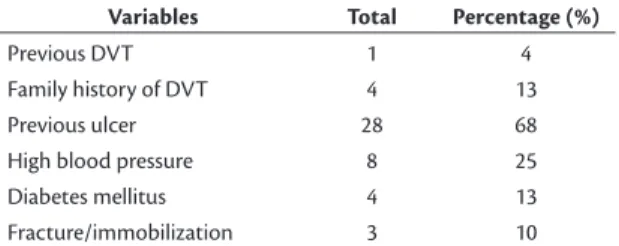
Thirty-two patients were recruited. Females accounted for 82% (26/32) of the patients and mean age was 52 years (range 36-76 years). The left lower limb was the most frequently affected limb, representing 57% (18/32) of the sample. None of the patients had undergone previous invasive treatment at the point of enrollment on the study. Other epidemiological data are shown in Table 1.
Family history of DVT was present in four patients (13%), and three (10%) had a history of fracture and/or limb immobilization. Healed or active venous ulcers were present in 68% of the sample. The most frequent comorbidities were high blood pressure in eight individuals (25%) and diabetes in four (13%).
The patients’ CEAP classiications broke down as follows: 10 (32%) were classiied as C4, seven (22%) as C5, and 15 (47%) as C6. All patients had a primary etiological classiication. Anatomical classiication was 62.5% for supericial veins and 37.5% for perforating veins. Relux was the pathophysiology in all patients.
The mean number of punctures required per patient was 3.96 (3-7), the mean number of sessions was 1.4 (1-3) and the mean volume of foam per session was 8.5 mL (10-23 mL).
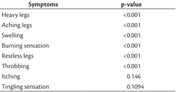
There were ive response options for questionnaire items referring to symptoms: “every day”, “several times a week”, “about once a week”, “less than once a week”, and “never”. There was signiicant improvement in all items, as shown in Figure 1. For the purposes of statistical analysis, the responses “every day”, “several times a week”, and “about once a week”
Table 1. Epidemiological data.
Variables Total Percentage (%)
Previous DVT 1 4
Family history of DVT 4 13
Previous ulcer 28 68
High blood pressure 8 25
Diabetes mellitus 4 13
Fracture/immobilization 3 10
were collapsed into a single category - group 1 - and the responses “less than once a week” and “never” were collapsed to form group 2. Group 1 responses were deined as indicating absence of improvement while group 2 responses represented improvements in the symptoms evaluated. All items exhibited statistical signiicance to p<0.001, except for the items “itching” and “tingling sensation”. The p values for improvements in symptoms are shown in Table 2.
Patients were also asked about the time of the day during which their leg problem was most severe. After treatment, 25% of the study population did not report any complaint during the entire day.
The next item on the questionnaire was: “Compared to one year ago, how would you rate your leg problem in general now?”. Before treatment, 56% of patients reported that the problem was somewhat worse or much worse than one year previously. After treatment, 100% of the patients reported that the problem was somewhat better or much better than one year previously.
Treatment resulted in improvement in patients’ work activities and the percentage of patients who answered that the problem did not limit their work activities at all increased from 34% to 75%. Additionally, none of the patients reported that the problem limited their activities a lot after treatment. When asked about limitations to daily household activities caused by their leg problem, 97% of patients answered that the problem did not cause any limitation to household activities. All items related to the impact of the disease on work or personal activities exhibited statistical signiicance to p<0.0001, as shown in Table 3.
There were improvements in activities performed both in standing and sitting positions, with the number of patients who answered that the problem did not limit these types of activity increasing from 34 to 94% and from 47 to 97% respectively.
These improvements attained statistical signiicance for both items (p<0.01), as shown in Table 4.
Patients who did not report any limitation to social activities with friends and family accounted for 94% of the sample after treatment (p<0.01), in contrast with 37% of the sample before treatment. Before treatment, patients who answered that the problem interfered “extremely”, “quite a bit” or “moderately” had accounted for 50% of the sample.
There was a speciic item to assess the symptom “pain”. Before treatment, 25% of patients had reported “very severe” or “severe” pain, whereas after treatment this igure was 12%. Conversely, 19% of patients had reported “very mild” or “no” pain before treatment and this igure had risen to 56% after the procedure. Although the sample exhibited improvement in complaints of pain, these were not statistically signiicant (p>0.05).
Figure 1. Improvement in symptoms.
Table 2. Improvement in symptoms after treatment with
ultrasound-guided foam sclerotherapy.
Symptoms p-value
Heavy legs <0.001
Aching legs <0.001
Swelling <0.001
Burning sensation <0.001
Restless legs <0.001
hrobbing <0.001
Itching 0.146
Tingling sensation 0.1094
Table 3. Improvement in work activities.
Work activities p-value
Reduction in the amount of time spent at work <0.0001 Reduction in the amount of work accomplished <0.0001
Limitation in work time <0.0001
The psychological impact of leg problems on patients’ daily lives was also analyzed. The percentage of patients who answered that they felt concerned about their appearance “all of the time” or “most of the time” decreased from 62% before treatment to
6% after treatment, while the percentage of those who reported feeling concerned about their appearance “a little of the time” or “none of the time” increased from 25% to 78% of the sample. After treatment, 87% of the patients reported feeling irritated “none of the time”. Half of the patients did not feel they were a burden to their family and friends before treatment and this igure had increased to 100% after treatment. Initially, 60% of the patients were worried about bumping into things “all of the time”. After intervention, this value had fallen to 6%. Additionally, before UGFS 72% of patients had answered that the appearance of their legs inluenced their choice of clothing “all of the time” and this percentage had decreased to 22% after the procedure.
The mean result for VRT before treatment was 6.7 seconds (2.0-15.3 seconds) and after treatment mean VRT was 22.2 seconds (6.0-53.2 seconds), which is an increase of more than 230%.
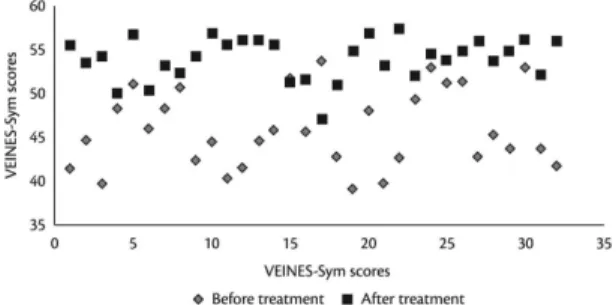
Before treatment, the mean VEINES-QOL score was 44.8 (40.2-53.4), and the mean VEINES- Sym score was 45.03 (39.1-53.1). After treatment, the mean VEINES-QOL and VEINES- Sym scores were 54.6 (48.2-57.3) and 54.1 (47.1-57.6) respectively.
Figure 2 compares VRT values before and after treatment, and Figures 3 and 4 show before and after results for VEINES-QOL and VEINES-Sym scores respectively. Statistically signiicant improvements (p<0.0001) were observed in VRT values and in both questionnaire scores after treatment.
There were no major complications such as DVT or pulmonary embolism during follow-up. Phlebitis (19%), local pain (90%), pigmentation (97%), and local induration (97%) were the most frequent minor complications.
Seven (47%) of the patients classiied as C6 exhibited healed ulcers at 45 days after treatment. There were no cases of ulcer recurrence in the C5 group.
DISCUSSION
Over the last decade, alternative treatments for CVI such as endovenous thermal ablation and UGFS have become popular. These are outpatient procedures performed with local tumescent anesthesia that do not require hospitalization or absence from daily activities and several studies have demonstrated that they are safe and effective for eliminating venous relux.19-22
Furthermore, the clinical impact of these treatments can be measured by quality of life questionnaires, whether speciic14, 23 or generic.24
Ambulatory venous pressure is considered the best parameter for evaluation of hemodynamic
Figure 2. VRT values before and after treatment with UGFS. VRT:
venous reilling time; UGFS: ultrasound-guided foam sclerotherapy.
Figure 3. VEINES-QOL scores before and after treatment with
UGFS. VEINES-QOL: Venous Insuiciency Epidemiological and Economic Study - Quality of Life; UGFS: ultrasound-guided foam sclerotherapy.
Table 4. Impact of the disease on personal activities.
Personal activities p-value
Daily work activities 0.002
Daily household activities <0.001
Social or leisure activities performed in the standing position
<0.001
Social or leisure activities performed in the sitting position
<0.001
Figure 4. VEINES-Sym scores before and after treatment with
improvement after treatment, but it is an invasive method with limited applications in routine practice.25
Air plethysmography is an effective alternative for measuring CVI severity,26 but it is a long test that
requires a large instrument.2 In turn, VRT as measured
by PPG offers good reproducibility and good correlation with direct measures of venous pressure.9 Moreover,
it is a rapid test performed with a portable device and can be widely used in clinical practice.
Our data consistently demonstrated that there was an improvement in symptoms after treatment and conirm indings reported by Bradbury et al.11 The portion of
the questionnaire related to symptoms (VEINES-Sym) identiied improvements in all nine items 45 days after treatment, with statistical signiicance for the following items: pain, heavy legs, swelling, burning sensation, restless legs, and throbbing (p<0.0001). The portion of the questionnaire related to quality of life (VEINES-QOL) conirmed these data, since it showed that 100% of patients reported being somewhat better (6%) or much better (94%) than one year before treatment. There were statistically signiicant improvements in the ability to perform both work and household activities, with 75% of patients reporting no limitation to work activities and 97% of patients reporting no dificulty in performing their usual household activities or social activities with friends and/or family.
We would like to draw attention to the improvement in pain, evidenced by the increase from 25 to 56% in the percentage of patients with no pain or complaining of very mild pain. Although analysis of this data did not reveal statistical signiicance, this improvement conirms indings previously reported in the literature.27
The analysis of patients’ psychological proiles also revealed improvements in all questionnaire items. The instrument is therefore an important tool for evaluating treatment outcomes, since patients have usually been dealing with CVI for a long time, often relying on palliative treatments that have failed to achieve deinitive resolution of the disease.28
There were no thromboembolic events or adverse effects resulting from sclerotherapy in this series of patients. Conversely, local complications were observed, including pigmentation, induration, and pain in the varicosities treated. These complications did not cause severe clinical repercussions, as conirmed by the high rate of patient satisfaction observed in the responses to the VEINES-QOL. Our data reveal higher local complication rates after UGFS than reported in other publications.29,30 Notwithstanding,
the majority of patients in those studies were classiied
as CEAP 2-3, which means less severe clinical CVI status. It is believed that the higher rate of complications found in this study is the result of actively searching for information and the fact that most of our cases presented thicker varicose veins (CEAP 5 and 6).
Mean VRT as measured by PPG increased from 6.7 before treatment to 22.2 after treatment (p<0.0001). This inding had a statistically signiicant direct relationship with the increase in VEINES-QOL/Sym scores, which conirms that VRT may have applications as an objective marker of short term clinical improvement in patients who have had UGFS treatment. As demonstrated by Kulkarni et al.,12 VRT
may be an effective marker of venous ulcer recurrence when it does not improve after treatment. There were no cases of deep venous relux in the sample. Therefore, the lack of improvement in VRT exhibited by patients can be explained by calf pump failure or restriction of ankle mobility as a result of trophic skin changes. In our experience, the improvement in VRT was associated with a rate of ulcer healing of 47% in patients classiied as C6, 45 days after treatment with UGFS. The sample size and the limited follow-up time are weaknesses of our study.
However, the VEINES QOL/Sym questionnaire has never been used before to assess the results of CVI treatment using UGFS. The VEINES-QOL/Sym was the only questionnaire available that had been translated and adapted for use with the Brazilian population at the start of this study. Although other more speciic questionnaires for venous ulcers exist, such as the Charing Cross Venous Ulcer Questionnaire,31
they were not available for use at the start of our research project.
This study is the first report of use of this questionnaire for this purpose and shows that the VEINES QOL/Sym questionnaire is a feasible tool that exhibited a direct relationship with the objective hemodynamic measure obtained by PPG, in addition to portraying the psychological impact of the treatment on quality of life in the short-term.
In turn, UGFS proved to be effective and resulted in high levels of satisfaction and low rates of major complications, conirmed by the VEINES QOL/Sym scores and by VRT as measured by PPG.
ACKNOWLEDGEMENT
The authors are grateful to Beatriz Helena Pavan Balducci Coelho, Olympio Teixeira, Sirleide Braga, and Rosana Falasque for their dedication and valuable advice during the execution of this project.
REFERENCES
1. Silva MC. Chronic venous insufficiency of the lower limbs and its socio-economic significance. Int Angiol. 1991;10(3):152-7. PMid:1765717.
2. Guex JJ, Isaacs MN. Comparison of surgery and ultrasound guided sclerotherapy for treatment of saphenous varicose veins: must the criteria for assessment be the same? Int Angiol. 2000;19(4):299-302. PMid:11305726.
3. Wright D, Gobin JP, Bradbury AW, et al. Varisolve® polidocanol microfoam compared with surgery or sclerotherapy in the management of varicose veins in the presence of trunk vein incompetence: European randomized controlled trial. Phlebology. 2006;21(4):180-90. http://dx.doi.org/10.1258/026835506779115807.
4. Orsini C, Brotto M. Immediate pathologic effects on the vein wall of foam sclerotherapy. Dermatol Surg. 2007;33(10):1250-4. PMid:17903159.
5. Figueiredo M, Araújo SP, Penha-Silva N. Microfoam ultrasound-guided sclerotherapy in primary trunk varicose veins. J Vasc Bras. 2006;5(3):177-83. http://dx.doi.org/10.1590/S1677-54492006000300005.
6. Lamping DL, Schroter S, Kurz X, Kahn SR, Abenhaim L. Evaluation of outcomes in chronic venous disorders of the leg: development of a scientifically rigorous, patient-reported measure of symptoms and quality of life. J Vasc Surg. 2003;37(2):410-9. http://dx.doi. org/10.1067/mva.2003.152. PMid:12563215.
7. Kahn SR, Lamping DL, Ducruet T, et al. VEINES-QOL/Sym questionnaire was a reliable and valid disease-specific quality of life measure for deep venous thrombosis. J Clin Epidemiol. 2006;59(10): 1056.e1–1056.e4. http://dx.doi.org/10.1016/j.jclinepi.2005.10.016. PMid:16980144.
8. Rosfors S. Venous photoplethysmography: relationship between transducer position and regional distribution of venous insufficiency. J Vasc Surg. 1990;11(3):436-40. http://dx.doi.org/10.1016/0741-5214(90)90244-5. PMid:2313832.
9. Abramowitz HB, Queral LA, Finn WR, et al. The use of photoplethysmography in the assessment of venous insufficiency: a comparison to venous pressure measurements. Surgery. 1979;86(3):434-41. PMid:473029.
10. Nicolaides AN, Miles C. Photoplethysmography in the assessment of venous insufficiency. J Vasc Surg. 1987;5(3):405-12. http://dx.doi. org/10.1016/0741-5214(87)90047-4. PMid:3334678.
11. Bradbury AW, Bate G, Pang K, Darvall KA, Adam DJ. Ultrasound-guided foam sclerotherapy is a safe and clinically effective treatment for superficial venous reflux. J Vasc Surg. 2010;52(4):939-45. PMid:20638224.
12. Kulkarni SR, Barwell JR, Gohel MS, Bulbulia RA, Whyman MR, Poskitt KR. Residual venous reflux after superficial venous surgery does not predict ulcer recurrence. Eur J Vasc Endovasc Surg. 2007;34(1):107-11. http://dx.doi.org/10.1016/j.ejvs.2006.12.033. PMid:17408990.
13. Eklöf B, Rutherford RB, Bergan JJ, et al. Revision of the CEAP classification for chronic venous disorders: consensus statement. J Vasc Surg. 2004;40(6):1248-52. http://dx.doi.org/10.1016/j. jvs.2004.09.027. PMid:15622385.
14. Labropoulos N, Tiongson J, Pryor L, et al. Definition of venous reflux in lower-extremity veins. J Vasc Surg. 2003;38(4):793-8. http://dx.doi.org/10.1016/S0741-5214(03)00424-5. PMid:14560232. 15. Sandri JL, Barros FS, Pontes S, Jacques C, Salles-Cunha SX. Diameter-reflux relationship in perforating veins of patients with varicose veins. J Vasc Surg. 1999;30(5):867-74. http://dx.doi.org/10.1016/ S0741-5214(99)70011-X. PMid:10550184.
16. Sam RC, Darvall KA, Adam DJ, Silverman SH, Bradbury AW. Digital venous photoplethysmography in the seated position is a reproducible noninvasive measure of lower limb venous function in patients with isolated superficial venous reflux. J Vasc Surg. 2006;43(2):335-41. http://dx.doi.org/10.1016/j.jvs.2005.10.039. PMid:16476612.
17. Moura RMF, Gonçalves GS, Navarro TP, Britto RR, Dias RC. Transcultural adaptation of VEINES/QOL-Sym questionnaire: evaluation of quality of life and symptoms in chronic venous disease. J Vasc Bras. 2011;10(1):17-23. http://dx.doi.org/10.1590/ S1677-54492011000100004.
18. Coelho F No, Araujo GR, Kessler IM, Amorim RF, Falcao DP. Treatment of severe chronic venous insufficiency with ultrasound-guided foam sclerotherapy: a two-year series in a single center in Brazil. Phlebology. 2015;30(2):113-8. http://dx.doi.org/10.1177/0268355513517225. PMid:24335090.
19. Darwood RJ, Gough MJ. Endovenous laser treatment for uncomplicated varicose veins. Phlebology. 2009;24(Suppl 1):50-61. http://dx.doi.org/10.1258/phleb.2009.09s006. PMid:19307441.
20. Gohel MS, Davies AH. Radiofrequency ablation for uncomplicated varicose veins. Phlebology. 2009;24(Suppl 1):42-9. http://dx.doi. org/10.1258/phleb.2009.09s005. PMid:19307440.
21. Smith PC. Foam and liquid sclerotherapy for varicose veins. Phlebology. 2009;24(Suppl 1):62-72. http://dx.doi.org/10.1258/ phleb.2009.09s007. PMid:19307442.
22. Proebstle TM, Vago B, Alm J, Göckeritz O, Lebard C, Pichot O. Treatment of the incompetent great saphenous vein by endovenous radiofrequency powered segmental thermal ablation: first clinical experience. J Vasc Surg. 2008;47(1):151.e1-6.e1. http://dx.doi. org/10.1016/j.jvs.2007.08.056. PMid:18178468.
23. Garratt AM, Macdonald LM, Ruta DA, Russell IT, Buckingham JK, Krukowski ZH. Towards measurement of outcome for patients with varicose veins. Qual Health Care. 1993;2(1):5-10. PMid:10132081.
24. Smith JJ, Garratt AM, Guest M, Greenhalgh RM, Davies AH. Evaluating and improving health-related quality of life in patients with varicose veins. J Vasc Surg. 1999;30(4):710-9. http://dx.doi. org/10.1016/S0741-5214(99)70110-2. PMid:10514210.
25. Nicolaides AN. Cardiovascular Disease Educational and Research Trust, European Society of Vascular Surgery, The International Angiology Scientific Activity Congress Organization, International Union of Angiology, Union Internationale de Phlebologie at the Abbaye des Vaux de Cernay. Investigation of chronic venous insufficiency: a consensus statement (France, March 5-9, 1997). Circulation. 2000;102(20): E126-63.
26. Engelhorn CA, Beffa CV, Bochi G, et al. Can air plethysmography evaluate the severity of chronic venous insufficiency?. J Vasc Bras. 2004;3:311-6.
27. Rasmussen LH, Lawaetz M, Bjoern L, Vennits B, Blemings A, Eklof B. Randomized clinical trial comparing endovenous laser ablation, radiofrequency ablation, foam sclerotherapy and surgical stripping for great saphenous varicose veins. Br J Surg. 2011;98(8):1079-87. http://dx.doi.org/10.1002/bjs.7555. PMid:21725957.
29. Cavezzi A, Parsi K. Complications of foam sclerotherapy. Phlebology. 2012;27(Suppl 1):46-51. http://dx.doi.org/10.1258/ phleb.2012.012S09. PMid:22312067.
30. Jia X, Mowatt G, Burr JM, Cassar K, Cook J, Fraser C. Systematic review of foam sclerotherapy for varicose veins. Br J Surg. 2007;94(8):925-36. http://dx.doi.org/10.1002/bjs.5891. PMid:17636511.
31. Couto RC, Leal FJ, Pitta GBB, Bezerra RCB, Segundo Walmir SS, Porto TM. Tradução e adaptação cultural do Charing Cross Venous Ulcer Questionnaire - Brasil. J Vasc Bras. 2012;11(2):102-7. http:// dx.doi.org/10.1590/S1677-54492012000200006.
*
Correspondence Felipe Coelho Neto AngioClínica Brasília SEPS 709/909, Centro Médico Julio Adnet, Salas 419-422, Bloco A CEP 70100-360 – Brasília (DF), Brazil Tel.: +55 (61) 3328-1940 E-mail: [email protected]
Author information FCN - vascular surgeon at the Vascular Surgery Unit, Hospital Regional da Asa; MSc in Medical Sciences from Universidade de Brasília (UnB). IMK - MSc and PhD in Medical Sciences from Universidade de Brasília (UnB). GRA - vascular surgeon at the Vascular Surgery Unit, Hospital Regional da Asa Norte.
Author contributions Conception and design: FCN Analysis and interpretation: FCN, GRA Data collection: FCN Writing the article: FCN, IMK Critical revision of the article: FCN, IMK Final approval of the article*: FCN, GRA, IMK Statistical analysis: FCN Overall responsibility: FCN