online | memorias.ioc.fiocruz.br
Cellular and genetic mechanisms involved in the generation of protective
and pathogenic immune responses in human Chagas disease
Walderez Ornelas Dutra1/+, Cristiane Alves Silva Menezes1,2, Fernanda Nobre Amaral Villani1,
Germano Carneiro da Costa1, Alexandre Barcelos Morais da Silveira3, Débora d’Ávila Reis1,
Kenneth J Gollob4
1Departamento de Morfologia 4Departamento de Bioquímica-Imunologia, Instituto de Ciências Biológicas, Universidade Federal de Minas Gerais, Av. Presidente Antônio Carlos 6627, 31.270-901 Belo Horizonte, MG, Brasil 2Robarts Research Institute, University of Western Ontario, London, Canadá 3Departamento de Morfologia, Instituto de Ciências Biomédicas, Universidade Federal de Uberlândia,
Uberlândia, MG, Brasil
Perhaps one of the most intriguing aspects of human Chagas disease is the complex network of events that under-lie the generation of protective versus pathogenic immune responses during the chronic phase of the disease. While most individuals do not develop patent disease, a large percentage may develop severe forms that eventually lead to death. Although many efforts have been devoted to deciphering these mechanisms, there is still much to be learned before we can fully understand the pathogenesis of Chagas disease. It is clear that the host’s immune response is decisive in this process. While characteristics of the parasite influence the immune response, it is becoming evident that the host genetic background plays a fundamental role in the establishment of pathogenic versus protective re-sponses. The involvement of three complex organisms, host, parasite and vector, is certainly one of the key aspects that calls for multidisciplinary approaches towards the understanding of Chagas disease. We believe that now, one hundred years after the discovery of Chagas disease, it is imperative to continue with highly interactive research in order to elucidate the immune response associated with disease evolution, which will be essential in designing prophylactic or therapeutic interventions.
Key words:pathology - protection - T-cells - immunoregulation - Chagas disease
Financial support: WHO/TDR Program, CNPq, FAPEMIG, NIH/NI-AID 1 R03 AI 066044-0181
+ Corresponding author: [email protected] Received 6 April 2009
Accepted 14 May 2009
Chagas disease: contemporary concerns of a cente-nary disease
The first records of human infection with Trypano-soma cruzi date back nine thousand years, to a time when the first humans peopled the Andean coast. Molecular analyses of tissues extracted from human mummies of the Northern Chile and Southern Peru regions showed the presence of the parasite’s kinetoplast DNA in 41% of the samples taken (Aufderheide et al. 2004). Contact of these human groups with other mammals infected with the parasite seemed to have been the main route of disease transmission. It is believed that upon the first human in-fections, the domiciliation process begun, which provided the vectors with protection to climate changes and preda-tors, consolidating the human disease (Guhl et al. 2000).
Chagas disease was first detected in the Andes and even today it remains mostly restricted to Latin Ameri-ca. Several factors contribute to this restricted distribu-tion, but the most important is likely related to
socio-economic factors. The close contact between the vector, the reservoirs and humans is critical for disease trans-mission. In fact, recent studies have shown that infected domestic animals are important reservoirs for sustaining natural transmission in endemic areas (Levy et al. 2006, Gurthler et al. 2007). Poor housing conditions offer an adequate nesting environment for the vectors and the proximity to mammals, including man, offers abundant food for these insects. Recent studies have demonstrated both the presence of potential vectors of T. cruzi in the United States and a large number of infected animal res-ervoirs (Beard et al. 2003, Hancock et al. 2005). Despite this, Chagas disease is not endemic in the United States, emphasising the importance of both natural components and socio-economic aspects in maintaining endemicity.
While natural transmission is still the most important form of disease transmission in Latin America, other forms now have epidemiological importance. Infection via blood transfusion or organ transplantation, directly associated with the lack of blood/organ screening for the parasite, has brought the disease to non-endemic coun-tries (Leiby et al. 2002). Moreover, ingestion of contami-nated non-pasteurised fruit juices was recently responsi-ble for new cases of Chagas disease in areas where acute cases had not been detected for over 15 years (Steindel et al. 2008), suggesting a repopulation by the vectors in such areas.
con-cern, even more so because of the threat of its emergence in non-endemic areas and re-emergence in some endem-ic areas where it was thought to have been controlled. Taken together, these data point to a critical need for Chagas disease control in the following areas: (i) vector control programs that are not restricted to certain areas, but rather extended to all countries with active transmis-sion; (ii) reliable blood bank surveillance, even in areas where the disease is considered non-endemic; (iii) im-provement of socio-economic conditions; (iv) efficient parasite-targeted veterinary care and (v) reliable prophy-laxis or therapeutic interventions. Multidisciplinary ap-proaches and consistent support are essential for achiev-ing these goals, which will ultimately lead to disease control at all levels.
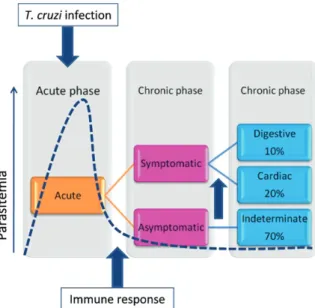
Clinical progression of human Chagas disease: from asymptomatic to severe forms
Following infection with T. cruzi, individuals un-dergo an acute phase that lasts between two and four months. With the exception of the mucosal edema that may appear at the site of infection (when in the eye, this is referred to as Romaña sign), the signs and symptoms associated to the acute phase of Chagas disease are most-ly non-specific, making it difficult to detect infection at early stages. While the acute phase is characterised by high numbers of parasites in the bloodstream, blood smear examination requires expertise and is only per-formed upon suspicion of T. cruzi infection. If the infec-tion is diagnosed, treatment is offered to the patients and cure may be observed in as many as 75% of cases (Sosa-Estani & Segura 2006, Rassi et al. 2007). This highlights the importance of developing reliable and easily avail-able tests for detecting acute infection, as well as pro-viding proper training to health professionals and care to the affected populations. Although a relatively high success rate is observed upon treatment, the currently available drugs are toxic and specific formulations for children, a group in which infection is highly prevalent, does not exist. Therefore, finding new parasiticidal com-pounds and new formulations will significantly improve disease treatment.
If the infection is not treated and cured, individuals will enter the chronic phase of the disease. The transition from acute to chronic phase is accompanied by a marked decrease in parasitaemia, due to the mounting of a rela-tively effective immune response, which keeps parasite frequency at below detectable levels in the host through-out the entire chronic phase of the disease. Despite the low parasite levels, it is during the chronic stage that patients may develop the most severe forms of Chagas disease. Figure 1 summarises the clinical progression of human Chagas disease.
As in most parasitic diseases, the vast majority of the individuals who are infected develop a relatively mild form of the disease, in which no clinical symptoms or signs are observed. Past or present infection with the parasite in many cases is only identified by the pres-ence of specific antibodies directed against the patho-gen. These individuals, who are serologically positive for anti-T. cruzi antibodies, are classified as
observe heart enlargement and loss of contractile func-tion, characteristic of chronic chagasic cardiomyopathy, which often leads to death (Rocha et al. 2007).
The differences in the clinical courses observed in chronic Chagas disease, as well as the variation observed within the same clinical form, suggest that different pathogenic mechanisms are associated with the clinical status of the patients. Thus, refined clinical characterisa-tion, obtained through well-defined patient groups, is a key element to be considered when analysing immuno-logical as well as epidemioimmuno-logical data. The combination of clinical and basic research is essential in determining strategies for better understanding disease progression and pathology.
The million-dollar question in human Chagas disease research is “how can we halt the establishment of pathol-ogy?”. In order to answer this question, researchers must first establish what factors mediate the different clinical progressions among patients. Given the complex nature of the host-pathogen relationship, this is not an easy task. However, it poses an exciting challenge and the pursuit of this knowledge will lead to concrete benefits to the large contingent of infected patients, as well as to those at risk of infection. In the following paragraphs, we will discuss the theories of pathology development and the immune response associated with pathology or protec-tion in human Chagas disease.
Mechanisms of pathogenesis in human Chagas dis-ease: controversies and consensus
There are two main hypotheses seeking to explain the mechanisms of pathogenesis in human Chagas dis-ease. The first of these defends the pivotal role of para-site’s persistence in the host as a major cause of pathol-ogy, while the other postulates that an immune response against self antigens is responsible for the tissue dam-age observed in affected organs of chagasic individuals (Kierszenbaum 2005, Hyland & Engman 2006, Dutra & Gollob 2008).
The parasite persistence hypothesis is supported by evidence showing that T. cruzi is present close to, or within, damaged areas (Fuenmayor et al. 2005). Al-though immunohistochemical/histological techniques often fail to reveal parasites at lesion sites, studies using sensitive techniques such as polymerase chain reaction (PCR). and in situhybridisation have shown T. cruzi per-sistence in affected organs (Jones et al. 1993, Vago et al. 1996). Benvenuti et al. (2005) detected T. cruzi DNA in endomyocardial biopsies after heart transplantation using PCR. In situ hybridisation using human cardiac tissue provided little evidence for the presence of intact
T. cruzi at sites of marked inflammation. Nevertheless, remnants of both T. cruzi kinetoplast and nuclear DNA were detected (Elias et al. 2003). Furthermore, the trans-mission of T. cruzi via blood transfusion from chronic chagasic donors and the observation that there is reac-tivation of parasitaemia in immuno-suppressed patients both support the assertion that parasites persist in many hosts (Bocchi et al. 1996, Ferreira et al. 1997). Consist-ent with the parasite persistence hypothesis is the ob-servation that patients treated with drugs that decrease
parasite load display concomitant decrease in disease se-verity. Viotti et al. (2006) observed that patients present-ing with chronic disease and no heart failure treated with benznidazole exhibited reduced progression of Chagas disease and increased negative seroconversion. Reduc-tion of electrocardiogram abnormalities were observed in chagasic patients treated with itraconazole or allopu-rinol (Apt et al. 2003). Thus, it seems beyond doubt that parasite presence is strongly tied to pathology, since T. cruzi infection is the initial event responsible for trigger-ing Chagas disease, persists in the host, and because an anti-parasite response is observed in chronic patients.
On the other hand, the contrast between the severity of the lesions observed during the chronic phase of Cha-gas disease and the low parasite load in the blood and tissues of chagasic patients suggests that the response to the parasite alone is insufficient to account for the ob-served pathology and that autoreactivity may contribute to disease aggravation. This idea led to the hypothesis that autoimmune responses take place during the devel-opment of pathology. Favouring this hypothesis is the fact that epitopes of parasite antigens elicit antibodies that cross react with epitopes of host tissues (Girones et al. 2005, Cunha-Neto et al. 2006). Furthermore, it was demonstrated that anti-parasite host-derived antibodies can mediate cellular reactivity (Gazzinelli et al. 1990, Reis et al. 1993a, Dutra et al. 2000). In addition to the presence of auto reactive antibodies, many studies dem-onstrated the existence of auto reactive T-cells in Cha-gas patients (Benoist & Mathis 2001, Cunha-Neto et al. 2006). To this end, molecular mimicry has been suggest-ed to exist between components of the host and T. cruzi
and, thus, strongly supports the participation of autoim-mune reactivity in the pathogenesis of Chagas disease (Cunha-Neto et al. 1996).
Although the theories seeking to explain the mecha-nisms underlying the pathogenesis of Chagas disease are controversial, autoreactivity and parasite persistence theories are not mutually exclusive. It is clear that, as the studies are not decisive in excluding one another, both should be considered when attempting to understand the establishment and maintenance of Chagas disease pa-thology. Regardless of the origin/source of the antigens that trigger the immune response during chronic infec-tion, there is a consensus that the host’s immune system, particularly T-cell subpopulations, plays a central role in pathology development. The increasing technical ability of researchers to phenotypically and functionally define T-cell subpopulations has provided more information about the role of differenT-cells during disease evolu-tion, allowing for a more refined clinical classification of patients. This, in turn, is critical for defining what leads to protective versus pathogenic responses.
The role of different cell populations and cytokines in establishing pathogenic versus protective re-sponses: association with clinical aspects
Interestingly, the parasite is present in symptomatic as well as in indeterminate patients and, to date, there is no published evidence that parasite load is higher in the symptomatic groups as compared to the indeterminate groups. Moreover, T-cells from all groups of patients, regardless of the clinical form, display characteristics of activation (Dutra et al. 1994, 1996, Lemos et al. 1998) and are capable of proliferating in vitro in response to parasite antigens (Dutra et al. 2000). The apparent lack of differences amongst patients with distinct clinical forms of Chagas disease led to the idea that particular parasite populations could lead to the establishment of different clinical forms. Molecular genetic analyses of different T. cruzi isolates have demonstrated that dis-tinct parasite populations are associated with different clinical forms of Chagas disease (Vago et al. 2000), suggesting that genetically distinct populations display characteristic tissue tropism and, thus, influence dis-ease outcome. However, the observation that genetically similar parasite isolates have been found in different or-gans suggests that the host immune response is a critical factor in determining the loutcome of infection (Lages-Silva et al. 2006). Also, as more refined clinical criteria are used, important immunological differences can be associated with patients with distinct clinical outcomes. Although studies of the human disease are still scarce in comparison to those using animal models, a collec-tion of data has been made available in the literature describing aspects of the immune response observed in patients with different clinical forms. We will present some of these data below, with emphasis on the cellular immune response.
Indeterminate form: equilibrium between host and parasite - The indeterminate clinical form represents the ideal situation for both the host and the parasite. In-dividuals with this clinical form harbour the parasite, as demonstrated by a variety of methods, but have no symptoms of disease whatsoever. Thus, the type of im-mune response induced in these individuals seems to be critical in maintaining a “healthy” balance between the parasite and host.
Indeterminate patients present positive serological standard tests (at least 2 positive results using different methods) and display no clinical signs and symptoms related to Chagas disease. According to WHO (2002) criteria, indeterminate patients have normal electro-cardiogram and radiological examination of the chest, oesophagus and colon. Patients with this form of the disease may progress to symptomatic forms eventually. However, many never develop clinical disease and die later in life of unrelated causes. Interestingly, despite the complete lack of clinical disease manifestations, in-determinate patients display quite a robust immune re-sponse. The idea that these individuals do not develop disease because they do not display cellular reactivity, therefore, is incorrect. Rather, the quality of this cellular reactivity is what seems to set these patients apart from other groups.
Most studies concerning the cellular immune re-sponse in Chagas disease, especially in indeterminate
patients, who do not display associated lesions, have been performed using peripheral blood cells (PBC). Ear-ly studies have demonstrated that PBC from indetermi-nate patients proliferate upon stimulation with T. cruzi -derived antigens and that this proliferative response does not differ quantitatively from those observed in patients with the cardiac clinical form of the disease (Morato et al. 1986, Dutra et al. 2000). Also, cells from indetermi-nate chagasic patients proliferate when stimulated with anti-epimastigote antibodies derived from patients with Chagas disease (Gazzinelli et al. 1990, Dutra et al. 2000). However, this particular response to the antibody stimu-lus seems to be lower in patients with the indeterminate form compared to cardiac patients. Reis et al. (1993a), studying the response to anti-epimastigote antibodies of chagasic individuals, showed that anti-epimastigote an-tibodies derived from indeterminate patients displayed a lower stimulatory capacity than antibodies isolated from cardiac patients. These data suggest that, although inde-terminate patients do display anti-epimastigote antibod-ies in their bloodstream that can stimulate T-cells, these antibodies are less stimulatory, which may contribute to a lower cellular response in vivo.
Analysis of the expression of activation markers by T-cells showed that indeterminate patients have a high frequency of CD4+ and CD8+ T-cells expressing
HLA-DR and CD45RO (Dutra et al. 1994). Moreover, the vast majority of these T-cells do not express the co-stimula-tory molecule CD28 (Dutra et al. 1996, Menezes et al. 2004, Albareda et al. 2006). Because CD28-negative T-cells were so frequent in indeterminate patients, fur-ther studies were designed to better characterise these cells with regards to their immunoregulatory potential through the analysis of cytokine expression. Interest-ingly, we found a positive correlation between the fre-quency of CD4+CD28- T-cells and the expression of
the anti-inflammatory cytokine IL-10 in indeterminate patients (Menezes et al. 2004). This suggested that this subpopulation of CD4+CD28- activated T-cells from
indeterminate patients displayed down modulatory ca-pacity. It is known that, upon activation and consequent down-regulation of CD28, T-cells express the co-stimu-latory molecule CTLA-4. This molecule recognises the same ligands as CD28 but, instead of leading to cell acti-vation, it leads to modulation of T-cell responses. When we evaluated the expression of CTLA-4 in T-cells from indeterminate patients, we observed an upregulation of CTLA-4, especially within the CD8+ T-cell population
(Souza et al. 2007). These data suggest that CD8+ T-cells
from indeterminate patients may be self-regulated, pos-sibly due to intrinsic regulation via CTLA-4. Given that CD8+ T-cells seem to be the best candidate for tissue
de-struction, as we will discuss below, it is possible that this regulatory mechanism, working in tandem with others, helps prevent pathology in indeterminate patients. Thus, activated T-cells from indeterminate patients, although present at similar levels as those in cardiac patients, are associated with modulatory capacities (e.g., IL-10 and CTLA-4 expression).
pre-senting cells are mechanistically essential for T-cell activation and cytokines produced by these cells may create an environment that will influence T-cell func-tion. Since unique T-cell characteristics have been ob-served in indeterminate patients, the question arises as to whether they were associated with characteristics of antigen presenting cells. It was observed that in vitro in-fection of monocytes from indeterminate patients with the trypomastigote form of T. cruzi led to a decrease in the expression of HLA-DR and, at the same time, an in-crease in the expression of CD80 (Souza et al. 2004). While the lower expression of HLA-DR may help keep T-cell activation at lower levels (since this molecule is important for antigen presentation), the increase of CD80, a ligand for CTLA-4 which is increased on the T-cells from these patients, will likely lead to a modula-tion of the T-cell response. Importantly, the exposure of monocytes from indeterminate patients to the parasite in vitro leads to a high expression of IL-10, consistent with a modulatory response. Other researchers have also shown that monocytes from indeterminate patients are an important source of this immunoregulatory cytokine (Gomes et al. 2003). An interesting study by Vitelli-Ave-lar et al. (2006), evaluating children at the early stages of the indeterminate form of Chagas disease, showed a high frequency of proinflammatory monocytes and regulatory cells, as compared to non-infected children. Thus, different kinetics of cytokine expression may be important for determining the fate of infection. Consid-ering all these data, we hypothesise that, at early stages of indeterminate disease (which follows recent infec-tion), expression of inflammatory cytokines is impor-tant to help control parasite levels. However, later on, it is critical to establish modulation of the inflammatory response to avoid tissue destruction. In this later stage, IL-10 may play an essential role in controlling disease. A recent study performed by our group demonstrated a biased distribution of the high expression the IL-10 allele amongst indeterminate chagasic patients (Costa et al. 2009). Thus, the ability to express IL-10 at sufficiently high levels may be genetically determined and may in-fluence disease outcome.
Whether the immunological characteristics associ-ated with the protective response observed in indeter-minate patients can be achieved via the use of immu-nological interventions is still unclear. Adding to the complexity of the host-parasite interaction is the host’s genetic background, which may be a key factor in dis-ease development. However, it is clear that some ele-ments of the overall immune response, such as IL-10, are consistently associated with the generation of this partially protective phenotype. Dissecting the mecha-nisms that control IL-10 expression, the expression of its functional receptor and the subsequent intracellular signalling triggered by IL-10 will certainly lead to im-portant information on how to achieve (or maintain) this desirable response.
Immune-pathology of megaoesophagus and mega-colon: consequences of neuronal destruction - Me- gaoesophagus and megacolon are the major causes of
morbidity in the digestive clinical form of chronic Cha-gas disease. Pathologically, both the oesophagus and co-lon exhibit striking luminal enlargement and muscular hypertrophy. Microscopically, inflammatory infiltrates and fibrosis are found associated with lesions of mus-cle cells and of the intramural nervous system (Koberle 1968, Adad et al. 2001). The inflammatory infiltrates are composed mainly of CD3+CD4+ T lymphocytes,
CD20+ B lymphocytes, CD57+ NK cells and CD68+
macrophage-like cells (Corbettet al. 2001, d’Avila Reis et al. 2001). The observation that T. cruzi kDNA per-sists in the chronic lesion suggests a role for the parasite in the maintenance of cell activation and of the late in-flammatory process (Joneset al. 1993, Vagoet al. 1996, Vagoet al. 2000).
Denervation, characterised by a striking reduction in the number of neurons, has been considered the hallmark of the chronic digestive disease (Adadet al. 1991, 2001). A reduction of about 85% in the number of neurons is necessary for the development of megaoesophagus, while megacolon is associated with a neuronal loss of at least 50% (Koberle 1968). Assessment of denervation in chagasic megacolon and megaoesophagus has also been performed by computerised morphometric analyses af-ter immunolabelling with anti-PGP-9.5 monoclonal an-tibody, specific for neurons and nerve fibres (da Silveira et al. 2005, 2007c, 2008a). In patients with megacolon or megaoesophagus, these studies have demonstrated both decreased expression of the integrated PGP-9.5 area and thinning of the nerves, which has been interpreted as a loss of axons in the fibre bundles.
More recently, the denervation process in chagasic megacolon has been further analysed by immunopheno-typing the enteric neurons in the colon. Since the en-teric nervous system contains between 10-100 million neurons with a great variety of neurotransmitters and/ or neuropeptides (Furness 2000), destruction of certain selective neuronal classes in Chagas disease could easily affect the peristalsis and vascular tonus, favouring the development of pathology. In fact, in chagasic megaco-lon, inhibitory motor neurons (VIP and NOS immuno-reactive) are preferentially destroyed. This may explain, at least in part, the partial inability of the involved colon and internal anal sphincter to relax, which seems to in-duce a mechanical obstruction and dilation of the organ (da Silveiraet al. 2007b, 2008b).
mega-colon (da Silveiraet al. 2007a). It is well known that these cells, when associated with inflammatory processes, can participate in tissue injury through the secretion of cy-tokines such as IL-1, TNF-alpha and IL-6, which acti-vate the cytotoxic process (Cardosoet al. 2006). In this context, it has already been demonstrated that NK cells and TIA-1+cytotoxic lymphocytes are prevalent in the oesophagus and colon of chronic chagasic patients, with or without digestive disease (d’Avila Reiset al. 2001, da Silveiraet al. 2005, 2007a). Moreover, eosinophils and masT-cells themselves can cause tissue injury by secret-ing, when activated, a variety of enzymes, nitric oxide and free radicals. MasT-cells and eosinophils are prob-ably also implicated in the development of fibrosis, an-other important factor in the pathogenesis of megacolon and megaoesophagus (Adadet al. 1991, 2001).
Another important co-factor in cell-mediated pathol-ogy of megaoesophagus and megacolon is the enteric glial cell. These cells are activated by inflammatory ac-tivity and may contribute actively to inflammatory pa-thology via antigen presentation and cytokine synthesis. The participation of enteric glial cells in the pathology of megacolon was suggested primarily by the demon-stration that these cells have different phenotypes based on whether or not they are associated with an intense inflammatory process, and whether they are in dilated or non-dilated portions of the organ. The non-dilated portion of chagasic megacolon exhibits increased ex-pression of glial fibrillary acidic protein (GFAP) com-pared with both the dilated portion of the colon in the same patient and also with the colons of non-infected individuals (da Silveiraet al. 2007c, 2009). As a constitu-ent of the intermediate filamconstitu-ents, one of the functions of GFAP is to increase the cohesion between enteric glial cells (Steinkampet al. 2003, von Boyenet al. 2004). It is thus tempting to speculate that the increased expression of GFAP on glial cells in chagasic patients creates a bar-rier of protection for the neuronal cell bodies and repre-sents an attempt to protect the neurons against destruc-tion by the inflammatory processes. Studies designed to identify signalling molecules, including cytokines, neurotransmitters and neurotrophic factors, which lead to GFAP expression by the enteroglial cells, are crucial for understanding, not only the pathogenesis of chagasic megacolon, but also the feedback of the enteric nervous system under inflammatory conditions.
The alterations suffered as a result of neuroimmune integration have been the subject of research in several different diseases that affect the gastrointestinal tract. In Chagas disease, it is possible that the study of the en-teric nervous system, as well as its association with the inflammatory process, could provide a basis for under-standing neuroimmune alterations that may somehow be involved in the development of mega-organs, allowing for the design of interventions to control immune and nervous cells, and prevent disease.
Cardiac disease: a lack of proper immunological modulation? - The pathology that characterises chronic chagasic cardiomyopathy is associated with the presence of an intense inflammatory infiltrate in the myocardium
of the patients, especially at sites where T. cruzi antigens are observed (Fuenmayor et al. 2005). This inflamma-tory infiltrate is mainly composed of mononuclear cells, especially CD8+ T-cells (Reis et al. 1993b). These CD8+
T-cells display characteristics of activated cells, since they are associated with the expression of inflamma-tory cytokines and cytotoxic molecules, such as TNF-alpha and granzyme A (Reis et al. 1993b). Recent studies have suggested that cytokines such as IL-7 and IL-15 are critical for maintenance of these cells and of their activation state in the heart tissue of cardiac chagasic patients (Fonseca et al. 2007).
The T-cell activation observed in situ is also ob-served in the circulating cells of cardiac patients. Sev-eral studies have shown that PBC from cardiac patients proliferate in vitro upon exposure to both parasite and host-derived antigens (Dutra et al. 2000). Both CD4+ and
CD8+ circulating T-cells from cardiac patients display
high expression of HLA-DR and lower expression of CD28, similar to what was described in indeterminate patients (Dutra et al. 1994, 1996). However, significant differences at the functional level distinguish these ac-tivated cell populations between the two clinical forms. While a modulatory profile in activated T-cells from in-determinate patients has been observed, CD28- T-cells from cardiac patients are associated with the expression of inflammatory cytokines such as TNF-alpha (Menezes et al. 2004). A correlation between serum levels of TNF-alpha and the occurrence of severe chagasic cardiomy-opathy has also been established (Ferreira et al. 2003). These data were expanded by findings demonstrating an inverse correlation between high levels of TNF-alpha or the chemokine CCL2 and the left ventricular ejection fraction (lower fractions, as assessed by echocardiog-raphy, indicate worse heart function) in severe cardiac chagasic patients (Talvani et al. 2004). Interestingly, the activated T-cells from cardiac patients, which lack CD28 at the same levels as cells from indeterminate patients, do not up-regulate CTLA-4 (Souza et al. 2007). While this molecule is expressed intracellularly, it is not seen on the cell membrane, suggesting a defect in CTLA-4 expression by T-cells from cardiac chagasic patients. This event could determine a lack of control in T-cell responses and aid in tissue destruction. This mechanism is under investigation in our laboratory.
Our lab demonstrated that CD4+ T-cells from
inde-terminate and cardiac patients display a biased expres-sion of the T-cell receptor region Vbeta5, suggesting the response to a dominant peptide or to a superantigen in the chronic phase of Chagas disease (Costa et al. 2000). We observed a positive correlation between the frequen-cy of CD4+CD28-Vbeta5+ T-cells and the frequency of
the severe form (mucosal) did not display this balance (Antonelli et al. 2004, Gaze et al. 2006, Gollob et al. 2008). The same rationale can be used to interpret these data, where patients with cardiac disease would display a lack of immunoregulatory control that may contribute to the establishment and maintenance of pathology.
Activated T-cells are not the only important source of TNF-alpha; monocytes from cardiac patients produce this important inflammatory mediator as well. We have shown that in vitro exposure to T. cruzi trypomastigotes induces expression of TNF-alpha by monocytes of cardi-ac patients, as opposed to IL-10 preferentially expressed by monocytes from indeterminate patients, under the same conditions (Souza et al. 2004).
Another inflammatory cytokine consistently asso-ciated with cardiac disease is IFN-gamma. It has been shown that PBC from cardiac patients express higher levels of IFN-gamma as compared to PBC from inde-terminate patients (Menezes et al. 2004) and that there is a direct correlation between disease severity (as de-termined by different degrees of cardiomyopathy) and expression of IFN-gamma (Gomes et al. 2003). In addi-tion, it has been shown that T-cell clones derived from the heart of cardiac patients produce predominantly IFN-gamma (Abel et al. 2001). It has been shown that the main sources of IFN-gamma in chagasic patients
are CD4+ T-cells (Gomes et al. 2003). However, recent
studies in our laboratory have suggested that other cell populations such as CD4-CD8- T-cells are an important source of this cytokine in cardiac chagasic patients (un-published data), which again was paralleled by a find-ing from our group in human leishmaniasis (Antonelli et al. 2006). Taken together, these findings concerning the expression of IFN-gamma and TNF-alpha are consist-ent with the inflammatory immune response observed in situ. However, others have found an opposite correla-tion between the expression of IFN-gamma and cardiac disease (Laucella et al. 2004). Moreover, Bahia-Oliveira et al. (2000). demonstrated that the levels of IFN-gamma were higher in cured former chagasic individuals than in those submitted to therapy, but not cured, suggesting a role for IFN-gamma in the mechanisms of disease resolu-tion. Again, this is suggestive that the balance of inflam-matory and anti-inflaminflam-matory cytokines determines the fate of infection and the progression of disease.
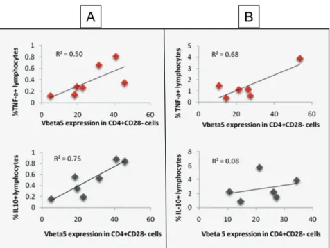
Although cells from cardiac patients are able to produce IL-10, the ratio of this cytokine to TNF-alpha seems to be lower in cardiac patients (Souza et al. 2004). The lower expression of IL-10 has been associated with the occurrence of a gene polymorphism in the promoter region of the IL-10 gene (Costa et al. 2009). The associa-tion of this polymorphism with cardiac Chagas disease Fig. 2: immunoregulation in human Chagas disease: distinct functions of CD28- T-cells. Correlative analysis of the frequency of CD4+
CD28-Vbeta5+ T-cells and TNF-alpha or IL-10-expressing cells was performed in indeterminate (A) and cardiac (B) chagasic patients. Peripheral
blood mononuclear cells were obtained from indeterminate and severe cardiac chagasic patients and analyzed using flow cytometry to de-termine the values obtained in X and Y axis, as previously done by us (Menezes et al. 2004). The data showed a positive correlation between the frequency of CD4+CD28-Vbeta5+ cells and IL-10 as well as TNF-alpha in indeterminate, suggesting a balance in the expression of these
cytokines by this cell sub-population, leading to a co-regulation of inflammatory/anti-inflammatory responses. On the other hand, a positive correlation was only observed between CD4+CD28-Vbeta5+ cells and TNF-alpha, but not IL-10, in cardiac patients. This suggests a
points to an important genetic susceptibility factor that could influence the outcome of the immune response in these patients.
Genetic susceptibility to development of cardiomy-opathy has been described with regards to polymorphism in genes that code for molecules involved in the control of the immune response, especially cytokines. Recently, an
association with polymorphisms in lymphotoxin, MCP-1, Bat-1 and NFkB genes, among others, was described for cardiac Chagas disease in the Brazilian population (Ramazawmy et al. 2006a, b, 2007, 2008). Taken togeth-er, these data show that genetic predisposition can also influence the outcome of Chagas disease. Thus, typing of gene polymorphisms could be an important approach
for identifying groups of individuals at risk of develop-ing severe disease.
The current picture shows that cardiac chagasic pa-tients display an inflammatory cytokine profile, consist-ent with the tissue damage observed in these individuals. Moreover, specific cell populations are involved in the establishment of the cytokine environment that favours inflammation. Most importantly, all of the data, taken together, suggest that a lack of control of the inflamma-tory response is a major cause of pathology establish-ment in cardiac Chagas disease.
A critical analysis of the data presented in this re-view suggests that the generation of protective or patho-genic responses in human Chagas disease is highly influenced by the complexity of the immune response generated during T. cruzi infection. The anti-parasitic response, crucial for chronification of infection, may work as a double-edged sword if not properly modu-lated. It is clear that while an activated, inflammatory, response may be beneficial in the early stages of infec-tion, lack of control of this response later on will allow for the establishment of pathology. Fig. 3 summarises the cellular immunological characteristics of the differ-ent clinical forms. Parasite, environmdiffer-ental and genetic factors are the key players in the establishment of the different responses. The use of genetic studies to iden-tify groups at risk of developing severe disease opens new perspectives for disease surveillance. However, it is likely that more than one approach needs to be taken towards designing strategies for disease control and prevention. Our ongoing quest for efficient diagnosis, therapy and prevention is fully justified.
ACKNOWLEDGEMENTS
To all the researchers whose scientific contributions, cited here or not, have allowed for great progress towards the under-standing of Chagas disease.
REFERENCES
Adad SJ, Andrade DC, Lopes ER, Chapadeiro E 1991. Pathological anatomy of chagasic megaesophagus. Rev Inst Med Trop São Paulo 33: 443-450.
Adad SJ, Cançado CG, Etchebehere RM, Teixeira VP, Gomes UA, Chapadeiro E, Lopes ER 2001. Neuron count reevaluation in the myoenteric plexus of chagasic megacolon after morphometric neuron analysis. Virchows Arch 438: 254-258.
Albareda MC, Laucella SA, Alvarez MG, Armenti AH, Bertochi G, Tarleton RL, Ponstan M 2006. T. cruzi modulates the profile of memory CD8+ T cells in chronic Chagas’ disease patients. Int
Immunol18: 465-471.
Antonelli LR, Dutra WO, Almeida RP, Bacellar O, Gollob KJ 2004. Antigen specific correlations of cellular immune responses in hu-man leishhu-maniasis suggests mechanisms for immunoregulation.
Clin Exp Immunol 136: 341-348.
Antonelli LR, Dutra WO, Oliveira RR, Torres KC, Guimaraes LH, Bacellar O, Gollob KJ 2006. Disparate immunoregulatory poten-tials for double-negative (CD4- CD8-) alphabeta and gammadelta T cells from human patients with cutaneous leishmaniasis. Infect Immun 74: 6317-6323.
Apt W, Arribada A, Zulantay I, Sanchez G, Vargas SL, Rodriguez J 2003. Itraconazole or allopurinol in the treatment of chronic American trypanosomiasis: the regression and prevention of
electrocardiographic abnormalities during 9 years of follow-up.
Ann Trop Med Parasitol 97: 23-29.
Aufderheide AC, Salo W, Madden M, Streitz J, Buikstra J, Guhl F, Arriaza B, Renier C, Wittmers LE Jr, Fornaciari G, Allison M 2004. A 9,000-year record of Chagas’ disease. Proc Natl Acad Sci USA 17: 2034-2039.
Bahia-Oliveira LM, Gomes JA, Cançado JR, Ferrari TC, Lemos EM, Luz ZM, Moreira MC, Gazzinelli G, Correa-Oliveira R 2000. Immunological and clinical evaluation of chagasic patients sub-jected to chemotherapy during the acute phase of Trypanosoma cruzi infection 14-30 years ago. J Infect Dis 182: 634-638.
Beard CB, Pye G, Steurer FJ, Rodriguez R, Campman R, Peterson AT, Ramsey J, Wirtz RA, Robinson LE 2003. Chagas disease in a domestic transmission cycle, Southern Texas, USA. Emerg Infect Dis 9: 103-105.
Benoist C, Mathis D 2001. Autoimmunity provoked by infection: how good is the case for T cell epitope mimicry? Nat Immunol 2: 797-801.
Benvenuti LA, Roggério A, Sambiase NV, Fiorelli A, Higuchi M de L 2005. Polymerase chain reaction in endomyocardial biopsies for monitoring reactivation of Chagas’ disease in heart transplanta-tion: a case report and review of the literature. Cardiovasc Pathol 14: 265-268.
Bocchi EA, Bellotti G, Mocelin AO, Uip D, Bacal F, Higuchi ML, Amato-Neto V, Fiorelli A, Stolf NA, Jatene AD, Pileggi F 1996. Heart transplantation for chronic Chagas’ heart disease. Ann Thorac Surg 61:1727-1733.
Cardoso GM, Morato MJ, Gomes JA, Rocha MO, Bonfim IP, Wil-liams-Blangero S, VandeBerg JL, Reis MR, Magalhães EF, Cor-rea-Oliveira R 2006. Comparative analysis of cell phenotypes in different severe clinical forms of Chagas’ disease. Front Biosci 11: 1158-1163.
Corbett CE, Ribeiro U Jr, Prianti MG, Habr-Gama A, Okumura M, Gama-Rodrigues J 2001. Cell-mediated immune response in megacolon from patients with chronic Chagas’ disease. Dis Co-lon Rectum44: 993-998.
Costa GC, da Costa Rocha MO, Moreira PR, Menezes CA, Silva MR, Gollob KJ, Dutra WO 2009. Functional IL-10 gene polymor-phism is associated with Chagas disease cardiomyopathy. J Infect Dis 199: 451-454.
Costa RP, Gollob KJ, Fonseca LL, Rocha MO, Chaves AC, Medrano-Mercado N, Araújo-Jorge TC, Antas PR, Colley DG, Correa-Ol-iveira R, Gazzinelli G, Carvalho-Parra J, Dutra WO 2000. T-cell repertoire analysis in acute and chronic human Chagas’ disease: differential frequencies of Vbeta5 expressing T cells. Scand J Im-munol 51: 511-519.
Cunha-Neto E, Bilate AM, Hyland KV, Fonseca SG, Kalil J, Engman DM 2006. Induction of cardiac autoimmunity in Chagas heart disease: a case for molecular mimicry. Autoimmunity 39: 41-54.
Cunha-Neto E, Coelho V, Guilherme L, Fiorelli A, Stolf N, Kalil J 1996. Autoimmunity in Chagas’ disease. Identification of car-diac myosin-B13 Trypanosoma cruzi protein crossreactive T cell clones in heart lesions of a chronic Chagas’ cardiomyopathy pa-tient. J Clin Invest 98: 1709-1712.
da Silveira AB, Adad SJ, Correa-Oliveira R, Furness JB, D’Avila Reis D 2007a. Morphometric study of eosinophils, mast cells, mac-rophages and fibrosis in the colon of chronic chagasic patients with and without megacolon. Parasitology 134: 789-796.
chagasic patients with and without megaesophagus. Parasitology 131: 627-634.
da Silveira AB, Correa-Oliveira R, Matsuyama H, de Oliveira EC, Neto SG, Luquetti AO, Furness JB, d’Avila Reis D 2008a. De-creased expression of IK channels in neurons from enteric ner-vous system is associated with the development of chagasic megacolon. Hum Pathol 39: 1406-1407.
da Silveira AB, D’Avila Reis D, de Oliveira EC, Neto SG, Luquetti AO, Poole D, Correa-Oliveira R, Furness JB 2007b. Neurochemi-cal coding of the enteric nervous system in chagasic patients with megacolon. Dig Dis Sci 52: 2877-2883.
da Silveira AB, de Araújo FF, Freitas MA, Gomes JA, Chaves AT, de Oliveira EC, Neto SG, Luquetti AO, da Cunha Souza G, Ber-nardino Júnior R, Fujiwara R, d’Avila Reis D, Correa-Oliveira R 2009. Characterization of the presence and distribution of Foxp3(+) cells in chagasic patients with and without megacolon.
Hum Immunol 70: 65-7.
da Silveira AB, Freitas MA, de Oliveira EC, Neto SG, Luquetti AO, Furness JB, Correa-Oliveira R, d’Avila Reis D 2008b. Neuronal plasticity of the enteric nervous system is correlated with cha-gasic megacolon development. Parasitology135: 1337-1342.
da Silveira AB, Lemos EM, Adad SJ, Correa-Oliveira R, Furness JB, D’Avila Reis D 2007c. Megacolon in Chagas disease: a study of inflammatory cells, enteric nerves, and glial cells. Hum Pathol 38: 1256-1264.
d’Avila Reis D, Lemos EM, Silva GC, Adad SJ, McCurley T, Correa-Oliveira R, Machado CR 2001. Phenotypic characterization of the inflammatory cells in chagasic megaoesophagus. Trans R Soc Trop Med Hyg 95: 177-178.
Dutra WO, Colley DG, Pinto-Dias JC, Gazzinelli G, Brener Z, Pereira ME, Coffman RL, Correa-Oliveira R, Carvalho-Parra JF 2000. Self and nonself stimulatory molecules induce preferential ex-pansion of CD5C B cells or activated T cells chagasic patients, respectively. Scand J Immunol 51: 91-97.
Dutra WO, Gollob KJ 2008. Current concepts in immunoregulation and pathology of human Chagas disease. Curr Opin Infect Dis 21: 287-292.
Dutra WO, Martins-Filho OA, Cançado JR, Pinto-Dias JC, Brener Z, Freeman Júnior GL, Colley DG, Gazzinelli G, Parra JC 1994. Activated T and B lymphocytes in peripheral blood of patients with Chagas disease. Int Immunol 6: 499-506.
Dutra WO, Martins-Filho OA, Cançado JR, Pinto-Dias JC, Brener Z, Gazzinelli G, Carvalho JF, Colley DG 1996. Chagasic patients lack CD28 expression on many of their circulating T lympho-cytes. Scand J Immunol 43: 88-93.
Elias FE, Vigliano CA, Laguens RP, Levin MJ, Berek C 2003. Analy-sis of the presence of Trypanosoma cruzi in the heart tissue of three patients with chronic Chagas’ heart disease. Am J Trop Med Hyg 68: 242-247.
Ferreira MS, Nishioka S, Silvestre MT, Borges AS, Nunes-Araujo FR, Rocha A 1997. Reactivation of Chagas’ disease in patients with AIDS: report of three new cases and review of the literature.
Clin Infect Dis 25: 1397-1400.
Ferreira RC, Ianni BM, Abel LC, Buck P, Mady C, Kalil J, Cunha-Neto E 2003. Increased plasma levels of tumor necrosis factor-alpha in asymptomatic/”indeterminate” and Chagas disease car-diomyopathy patients. Mem Inst Oswaldo Cruz98: 407-411.
Fonseca SG, Reis MM, Coelho V, Nogueira LG, Monteiro SM, Maire-na EC, Bacal F, Bocchi E, Guilherme L, Zheng XX, Liew FY, Higuchi ML, Kalil J, Cunha-Neto E 2007. Locally produced sur-vival cytokines IL-15 and IL-7 may be associated to the
predomi-nance of CD8+ T cells at heart lesions of human chronic Chagas
disease cardiomyopathy. Scand J Immunol66: 362-371.
Fuenmayor C, Higuchi ML, Carrasco H, Parada H, Gutierrez P, Aiello V, Palomino S 2005. Acute Chagas’ disease: immunohistochemi-cal characteristics of T cell infiltrate and its relationship with T. cruzi parasitic antigens. Acta Cardiol 60: 33-67.
Furness JB 2000. Types of neurons in the enteric nervous system.
J Auton Nerv Syst 81: 87-96.
Gaze ST, Dutra WO, Lessa M, Lessa H, Guimarães LH, Jesus AR, Carvalho LP, Machado P, Carvalho EM, Gollob KJ 2006. Mu-cosal leishmaniasis patients display an activated inflammatory T-cell phenotype associated with a nonbalanced monocyte popu-lation. Scand J Immunol 63: 70-78.
Gazzinelli RT, Gazzinelli G, Cançado JR, Cardoso JE, Brener Z, Col-ley DG 1990. Two models of idiotypic stimulation of T lympho-cytes from patients with Chagas disease: correlations with clini-cal forms of infection. Res Immunol 140: 757-761.
Girones N, Cuervo H, Fresno M 2005. T. cruzi-induced molecular mimicry and Chagas’ disease. Curr Top Microbiol Immunol 296: 89-123.
Gollob KJ, Antonelli LR, Faria DR, Keesen TS, Dutra WO 2008. Im-munoregulatory mechanisms and CD4-CD8- (double negative) T cell subpopulations in human cutaneous leishmaniasis: a balanc-ing act between protection and pathology. Int Immunopharmacol 8: 1338-1343.
Gomes JA, Bahia-Oliveira LM, Rocha MO, Martins-Filho OA, Gazzinelli G, Correa-Oliveira R 2003. Evidence that develop-ment of severe cardiomyopathy in human Chagas’ disease is due to a Th1-specific immune response. Infect Immun 71: 1185-1193.
Guhl F, Jaramillo C, Vallejo GA, Cárdenas A-Arroyo F, Aufderheide A 2000. Chagas disease and human migration. Mem Inst Oswal-do Cruz 95: 553-555.
Gurtler RE, Cecere MC, Lauricella MA, Cardinal MV, Kitron U, Co-hen JE 2007. Domestic dogs and cats as sources of T. cruzi infec-tion in rural Northwestern Argentina. Parasitology 134: 69-82.
Hancock K, Zajac AM, Pung OJ, Elvinger F, Rosypal AC, Lindsay DS 2005. Prevalence of antibodies to T. cruzi in raccoons ( Pro-cyon lotor) from an urban area of Northern Virginia. J Parasitol 91: 470-472.
Hyland KV, Engman DM 2006. Further thoughts on where we stand on the autoimmunity hypothesis of Chagas disease. Trends Para-sitol 22: 101-102.
Jones EM, Colley DG, Tostes S, Lopes ER, Vnencak-Jones CL, Mc-Curley TL 1993. Amplification of a T. cruzi DNA sequence from inflammatory lesions in human chagasic cardiomyopathy. Am J Trop Med Hyg 48: 348-357.
Kierszenbaum F 2005. Where do we stand on the autoimmunity hy-pothesis of Chagas disease? Trends Parasitol 21: 513-516.
Köberle F 1968. Chagas’ disease and Chagas’ syndromes: the pathol-ogy of American trypanosomiasis. Adv Parasitol 6: 63-116.
Lages-Silva E, Ramírez LE, Pedrosa AL, Crema E, da Cunha Galvão LM, Junho Pena SD, Macedo AM, Chiari E 2006. Variability of kinetoplast DNA gene signatures of Trypanosoma cruzi II strains from patients with different clinical forms of Chagas’ disease in Brazil. J Clin Microbiol 44: 2167-2171.
Leiby DA, Herron RM Jr, Read EJ, Lenes BA, Stumpf RJ 2002. T. cruzi in Los Angeles and Miami blood donors: impact of evolv-ing donor demographics on seroprevalence and implications for transfusion transmission. Transfusion 42: 549-555.
Lemos EM, Reis D, Adad SJ, Silva GC, Crema E, Correa-Oliveira R 1998. Decreased CD4(+) circulating T lymphocytes in patients with gastrointestinal Chagas disease. Clin Immunol Immuno- pathol 88: 150-155.
Levy MZ, Bowman NM, Kawai V, Waller LA, Cornejo del Carpio JG, Cordova Benzaquen E 2006. Periurban T. cruzi-infected Tria-toma infestans, Arequipa, Peru. Emerg Infect Dis 12: 1345-1352.
Menezes CA, Rocha MO, Souza PE, Chaves AC, Gollob KJ, Dutra WO 2004. Phenotypic and functional characteristics of CD28C and CD28K cells from chagasic patients: distinct repertoire and cytokine expression. Clin Exp Immunol 137: 129-138.
Morato MJ, Brener Z, Cançado JR, Nunes RM, Chiari E, Gazzinelli G 1986. Cellular immune responses of chagasic patients to anti-gens derived from different T. cruzi strains and clones. Am J Trop Med Hyg 35: 505-511.
Ramasawmy R, Cunha-Neto E, Fae KC, Martello FG, Müller NG, Cavalcanti VL, Ianni B, Mady C, Kalil J, Goldberg AC 2006a. The monocyte chemoattractant protein-1 gene polymorphism is associated with cardiomyopathy in human Chagas disease. Clin Infect Dis 43: 305-311.
Ramasawmy R, Cunha-Neto E, Fae KC, Müller NG, Cavalcanti VL, Drigo SA, Ianni B, Mady C, Kalil J, Goldberg AC 2006b.BAT1, a putative anti-inflammatory gene, is associated with chronic Cha-gas cardiomyopathy. J Infect Dis 193: 1394-1399.
Ramasawmy R, Fae KC, Cunha-Neto E, Borba SC, Ianni B, Mady C, Goldberg AC, Kalil J 2008. Variants in the promoter region of
IKBL/NFKBIL1 gene may mark susceptibility to the development of chronic Chagas’ cardiomyopathy among T. cruzi-infected indi-viduals. Mol Immunol 45: 283-288.
Ramasawmy R, Fae KC, Cunha-Neto E, Müller NG, Cavalcanti VL, Ferreira RC, Drigo SA, Ianni B, Mady C, Goldberg AC, Kalil J 2007. Polymorphisms in the gene for lymphotoxin-alpha predispose to chronic Chagas cardiomyopathy. J Infect Dis 196: 1836-1843.
Rassi A Jr, Rassi A, Rassi SG 2007. Predictors of mortality in chronic Chagas disease: a systematic review of observational studies.
Circulation 115: 1101-1108.
Reis DD, Gazzinelli RT, Gazzinelli G, Colley DG 1993a. Antibodies to T. cruzi express idiotypic patterns that can differentiate be-tween patients with asymptomatic or severe Chagas disease. J Immunol 150: 1611-1618.
Reis DD, Jones EM, Tostes S Jr, Lopes ER, Gazzinelli G, Colley DG, McCurley TL 1993b. Characterization of inflammatory infil-trates in chronic chagasic myocardial lesions: presence of TNF-α cells and dominance of granzyme AC, CD8C lymphocytes. Am J Trop Med Hyg 48: 637-642.
Rocha MO, Teixeira MM, Ribeiro AL 2007. An update on the man-agement of Chagas cardiomyopathy. Expert Rev Anti Infect Ther 5: 727-743.
Sosa-Estani S, Segura EL 2006. Etiological treatment in patients in-fected by T. cruzi: experiences in Argentina. Curr Opin Infect Dis 19: 583-587.
Souza PE, Rocha MO, Menezes CA, Coelho JS, Chaves AC, Gollob KJ, Dutra WO 2007. T. cruzi infection induces differential modu-lation of costimulatory molecules and cytokines by monocytes and T cells from patients with indeterminate and cardiac Chagas’ disease. Infect Immun 75: 1886-1894.
Souza PE, Rocha MO, Rocha-Vieira E, Menezes CA, Chaves AC, Gollob KJ, Dutra WO 2004. Monocytes from patients with inde-terminate and cardiac forms of Chagas’ disease display distinct phenotypic and functional characteristics associated with mor-bidity. Infect Immun 72: 5283-5291.
Steindel M, Kramer Pacheco L, Scholl D, Soares M, de Moraes MH, Eger I, Kosmann C, Sincero TC, Stoco PH, Murta SM, de Car-valho-Pinto CJ, Grisard EC 2008. Characterization of T. cruzi
isolated from humans, vectors, and animal reservoirs following an outbreak of acute human Chagas disease in Santa Catarina State, Brazil. Diagn Microbiol Infect Dis 60: 25-32.
Steinkamp M, Geerling I, Seufferlein T, von Boyen G, Egger B, Grossmann J, Ludwig L, Adler G, Reinshagen M 2003. Glial-de-rived neurotrophic factor regulates apoptosis in colonic epithelial cells. Gastroenterology 124: 1748-1757.
Talvani A, Rocha MO, Barcelos LS, Gomes YM, Ribeiro AL, Teix-eira MM 2004. Elevated concentrations of CCL2 and tumor ne-crosis factor-alpha in chagasic cardiomyopathy. Clin Infect Dis 38: 943-950.
Vago AR, Andrade LO, Leite AA, d’Avila Reis D, Macedo AM, Adad SJ, Tostes S Jr, Moreira MC, Filho GB, Pena SD 2000. Genetic characterization of Trypanosoma cruzi directly from tissues of patients with chronic Chagas disease: differential distribution of genetic types into diverse organs. Am J Pathol 156: 1805-1809.
Vago AR, Macedo AM, Adad SJ, Reis DD, Corrêa-Oliveira R 1996. PCR detection of Trypanosoma cruzi DNA in oesophageal tis-sues of patients with chronic digestive Chagas’ disease. Lancet 348: 891-892.
Viotti R, Vigliano C, Lococo B, Bertocchi G, Petti M, Alvarez MG, Postan M, Armenti A 2006. Long-term cardiac outcomes of ing chronic Chagas disease with benznidazole versus no treat-ment: a nonrandomized trial. Ann Intern Med 144: 724-734.
Vitelli-Avelar DM, Sathler-Avelar R, Massara RL, Borges JD, Lage PS, Lana M, Teixeira-Carvalho A, Dias JC, Elói-Santos SM, Martins-Filho OA 2006. Are increased frequency of mac-rophage-like and natural killer (NK) cells, together with high levels of NKT and CD4+CD25high T cells balancing activated
CD8+ T cells, the key to control Chagas’ disease morbidity? Clin
Exp Immunol 145: 81-92.
von Boyen GB, Steinkamp M, Reinshagen M, Schäfer KH, Adler G, Kirsch J 2004. Proinflammatory cytokines increase glial fibril-lary acidic protein expression in enteric glia. Gut 53: 222-228.