Antibiotic resistance in lactic acid bacteria isolated from some pharmaceutical
and dairy products
Gamal Fadl M. Gad
1, Ahmed M. Abdel-Hamid
2, Zeinab Shawky H. Farag
1 1Microbiology Department, Faculty of Pharmacy, El-Minia University, Egypt.
2
Botany and Microbiology Department, Faculty of Science, El-Minia University, Egypt.
Submitted: April 17, 2012; Approved: September 9, 2013.
Abstract
A total of 244 lactic acid bacteria (LAB) strains were isolated from 180 dairy and pharmaceutical products that were collected from different areas in Minia governorate, Egypt. LAB were identified phenotypically on basis of morphological, physiological and biochemical characteristics. Lactobacillusisolates were further confirmed using PCR-based assay. By combination of phenotypic with molecular identificationLactobacillusspp. were found to be the dominant genus (138, 76.7%) followed byStreptococcusspp. (65, 36.1%) andLactococcusspp. (27, 15%). Some contaminant or-ganisms such as (Staphylococcusspp.,Escherichia coli,Salmonellaspp., mould and yeast) were iso-lated from the collected dairy samples but pharmaceutical products were free of such contaminants. Susceptibility of LAB isolates to antibiotics representing all major classes was tested by agar dilution method. Generally, LAB were highly susceptible to Beta-lactams except penicillin. Lactobacilli were resistant to vancomycin, however lactococci and streptococci proved to be very susceptible. Most strains were susceptible to tetracycline and showed a wide range of streptomycin MICs. The MICs of erythromycin and clindamycin for most of the LAB were within the normal range of susceptibility. SixteenLactobacillus, 8Lactococcusand 8Streptococcusisolates including all tetracycline and/or erythromycin resistant strains were tested for the presence of tetracycline and/or erythromycin resis-tant genes [tet(M) and/orerm(B)]. PCR assays shows that some resistant strains harbortet(M) and/or erm(B) resistance genes.
Key words:lactic acid bacteria, phenotypic and molecular identification, PCR assay, antibiotic re-sistance genes.
Introduction
The Food and Agriculture Organization of the United Nations and the World Health Organization (FAO/WHO) defined a probiotic as ‘live microorganisms which when administered in adequate amounts confer a health benefit on the host’ (FAO/WHO, 2002). Lactic acid bacteria (LAB) have received considerable attention as probiotics over the past few years. This concept has grown from tradi-tional dairy products to a profitable market of probiotic health supplements and functional foods. Extensive re-search is done on novel potential probiotic strains, with spe-cific emphasis on their health benefits and mode of action (Dicks and Botes, 2010).
Probiotic strains of lactobacilli are used in different medical and health-related areas including the control of in-testinal inflammation; treating infections during preg-nancy; management of allergic diseases; control of antibi-otic-related diarrhea and prevention of urinary tract infections (Bernardeauet al., 2008). Also, LAB have a role in the treatment of people suffering with tumors and immunocompromised subjects. This may add many com-ponents to conventional therapies, which have relatively low toxicity compared to other treatments (Wood, 1992). Commensal LAB can be used to deliver vaccines and other biologically active material to the gastrointestinal tract. Their use in vaccine delivery is of special value in stimulat-ing mucosal immunity that is protective at the site of patho-gen entry (Hollmannet al., 2010).
Send correspondence to Z.S.H. Farag. Microbiology Department, Faculty of Pharmacy, El-Minia University, Egypt. E-mail: zeinab.shawky@ya-hoo.com.
Regarding the safety assurance of probiotic organ-isms in food, FAO/WHO (2002) guidelines suggest testing probiotic strains for antibiotic resistance patterns. Investi-gation of the antibiotic resistance profiles of LAB is moti-vated by three fundamental reasons. First is the possibility of exchange of resistance factors with other microorgan-isms, with the risk of transferring these genes to many pathogenic bacteria. Second, lactobacilli have been re-ported as the etiological agents in some cases of endo-carditis that can be only controlled by antibiotic therapy (Salvana and Frank, 2006). Finally, the optimization of the use of probiotic lactobacilli in cases of gastrointestinal dis-orders requires the knowledge of their antibiotic resistance to reinforce the concomitant antibiotic therapy (Salminen et al., 1998).
Selective pressure of using antibiotic organisms in both human and animal treatment, and dissemination of an-tibiotic resistance bacteria has the possibility to aggravate acquisition and spread of resistant genes. In this context, probiotic organisms are considered to pool the resistant genes and transfer these to pathogenic bacteria. In order to eliminate this possibility, MIC of the most relevant anti-microbials for each strain used as a probiotic organism could be determined (Rabia and Shah, 2011). Phenotypic assays for characterizing LAB as being either susceptible or resistant to antibiotics have now been complemented by molecular methods, which directly screen for the presence of antibiotic resistance determinants (Perretenet al., 2005). Two of the most commonly observed resistance genes found in LAB so far aretet(M) for tetracycline anderm(B) for erythromycin resistance (Cataloluk and Gogebakan, 2004).
The aim of this study was to accurately identify the dominant LAB that occur in pharmaceutical and dairy products. We use both phenotypic methods and genotypic methods. In addition, to assess the safety of the collected products by detection of contaminants, determination of the antibiotic resistance patterns of LAB strains and to identify the antibiotic resistance determinants through the use of PCR-based molecular methods.
Material and Methods
Bacterial strains
A total of 176 dairy and 4 pharmaceutical probiotic products, were collected during the study period from No-vember 2009 to May 2010 from different markets and phar-macies in Minia governorate, Egypt. Lactobacillus and Lactococcus strains were isolated onto de Man Rogosa Sharpe (MRS) agar (Oxoid, UK), whereas streptococci were isolated onto M17 medium (Oxoid, UK). The isolated strains were stored on selective broth supplemented with 15% glycerol at -20 °C.
Identification of LAB
Phenotypic identification
The identification of LAB was performed according to their morphological, cultural and biochemical character-istics as described (Collins and Lyne, 1980).
Genotypic Identification of Lactobacillus isolates
Lactobacillus isolates were identified to the genus level using PCR-based assay:
a. Bacterial DNA extraction. Bacterial cells were grown in 10 mL MRS broth for 18 h at 37 °C. A 500-mL aliquot of each culture were mixed with 500 mL cetyl-trimethyl ammonium bromide (CTAB) buffer (50 mM hexadecyltrimethyl ammonium bromide, 1.4 mol L-1NaCl 100 mmol L-1 Tris-HCl at pH 8.0, 20 mmol L-1 EDTA, 0.2%b-mercaptoethanol), incubated at 65 °C for 30 min and then centrifuged at 12,000g for 10 min. The super-natant was transferred to a new 1.5 mL Eppendorf tube, precipitated with one volume of isopropanol and centri-fuged at 12,000gfor 10 min. After discarding the super-natant, the pellet was washed with 500 mL of 70% v/v ethanol before drying for 10 min. The pellet was dissolved in 100mL Tris-EDTA buffer (10 mmol L-1Tris-HCl at pH 8.0, 1 mmol L-1EDTA) and stored at -18 °C (Picozziet al., 2006).
b.PCR for identification of the genusLactobacillus. Identification of Lactobacillus strains was performed by genus-specific PCR primers targeted to the 16S/23S ribo-somal RNA intergenic spacer region (Dubernet et al., 2002). The sequences of the primers were taken from GenBank sequence database of the National Center for Biotechnology Information. 5’ - CTC AAA ACT AAA CAA AGT TTC -3’ was used as a forward primer and 5’ -CTT GTA CAC ACC GCC CGT CA- 3’ was used as a re-verse primer. The primers were synthesized by the Midland Certified Reagent Company Inc. (Texas, USA).
The reaction mixture (50mL) contains 2.5mL of each forward and reverse primer (20 pmol of each), 100 ng (4 mL) of the extracted DNA, 25 mL of GoTaq® Green Master Mix (Promega) and 16mL of distilled water. PCR amplification was performed in a DNA thermal cycler (UNO II Thermocycler; Biometra GmbH, Gottingen, Ger-many) with the following temperature program: initial de-naturation at 95 °C for 5 min; 20 cycles of 95 °C for 30 s (denaturation), 55 °C for 30 s (annealing), and 72 °C for 30 s (extension); and a final extension step at 72 °C for 7 min.
Agarose gel electrophoresis was conducted using 1% agarose. The Gene Ruler 100 bp DNA ladder (Fermentas, USA) was used. The gel was stained with ethidium bromide 0.5 mg/mL and observed under UV transilluminator for the presence of DNA bands.
Isolation and identification of contaminant strains. Some contaminants were isolated from the previous 180
pharmaceutical and dairy samples. Identification of these strains was performed according to the procedures de-scribed (Benson, 2002).
Determination of antimicrobial susceptibilities of LAB. Minimum inhibitory concentrations (MICs) of 19 an-tibiotics (Oxoid, UK) were determined by the agar dilution method, according to the Clinical and Laboratory Stan-dards Institute (CLSI) (2007).
PCR detection oftet(M) anderm(B) genes. Isolates that were resistant to tetracycline or erythromycin were subjected for PCR-based detection oftet(M) and erm(B) genes respectively. DNA extraction was performed accord-ing to the standard protocols mentioned earlier. Each PCR reaction (total volume, 50 mL) contained 2 mL of each primer (20 pmol of each), with 50 ng (4mL) of the extracted DNA and 25mL of GoTaq® Green Master Mix (Promega) and 17mL of distilled water. The sequences of the primer used and their amplicon sizes are listed in Table 1. The primers were synthesized by the Midland Certified Reagent Company Inc. (Texas, USA).
PCR-based detection of the tet(M) gene was per-formed using the following thermal cycles: 95 °C for 5 min; 95 °C for 45 s, 52 °C for 45 s and 72 °C for 45 s (25 cycles); and 72 °C for 7 min. While forerm(B) the thermal cycling
program was as follows: 94 °C for 5 min; 94 °C for 1 min, 55 °C for 1 min and 72 °C for 2 min (30 cycles); and 72 °C for 10 min. Amplification products were detected by elec-trophoresis on a 1% agarose and subsequent staining with ethidium bromide solution.
Results
Isolation and identification of LAB
A total of 244 LAB isolates were recovered from 180 dairy and pharmaceutical samples collected from different markets and pharmacies. Out of the 244 isolates, 152 were Lactobacillusspp., 27 wereLactococcusspp. and 65 were Streptococcusspp.
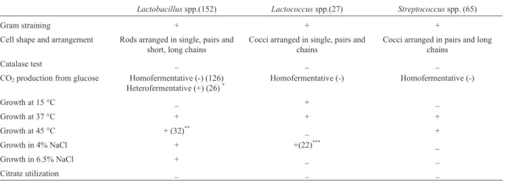
Physiological and biochemical tests for identification of LAB
The isolates were identified phenotypically as illus-trated in Table 2. Based on these characters, the coccid LAB isolates were characterized as mesophilic homofer-mentative cocci, 27 isolates, and so they belonged to Lactococcus spp. and thermophilic homofermentative cocci; 65 isolates. All strains of second group promoted growth at thermophilic conditions, failed to grow in 4 and 6.5% NaCl concentration and so they belonged to
Strepto-Table 1- Primers for PCR detection oftet(M)and erm(B) genes.
Primers Sequence (5’-3’) Amplicon size (bp) PCR type Reference for primers
tet(M) forward GGTGAACATCATAGACACGC 401 tet(M) (Werneret al., 2003)
tet(M) reverse CTTGTTCGAGTTCCAATGC
erm(B) forward CATTTAACGACGAAACTGGC 405 erm(B) (Jensenet al., 1999)
erm(B) reverse GGAACATCTGTGGTATGGCG
Table 2- Phenotypic characteristics of the isolated LAB strains.
Lactobacillusspp.(152) Lactococcusspp.(27) Streptococcusspp. (65)
Gram straining + + +
Cell shape and arrangement Rods arranged in single, pairs and short, long chains
Cocci arranged in single, pairs and chains
Cocci arranged in pairs and long chains
Catalase test _ _ _
CO2production from glucose Homofermentative (-) (126)
Heterofermentative (+) (26)*
Homofermentative (-) Homofermentative (-)
Growth at 15 °C _ + _
Growth at 37 °C + + +
Growth at 45 °C + (32)** _ +
Growth in 4% NaCl + +(22)*** _
Growth in 6.5% NaCl + _ _
Citrate utilization _ _ _
*
126 homofermentative, 26 heterofermentative lactobacilli (152).
**
32 thermophilic lactobacilli (152).
coccus spp.. All the isolates lacked reduction of citrate. Lactobacilli bacteria (152 isolates) were represented by 3 groups; (i), mesophilic homofermentative lactobacilli, 94 isolates; (ii), thermophilic homofermentative lactobacilli, 32 isolates and (iii) mesophilic heterofermentative lacto-bacilli, 26 isolates. All strains grow in 4 and 6.5% NaCl concentration.
Genotypic identification ofLactobacillusspp.
Out of the 152Lactobacillusisolates 138 were con-firmed using PCR-based assay. When DNA from the Lactobacillusstrains were used as a template, a 250 bp PCR product was obtained for all the tested strains (Fig-ure 1).
So by combination of both phenotypic and genotypic identification it was found that theLactobacillusisolates were the most dominant genus (138, 76.7%) followed by Streptococcus(65, 36.1%) and Lactococcus isolates (27, 15%). Table 3 illustrates the incidence of the isolated LAB in relation to the different samples.
Incidence of contaminant strains in pharmaceutical and dairy samples. Out of the 180 pharmaceutical and dairy samples, 123 (68.3%) contaminant strains were isolated and identified. Out of the 123 isolates, 48 were
Staphylo-coccusspp. (39%), 16 wereE.coli(13%), 8 were Salmo-nellaspp. (6.5%) and 51 were mould and yeast (41.5%). No contamination was observed in pharmaceutical samples.
Antibiotic susceptibility and determination of MICs. Tested strains of LAB demonstrated different profiles of antibiotic resistance. Table 4 shows the distributions of MICs of different antibiotics among LAB isolates. When resistance to Beta-lactams was tested, most LAB isolates showed more susceptibility to ampicillin and amoxicillin. The highest prevalence of penicillin resistance was shown among the isolates ofLactobacillusspp. (20.3%). Nearly high percentage ofLactobacillusisolates showed interme-diate resistance to cephalexin and a low percentage were re-sistant to cefoperazone. All Lactococcus isolates were sensitive to cefoperazone.
Lactobacillus strains were highly resistant to vancomycin (40.6%) and streptomycin (17.4%). All LactococcusandStreptococcusisolates were susceptible to vancomycin. High-level of resistance to nalidixic acid, ciprofloxacin and norfloxacin and low-level of resistance to chlormphenicol was detected in LAB isolates. All Lactococcusisolates were susceptible to chlormphenicol. Variations in the susceptibility of erythromycin against LAB were observed. Higher percentage of erythromycin
28 Gadet al.
Figure 1- PCR analysis of someLactobacillusspp. Lane M: 100-bp marker, Lane NC: Negative control, Lane 1-8: 250-bp band ofLactobacillusspp.
Table 3- Incidence of the isolated LAB in relation to the different types of samples.
Sample type Number of
sam-ples (n = 180)
LAB isolates (n = 230)
Lactobacillusisolates (138)
Lactococcusisolates (27)
Streptococcusisolates (65)
No. %* No. %* No. %*
Cheese 68 91 55 80.9 13 19.1 23 33.8
Yogurt 35 71 33 94.3 6 17.1 32 91.4
Fresh Milk 33 22 16 48.5 3 9.1 3 9.1
Fermented milk 30 34 25 83.3 4 13.3 5 16.7
Cream 10 8 5 50 1 10 2 20
Pharmaceutical products 4 4 4 100 0 0 0 0
*
resistance
in
lactic
acid
bacteria
29
Antibiotic Genus Break point*
mg/mL
MIC (mg/mL)** MIC90*** Resistant %****
£0.25 0.5 1 2 4 8 16 32 64 128 256 ³512
Penicillin G Lb 4 36 20 19 14 21 14 12 2 8 28 20.3
Lc 4 20 4 0 1 2 1 2 7.4
St 4 10 22 20 10 2 1 2 1 1.5
Ampicillin Lb 4 40 31 32 29 4 2 2 2 1.4
Lc 4 3 9 8 7 2 0 0
St 4 17 24 12 11 1 2 1 1.5
Amoxycillin Lb 4 42 35 20 22 14 3 2 4 5 3.6
Lc 4 5 8 6 5 2 1 2 1 3.7
St 4 16 18 20 7 2 2 2 2 3.1
Amp/Sul***** Lb 8 49 43 22 21 2 1 2 1 0.7
Lc 8 11 10 4 2 1 0 0
St 8 32 15 9 8 1 2 0 0
Amox/Clav***** Lb 4 66 34 13 16 5 2 2 2 4 2.9
Lc 4 8 10 4 4 1 2 0 0
St 4 12 23 23 5 1 1 1 1 1.5
Cephalexin Lb 16 4 29 26 25 13 12 18 7 4 32 29 21
Lc 16 1 5 8 4 2 1 3 3 16 3 11.1
St 16 15 15 12 10 6 5 2 16 7 10.8
Cefuroxime Lb 8 36 23 24 20 15 10 4 3 3 8 10 7.2
Lc 8 2 2 6 7 4 3 2 1 8 3 11.1
St 8 13 17 14 13 5 3 8 3 4.6
Cefoperazone Lb 16 38 24 21 20 11 9 7 5 3 16 8 5.8
Lc 16 9 5 2 3 4 2 4 0 0
St 16 18 11 12 3 7 6 5 3 16 3 4.6
Vancomycin Lb 4 18 28 23 8 5 6 16 21 13 256 56 40.6
Lc 4 10 8 6 3 1 0 0
St 4 16 18 13 13 5 2 0 0
Gentamicin Lb 8 25 26 11 19 20 16 12 8 1 16 21 15.2
Lc 8 7 8 6 2 2 1 1 16 4 14.8
St 8 11 31 10 5 3 4 1 16 8 12.3
30
Gad
et
al.
Antibiotic Genus Break point*
mg/mL
MIC (mg/mL)** MIC90*** Resistant %****
£0.25 0.5 1 2 4 8 16 32 64 128 256 ³512
Lc 16 2 3 9 6 4 1 2 16 3 11.1
St 16 13 12 10 9 8 8 2 2 1 16 5 7.7
Nalidixic acid Lb 4 23 30 23 16 3 12 15 5 7 2 2 64 46 33.3
Lc 4 4 7 8 2 1 3 2 32 6 22.2
St 4 4 25 9 11 5 3 4 1 3 16 11 16.9
Ciprofloxacin Lb 4 39 26 17 16 9 10 7 9 5 16 31 22.5
Lc 4 6 7 6 4 1 1 1 1 4 3 11.1
St 4 14 3 15 18 9 5 1 4 6 9.2
Norfloxacin Lb 4 21 27 29 14 12 12 4 4 13 2 64 35 25.4
Lc 4 5 8 3 7 3 1 16 4 14.8
St 4 13 17 20 9 2 2 2 4 6 9.2
Erythromycin Lb 4 31 37 32 21 9 1 2 1 3 1 4 8 5.8
Lc 4 6 12 5 2 4 1 1 2 6 22.2
St 4 11 20 12 10 6 2 4 2 3.1
Clindamycin Lb 4 20 24 35 32 11 2 4 3 4 2 1 8 16 11.6
Lc 4 5 5 8 7 2 2 0 0
St 4 10 22 12 11 8 1 1 4 2 3.1
Tetracycline Lb 8 11 26 31 33 11 10 8 3 3 2 64 16 11.6
Lc 4 9 4 3 3 2 4 2 64 8 29.6
St 4 2 8 11 19 17 5 1 1 1 32 8 12.3
Chloramphenicol Lb 4 31 44 39 19 3 2 4 5 3.6
Lc 8 8 13 5 1 4 0 0
St 8 8 5 17 25 10 7 1 4 1 1.5
Rifampicin Lb 4 41 33 20 12 16 2 6 6 2 8 16 11.6
Lc 4 16 2 6 1 1 1 1 2 7.4
St 4 23 5 5 12 13 7 4 7 10.7
*Break points of different antibiotics according to (CLSI, 2007). **
MIC: minimum inhibitory concentration.
***MIC
90: minimum inhibitory concentration for 90% of isolates.
****Percents were correlated to the number of isolates of each microorganism.
Lb:Lactobacillusspp., Lc:Lactococcusspp., St:Streptococcusspp.
resistant strains was observed among lactococci (22.2%). Lactococcusisolates showed high resistance to tetracycline (29.6%) followed by Streptococcus (12.3%) and Lactobacillusisolates (11.6%).
PCR detection oftet(M) anderm(B) resistance genes
EighteenLactobacillusspp. isolates, 9Lactococcus spp. isolates and 9Streptococcusspp. isolates including all tetracycline and/or erythromycin resistant strains were tested for the presence oftet(M) anderm(B) antibiotic re-sistance genes corresponding to their rere-sistance pheno-types. Some strains were to be positive for one or both genes, giving a 401-bp band fortet(M), and a 405-bp band forerm(B) genes (Table 5 and Figures 2, 3).
Discussion
LAB are regarded as a major group of probiotic bac-teria (Bernardeauet al., 2008). In the present work, the inci-dence of LAB was studied. Lactobacillus was the most prevalent genus isolated from dairy and pharmaceutical products (76.7% of total samples). This consistent with the finding of Raquibet al.(2003). On the other hand, the re-sults of Harun-ur-Rashidet al.(2007) contradict with our finding. They isolated a total of 266 strains of LAB from 28 Dahi samples withStreptococcus(50%) as the most domi-nant genus followed by Lactobacillus (27%), and Lactococcus(5%).
LAB were initially identified phenotypically on basis of morphological, physiological and biochemical charac-teristics. PCR analysis was then used for identification of Lactobacillusisolates. Out of the 152 isolates identified phenotypically, 138 were confirmed asLactobacillus rep-resenting only 56.6% of total LAB isolates. We observed
some false positive results after phenotypic characteriza-tion compared with molecular identificacharacteriza-tion of genus Lactobacillusthat could be attributed to the experimental conditions used in isolation and identification. Also Wang et al.(2008) agreed with us in the necessity of combination of conventional identification with molecular techniques in order to obtain more exact results.
In order to examine the safety of collected products, the percentage of contamination was determined in the col-lected samples. We found that 123 contaminant strains were recovered from the dairy samples, whereas pharma-ceutical products were contaminant free. These results can be explained by the fact that the methods of production of the various traditional foods are usually primitive and the major risk enhancing factors such as the use of contami-nated raw materials, lack of pasteurization and inadequate fermentation and storage conditions. These conditions with the antibiotic resistances among LAB require more atten-tion to be focused on the usage and safety of these benefi-cial strains in dairy samples. Similar results were obtained by Soomro and Masud (2007) who isolated some contami-nants such as Staphylococcus, Micrococcus and Saccharomycesspp. from randomly collected market dahi samples from Rawalpindi, Pakistan.
Our study revealed high susceptibility of LAB iso-lates to ampicillin and amoxicillin and more resistance to cephalosporins, also, high vancomycin resistance rate was observed. This is corroborated by data from other groups (Ammor et al., 2007). The strains tested in this study showed also a high susceptibility toward erythromycin and tetracycline. These observations confirmed the data ported by Danielsen and Wind (2003). In contrast to our re-sults Hoqueet al.(2010) found that theLactobacillusspp. were sensitive to clindamycin and highly resistant to tetra-cycline. Our results agreed with those of Ammor et al.
Table 5-tet(M) anderm(B) antibiotic resistance genes detected in LAB isolates.
Genus Number of examined strains Relevant phenotype Genes detected by PCR
Lactobacillusspp. 3 Tetr, Err tet(M),erm(B)
3 Tetr, Err tet(M)
9 Tetr tet(M)
1 Tetr ——
2 Err erm(B)
Lactococcusspp. 1 Tetr, Err tet(M),erm(B)
4 Tetr, Err tet(M)
1 Tetr tet(M)
2 Tetr ——
1 Err erm(B)
Streptococcusspp. 1 Tetr, Err tet(M)
3 Tetr tet(M)
4 Tetr
——-(2007) who reported that the resistance of many Lactobacillusspecies toward vancomycin has been often described as intrinsic. However, Limet al.(1995) found that isolatedLactobacillusspp. were susceptible to vanco-mycin but resistant to gentamicin and streptovanco-mycin.
Phenotypic assays that used to determine the antibi-otic susceptible/resistant patterns have been complemented by molecular methods in which bacterial strains are directly screened for the presence of antibiotic resistance determi-nants. We use PCR for detection oftet(M) anderm(B) re-sistance genes in LAB. It was found that some strains harbor tet(M) and/orerm(B) genes and others that were previously showed tetracycline or erythromycin resistant patterns, were found to be negative fortet(M) orerm(B) genes respectively. These false results can be explained by the fact that there is currently no standard method for anti-biotic susceptibility testing of LAB, although several microdilution methods have been used. Also, many factors may affect the susceptibility results such as the inoculum size, the incubation time, the incubation temperature, the composition of the atmosphere and the growth medium. An
increased inoculum size and an extended incubation time resulted in elevated antibiotic MICs for some species (Egervärnet al., 2007).
Nawaz et al. (2011) reported that out of 84 LAB strains,erm(B) gene was detected in eightLactobacillus strains and oneStreptococcus thermophilusstrain. Thetet genes were identified in 12 strains of lactobacilli from tra-ditional foods which is consistent with our results. Also, de-tection oftet(M) anderm(B) resistance genes have been previously investigated (Devirgiliiset al., 2010; Toomeyet al., 2010).
Conclusion
This study had established that wide variety of LAB are present in the Egyptian products and lactobacilli are considered to be one of the most important potential probiotics. Accurate characterization and identification of LAB and the precise screening for the presence of antibi-otic resistance determinants requires the combined use of phenotypic properties and molecular methods since, con-ventional methods are time-consuming and not fully
reli-32 Gadet al.
Figure 3- PCR detection ofermBresistance gene inLactobacillusspp. Lane M: 100-bp marker, Lane NC: Negative control, Lane 1, 4, 5, 6 and 7: 405-bp band ofermBgene, Lane 2, 3 and 8: no bands with DNA (noermB gene).
able. Also, isolated and identified LAB from pharmaceuti-cal products show higher safety properties regarding contamination and antibiotic resistance if compared with commercial dairy products in this study. This is attributed to the more strict quality control measures and the proper characterization and maintenance of starter culture strains during the production of pharmaceutical products.
References
Ammor MS, Florez AB, Mayo B (2007) Antibiotic resistance in non-enterococcal lactic acid bacteria and bifidobacteria. Food Microbiol 24:559-70.
Benson HJ (2002) Microbiological Application: Laboratory man-ual in general microbiology, 11thed., McGram-Hill Higher Education, Sanfrancisco.
Bernardeau M, Vernoux JP, Henri-Dubernet S, Gueguen M (2008) Safety assessment of dairy microorganisms: the
Lactobacillusgenus. Int J Food Microbiol 126:278-85. Cataloluk O, Gogebakan B (2004) Presence of drug resistance in
intestinal lactobacilli of dairy and human origin in Turkey. FEMS Microbiol Lett 236:7-12.
Clinical and Laboratory Standards Institute (CLSI) (2007) Perfor-mance standards for antimicrobial susceptibility testing. 17th ed. Wayne, PA.
Collins CH, Lyne PM (1980) Microbiological Methods, Vol. IV, Butterworths, London, UK.
Danielsen M, Wind A (2003) Susceptibility ofLactobacillusspp. to antimicrobial agents. Int J Food Microbiol 82:1-11. Devirgiliis C, Barile S, Caravelli A, Coppola D, Perozzi G (2010)
Identification of tetracycline- and erythromycin-resistant Gram-positive cocci within the fermenting microflora of an Italian dairy food product. J Appl Microbiol 109:313-323. Dicks LMT, Botes M (2010) Probiotic lactic acid bacteria in the
gastro-intestinal tract: health benefits, safety and mode of action. Beneficial Microbes 1:11-29.
Dubernet S, Desmasures N, Guéguen M (2002) A PCR-based method for identification of lactobacilli at the genus level. FEMS Microbiol Lett 214:271-75.
Egervärn M, Lindmark H, Roos S, Huys G, Lindgren S (2007) Ef-fects of Inoculum size and incubation time on broth micro-dilution susceptibility testing of lactic acid bacteria. Antimicrob Agents Chemother 51:394-396.
FAO/WHO (Food and Agriculture Organization/World Health Organization) (2002) Guidelines for the evaluation of probiotics in food. Report of a Joint FAO/WHO Working Group on Drafting Guidelines for the Evaluation of Probiotics in Food; Ontario, Canada.
Harun-ur-Rashid M, Togo K, Ueda M, Miyamoto T (2007) Identi-fication and characterization of dominant lactic acid bacteria isolated from traditional fermented milk Dahi in Bangla-desh. World J Microbiol Biotechnol 23:125-133.
Hollmann A, Delfederico L, Miyoshi A, Disalvo EA, De Antoni G, Semorile L, Azevedo V (2010) S-layer proteins from
lactobacilli as vaccine delivery systems. Int J Microbiol Res 2:30-43.
Hoque MZ, Akter F, Hossain KM, Rahman MSM, Billah MM, Is-lam KMD (2010) Isolation, identification and analysis of probiotic properties ofLactobacillus Spp.from selective re-gional yoghurts. World J Dairy and Food Sciences 5:39-46. Jensen LB, Frimodt-Moller N, Aarestrup FM (1999) Presence of
ermgene classes in Gram-positive bacteria of animal and
human origin in Denmark. FEMS Microbiol Lett
170:151-158.
Lim KS, Huh CS, Baek YJ (1995) Studies on the antimicrobial susceptibility of lactic acid bacteria in cultured milk prod-ucts. Dairy Science Abstracts 57:992-993.
Nawaz M, Wang J, Zhou A, Ma C, Wu X, Moore JE, Millar BC, Xu J (2011) Characterization and transfer of antibiotic resis-tance in lactic acid bacteria from fermented food products. Curr Microbiol 62:1081-1089.
Perreten V, Vorlet-Fawer L, Slickers P, Ehricht R, Kuhnert P, Frey J (2005) Microarray-based detection of 90 antibiotic resistance genes of Gram-positive bacteria. J Clin Microbiol 43:2291-302.
Picozzi C, D’Anchise F, R Foschino (2006) PCR detection of
Lactobacillus sanfranciscensisin sourdough and Panettone baked product. Eur Food Res Technol 222:330-35. Rabia A, Shah NP (2011) Antibiotic resistance of probiotic
organ-isms and safety of probiotic dairy products. Int Food Res J 18:59-75.
Raquib M, Trishna B, Choudhary RK, Rahaman H, Borpuzari T (2003) Isolation and characterization of lactobacilli isolated from market sample of sour dahi. Indian Vet J 80:791-794. Salminen S, Von Wright A, Morelli L, Marteau P, Brassart D, De
Vos WM, Fonden R, Saxelin M, Collins K, Mogensen G, Birkeland SE, Mattila-Sandholm T (1998) Demonstration of safety of probiotics - A review. Int J Food Microbiol 44:93-106.
Salvana EMT, Frank M (2006)Lactobacillusendocarditis: case report and review of cases reported since 1992. J Infect 53:5-10.
Soomro AH, Masud T (2007) Protein Pattern and Plasmid Profile of LAB, Food Technol. Biotechnol 45:447-453.
Toomey N, Bolton D, Fanning S (2010) Characterisation and transferability of antibiotic resistance genes from lactic acid bacteria isolated from Irish pork and beef abattoirs. Re-search in Microbiology 161:127-135.
Wang J, Chen X, Liu W, Yang M, Zhang H (2008) Identification ofLactobacillusfrom koumiss by conventional and molecu-lar methods. Eur Food Res Technol 227:1555-61.
Werner G, Willems RJ, Hildebrandt B, Klare I, Witte W (2003) Influence of transferable genetic determinants on the out-come of typing methods commonly used forEnterococcus faecium. J Clin Microbiol 41:1499-506.
Wood JB (1992) The lactic acid bacteria in health & diseases. Elsevier Applied Science 1:23-27.


![Figure 2 - PCR detection of tet(M) resistance gene in some Lactobacillus spp. Lane M: 100-bp marker, lane NC: Negative control, lane 2-8: 401-bp band of tet(M) gene, lane 1: no bands with DNA [no tet(M) gene].](https://thumb-eu.123doks.com/thumbv2/123dok_br/15804631.649601/8.918.252.668.425.651/figure-detection-resistance-lactobacillus-lane-marker-negative-control.webp)
