Molecular typing of
Mycobacterium bovis
isolates
:
A review
Daniela Fernandes Ramos
1, Lucas Tavares
1, Pedro Eduardo Almeida da Silva
2,
Odir Antônio Dellagostin
11
Núcleo de Biotecnologia, Universidade Federal de Pelotas, Pelotas, RS, Brazil.
2
Faculdade de Medicina, Universidade Federal de Rio Grande, Rio Grande, RS, Brazil.
Submitted: November 12, 2012; Approved: September 9, 2013.
Abstract
Mycobacterium bovisis the main causative agent of animal tuberculosis (TB) and it may cause TB in humans. Molecular typing ofM. bovisisolates provides precise epidemiological data on issues of inter- or intra-herd transmission and wildlife reservoirs. Techniques used for typingM. bovishave evolved over the last 2 decades, and PCR-based methods such as spoligotyping and mycobacterial in-terspersed repetitive unit-variable number tandem repeat (MIRU-VNTR) have been extensively used. These techniques can provide epidemiological information about isolates ofM. Bovisthat may help control bovine TB by indicating possible links between diseased animals, detecting and sam-pling outbreaks, and even demonstrating cases of laboratory cross-contamination between samples. This review will focus on techniques used for the molecular typing ofM. bovisand discuss their gen-eral aspects and applications.
Key words:tuberculosis, bovine, diagnosis, genotyping,Mycobacterium bovis.
Introduction
Bovine tuberculosis (BTB) has been detected in cattle throughout the world. According to disease timelines avail-able in the Worldwide Animal Health Information Data-base (OIE, 2009), 109 countries reported the presence of Mycobacterium bovisinfections and/or clinical diseases in their cattle herds at some time in 2005-2010. In developed countries that have a tradition of cattle farming, the preva-lence of BTB has reached very low levels because of strict control policies. In several of these countries, the disease has been eradicated. Conversely, in developing countries, despite recently implanted control mea-sures, considerable economic losses consistently occur in regions such as Brazil and Argentina, where there is intense cattle breeding (Ruggieroet al., 2007).
The first attempt to differentiate M. bovis isolates based on DNA sequence polymorphism was done by Col-lins and de Lisle in 1985 (ColCol-lins and De Leslie, 1985). They performed restriction digestion and agarose gel elec-trophoresis of genomic DNA. Identification of insertion
se-quences inM. tuberculosisandM. bovisgenomes led to the development of the restriction fragment length polymor-phism (RFLP) typing method (Thierryet al., 1990), a tech-nique still in use nowadays. Knowledge of the genomic sequence ofM. bovisandM. tuberculosishas facilitated the development of high-throughput molecular typing tech-niques that allow greater insight into the epidemiology, evolution, and population structure ofM. bovis. Polymor-phic GC Repeat Sequence (Roringet al., 1998; Rosset al., 1992), direct repeats (DR) regions (Hermanset al., 1991), and variable number of tandem repeats (VNTR) (Supplyet al., 2000) have all been exploited as typing methods.
These tools can help to determine the source of infec-tion and outbreaks, understand the relainfec-tionship between different outbreaks, and identify wild animal reservoir of M. bovis. In addition, they can provide insight into the risk factors for BTB transmission by allowing identification of the dynamics of this disease (Broschet al., 2002; Garnieret al., 2003; Zumarragaet al., 2005). In this review, we de-scribe the main techniques used for genotyping and their application in characterizingM. bovisisolates.
Send correspondence to O.A. Dellagostin. Núcleo de Biotecnologia, Universidade Federal de Pelotas, Pelotas, RS. E-mail: odirad@terra.com.br, odirad@gmail.com.
Restriction Fragment Length Polymorphism
(RFLP)
Strain differentiation by using RFLP analysis has proven to be a very useful tool for epidemiologic studies of tuberculosis. RFLP based on the presence of the insertion sequenceIS6110has been widely used as a genetic marker (Otal et al., 1991). IS6110 fingerprinting via RFLP has been standardized by usingPvuII as the restriction enzyme of choice to digest mycobacterial genomic DNA (Figure 1) (Broschet al., 2002; Gutierrezet al., 1995; Thierryet al., 1990). After electrophoresis of digested DNA on agarose gel, Southern blotting is carried out. Polymorphic banding patterns is revealed after hybridization by using a fragment ofIS6110as a probe (Durret al., 2000). This insertion se-quence is present in up to 20 copies inM. tuberculosis, thus enabling the application ofIS6110-RFLP as the gold stan-dard genotyping technique for this organism. In contrast, only 1-5 copies ofIS6110are found inM. bovis, which lim-its the ability of this element to discriminate between differ-ent M. bovis strains (Aranaz et al., 1996; Aranaz et al., 1999; vanet al., 1994; van, 2001).
One additional limitation of the RFLP-based typing systems is that they require a well-grown culture for DNA extraction. The time lag between isolation ofM. bovisfrom a clinical sample and the growth of a mycobacterial culture
is often too long. This problem can be circumvented with the use of several complementary biomarkers such as those based in the polymorphicIS6110region, and by using di-rect repeats (DR) and polymorphic GC-rich repeat se-quences (PGRS) as probes (Cousins et al., 1998; van Embdenet al., 1996).
Polymorphic GC-Rich Repeat Sequence
(PGRS)
The PGRS method is similar to standardizedIS6110 fingerprinting in that it requires purified DNA for Southern blot hybridization and banding pattern analysis. PGRS fin-gerprinting has proven to be useful for differentiating strains with fewer than 6 copies ofIS6110 that could not readily be differentiated byIS6110fingerprinting (van et al., 1993; Yanget al., 2000).
The PGRS-based RFLP probe is the single most dis-criminatory of the probes currently available forM. bovis strain typing and can be present in up to 30 copies in mem-bers of theM. tuberculosiscomplex. PGRS is present in multiple copies interspersed throughout the genome and it exhibits a high level of polymorphism between unrelated isolates. However, the result of a PGRS DNA fingerprint is relatively complex because it contains many bands, making it potentially difficult to interpret. For the same reason,
computer-assisted band analysis, particularly when the fi-nal image is less than ideal (O’Brienet al., 2000), can also be difficult.
Polymerase Chain Reaction (PCR) Based
Techniques
Techniques based on DNA amplification via PCR, such as spoligotyping and MIRU-VNTR (Burgos et al., 2004), have become tools for epidemiological studies of bovine tuberculosis transmission, and have given promi-nence to a modern field of research known as molecular ep-idemiology. The agility and speed in detectingM. boviscan be decisive in the choice of these methods, which differen-tiate the species and different isolates of the same species at the DNA level.
Spoligotyping
Spoligotyping (from “spacer oligotyping”) is based on the direct repeat region (DR), a DNA polymorphism present in a particular chromosomal locus that was first de-scribed by Hermanset al...(1991). This chromosomal re-gion contains a large number of DRs of 36 bp each, interspersed with a spacer DNA of 35-41 bp in length. When DR regions of several isolates are compared, it is ob-served that the order of the spacers is about the same in all isolates; however, deletions and insertions of DRs occur (Figure 2). Polymorphisms in various isolates comprise the presence or absence of spacers of known sequence. This characteristic is used to determine genetic similarity among strains (Kontsevayaet al., 2011). Spoligotyping can easily distinguish betweenM. tuberculosisandM. bovis, can be used with DNA extracted from a bacterial culture as well as directly from a specimen, and has been used to identify the clonal nature of the isolates (O’Brienet al., 2000; Zaniniet al., 2001; Zumarragaet al., 1999).
Spoligotyping is reportedly useful for identifying sources of infection, transmission of tuberculosis (TB) be-tween species, and the stability of tuberculosis strains for long periods of time in closed populations, indicating that Mycobacteriumis clonal (Cousinset al., 1998). In an eco-logical setting, spoligotyping is a rapid and inexpensive op-tion that can be used to search for a relaop-tionship between strains (Zumarraga et al., 2012). Because strains of M. bovis from cattle usually contain few copies of IS6110, IS6110-RFLP is not the best method for distinguishing strains ofM. bovis (Allixet al., 2006). Spoligotyping has proven to be a practical and discriminatory method for large-scale studies of the epidemiology ofM. bovisas well as for the differentiation ofM. bovisfromM. tuberculosis, because the former lacks spacers 39-43 (Kamerbeeket al., 1997). Furthermore, there is an international database hold-ing over 1900 spoligotype patterns from around the world (Lazzariniet al., 2012).
The main disadvantage of spoligotyping is that all ge-netic polymorphisms are restricted to a single genomic lo-cus, the DR cluster, which limits the resolution. While having the advantages of being considerably faster and less labor-intensive than RFLP analysis, spoligotyping alone does not usually provide sufficient discrimination among strains ofM. bovisto be used as a sole typing method, and is thus often combined with supplementary techniques (Cos-telloet al., 1999; Cousins et al., 1998; McLernonet al., 2010; Roringet al., 1998).
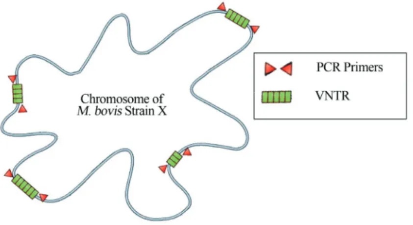
Variable Number Tandem Repeat (VNTR)
Tandemly repeated sequences are dispersed by thou-sands of copies in virtually all higher eukaryote genomes. Loci with short sequence repeats of 1±13 bp are generally referred to as microsatellites, and those with 10±100 bp se-quence repeats as minisatellites (Figure 3). Many of these
loci show hypervariability in their repeat numbers in hu-mans and in animals, and are therefore called VNTR loci (Supplyet al., 2000). VNTR sequences are also found in bacteria, and have been used to genotype many species. Polymorphism at a tandem repeat (TR) locus can occur ei-ther as a result of nucleotide sequence changes between in-dividual repeat units or as a result of variations in the number of repeat units, thereby creating allelic variants. VNTR typing is based upon repeat number polymorphisms within these tandemly arranged repetitive DNA sequences.
There are several VNTR loci in the genome ofM. bovis, and hence VNTR typing provides a greater resolu-tion than spoligotyping alone (Roringet al., 2002). Many of these TR loci display hypervariability, enabling their ex-ploitation for strain typing in numerous bacterial species. Originally, 6 VNTR loci, described as exact TR A through F (ETR-A, -B, -C, -D, -E, and -F), were reported and ap-plied to M. tuberculosis isolates (Frothingham and Meeker-O’Connell, 1998).
However, the level of discrimination found inM. tu-berculosisorM. bovisisolates using the 5 ETRs (A through E) was not as good as that achieved with either spoli-gotyping or IS6110-RFLP typing (Collins et al., 1994; Good and Duignan, 2011). Therefore, another set of poly-morphic repeats, termed as mycobacterial interspersed re-petitive units (MIRU-VNTR) (Roringet al., 2002; Supply et al., 2000; Supplyet al., 2006), has been proposed. Ini-tially, these involved a 12-loci set, which was considered efficient for epidemiological purposes (Supply et al., 2001a), but some limitations were found with regard to the discriminatory power (Garcia, Vet al., 2006; Scottet al., 2005). Furthermore, a 15- or 24-loci subset has been shown to ensure better discrimination (Supplyet al., 2006).
The challenge is to compile standardized molecular fingerprinting patterns originating from highly networked, multi-centric, genotypic analysis in databases for inter-laboratory use and for further references. Rapid genotyping methods are needed to overcome low reproducibility, not
proven application-less discriminatory methods such as MIRU-VNTR (Viedmaet al., 2011). By analyzing the in-tended purpose and possibilities for each method (Table 1), it is possible to use a combination of typing methods and thus accurately identify differences among strains.
Implications of Molecular Typing in
Epidemiology
Within the last 10 years, many techniques have been developed or adapted for typingM. tuberculosiscomplex isolates. In this review, we have presented an overview of the main techniques used for the differentiation ofM. bovis isolates. For the majority of them, a specific and polymor-phic genetic region is involved. In comparison to the major-ity of other bacterial groups, the genome of mycobacteria has a high GC content (GC% is 65%), and its polymor-phism is very limited compared to its genome size (4.4 Mb). However, some regions are highly polymorphic, either due to variations in number and/or position, or be-cause of variations in primary structure. These areas of higher polymorphism appear to correspond essentially to segments of genes encoding proteins where variability pro-vides a selective advantage to the bacteria, such as antibi-otic-resistance proteins, antigens involved in escaping the immune response, or non-coding sequences (insertion se-quences or repeated sese-quences) that are probably involved in inducing variability in neighboring genetic areas.
laboratory and the particular features of a geographic re-gion (Rozo and Ribón, 2010).
Genotyping of bacterial isolates or PCR products is increasingly becoming a standard tool for epidemiological disease control and eradication. DistinguishingM. bovis strains at the molecular level provides important insights into the sources of infection and identification of practices or environments, thereby aiding the spread and mainte-nance of tuberculosis (Medeiroset al., 2010). More impor-tantly, transmission routes between livestock and wildlife may be identified by strain typing. In addition, transmission routes of BTB within livestock via animal movements be-come evident; this is a prerequisite for targeted disease con-trol aiming at testing all potentially exposed animals (Schilleret al., 2010).
PCR-based techniques used for strain fingerprinting have proven to be of useful in relating outbreaks of TB to sources of infection. Epidemiologically related isolates have similar fingerprints that differ from those that are epidemiologically unrelated. Therefore, a desirable charac-teristic for typing is related to its stability within a strain and diversity within a species.
DNA fingerprinting ofM. bovisfor molecular epide-miology has been used to study transmission of bovine tu-berculosis in Latin America and other parts of the world (PARREIRASet al., 2012). However, there are a few re-ports in Brazil with regard to characterization ofM. bovis (Figueiredoet al., 2011; Rodriguezet al., 2004; Zaniniet al., 2005) that revealed the occurrence of a high genetic di-versity; however, these studies were conducted using a lim-ited number of isolates (Zumarragaet al., 1999).
Conclusion
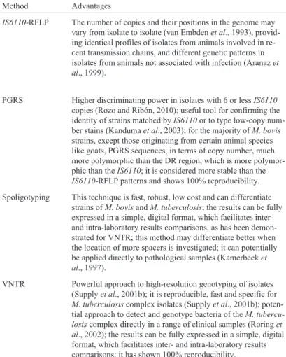
Tuberculosis caused byM. bovisis important for pub-lic health, animal health, and animal production. Whatever the epidemiological context, the need for techniques per-mitting differentiation of isolates at the molecular level is evident. These tools would help determine the origin of out-breaks, increase the understanding with regard to the link between different outbreaks, show the relationship between domestic TB and wild TB, and identify the source of infec-tion. No typing technique developed so far can be used on its own. Each technique has its advantages and disadvan-tages that must be considered when choosing the one to be Table 1- Molecular typing methodsfor Mycobacterium bovis.
Method Advantages Disadvantages
IS6110-RFLP The number of copies and their positions in the genome may vary from isolate to isolate (van Embdenet al., 1993), provid-ing identical profiles of isolates from animals involved in re-cent transmission chains, and different genetic patterns in isolates from animals not associated with infection (Aranazet al., 1999).
Requires large amounts of DNA (1-2mg) and technical skills; it is slow and has little discriminating power in isolates with less than 6IS6110copies (Gutackeret al., 2002); there is diffi-culty in reproducing results and comparing them among differ-ent laboratories (Supplyet al., 2001b); and the majority ofM. bovisisolates have a low number ofIS6110copies-in gen-eral, only 1 or 2 (Haddadet al., 2004).
PGRS Higher discriminating power in isolates with 6 or lessIS6110
copies (Rozo and Ribón, 2010); useful tool for confirming the identity of strains matched byIS6110or to type low-copy num-ber stains (Kandumaet al., 2003); for the majority ofM. bovis
strains, except those originating from certain animal species like goats, PGRS sequences, in terms of copy number, much more polymorphic than the DR region, which is more polymor-phic than theIS6110; it is considered more stable than the
IS6110-RFLP patterns and shows 100% reproducibility.
Requires large amounts of high quality DNA (Asgharzadeh and Kafil, 2007; Doroudchiet al., 2000) and technical skills; it has lower discriminating power in isolates with multiple copies (Baueret al., 1999); the large number of bands produced by this technique makes interpretation of the gels difficult, limit-ing its application as a primary typlimit-ing technique (Kandumaet al., 2003).
Spoligotyping This technique is fast, robust, low cost and can differentiate strains ofM. bovisandM. tuberculosis; the results can be fully expressed in a simple, digital format, which facilitates inter-and intra-laboratory results comparisons, as has been demon-strated for VNTR; this method may differentiate better when the location of more spacers is investigated; it can potentially be applied directly to pathological samples (Kamerbeeket al., 1997).
The discriminatory power of this method is lower than
IS6110-RFLP typing when high copy number strains are being analyzed.
VNTR Powerful approach to high-resolution genotyping of isolates (Supplyet al., 2001b); it is reproducible, fast and specific for
M. tuberculosiscomplex isolates (Supplyet al., 2001b); poten-tial approach to detect and genotype bacteria of theM. tubercu-losiscomplex directly in a range of clinical samples (Roringet al., 2002); the results can be fully expressed in a simple, digital format, which facilitates inter- and intra-laboratory results comparisons; it has shown 100% reproducibility.
The discriminatory power of this method is lower than
implemented in the laboratory. Using spoligotyping in combination with MIRU-VNTR seems to be the best choice as both have the advantages of being PCR-based, with improved discriminatory power when combined.
Hopefully, in the future we will have new and im-proved techniques for typingM. bovis. It is conceivable that a lab-on-a-chip approach will be capable of not only detect-ing M. bovis from a clinical sample but also typing the pathogen at the same time. Regarding the target locus, it is likely that single nucleotide polymorphisms in specific genes will be used for molecular epidemiology.
References
Allix C, Walravens K, Saegerman C, Godfroid J, Supply P, Fauville-Dufaux M (2006) Evaluation of the epidemiologi-cal relevance of variable-number tandem-repeat genotyping ofMycobacterium bovisand comparison of the method with IS6110 restriction fragment length polymorphism analysis and spoligotyping. J Clin Microbiol 44:1951-1962. Aranaz A, Liebana E, Gomez-Mampaso E, Galan JC, Cousins D,
Ortega A, Blazquez J, Baquero F, Mateos A, Suarez G, Dominguez L (1999) Mycobacterium tuberculosissubsp. caprae subsp. nov.: a taxonomic study of a new member of the Mycobacterium tuberculosis complex isolated from goats in Spain. Int J Syst Bacteriol 49:1263-1273. Aranaz A, Liebana E, Mateos A, Dominguez L, Vidal D,
Domingo M, Gonzolez O, Rodriguez-Ferri EF, Bunschoten AE, Van Embden JD, Cousins D (1996) Spacer oligo-nucleotide typing ofMycobacterium bovisstrains from cat-tle and other animals: a tool for studying epidemiology of tu-berculosis. J Clin Microbiol 34:2734-2740.
Asgharzadeh M, Kafil HS (2007) Current trends in Molecular Ep-idemiology Studies of Mycobacterium tuberculosis, pp. 108-115.
Bauer J, Andersen AB, Kremer K, Miorner H (1999) Usefulness of spoligotyping To discriminate IS6110 low-copy-number
Mycobacterium tuberculosis complex strains cultured in Denmark. J Clin Microbiol 37:2602-2606.
Brosch R, Gordon SV, Marmiesse M, Brodin P, Buchrieser C, Eiglmeier K, Garnier T, Gutierrez C, Hewinson G, Kremer K, Parsons LM, Pym AS, Samper S, van SD, Cole ST (2002) A new evolutionary scenario for theMycobacterium tuber-culosiscomplex. Proc Natl Acad Sci USA 99:3684-3689. Burgos MV, Mendez JC, Ribon W (2004) Molecular
epidemiol-ogy of tuberculosis: methodolepidemiol-ogy and applications. Bio-medica. 24 Supp 1:188-201.
Collins DM, De Leslie GW (1985) DNA restriction endonuclease analysis ofMycobacterium bovisand other members of the tuberculosis complex. J Clin Microbiol 21:562-564. Collins DM, Radford AJ, de Leisle GW, Billman-Jacobe H (1994)
Diagnosis and epidemiology of bovine tuberculosis using molecular biological approaches. Vet Microbiol 40:83-94. Costello E, O’Grady D, Flynn O, O’Brien R, Rogers M, Quigley
F, Egan J, Griffin J (1999) Study of restriction fragment length polymorphism analysis and spoligotyping for epide-miological investigation ofMycobacterium bovisinfection. J Clin Microbiol 37:3217-3222.
Cousins DV, Skuce RA, Kazwala RR, Van Embden JD (1998) Towards a standardized approach to DNA fingerprinting of
Mycobacterium bovis. International Union Against Tuber-culosis and Lung Disease, TuberTuber-culosis in Animals Subsection. Int J Tuberc Lung Dis 2:471-478.
Doroudchi M, Kremer K, Basiri EA, Kadivar MR, van SD, Ghaderi AA (2000) IS6110-RFLP and spoligotyping of My-cobacterium tuberculosisisolates in Iran. Scand J Infect Dis 32:663-668.
Durr PA, Hewinson RG, Clifton-Hadley RS (2000) Molecular ep-idemiology of bovine tuberculosis. I.Mycobacterium bovis
genotyping. Rev Sci Tech 19:675-688.
Figueiredo EE, Ramos DF, Medeiros L, Silvestre FG, Lilenbaum W, Silva JT, Paschoalin VM, Dellagostin OA (2011) Multi-ple strains ofMycobacterium bovisrevealed by molecular typing in a herd of cattle. Vet J 193:296-298.
Frothingham R, Meeker-O’Connell WA (1998) Genetic diversity in theMycobacterium tuberculosiscomplex based on vari-able numbers of tandem DNA repeats. Microbiology 144:1189-1196.
Garcia dV, Alonso RN, Andres S, Martinez LM, Ruiz Serrano MJ, Bouza E (2006) Evaluation of alternatives to RFLP for the analysis of clustered cases of tuberculosis. Int. J. Tuberc. Lung Dis 10:454-459.
Garnier T, Eiglmeier K, Camus JC, Medina N, Mansoor H, Pryor M, Duthoy S, Grondin S, Lacroix C, Monsempe C, Simon S, Harris B, Atkin R, Doggett J, Mayes R, Keating L, Wheeler PR, Parkhill J, Barrell BG, Cole ST, Gordon SV, Hewinson RG (2003) The complete genome sequence of Mycobacte-rium bovis. Proc Natl Acad Sci USA 100:7877-7882. Good M, Duignan A (2011) Perspectives on the History of Bovine
TB and the Role of Tuberculin in Bovine TB Eradication. Vet Med Int 2011:410-470.
Gutacker MM, Smoot JC, Migliaccio CA, Ricklefs SM, Hua S, Cousins DV, Graviss EA, Shashkina E, Kreiswirth BN, Musser JM (2002) Genome-wide analysis of synonymous single nucleotide polymorphisms inMycobacterium tuber-culosiscomplex organisms: resolution of genetic relation-ships among closely related microbial strains. Genetics 162:1533-1543.
Gutierrez M, Samper S, Gavigan JA, Garcia Marin JF, Martin C (1995) Differentiation by molecular typing of Mycobacte-rium bovisstrains causing tuberculosis in cattle and goats. J Clin Microbiol 33:2953-2956.
Haddad N, Masselot M, Durand B (2004) Molecular differentia-tion ofMycobacterium bovisisolates. Review of main tech-niques and applications. Res Vet Sci 76:1-18.
Hermans PW, van SD, Bik EM, de Haas PE, Dale JW, Van Embden JD (1991) Insertion element IS987 from Mycobac-terium bovisBCG is located in a hot-spot integration region for insertion elements inMycobacterium tuberculosis com-plex strains. Infect Immun 59:2695-2705.
Kamerbeek J, Schouls L, Kolk A, van AM, van SD, Kuijper S, Bunschoten A, Molhuizen H, Shaw R, Goyal M, Van EJ (1997) Simultaneous detection and strain differentiation of
Mycobacterium tuberculosisfor diagnosis and epidemiol-ogy. J Clin Microbiol 35:907-914.
Kanduma E, McHugh TD, Gillespie SH (2003) Molecular meth-ods for Mycobacterium tuberculosisstrain typing: a users guide. J Appl Microbiol 94:781-791.
Kremer K, van SD, Frothingham R, Haas WH, Hermans PW, Martin C, Palittapongarnpim P, Plikaytis BB, Riley LW, Yakrus MA, Musser JM, van Embden JD (1999) Compari-son of methods based on different molecular epidemiologi-cal markers for typing ofMycobacterium tuberculosis com-plex strains: interlaboratory study of discriminatory power and reproducibility. J Clin Microbiol 37:2607-2618. Lazzarini LC, Rosenfeld J, Huard RC, Hill V, Lapa e Silva JR,
DeSalle R, Rastogi N, Ho JL (2012)Mycobacterium tuber-culosisspoligotypes that may derive from mixed strain in-fections are revealed by a novel computational approach. In-fect Genet Evol 12:798-806.
McLernon J, Costello E, Flynn O, Madigan G, Ryan F (2010) Evaluation of mycobacterial interspersed repetitive-unit-variable-number tandem-repeat analysis and spoligotyping for genotyping ofMycobacterium bovisisolates and a com-parison with restriction fragment length polymorphism typ-ing. J Clin Microbiol 48:4541-4545.
Medeiros LS, Marassi CD, Figueiredo EES, Lilenbaum W (2010) Potential application of new diagnostic methods for control-ling bovine Tuberculosis in Brazil. Braz J Microbiol 41:531-541.
O’Brien R, Flynn O, Costello E, O’Grady D, Rogers M (2000) Identification of a novel DNA probe for strain typing Myco-bacterium bovis by restriction fragment length polymor-phism analysis. J Clin Microbiol 38:1723-1730.
OIE (2009) Annual Animal Disease Status, Bovine Tuberculosis. Otal I, Martin C, Vincent-Levy-Frebault V, Thierry D, Gicquel B
(1991) Restriction fragment length polymorphism analysis using IS6110 as an epidemiological marker in tuberculosis. J Clin Microbiol 29:1252-1254.
Parreiras PM, Andrade GI, Nascimento TF, Oelemann MC, Go-mes HM, Alencan AP, Assis RA, Mota PMPC, Pereiras MAS, Lobato FCF, Lage AP, Suffys PN (2012) Spoli-gotyping and variable number tandem repeat analysis of My-cobacterium bovisisolates from cattle in Brazil. Mem Inst Oswaldo Cruz 107:64-73.
Pheiffer C, Betts JC, Flynn HR, Lukey PT, van HP (2005) Protein expression by a Beijing strain differs from that of another clinical isolate and Mycobacterium tuberculosis H37Rv. Microbiology 151:1139-1150.
Rodriguez CAR, Zumarraga M, Oliveira EMD, Cataldi A, Ro-mano MI, Otto HH, Bonafé VL, Ferreira Neto JS (2004) Caracterização molecular de isolados de Mycobacterium bovisdo Estado de São Paulo Brasil, utilizando a técnica de
spoligotyping. Arq Inst Biol 71:277-282.
Roring S, Brittain D, Bunschoten AE, Hughes MS, Skuce RA, Van Embden JD, Neill SD (1998) Spacer oligotyping of My-cobacterium bovisisolates compared to typing by restriction fragment length polymorphism using PGRS, DR and IS6110 probes. Vet Microbiol 61:111-120.
Roring S, Scott A, Brittain D, Walker I, Hewinson G, Neill S, Skuce R (2002) Development of variable-number tandem repeat typing of Mycobacterium bovis: comparison of sults with those obtained by using existing exact tandem re-peats and spoligotyping. J Clin Microbiol 40:2126-2133. Ross BC, Raios K, Jackson K, Dwyer B (1992) Molecular cloning
of a highly repeated DNA element fromMycobacterium tu-berculosis and its use as an epidemiological tool. J Clin Microbiol 30:942-946.
Rozo AJC, Ribón W (2010) Molecular tools forMycobacterium tuberculosisgenotyping. Rev salud pública 12:510-521. Ruggiero AP, Ikuno AA, Ferreira VCA, Roxo E (2007)
Tubercu-lose bovina: Alternativas para o diagnóstico. Arq Inst Biol 74:55-65.
Schiller I, Oesch B, Vordermeier HM, Palmer MV, Harris BN, Orloski KA, Buddle BM, Thacker TC, Lyashchenko KP, Waters WR (2010) Bovine tuberculosis: a review of current and emerging diagnostic techniques in view of their rele-vance for disease control and eradication. Transbound Emerg Dis 57:205-220.
Scott AN, Menzies D, Tannenbaum TN, Thibert L, Kozak R, Jo-seph L, Schwartzman K, Behr MA (2005) Sensitivities and specificities of spoligotyping and mycobacterial inter-spersed repetitive unit-variable-number tandem repeat typ-ing methods for studytyp-ing molecular epidemiology of tuber-culosis. J Clin Microbiol 43:89-94.
Supply P, Allix C, Lesjean S, Cardoso-Oelemann M, Rusch-Gerdes S, Willery E, Savine E, de HP, van DH, Roring S, Bifani P, Kurepina N, Kreiswirth B, Sola C, Rastogi N, Vatin V, Gutierrez MC, Fauville M, Niemann S, Skuce R, Kremer K, Locht C, van SD (2006) Proposal for standard-ization of optimized mycobacterial interspersed repetitive unit-variable-number tandem repeat typing of Mycobacte-rium tuberculosis. J Clin Microbiol 44:4498-4510. Supply P, Lesjean S, Savine E, Kremer K, van SD, and Locht C
(2001) Automated high-throughput genotyping for study of global epidemiology ofMycobacterium tuberculosisbased on mycobacterial interspersed repetitive units. J Clin Micro-biol 39:3563-3571.
Supply P, Lesjean S, Savine E, Kremer K, van SD, Locht C (2001) Automated high-throughput genotyping for study of global epidemiology of Mycobacterium tuberculosis based on mycobacterial interspersed repetitive units. J Clin Microbiol 39:3563-3571.
Supply P, Mazars E, Lesjean S, Vincent V, Gicquel B, Locht C (2000) Variable human minisatellite-like regions in the My-cobacterium tuberculosis genome. Mol Microbiol 36:762-771.
Thierry D, Brisson-Noel A, Vincent-Levy-Frebault V, Nguyen S, Guesdon JL, Gicquel B (1990) Characterization of a Myco-bacterium tuberculosisinsertion sequence, IS6110, and its application in diagnosis. J Clin Microbiol 28:2668-2673. van Embden JD, Cave MD, Crawford JT, Dale JW, Eisenach KD,
Gicquel B, Hermans P, Martin C, McAdam R, Shinnick TM (1993) Strain identification of Mycobacterium tuberculosis by DNA fingerprinting: recommendations for a standard-ized methodology. J Clin Microbiol 31:406-409.
van Embden JD, van SD, Heersma HF, De Neeling AJ, Jones ME, Steiert M, Grek V, Mooi FR, Verhoef J (1996) Establish-ment of a European network for the surveillance of Myco-bacterium tuberculosis, MRSA and penicillin-resistant pneumococci. J Antimicrob Chemother 38:905-907. van SD (2001) Molecular epidemiology of tuberculosis and other
mycobacterial infections: main methodologies and achieve-ments. J Intern Med 249:1-26.
van SD, de Haas PE, Hermans PW, Groenen PM, Van Embden JD (1993) Comparison of various repetitive DNA elements as genetic markers for strain differentiation and epidemiology ofMycobacterium tuberculosis. J Clin Microbiol 31:1987-1995.
Viedma DG, Mokrousov I, Rastogi N (2011) Innovations in the molecular epidemiology of tuberculosis. Enferm Infecc Microbiol Clin 29:8-13.
Yang ZH, Ijaz K, Bates JH, Eisenach KD, Cave MD (2000) Spoligotyping and polymorphic GC-rich repetitive se-quence fingerprinting of Mycobacterium tuberculosis
strains having few copies of IS6110. J Clin Microbiol 38:3572-3576.
Zanini MS, Moreira EC, Lopes MT, Oliveira RS, Leao SC, Fiora-vanti RL, Roxo E, Zumarraga M, Romano MI, Cataldi A, Salas CE (2001) Mycobacterium bovis: polymerase chain reaction identification in bovine lymphonode biopsies and genotyping in isolates from Southeast Brazil by spolygo-typing and restriction fragment length polymorphism. Mem Inst Oswaldo Cruz 96:809-813.
Zanini MS, Moreira EC, Salas CE, Lopes MT, Barouni AS, Roxo E, Telles MA, Zumarraga MJ (2005) Molecular typing of
Mycobacterium bovis isolates from south-east Brazil by spoligotyping and RFLP. J Vet Med B Infec Dis Vet Public Health 52:129-133.
Zumarraga MJ, Arriaga C, Barandiaran S, Cobos-Marin L, de WJ, Estrada-Garcia I, Figueiredo T, Figueroa A, Gimenez F, GOMES HM, Gonzalez YMJ, Macias A, Milian-Suazo F, Rodriguez CA, Santillan MA, SUFFYS PN, Trangoni MD, Zarraga AM, Cataldi A (2012) Understanding the relation-ship betweenMycobacterium bovisspoligotypes from cattle in Latin American Countries. Res Vet Sci.
Zumarraga MJ, Martin C, Samper S, Alito A, Latini O, Bigi F, Roxo E, Cicuta ME, Errico F, Ramos MC, Cataldi A, van SD, Romano MI (1999) Usefulness of spoligotyping in mo-lecular epidemiology ofMycobacterium bovis-related infec-tions in South America. J Clin Microbiol 37:296-303. Zumarraga MJ, Meikle V, Bernardelli A, Abdala A, Tarabla H,
Romano MI, Cataldi A (2005) Use of touch-down polymer-ase chain reaction to enhance the sensitivity of Mycobacte-rium bovisdetection. J Vet Diagn Invest 17:232-238.