online | memorias.ioc.fiocruz.br
Gestation and breastfeeding in schistosomotic mothers differently
modulate the immune response of adult offspring to
postnatal
Schistosoma mansoni
infection
Patrícia d‘Emery Alves Santos1, Virgínia Maria Barros de Lorena2, Érica de Souza Fernandes1,
Iana Rafaela Fernandes Sales1, Wheverton Ricardo Correia do Nascimento1,2,
Yara de Miranda Gomes2, Mônica Camelo Pessoa de Azevedo Albuquerque1,3,
Vlaudia Maria Assis Costa1,3, Valdênia Maria Oliveira de Souza1,3/ +
1Universidade Federal de Pernambuco, Laboratório de Imunopatologia Keizo Asami, Setor de Imunologia, Recife, PE, Brasil 2Fundação Oswaldo Cruz,Centro de Pesquisas Aggeu Magalhães, Recife, PE, Brasil 3 Universidade Federal de Pernambuco,
Centro de Ciências da Saúde, Departamento de Medicina Tropical, Recife, PE, Brasil
Schistosoma mansoni antigens in the early life alter homologous and heterologous immunity during postnatal infections. We evaluate the immunity to parasite antigens and ovalbumin (OA) in adult mice born/suckled by schisto-somotic mothers. Newborns were divided into: born (BIM), suckled (SIM) or born/suckled (BSIM) in schistoschisto-somotic mothers, and animals from noninfected mothers (control). When adults, the mice were infected and compared the hepatic granuloma size and cellularity. Some animals were OA + adjuvant immunised. We evaluated hypersensitiv-ity reactions (HR), antibodies levels (IgG1/IgG2a) anti-soluble egg antigen and anti-soluble worm antigen prepa-ration, and anti-OA, cytokine production, and CD4+FoxP3+T-cells by splenocytes. Compared to control group, BIM
mice showed a greater quantity of granulomas and collagen deposition, whereas SIM and BSIM presented smaller granulomas. BSIM group exhibited the lowest levels of anti-parasite antibodies. For anti-OAimmunity, immediate HR was suppressed in all groups, with greater intensity in SIM mice accompanied of the remarkable level of basal CD4+FoxP3+T-cells. BIM and SIM groupsproduced less interleukin (IL)-4 and interferon (IFN)-g. In BSIM, there
was higher production of IL-10 and IFN-g, but lower levels of IL-4 and CD4+FoxP3+T-cells. Thus, pregnancy in
schistosomotic mothers intensified hepatic fibrosis, whereas breastfeeding diminished granulomas in descendants. Separately, pregnancy and breastfeeding could suppress heterologous immunity; however, when combined, the re-sponses could be partially restored in infected descendants.
Key words: schistosomiasis - pregnancy - breastfeeding - postnatal infection - ovalbumin
doi: 10.1590/0074-02760150293
Financial support: FACEPE, CNPq, UFPE + Corresponding author: [email protected] Received 5 August 2015
Accepted 6 January 2016
Schistosomiasis is endemic in 78 countries and at least 261 million people are estimated to be infected worldwide (WHO 2015). In Brazil, the only known caus-ative species is Schistosoma mansoni (do Amaral et al. 2006). The infection is chronic because of S. mansoni
eggs, which remain lodged in intestinal and hepatic tis-sues and provoke typical eosinophilic inflammation that later becomes dominated by fibrotic deposits (Gryseels et al. 2006). However, in the chronic phase, most patients living in endemic areas are asymptomatic (only 4-12% show severe manifestations of the disease) (Caldas et al. 2008) due to an immunomodulatory phenomenon. The initial T-helper (Th)1 immune response [interferon (IF-N)-g, interleukin (IL)-2 and IgG2a]against infective lar-vae antigens is inhibited on the 60th day post-infection (dpi) by a predominant Th2 immune response, with the
production of IL-4, IL-5, and IL-10 that are induced by egg antigens and promote the IgE and IgG1 production (Finkelman et al. 1991, Pearce et al. 1991, Moore et al. 2001). This response is associated with the stimulation of T regulatory (Treg) cells and IL-10, which control the granulomatous reaction around eggs in the host (McKee & Pearce 2004). Studies in animals and humans have shown that this immunosuppressive profile extends to heterologous antigens. There are reduced Th1 and Th17 responses to viral, bacterial, and self-antigens (Actor et al. 1993, Sabin et al. 1996, La Flamme et al. 2003, Osada et al. 2009, Ruyssers et al. 2010), and Th2 responses to allergens have also been shown to be attenuated (Medei-ros Jr et al. 2003, Smits et al. 2007).
transforming growth factor (TGF)-β genes, which encode cytokines with potential inflammatory and regulatory ac-tivities, are significantly higher in animals exposed to pre-natal infection (Lenzi et al. 1987, Attallah et al. 2006, Oth-man et al. 2010). However, whether this protection from a granulomatous reaction in adult descendants is established during pregnancy or by breastfeeding from an infected mother remains unclear. Likewise, it is not known whether negative modulation of immune responses to heterologous antigens is preserved in postnatal infections of mice that were born to or breastfed by infected mothers.
To address these questions, after adoptive breastfeed-ing, mice were divided into the following groups: born (BIM), suckled (SIM), or born/suckled (BSIM) in schisto-somotic mothers, as well as animals born/suckled by non-infected mothers (control). When adults, the animals were infected and some of them were immunised with oval-bumin (OA) in adjuvant. We found that offspring from
S. mansoni-infected mothers that were then breastfed by noninfected mothers presented a granulomatous reaction worsened, which was strongly ameliorated by breast milk from these mothers. Related to the response to OA dur-ing postnatal infections, previous exposure to parasitic antigens in utero or through breast milk diminishes the heterologous response, favoring strong anti-OA immu-nosuppressive potential. However, when associated with pregnancy followed by breastfeeding, heterologous im-munity in the infected descendants is partially restored.
MATERIALS AND METHODS
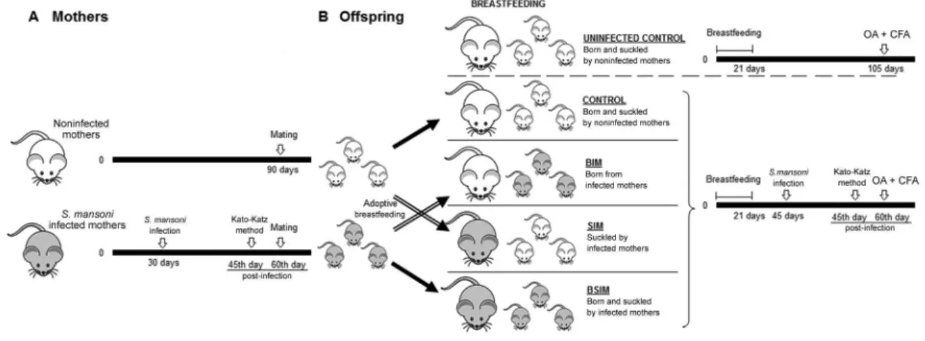
Animals and S. mansoni infection - Swiss Webster four-week-old female mice were infected subcutaneous-ly (s.c.) with 20 S. mansoni cercariae, São Lourenço da Mata (SLM) strain. On the 45th day the infection was confirmed by Kato-Katz method (Katz et al. 1972). On the 60th dpi, estruses were synchronised by administra-tion of 5 i.u. (100 mL) of equine chorionic gonadotrophin
hormone plus, after 48 h, injection of an additional 5 i.u. (100 mL) of human chorionic gonadotrophin. The females were caged with male mice at a 1:1 ratio and successful mating was checked by presence of a vaginal plug. The same procedure was performed in noninfected females (Fig. 1A). Six-week-old offspring males were taken for the experimental and control groups. The mice were housed in the animal care facility at the Aggeu Magalhães Re-search Centre, Oswaldo Cruz Foundation (Fiocruz), mu-nicipality of Recife, state of Pernambuco, Brazil.
Infection/immunisation protocol and study groups - Immediately after birth, the newborns from S. manso-ni-infected or noninfected mothers were housed in cages with interchanged mothers. After adoptive breastfeeding, offspring BIM were suckled by noninfected mothers and offspring SIM were suckled by infected mothers. Another group of animals was born and suckled by schistosomotic mothers (BSIM). Animals born from noninfected females were also suckled by their own mothers (control).
Six-week-old male offspring were infected with 80 S. mansoni cercariae, SLM strain (confirmed by Kato-Katz method) and, 60 dpi, immunised s.c. with 100 mg of OA (grade V; Sigma-Aldrich, USA) and emulsified in com-plete Freund’s adjuvant (CFA) (Sigma-Aldrich) at the base of the tail (0.1 mL/animal). The immunisation was also carried out in noninfected control offspring. Mice were divided into five groups (n = 10): (i) mice BIM; (ii) mice SIM; (iii) mice BSIM, (iv) mice control subse-quently infected with S. mansoni and immunised with OA + CFA, (v) mice uninfected control and immunised with OA + CFA (Fig. 1B).
Histomorphometric study of liver tissue - On the 60th dpi, animals from BIM, SIM, BSIM, and control groups were immunised s.c and, nine days after immunisation, the parasite burden was determined and the livers were harvested after anaesthesia and euthanasia and fixed in
10% buffered formalin. Three fragments of liver tissue in transverse sections from three distinct lobes were collected from each animal. Horizontal histological sec-tions (4 μm) were cut using a microtome Yamato (Japan) and the slides were stained with haematoxylin-eosin and Masson trichrome (selective for collagen) for morpho-metric study. The study was performed using ImageJ Software (National Institutes of Health, USA) for meas-uring the average diameter (micrometer - μm) of gran -ulomas, with subsequent calculation of the area (μm2) and intensity of blue stain (specific for collagen) in his-tograms. Analyses were performed on images randomly obtained in 10-20 fields/animal (100X). The histomor-phometric study was performed in five animals/group.
Hypersensitivity reactions (HR) - Immediate and cell-mediated HR in the different groups were elicited eight days after OA immunisation. Briefly, 30 μL of 2% aggregated OA was injected into one hind footpad and the same volume of saline in the other. Footpad swell-ing was periodically measured from 0.5-24 h usswell-ing a pocket thickness gauge (Mitutoyo Mfg Co Ltd, Japan) and expressed as the increase in thickness relative to the saline-injected paw. The results are expressed as the me-dian ± standard error (SE) for each group (n = 10). Mice nonimmunised with OA were equally challenged to test the controls for nonspecific swelling (data not shown).
Detection of soluble egg antigen (SEA), soluble worm antigen preparation (SWAP), and OA-specific an-tibodies by ELISA - S. mansoni SEA and SWAP were prepared as described by Boros and Warren (1970) and Pearce et al. (1988), respectively, and were used for par-asite-specific antibodies production analysis. For heter-ologous antibody analysis was used OA. On the ninth day after immunisation, blood samples were taken by cardiac puncture from each group under intramuscular anaesthesia with xilazine HCl/ketamine HCl. Plasma samples were tested individually for IgG1 and IgG2a an-tibodies using SEA (1.25 μg/mL), SWAP (5 μg/mL), or OA (20 μg/mL)-coated 96-well plates (Nunc MaxiSorp, Denmark), and biotinylated goat antimouse IgG1 or Ig-G2a (Southern Biotechnology Associates Inc, USA). The reactions were developed with a streptavidin-perox-idase conjugate (Sigma-Aldrich) and an O-phenylenedi-amine (Sigma, USA) solution in 0.1 M citrate buffer plus H2O2. The plates were read (450 nm) in an automated ELISA reader. Titration curves were carried out for all the samples. The results are expressed as the median of the sample optical density from each group (n = 10) in an appropriated dilution (within the linear part of the titra-tion curve) for each isotype ± SE (SEA 1:256 for IgG1 or 1:16 for IgG2a, SWAP 1:64 for IgG1 or 1:8 for IgG2a, OA 1:2.048 for IgG1 or 1:16 for IgG2a).
Cell culture - Nine days after immunisation, the spleen of each animal was harvested after euthanasia by cervical dislocation. Cell suspensions were prepared in RPMI-1640 (Sigma-Aldrich) supplemented with HEPES (10 mM), 2-mercaptoethanol (0.05 mM), 216 mg of L-glutamine/L, gentamicin (50 mg/L) and 5% of foetal bovine serum (FBS) (Sigma-Aldrich). The spleen cells from each group (n= 10) were cultivated at a final con-centration of 107 (24 h) or 6 × 10⁶ (72 h) cells/mL in
24-well tissue culture plates (Costar Culture Plates, USA) and subsequently stimulated with OA (500 μg/mL) or concanavalin-A (Con-A) (5 μg/mL) at 37ºC in 5% CO2. Supernatants were harvested after 24 h or 72 h and as-sayed for cytokine content: IL-4 (24 h), IFN-γ, and IL-10 (72 h). Cells cultured for 72 h were collected and labelled for CD4+ and FoxP3+ T-cells detection.
Cytokine and CD4+FoxP3+ T-cells measurements - The cytokines were measured using specific two-site sandwich ELISA using the following monoclonal anti-bodies: for IFN-γ, XMG 1.2 and biotinylated AN18, for 4, 11B11 and biotinylated BVD6.24G2, and for IL-10, C252-2A5 and biotinylated SXC-1 (BD Biosciences Pharmingen, USA). Binding of biotinylated antibodies was detected using a streptavidin-peroxidase conju-gate (Sigma-Aldrich) and a 2-2′-azinobis (3-ethylben -zene-thiazoline-6-sulphonic acid) (Sigma) solution in 0.1 M citrate buffer plus H2O2. The plates were read (405 nm) in an automated ELISA reader. The samples were quantified by comparison with the standard curves of purified recombinant cytokines (rIFN-γ, 4, or rIL-10), with resulting detection limits of 2.5 ng/mL for IFN-γ and 0.3125 ng/mL for IL-4 or IL-10.
Spleen cells were subjected to double-labelling with fluorochrome-labelled antibody solutions at a concen-tration of 0.5 mg/106 cells: PE antimouse FoxP3 plus PE-Cy5 antimouse CD4 (BD Biosciences Pharmingen). After staining, the preparations were washed with phos-phate-buffered saline (PBS) containing azide (0.1%) and FBS (3%). After centrifugation, the cell pellet was re-suspended in PBS with paraformaldehyde (0.5%) and maintained at 4ºC until the moment of data acquisition. Data acquisition was performed using a flow cytometry FACSCalibur (BD-Pharmingen, USA) by collecting a minimum of 10,000 events per sample. The frequency of positive cells was analysed using the program Cell Quest Pro and the limits for the quadrant markers were always set based on negative populations and isotype controls. A descriptive analysis of the frequency of cells in the upper right quadrant (double-positive cells) was performed. The results are expressed as the mean of the frequency of cells double-labelled from each group ± standard deviation.
Statistical analysis - For HR analysis, the Wilcoxon test (treatment × time) was used to evaluate the differ-ences among groups, whereas for antibody production and histomorphometric analysis of liver sections, the Kruskal-Wallis test. The multiple comparisons were per-formed by Mann-Whitney U test. For cytokine analysis and flow cytometry, an one-way analysis of variance followed by Tukey’s method were used. For statistical analysis, we used GraphPad Prism v.5.0 (GraphPad Soft-ware, USA) and all findings were considered significant at p < 0.05. All procedures were repeated three times to evaluate the reproducibility of the results and it was showed one representative of three independent studies.
RESULTS
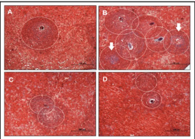
BIM mice show more intense granulomatous re-actions, whereas previous intake of breast milk from schistosomotic mothers strongly reduced hepatic in-flammation - To verify the intensity of the granuloma-tous reaction, a hepatic histomorphometric analysis was performed on the 69th dpi. We noted significantly more granulomas, including a greater quantity of collagen in the BIM group compared to the control group (Fig. 2B, Table). The livers of mice that received breast milk from
infected mothers (SIM and BSIM) showed a similar num-ber of granulomas and collagen quantity compared with the control group; however, the granuloma sizes were smaller (Fig. 2C, D, Table). Additionally, we observed no significant difference in the number of eggs in faeces or worm recovery of BIM or SIM descendants compared with the control group (Table). By contrast, in BSIM an-imals there was a 72% reduction in the quantity of eggs per gram of faeces and 54% reduction in the worm num-bers. It was also observed in mice that received mater-nal milk a lower number (BSIM groups) and less size of granulomas (SIM and BSIM groups) in the intestinal histomorphometric analysis (data not shown).
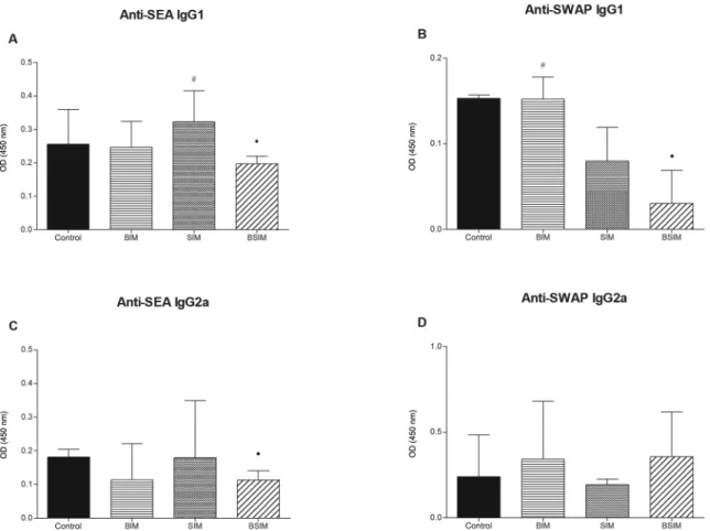
Descendants BSIM had lower levels of anti-SEA and anti-SWAP antibodies - When analysing anti-SEA (Fig. 3A, C) and anti-SWAP (Fig. 3B, D) IgG1 and IgG2a pro-duction in the descendants of schistosomotic or uninfected mothers after infection as adults, we observed decreased production of anti-SEA IgG1 and IgG2a and anti-SWAP IgG1 in the BSIM group compared to the control group. Furthermore, we observed greater production of anti-SEA IgG1 in the SIM group and anti-SWAP IgG1 in the BIM group compared to the BSIM group.
Anti-OA immediate HR are suppressed during postna-tal infection, mainly in descendants SIM - The BIM, SIM, BSIM, and control groups of mice were subjected to post-natal S. mansoni infection and, 60 days later, were immu-nised with OA in adjuvant. To evaluate in vivo anti-OA HR, all groups were challenged with OA aggregates in the footpad and swelling was measured. The same analysis was performed in newborns born/suckled by noninfected mothers that were not infected (uninfected control).
As shown in Fig. 4, all infected groups had imme-diate HR (at 0.5-6 h) that were significantly lower than that in the uninfected group (uninfected control). At 6 h, the SIM group showed significantly less HR than the Fig. 2: histomorphometric study of liver tissue. Analysis of hepatic
granulomas in Swiss Webster mice born and suckled from uninfected mothers (control) (A), born from infected mothers (B), suckled by in-fected mothers (C), and born and suckled by schistosomotic mothers (D) and 69thday post-infection with 80 S. mansoni cercariae. The slides were stained with Masson trichrome (selective for collagen) and the analyses were performed on images randomly obtained in 10-20 fields/animal (100X). The histomorphometric study was performed in five animals/group. Circles defining the granuloma size (A, B, C and D) and arrows indicate areas with increased collagen deposition (B).
TABLE
Number and size of granulomas and hepatic fibrosis developed in mice infected with 80 Schistosoma mansoni cercariae born from infected mothers (BIM), suckled by infected mothers (SIM), and born and suckled by schistosomotic mothers (BSIM)
Groupsa
Kato-Katz (epg)b
Worm recoveryc
Number of hepatic granulomasd
Granuloma sizee
(MT)
Collagenf
(MT) (H&E) (MT)
Control 509.1 ± 359.1 15.00 ± 1.83 2.89 ± 1.22 3.13 ± 1.95 23237 ± 7934 78.67 ± 6.64 BIM 408.0 ± 159.2 10.14 ± 2.27 4.63 ± 1.38g 5.73 ± 2.25g 21451 ± 9454 87.46 ± 10.46g
SIM 332.6 ± 202.7 12.67 ± 3.20 3.11 ± 1.07 3.84 ± 2.21 18407 ± 6674g 79.33 ± 7.98
BSIM 142.3 ± 103.7g 6.88 ± 3.44g 3.74 ± 1.45 4.16 ± 2.10 20467 ± 8577g 83.91 ± 9.01
a: Swiss Webster mice infected with 80 S. mansoni cercariae, BIM, SIM, or BSIM from S. mansoni infected mothers immunised
subcutaneously with ovalbumin (100 μg/animal) in complete Freund`s adjuvant 60 days post-infection had their liver subjected to
morphometric study nine days after immunisation. Born and suckled mice from uninfected mothers (control) were also analysed under the same conditions [haematoxylin-eosin (H&E) and Masson’s trichrome (MT) stains]; b: median ± standard error (SE) of number of the eggs per gram (epg) faeces. Analysis was conducted 49 days post-infection; c: median ± SE of parasite burden. Analysis was conducted 69 days post-infection; d: median ± SE of number of hepatic granulomas per field; e: median ± SE of size (sectional area) of hepatic granulomas in µm2. Analysis of 20 granulomas per animal (n = 5) totalling 100 granulomas/group; f:
group of infected descendants from nonschistosomotic mothers (control). This suppression was also observed at 9 h in the SIM group.
Delayed anti-OA HR (24 h) was observed in unin-fected control group and was similar to groups of infect-ed descendants from nonschistosomotic and schistoso-motic mothers.
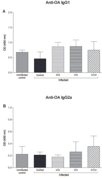
S. mansoni infection did not change the production of anti-OA IgG1 and IgG2a in the descendants of schistoso-motic mothers as adults - Nine days after immunisation, all groups were bled, and the serum antibody levels were measured by ELISA. Anti-OA IgG1 (Fig. 5A) and IgG2a (Fig. 5B) levels were similar between all of the groups in-fected as adults and the uninin-fected control group.
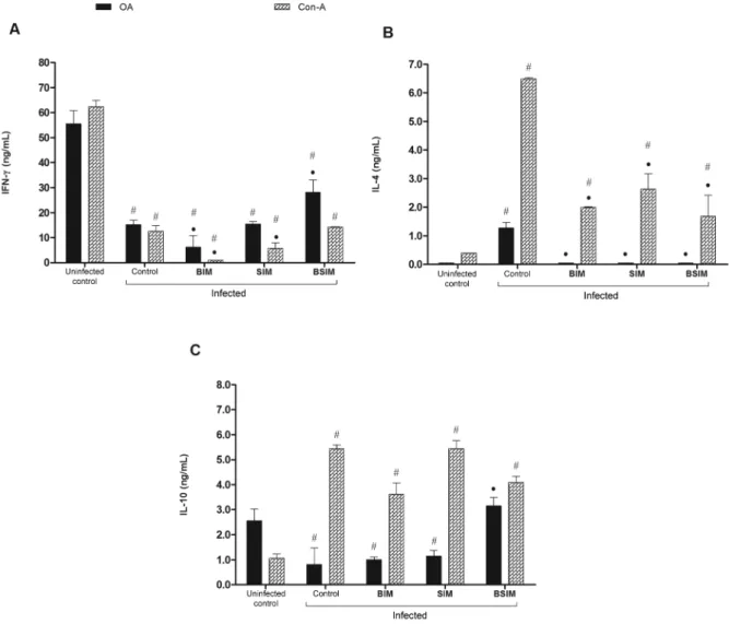
IFN-g, IL-4, and IL-10 production in adult descend-ants of schistosomotic mothers after postnatal infec-tion - Splenocytes from animals of the groups studied were cultured in the presence of OA or Con-A and the supernatants were collected to measure the secreted cy-tokines. In these conditions, IFN-g production was
sig-nificantly lower in all infected groups compared with the uninfected control group (Fig. 6A). However, in an-imals born from schistosomotic mothers, there was also a lower quantity of this cytokine compared with the con-trol group. This finding was observed in the group that received only breast milk from schistosomotic moth-ers and was cultured with a mitogenic stimulus. In the BSIM group, the production of IFN-g in response to OA was significantly higher than in the control animals.
IL-4 production, in an in vitroresponse to OA, was only detected in the supernatant of splenocytes from con-trol animals (Fig. 6B). In response to mitogen, all groups of infected animals produced significantly more IL-4 than the uninfected control group. Still, in the groups of descendants from schistosomotic mothers, IL-4 produc-tion was significantly lower than in the group of infected descendants from noninfected mothers (control).
ly lower in infected animals (control) and in animals that were born (BIM) or breastfed (SIM) by schistosomotic mothers compared with the uninfected control mice. By contrast, there was a higher production of this cytokine in the BSIM group compared with the control group.
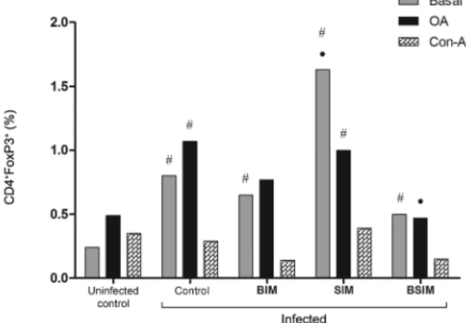
Postnatal infection lead to increased CD4+FoxP3+
T-cells frequencies in adult descendants, mostly in SIM mice - In unstimulated cultures, the infected groups showed a higher frequency of CD4+FoxP3+ T-cells com-pared with the uninfected control group (Fig. 7). When only the infected groups were compared, the SIM mice showed the highest CD4+FoxP3+T-cells frequency (BIM = 0.65%, SIM = 1.63%, BSIM = 0.50%, control = 0.80%, and uninfected control = 0.25%). In Con-A-stimulated cultures, the frequencies of CD4+FoxP3+ T-cells were similar (BIM = 0.14%, SIM = 0.39%, BSIM = 0.15%, control = 0.29%, and uninfected control = 0.35%). In OA-stimulated cultures, the infected SIM and control groups showed an increased frequency of CD4+FoxP3+ T-cells compared with the uninfected control group (BIM = 0.77%, SIM = 1%, BSIM = 0.47%, control = 1.07%, and uninfected control = 0.49%). Splenocytes from the BSIM group that were stimulated with OA showed a significantly lower frequency of CD4+FoxP3+ T-cells compared with the control group.
DISCUSSION
In this study we evaluated the effect of pregnancy, separately from the breast milk, on the intensity of the hepatic granulomatous reaction, as well as on immunity to heterologous antigen during the postnatal infection of descendants from schistosomotic mothers. For compari-son, group of mice that were born and suckled by nonin-fected mothers were postnatally innonin-fected. As expected, in infected descendants from noninfected mothers we observed the hepatic granulomas induced by S. mansoni
eggs, such as skewing of the immune response to a
het-Fig. 4: hypersensitivity reactions to ovalbumin (OA). Swiss Webster mice born from infected mothers (BIM), suckled by infected mothers (SIM), and born and suckled by schistosomotic mothers (BSIM) were immunised with OA (100 μg/animal) in complete Freund’s adjuvant 60 days after postnatal infection (80 Schistosoma mansoni cercariae). All the mice were challenged with OA aggregated in the footpad eight days after immunisation. Postnatal infected or uninfected mice born and suckled by uninfected mothers (control) were also immunised with OA and equally challenged. The results represent the median of the net increase in footpad thickness of 10 mice/group ± standard error. The results are showing one representative of three independent experiments. •: p < 0.05 compared with the control group; #: p < 0.05 compared with the uninfected control group.
Fig. 6: interferon (IFN)-γ (A), interleukin (IL)-4 (B), and IL-10 (C) secreted by spleen cells. Swiss Webster mice born from infected mothers (BIM), suckled by infected mothers (SIM), and born and suckled by schistosomotic mothers (BSIM) were immunised with ovalbumin (OA) (100 μg/animal) in complete Freund`s adjuvant 60 days after postnatal infection (80 Schistosoma mansoni cercariae). Postnatal infected or uninfected mice born and suckled by uninfected mothers (control) were also immunised with OA. On 9th day, 107 cells (IL-4) or 6 x 10⁶ cells (IFN-g and
IL-10) were stimulated with OA (500 μg/ml) or concanavalin-A (Con-A) (5 μg/mL) for 24 h (IL-4) or 72 h (IFN-g and IL-10). Cytokines were quantified in supernatants harvested by sandwich ELISA. The results represent the mean ± standard deviation for 10 animals/group. Nonstim-ulated cells produced < 2.5 ng/mL of IFN-g, < 0.44 ng/mL of IL-4, and < 0.625 ng/mL of IL-10. The results are showing one representative of three independent experiments. •: p < 0.05 compared with the control group: #: p < 0.05 compared with the uninfected control group.
erologous antigen towards a Th2/IL-10 profile (IL-4 and IL-10, suppression of immediate HR, IFN-g production, and a optimal frequency of CD4+FoxP3+ T-cells) (Stavit-sky 2004, Gryseels et al. 2006, Barsoum et al. 2013, Lundy & Lukacs 2013). However, this scenario was al-tered by previous contact with schistosomotic mothers, either in utero or through breast milk.
BIM group presented greater quantity of hepatic granulomas and remarkable fibrosis intensity, which can be due to uterine conditions in infected mothers. It is widely accepted that alternatively activated macrophag-es (M2) are induced in the uterine mucosa to favour foetal tolerance (Gustafsson et al. 2006, Svensson et al. 2011) and are committed to tissue remodelling (Gordon & Martinez 2010). In infected mothers, these conditions
could be amplified by S. mansoni antigens which are strong inducers of M2 (Wilson et al. 2007, Joshi et al. 2008) and imprint long-term predisposition for collagen production in adult life. Currently, this hypothesis is be-ing tested in our experimental model. Postnatal infec-tion of animals pre-exposed to parasite antigens in utero
strongly impairs both anti-OA Th1/IFN-g and Th2/IL-4 responses. Recently, we reported reduced expression of the co-stimulatory molecule CD86 in response to OA by CD11c+ cells of adult animals born from infected moth-ers (Santos et al. 2014) that may have reduced capacity to prime anti-OA Th1 and Th2 responses.
Fig. 7: splenic cells expressing CD4+FoxP3+. Swiss Webster mice born from infected mothers (BIM), suckled by infected mothers (SIM), and born and suckled by schistosomotic mothers (BSIM) were immunised with ovalbumin (OA) (100 μg/animal) in complete Freund’s adjuvant 60 days after postnatal infection (80 Schistosoma mansoni cercariae). Postnatal infected or uninfected mice born and suckled by uninfected mothers (control) were also immunised with OA. Nine days after im-munisation, their spleen cells were unstimulated or stimulated with OA (500 μg/mL) or concanavalin-A (Con-A) (5 μg/mL) for 72 h, la -belled and analysed by flow cytometry. The results represent the mean of the frequency of spleen cells double-labelled ± standard deviation for 10 animals/group. The results are showing one representative of three independent experiments. •: p < 0.05 compared with the control group; #: p < 0.05 compared with the uninfected control group.
and Th2 immunity. In the cell culture of these offspring, there was a remarkable background level for CD4+FoxP3+ T-cells. Although, we would have to label more mole-cules to confirm the Treg cells phenotype (von Boehmer 2005, Taylor et al. 2006, Collison et al. 2009), these basal conditions could downregulate the immune responses to both homologous (McKee & Pearce 2004, Taylor et al. 2006, Wilson et al. 2007) and heterologous antigens (Smits et al. 2007, Cardoso et al. 2012). Whether parasite antigens in the breast milk in contact with the suppres-sive intestinal mucosa microenvironment (Weiner et al. 2011) may collaborate for enhanced CD4+FoxP3+ T-cells generation after antigenic re-exposure as adults deserve further investigations. Intriguingly, in SIM mice, we observed higher CD40+CD80+ B-cells frequency and greater IL-2 production after OA stimulation (Santos et al. 2010, 2014), which are conditions to favour increased frequency of CD4+FoxP3+ T-cells (Zheng et al. 2010). Besides of this, the contact with anti-SEA antibodies at a young age can generate idiotypes and antiidiotypes that negatively modulate granulomatous reactions (Caldas et al. 2008), and only passive transfer by breastfeeding, but not pregnancy, maintains the levels of anti-SEA IgG1 in the early in life (Nóbrega et al. 2012). Therefore, the modulation of granuloma by this mechanism must not be too ruled out in the SIM group.
Attallah et al. (2006) and Othman et al. (2010) previous-ly reported a reduced granulomatous reaction in animals born and suckled by infected mothers with a high parasite load. Here, the results in BSIM group corroborated these
data and showed that continuous contact with parasite an-tigens during breastfeeding in infected mothers reverted the strong hepatic damage that was obtained in prenatal phase. In our study, this phenomenon was achieved using a low maternal parasite load, which is similar to the con-ditions of endemic populations in the Northeast Region of Brazil (Tanabe et al. 1997, da Frota et al. 2011). Othman et al. (2010) showed increased levels of the cytokines IL-12 and TGF-β, which can counteract immune regulation. In our studies, we observed, in the cell culture, increased IL-10 (upon mitogenic stimulus) and CD4+FoxP3+ T-cells. Taken together, these findings corroborate that controlled Th1 and Th2 responses is required to minimise the sever-ity of the hepatic pathology (Wilson et al. 2007). Besides of this, in BSIM group there was reduction in the eggs quantity and worm numbers. This finding reflects con-comitant immunity, in which adult worms and egg anti-gens stimulate a protective immune response against new infections (Salim & Al-Humiany 2013) by reaching the intestinal epithelium (Schramm & Haas 2010). This situ-ation mimics the antigens present in the breast milk that continuously act during lactation.
For anti-OA immune responses, there was a partial recovery of anti-OA immunity in animals from the BSIM group, which was observed by increased IFN-g and IL-10 levels. Thus, these cytokines may be more involved with the reduced IL-4 levels in BSIM animals, since anti-OA CD4+FoxP3+ T-cells were found at a lower frequency in this group compared to the infected control group.
In regard to the antibodies against parasite antigens, our results were in agreement to study of the Attallah et al. (2006), in which were detected low levels of antibod-ies in the BSIM group in comparison to control. How-ever, in postnatal infection of offspring that breastfed or pregnancy in separate way, anti-SEA and anti-SWAP IgG1 levels were similar to control group, respectively.
The immunomodulatory actions of infection on het-erologous humoral immune responses are contradictory (Kullberg et al. 1992, Curry et al. 1995, Montesano et al. 1999, Smits et al. 2007, Cardoso et al. 2010). In this study, there was no significant difference in the levels of anti-OA IgG1 and IgG2a in the groups. However, some considerations about the BIM and SIM groups must be highlighted. Previously, we have shown an increase in anti-OA antibody levels in the SIM group (Santos et al. 2010), and those with postnatal infection showed sim-ilar levels to the uninfected control group. Therefore, this reduction of the anti-OA immune response supports the immunosuppressive profile that results from postna-tal infection in the SIM group. In the BIM group, there was reduced anti-OA antibody production (Santos et al. 2010), which was recovered after infection of the mice as adults. Thus, the levels of anti-OA antibodies were simi-lar between groups. These data could reflect the control of immunologic diseases that are mediated by antibod-ies, such as allergies and autoimmune diseases.
het-erologous anti-OA immunity (IFN-g production). Based on these results, we suggest that nonadoptive breastfeed-ing, i.e., from the biological mother, is more effective for immunomodulation of the granulomatous reaction in individuals from endemic areas who are at risk of postnatal infection. Nonadoptive breastfeeding can also guarantees better protection for nonrelated antigens, in-fections, and responses to vaccines, in these individuals.
ACKNOWLEDGEMENTS
To Gerlane Tavares de Souza Chioratto, for veterinary support, to Maria da Conceição Batista and Laurimar Thomé da Rocha, for technical assistance, and to the PDTIS/Fiocruz, for the use of its facilities.
REFERENCES
Actor JK, Shirai M, Kullberg MC, Buller RM, Sher A, Berzofsky JA 1993. Helminth infection results in decreased virus-specific CD8+ cytotoxic T-cell and Th1 cytokine responses as well as de-layed virus clearance. Proc Natl Acad Sci USA 90: 948-952. Attallah AM, Abbas AT, Dessouky MI, El-Emshaty HM, Elsheikha
HM 2006. Susceptibility of neonate mice born to Schistosoma mansoni-infected and noninfected mothers to subsequent S. man-soni infection. Parasitol Res 99: 137-145.
Barsoum RS, Esmat G, El-Baz T 2013. Human schistosomiasis: clini-cal perspective: review. J Adv Res 4: 433-444.
Boros DL, Warren KS 1970. Delayed hypersensitivity-type granu-loma formation and dermal reaction induced and elicited by a soluble factor isolated from Schistosoma mansoni eggs. J Exp Med 132: 488-507.
Caldas IR, Campi-Azevedo AC, Oliveira LFA, Silveira AMS, Olivei-ra RC, Gazzinelli G 2008. Human schistosomiasis mansoni: im-mune responses during acute and chronic phases of the infection.
Acta Trop 108: 109-117.
Cardoso LS, Oliveira SC, Araújo MI 2012. Schistosoma mansoni
antigens as modulators of the allergic inflammatory response in asthma. Endocr Metab Immune Disord Drug Targets 12: 24-32. Cardoso LS, Olivera SC, Góes AM, Oliveira RR, Pacífico LG,
Marin-ho FV, Fonseca CT, Cardoso FC, CarvalMarin-ho EM, Araújo MI 2010.
Schistosoma mansoni antigens modulate the allergic response in a murine model of ovalbumin-induced airway inflammation.
Clin Exp Immunol 160: 266-274.
Collison LW, Pillai MR, Chaturvedi V, Vignali DAA 2009. Regulatory T cell suppression is potentiated by target T-cells in a cell contact, IL-35, and IL-10-dependent manner. J Immunol 182: 6121-6128. Curry AJ, Else KL, Jones F, Bancroft A, Grencis RK, Dunne DW 1995.
Evidence that cytokine-mediated immune interactions induced by
Schistosoma mansoni alter disease outcome in mice concurrently infected with Trichuris muris. J Exp Med 181: 769-774.
da Frota SM, Carneiro TR, Queiroz JA, Alencar LM, Heukelbach J, Bezerra FS 2011. Combination of Kato-Katz faecal examina-tions and ELISA to improve accuracy of diagnosis of intestinal schistosomiasis in a low-endemic setting in Brazil. Acta Trop 120
(Suppl. 1): S138-S141.
do Amaral RS, Tauil PL, Lima DD, Engels D 2006. An analysis of the impact of the Schistosomiasis Control Programme in Brazil.
Mem Inst Oswaldo Cruz 101 (Suppl. I): 79-85.
Finkelman FD, Pearce EJ, Urban Jr JF, Sher A 1991. Regulation and biologic function of helminth-induced cytokine response. Immu-nol Today 12: A62-A66.
Friedman JF, Mital P, Kanzaria HK, Olds GR, Kurtis JD 2007. Schis-tosomiasis and pregnancy. Trends Parasitol 23: 159-164. Gordon S, Martinez FO 2010. Alternative activation of macrophages:
mechanism and functions. Immunity 32: 593-604.
Gryseels B, Polman K, Clerinx J, Kestens L 2006. Human schistoso-miasis. Lancet 368: 1106-1118.
Gustafsson C, Hummerdal P, Matthiesen L, Berg G, Ekerfelt C, Erner-udh J 2006. Cytokine secretion in decidual mononuclear cells from term human pregnancy with or without labour: ELISPOT detection of IFN-gamma, IL-4, IL-10, TGF-beta, and TNF-alpha.
J Reprod Immunol 71: 41-56.
Hillier SD, Booth M, Muhangi L, Nkurunzuza P, Khihembo M, Ka-kande M, Sewankambo M, Kizindo R, Kizza M, Muwanga M, Elliot AM 2008. Plasmodium falciparum and helminth co-infec-tion in a semi-urban populaco-infec-tion of pregnant women in Uganda. J Infect Dis 198: 920-927.
Joshi AD, Raymond T, Coelho AL, Kunkel SL, Hogaboam CM 2008. A systemic granulomatous response to Schistosoma mansoni
eggs alters responsiveness of bone-marrow-derived macrophages to Toll-like receptor agonists. J Leukoc Biol 83: 314-324. Katz N, Chaves A, Pellegrino J 1972. A simple device for quantitative
stool thick smear technique in schistosomiasis mansoni. Rev Inst Med Trop Sao Paulo 14: 397-400.
Kullberg MC, Pearce EJ, Hieny SE, Sher A, Berzofsky JA 1992. In-fection with Schistosoma mansoni alters Th1/Th2 cytokine re-sponse to a non-parasite antigen. J Immunol 148: 3264-3270. La Flamme AC, Ruddenklau K, Backstrom BT 2003.
Schistosomia-sis decreases central nervous system inflammation and alters the progression of experimental autoimmune encephalomyelitis. In-fect Immun 71: 4996-5004.
Lenzi JA, Sobral ACL, Araripe JR, Grimaldi Filho G, Lenzi HL 1987. Congenital and nursing effects on the evolution of Schistosoma mansoni infection in mice. Mem Inst Oswaldo Cruz 82 (Suppl. IV): 257-267.
Lundy SK, Lukacs NW 2013. Chronic schistosome infection leads to modulation of granuloma formation and systemic immune sup-pression. Front Immunol 4: 39.
McKee AS, Pearce EJ 2004. CD25+CD4+ cells contribute to Th2 po-larization during helminth infection by suppressing Th1 response development. J Immunol 173: 1224-1231.
Medeiros Jr M, Figueiredo JP, Almeida MC, Matos MA, Araújo MI, Cruz AA, Atta AM, Rego MA, de Jesus AR, Taketomi EA, Carv-alho EM 2003. Schistosoma mansoni infection is associated with a reduced course of asthma. J Allergy Clin Immunol 111: 947-951. Montesano MA, Colley DG, Freeman GL, Secor WE 1999. Neonatal ex-posure to idiotype induces Schistosoma mansoni egg antigen-specif-ic cellular and humoral immune responses. J Immunol 163: 898-905. Moore KW, Malefyt RW, Coffman RL, O’Garra A 2001. Interleukin-10
and the interleukin-10 receptor. Annu Rev Immunol 19: 683-765. Nóbrega CGO, Fernandes ES, Nascimento WRC, Sales IRF, Santos
PDA, Schirato GV, Albuquerque MCPA, Costa VMA, Souza VMO 2012. Transferência passiva de anticorpos específicos para antígenos de Schistosoma mansoni em camundongos nascidos ou amamenta-dos em mães esquistossomóticas. J Health Sci Inst 30: 17-21. Osada Y, Shimizu S, Kumagai T, Yamada S, Kanazawa T 2009.
Othman AA, Shoheib ZS, Saied EM, Soliman RH 2010. Congenital exposure to Schistosoma mansoni infection: impact on the future immune response and the disease outcome. Immunobiology 215: 101-112.
Pearce EJ, Caspar P, Grzych JM, Lewis FA, Sher A 1991. Downregu-lation of Th1 cytokine production accompanies induction of Th2 responses by a parasitic helminth, Schistosoma mansoni. J Exp Med 173: 159-166.
Pearce EJ, James SL, Hieny S, Lanar DE, Sher A 1988. Induction of protective immunity against Schistosoma mansoni by vaccina-tion with schistosome paramyosin (Sm97), a nonsurface parasite antigen. Proc Natl Acad Sci USA 85: 5678-5682.
Ruyssers NE, De Winter BY, De Man JG, Ruysser ND, Van Gils AJ, Loukas A, Pearson MS, Weinstock JV, Pelckmans PA, Moreels TG 2010. Schistosoma mansoni proteins attenuate gastrointes-tinal motility disturbances during experimental colitis in mice.
World J Gastroenterol 16: 703-712.
Sabin EA, Araújo MI, Carvalho EM, Pearce EJ 1996. Impairment of tetanus toxoid-specific Th1-like immune responses in humans infected with Schistosoma mansoni. J Infect Dis 173: 269-272. Salim AM, Al-Humiany AR 2013. Concomitant immunity to
Schisto-soma mansoni in mice. Turkiye Parazitol Derg 37: 19-22. Santos PD, Lorena VM, Fernandes E, Sales IR, Albuquerque MC,
Gomes Y, Costa VM, Souza VM 2014. Maternal schistosomiasis alters costimulatory molecules expression in antigen-presenting cells from adult offspring mice. Exp Parasitol 141: 62-67. Santos PD, Sales IR, Schirato GV, Costa VM, Albuquerque MC, Souza
VM, Malagueño E 2010. Influence of maternal schistosomiasis on the immunity of adult offspring mice. Parasitol Res 107: 95-102. Schramm G, Haas H 2010. Th2 immune response against
Schisto-soma mansoni infection. Microbes Infect 12: 881-888.
Smits HH, Hammad H, van Nimwegen M, Soullie T, Willart MA, Lievers E, Kadouch J, Kool M, Oosterhoud JK, Deelder AM, Lambrecht BN, Yazdanbakhsh M 2007. Protective effect of
Schistosoma mansoni infection on allergic airway inflammation depends on the intensity and chronicity of infection. J Allergy Clin Immunol 120: 932-940.
Stavitsky AB 2004. Regulation of granulomatous inflammation in experimental models of schistosomiasis. Infect Immun 72: 1-12. Svensson J, Jenmalm MC, Matussek A, Geffers R, Berg G,
Erner-udh J 2011. Macrophages at the fetal-maternal interface express markers of alternative activation and are induced by M-CSF and IL-10. J Immunol 187: 3671-3682.
Tanabe M, Gonçalves JF, Gonçalves FJ, Tateno S, Takeuchi T 1997. Occurrence of a community with high morbidity associated with
Schistosoma mansoni infection regardless of low infection inten-sity in north-east Brazil. Trans R Soc Trop Med Hyg 91: 144-149. Taylor JJ, Mohrs M, Pearce EJ 2006. Regulatory T cell responses devel-op in parallel to Th responses and control the magnitude and phe-notype of the Th effector population. J Immunol 176: 5839-5847. von Boehmer H 2005. Mechanisms of suppression by suppressor T
cells. Nat Immunol 6: 338-344.
Weiner HL, da Cunha AP, Quintana F, Wu H 2011. Oral tolerance.
Immunol Rev 241: 241-259.
WHO - World Health Organization 2015. Schistosomiasis. Available from: who.int/mediacentre/factsheets/fs115/en/.
Wilson MS, Mentink-Kane MM, Pesce JT, Ramalingam TR, Thomp-son R, Wynn TA 2007. Immunopathology of schistosomiasis. Im-munol Cell Biol 85: 148-154.