Mat. Res. vol.8 número2
Texto
Imagem

Documentos relacionados
Desde os anos 90 tem-se observado que a maioria das infecções causadas por fungos vem do gênero Candida e cada ano que passa está mais resistente frente aos
We conducted scanning electron microscopy, x-ray diffraction, and infrared spectroscopy analyses, which evidenced that the sample treated at 110°C contained calcium
Starting from the scanning electronic microscopy characterizations, the chemical analysis for energy dispersive spectroscopy, and phase analysis by X-ray diffraction, the
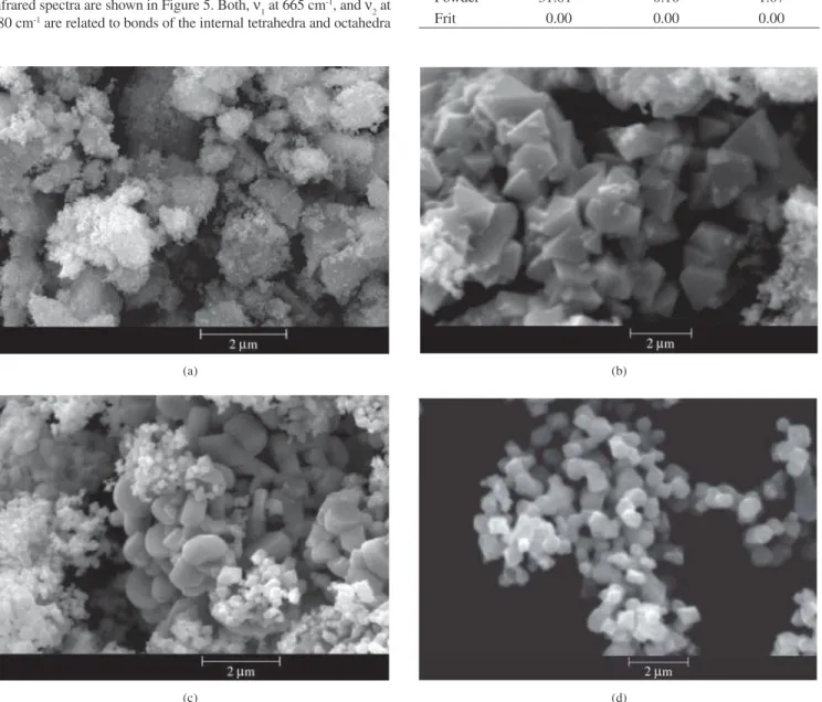
Samples were produced by different routes and characterized by scanning electron microscopy, differential thermal analysis, thermogravimetric analysis and X-ray diffraction, whereas
Using the optical and electron microscopy, X-ray powder diffraction and Fourier transform infrared spectroscopy we attempted to correlate the results obtained on 8
Its composition was determined by X-ray diffractometry (XRD), Fourier transform infrared spectroscopy (FTIR) and Raman micro-spectroscopy, and its topography by atomic
Physical and chemical tests were performed involving Fourier transform infrared spectroscopy (FTIR), Scanning Electron Microscopy (SEM) and Raman spectroscopy with the purpose of
For characterization, tests of water vapor permeability, swelling index, infrared absorption spectroscopy, thermogravimetric analysis, scanning electron microscopy and mechanical
