ORIGINAL ARTICLE
Environmental enrichment restores cognitive deficits
induced by experimental childhood meningitis
Tatiana Barichello,
1,2Glauco D. Fagundes,
1Jaqueline S. Generoso,
1Caroline S. Dagostin,
1Lutiana R. Simo˜es,
1Ma´rcia C. Vilela,
3Clarissa M. Comim,
4Fabricia Petronilho,
2,5Joa˜o Quevedo,
2,6Antonio L. Teixeira
71Laboratory of Experimental Microbiology, Universidade do Extremo Sul Catarinense (UNESC), Criciu´ma, SC, Brazil.2Center for Experimental
Models in Psychiatry, The University of Texas Medical School at Houston, Houston, TX, USA.3Department of Animal Biology, Universidade
Federal de Vic¸osa (UFV), Vic¸osa, MG, Brazil.4Laboratory of Experimental Neurosciences, Universidade do Sul de Santa Catarina (UNISUL),
Palhoc¸a, SC, Brazil.5Laboratory of Clinical and Experimental Pathophysiology, UNISUL, Tubara˜o, SC, Brazil.6Laboratory of Neurosciences,
UNESC, Criciu´ma, SC, Brazil.7Interdisciplinary Laboratory of Medical Investigation, Universidade Federal de Minas Gerais (UFMG), Belo
Horizonte, MG, Brazil.
Objective:To evaluate the influence of environmental enrichment (EE) on memory, cytokines, and brain-derived neurotrophic factor (BDNF) in the brain of adult rats subjected to experimental pneumococcal meningitis during infancy.
Methods: On postnatal day 11, the animals received either artificial cerebrospinal fluid (CSF) or
Streptococcus pneumoniaesuspension intracisternally at 16106CFU/mL and remained with their mothers until age 21 days. Animals were divided into the following groups: control, control + EE, meningitis, and meningitis + EE. EE began at 21 days and continued until 60 days of age (adulthood). EE consisted of a large cage with three floors, ramps, running wheels, and objects of different shapes and textures. At 60 days, animals were randomized and subjected to habituation to the open-field task and the step-down inhibitory avoidance task. After the tasks, the hippocampus and CSF were isolated for analysis.
Results: The meningitis group showed no difference in performance between training and test sessions of the open-field task, suggesting habituation memory impairment; in the meningitis + EE group, performance was significantly different, showing preservation of habituation memory. In the step-down inhibitory avoidance task, there were no differences in behavior between training and test sessions in the meningitis group, showing aversive memory impairment; conversely, differences were observed in the meningitis + EE group, demonstrating aversive memory preservation. In the two meningitis groups, IL-4, IL-10, and BDNF levels were increased in the hippocampus, and BDNF levels in the CSF.
Conclusions:The data presented suggest that EE, a non-invasive therapy, enables recovery from memory deficits caused by neonatal meningitis.
Keywords: Pneumococcal meningitis; environmental enrichment; cytokines; BDNF
Introduction
Meningitis is a life-threatening condition with a high mortality rate, particularly in neonates and children.1 Several microorganisms that are pathogenic to humans can cause meningitis, but Streptococcus pneumoniae, Neisseria meningitis, and Haemophilus influenzae are generally reported in children with acute bacterial meningitis.2,3 In young children, colonization of the na-sopharynx, oropharynx, or paranasal sinuses by virulent strains of bacteria is a prerequisite for the development of
meningitis. Transmission usually occurs from person to person via large-droplet secretions and the respiratory route.2 WhenS. pneumoniae reaches the subarachnoid space, it reproduces rapidly, releasing cell wall frag-ments, lipoteichoic acid, teichoic acid, pneumolysin, and peptidoglycans.4 These bacterial compounds are highly immunogenic and may be recognized by antigen-presenting cells through toll-like receptors.5,6 This cas-cade induces translocation of nuclear factor kappa B (NF-kB) to the nucleus, which stimulates the synthesis of cytokines, chemokines, and other pro-inflammatory molecules in response to bacterial stimuli. As a result, polymorphonuclear cells are attracted and activated, releasing large amounts of superoxide anions and ni-tric oxide and leading to peroxynitrite formation and oxidative stress. This cascade leads to lipid peroxidation, mitochondrial damage, and breakdown of the blood-brain barrier, which contributes to cell injury during
Correspondence: Profa. Tatiana Barichello, PhD, Laborato´rio de Microbiologia Experimental, Universidade do Extremo Sul Catarinense, Av. Universitaria, 1105, CEP 88806-000, Criciu´ma, SC, Brazil.
E-mail: [email protected]
Submitted Nov 14 2013, accepted May 01 2014.
ß2014 Associac¸a˜o Brasileira de Psiquiatria
pneumococcal meningitis.2,7 Long-term neurological se-quelae are found in patients who survive bacterial meningitis during infancy, including deafness, sensor-imotor deficits, seizure disorders, and cognitive impair-ments, such as deficits in learning and memory.8 In
animal models of meningitis, adjuvant treatment with antioxidant,9cannabidiol,10or dexamethasone has been shown to prevent cognitive impairment.11 In addition, when utilized in an attempt to minimize cognitive damage, environmental enrichment (EE) is a novel and promising non-pharmacological approach and has been shown to stimulate brain plasticity, neurogenesis, and increased neurotrophic factor expression, as well as protect against the effects of brain insults.12
EE refers to housing conditions in which the animals are kept in spacious cages containing toys, running wheels, climbing ropes, and objects with different shapes and textures.13 This environment promotes visual and
sensory stimulation, minimizes stressful social interac-tions, increases voluntary exercise, and may have an effect on cognitive functions compromised in disorders of the central nervous system (CNS).12-14 Compared with rats reared in groups in conventional cages, rats housed in an enriched environment exhibit increased cortical thickness, hippocampal neurogenesis, and hippocampal levels of transcripts that encode various genes involved in tissue plasticity and remodeling. EE rats also perform better on learning and memory tasks.12During the early postnatal period, EE increases cell proliferation in the dentate gyrus of guinea pigs.15The stimulation provided by EE fosters recovery from spatial memory deficits,16 improves memory impairment on object recognition tests, and preserves hippocampal dendritic spine density following neonatal hypoxia-ischemia injury in rats.17 EE
has also been shown to attenuate the motor and cognitive deficits that result from traumatic brain injury.18 We hypothesize that cognitive impairment in adult Wistar rats subjected to experimental pneumococcal meningitis during childhood will be reversed or mitigated by EE exposure. In the present study, we evaluated the influence of EE on habituation and aversive memories and cytokine and brain-derived neurotrophic factor (BDNF) levels in the brain of adult survivors of childhood pneumococcal meningitis.
Materials and methods
Infecting organism
S. pneumoniae(serotype 3) was cultured overnight in 10 mL of Todd Hewitt Broth (HimediaH), diluted in fresh medium, and grown to exponential phase. These cultures were centrifuged for 10 min at 5,000 6 g and resuspended in sterile saline solution at a concentration of 1 6 106 CFU/mL. The size of the inoculum was confirmed using quantitative cultures.19
Animal model of meningitis
Infant male Wistar rats (body weight 15-20 g) from our breeding colony were used for the experiments on
postnatal day 11. All procedures were approved by the Animal Care and Experimentation Committee of Universidade do Extremo Sul Catarinense (UNESC), Brazil, under protocol no. 88/2012, and were conducted in accordance with the National Institutes of Health Guide for the Care and Use of Laboratory Animals (NIH Publication No. 80-23, revised in 1996). All surgical procedures and bacterial inoculations were performed under anesthesia, which consisted of the intraperitoneal administration of ketamine (6.6 mg/kg), xylazine (0.3 mg/ kg), and acepromazine (0.16 mg/kg).20Rats underwent a cisterna magna tap with a 23-gauge needle. The animals received either 10 mL artificial cerebrospinal fluid (CSF)
as a placebo or an equivalent volume ofS. pneumoniae suspension. At the time of inoculation, the animals received fluid replacement (1 mL saline subcutaneously) and were then returned to their cages and kept under a 12-h light/dark cycle (lights on at 7 a.m.). Meningitis was documented by a quantitative culture of 5mL CSF, which
was obtained via puncture of the cisterna magna 18 h after experimental infection.20After the animals received ceftriaxone (ceftriaxone, 100 mg/kg, during 7 days).
Organization of the experimental groups
The animals were divided into the following experimental groups: control, control + EE, meningitis, and meningitis + EE (10-13 animals per group). Eighteen hours after induction of pneumococcal meningitis or intracisternal administration of artificial CSF as a placebo, all animals were treated with ceftriaxone for 7 days (100 mg/kg twice daily until 168 h after documented meningitis), and remained with their mothers until age 21 days.
Environmental enrichment
EE began when rats reached 21 days of age and continued until they reached 60 days of age, equivalent to adulthood.16,21EE consisted of a large cage (40660
6 90 cm) with three floors, ramps, running wheels, and several objects of different shapes and textures (Figure 1). Small changes were made once a week by adding new objects and withdrawing others.16,22 The running wheels and stairs enhanced voluntary exercise, a seesaw provided somatosensory stimulation, and large tubes, a set of tunnels, LEGOHblocks, wood pieces, and hanging items provided cognitive stimulation13(Figure 1).
Standard housing
Standard environmental housing consisted of non-enriched cages (49 6 34 6 16 cm) containing only bedding. The animals were housed in groups of six and provided free access to food and water.
Behavioral tasks
At 60 days of age, the animals were randomized and subjected to habituation to the open-field task and the
step-down inhibitory avoidance task. After the behavioral tests, the animals were sacrificed by decapitation.
Open-field test
Behavior was assessed in an open-field apparatus to evaluate both locomotor and exploratory activity. The apparatus was a 40 660 cm open field surrounded by 50-cm-high dark grey walls and a glass front wall. Black lines divided the floor of the open field into nine rectangles. Each animal was gently placed in the center of the open field and was left to explore the arena for 5 min (training session). The number of crossings (i.e., the number of times that each animal crossed the black lines, an assessment of locomotor activity) and rearing move-ments (i.e., the exploratory behavior observed in rats subjected to a new environment) were measured. Immediately after this procedure, the animals were taken back to their home cage. Twenty-four hours later, they were subjected to a second open-field session (test session). In both sessions, the number of crossings and rearings was counted during a 5-min period. A reduction in the number of crossings and rearings between the two sessions was considered as a measure of the retention of memory. The same experimenter, who was blind to group allocation, performed all of the behavioral testing and manual scoring.23The experimental box was thoroughly cleaned with 70% ethanol between testing sessions.
Step-down inhibitory avoidance task
The apparatus and procedures used for this task have been described in previous reports.24,25 Briefly, the training apparatus was a 506 25625 cm acrylic box
(Albarsch, Porto Alegre, Brazil), the floor of which consisted of parallel stainless steel bars (1 mm diameter) spaced 1 cm apart. A 7-cm-wide, 2.5-cm-high platform was placed on the floor of the box against the left wall. In the training trial, the animals were placed on the platform, and their latency to step down on the grid with all four paws was measured with an automatic device. Immediately after stepping down on the grid, the animals received a 0.4-mA, 2.0-s foot shock and were returned to their home cage. A retention test trial was performed 24 h after the training trial (long-term memory). The retention test trial was procedurally identical to the training trial, except that no foot shock was administered. The step-down latency during the retention test (maximum 180 s) was used as a measure of the retention of the inhibitory avoidance memory.24,25
Assessment of levels of cytokines, chemokines, and brain-derived neurotrophic factor
After the behavioral tests, the animals were anaesthe-tized and killed by decapitation. The hippocampus and CSF were immediately isolated on dry ice and stored at -806C for analysis of cytokine and BDNF levels.
Assessment of the concentration of tumor necrosis factor-alpha (TNF-a), interleukin (IL)-4, IL-6, IL-10, and
cytokine-induced neutrophil chemoattractant-1 (CINC-1)
Briefly, the hippocampus was homogenized in extraction solution containing aprotinin (100 mg of tissue per 1 mL). The levels of cytokines and chemokines in the hippo-campus were determined using commercially available enzyme-linked immunosorbent assays (ELISA), following
the instructions supplied by the manufacturer (DuoSet kits, R&D Systems, Minneapolis, MN, USA). The results are expressed as pg/100 mg of hippocampal tissue and pg/100 mL CSF.
Assessment of BDNF
Serum BDNF levels were measured by sandwich-ELISA, using a commercial kit in accordance with manufacturer instructions (Millipore, Billerica, MA, USA). Briefly, 96-well, flat-bottom microtiter plates were coated overnight at 46C with the samples, which were diluted 1:100 in sample diluent. The standard curve ranged from 7.8 to 500 pg of BDNF/mL. Plates were washed four times with wash buffer before biotinylated mouse anti-human BDNF monoclonal antibody (diluted 1:1000 in sample diluent) was added and the plates incubated for 3 h at room
temperature. After washing, a second incubation with streptavidin-peroxidase conjugate solution (diluted 1:1000) was carried out for 1 h at room temperature. After the addition of substrate and stop solution, the amount of BDNF present was quantified (absorbance set at 450 nm). The standard curve represents a direct relationship between optical density and BDNF concen-tration. Total protein was measured by Bradford’s method26 (samples diluted 1:200) using bovine serum albumin as the standard.
Statistics
For the cytokine and chemokine analyses, the variables are presented as the means6SEM of 10-13 animals per group. Differences between groups were measured on postnatal day 21 and evaluated using analysis of variance (ANOVA) followed by Tukey’s post-hoc test (p-values:
*p , 0.05, {p
, 0.01, and {p , 0.001). Data from the habituation to the open field task are reported as the means 6 SEM of 10-13 animals per group and were analyzed using a paired Student’s t test and ANOVA followed by Tukey’s post-hoc test. Data from the step-down inhibitory avoidance task are reported as the median and interquartile ranges of 10-13 animals per group, and comparisons among groups were performed using Mann-WhitneyUtests. Differences within individual groups were analyzed using Wilcoxon’s tests. For all comparisons, p,0.05 indicated statistical significance. All analyses were performed using SPSS version 20.0.
Results
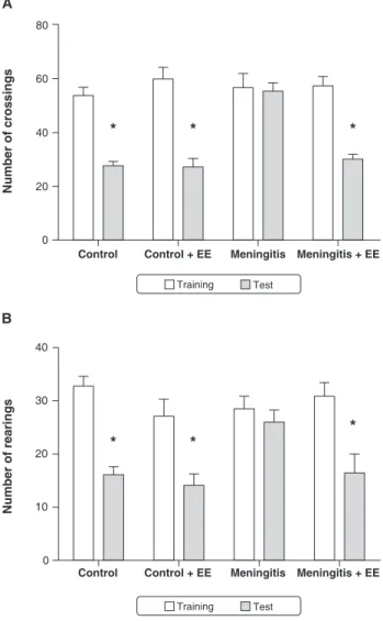
In the open-field task, there were no differences between groups in the number of crossings and rearings in the training session (p.0.05) (Figure 2), demonstrating that there was no difference in motor and exploratory activity
Figure 2 Habituation to the open-field task on postnatal day 60 in animals subjected to experimental pneumococcal meningitis during infancy. Data analyzed by a paired Student’s t test or ANOVA followed by Tukey’s post-hoc test and expressed as means6SEM of 10-13 animals per group. Symbols indicate statistical significance vs. the control group. EE = enriched environment.*p,0.05.
Figure 3 Performance of animals subjected to experimental pneumococcal meningitis during infancy on the step-down inhibitory avoidance task at postnatal day 60. Data expressed as median and interquartile ranges. Comparisons between groups performed using Mann-WhitneyUtests, with 10-13 animals per group. Differences within individual groups were analyzed by Wilcoxon’s tests. Symbols indicate statis-tically significant differences vs. the control group. EE = enriched environment.*p,0.05.
between the groups. In the meningitis group, there was no difference in behavior between the training and test sessions, demonstrating the presence of memory impair-ment in this group. In contrast, in the control, control + EE, and meningitis + EE groups, behavior was signifi-cantly different between the training and test sessions (p , 0.0001), suggesting that animals subjected to EE exhibited memory of the previous experience.
With regard to step-down latency (Figure 3), there was no statistically significant difference in latency between
any of the groups in the training test session (p.0.05). In the meningitis group, there was no difference in latency between the training and test sessions, demonstrating memory impairment in this group. However, in the control, control + EE, and meningitis + EE groups, the latencies were significantly different between the training and test sessions (p , 0.0001), demonstrating that the animals exhibited aversive memory.
Figures 4 and 5 show the effects of the EE on cytokine, chemokine, and BDNF levels in the hippocampus and
CSF, respectively, in adult rats subjected to pneumococ-cal meningitis during infancy. In the hippocampus, IL-10 and IL-4 levels were increased in the meningitis and meningitis + EE groups as compared with the control and control + EE groups (F3,41= 34.48, p,0.001 and F3,42=
75.77, p , 0.001, respectively) (Figure 4C and 4D). BDNF levels were also increased in the meningitis and meningitis + EE groups compared to the control + EE group (F3,41= 5,360, p,0.05 and p,0.01, respectively)
(Figure 4F). However, the TNF-a, IL-6, and CINC-1 levels did not change (F3,39= 0.05049, F3,42= 1,130, and F3,41=
0.6895, respectively).
In the CSF, TNF-alevels did not change (F3,42= 1.190)
(Figure 5A). In addition, BDNF levels were increased in the meningitis and meningitis + EE groups as compared with the control and control + EE groups (F3,42= 278.6,
p,0.001) (Figure 5B).
Discussion
The present study showed that environmental enrichment (EE) improved cognition in rats that had pneumococcal meningitis in their 11th day of life. EE did not change the expression profile of BDNF or of cytokines in adult rats that had meningitis during infancy. The present findings are consistent with previous studies from our group showing that rats that survived neonatal pneumococcal meningitis orStreptococcus agalactiaemeningitis exhib-ited learning and memory impairment in adulthood.19,27
Compared with standard housing, EE provides greater opportunities for voluntary exercise and generation of novelty and complexity in animal housing conditions, which facilitates enhanced sensory and cognitive stimula-tion as well as physical activity. In several animal models of neurological disorders, EE and exercise have been found to have advantageous effects, including beneficial effects on learning and memory, improved cellular plasticity, BDNF expression, adult neurogenesis, and associated molecular processes.12,14,28-30 Our study showed that animals subjected to pneumococcal menin-gitis during infancy presented memory impairment in adulthood. In the animals that were subjected to EE (promoting cognitive stimulation through motor activity, visual stimuli, object recognition, novelty, and the modulation of attention), these impairments of habituation and aversive memories were reversed. EE was also associated with enhanced spatial learning performance, neuroplasticity, and increased BDNF levels in the rat brain.30This non-invasive adjuvant treatment was effec-tive in inducing recovery from impaired declaraeffec-tive memory for object recognition and in preserving hippo-campal dendritic spine density in neonatal rats following hypoxia-ischemia.17EE has been shown to confer long-term cognitive benefits after traumatic brain injury in rats, including increasing the expression of genes important for signal transduction, calcium signaling pathways, mem-brane homeostasis, and metabolism in particular.31,32EE attenuated the production of cytokines and chemokines in response to lipopolysaccharides within the hippocampus and decreased hippocampal neuroinflammation during influenza infection in adult rodents.33,34In contrast, in an animal model of social defeat, EE does not change the expression levels of IL-1b, IL-1r, or TNF-a.35However, in
the present study, the levels of inflammatory mediators did not differ significantly between the meningitis group and meningitis + EE group; thus, the prevention of cognitive damage was not associated with these para-meters.
As an important neurotrophin, BDNF plays a role in neuronal development, differentiation, and survival. Exposure to EE restored normal expression of BDNF after reduction thereof by chronic cerebral hypoperfusion in rats.36 EE also increased BDNF expression during influenza infection and increased BDNF levels in the basal forebrain, cerebral cortex, and hippocampus in adult rats.30,33 On the other hand, in adulthood, BDNF levels were increased in the CSF and hippocampus of both meningitis groups. This increase in BDNF levels in both of the meningitis groups may be related to the
Figure 5 Expression of TNF-a and BDNF in the CSF at postnatal day 60 in animals subjected to experimental pneumococcal meningitis during infancy. Levels of cytokines and BDNF were assessed by ELISA, and the results are expressed as pg per 100 mL CSF. Results show the means 6SEM of 10-13 animals per group. Statistical significance was assessed by ANOVA followed by Tukey’s post-hoc test. Symbols indicate statistically significant differences as compared with the appropriate control group. BDNF = brain-derived neurotrophic factor; CSF = cerebrospinal fluid; EE = enriched environment; SEM = standard error of the mean; TNF-a= tumor necrosis factor-alpha.*p,0.05,
{p
,0.01, and{p
,0.001 vs. control group, and1p
,0.05,
|| p
, 0.01, and " p
, 0.001 vs. control + environmental enrichment group.
maintenance of cytokine levels during adulthood, con-sidering that immune cells can express BDNF and that neurons are the major source of this neurotrophin in the CNS.37Correlating with clinical findings, in pediatric
patients with bacterial meningoencephalitis and meningi-tis, elevated BDNF levels have been found in CSF and serum.38
In conclusion, EE can be a non-invasive experimental strategy for neurological recovery after bacterial meningi-tis in childhood. Further insight is necessary to elucidate the mechanisms underlying the effects of EE in the prevention of neuronal dysfunction.
Acknowledgements
The Laboratory of Experimental Microbiology is one of the centers of the Brazilian National Science and Technology Institutes for Translational Medicine (INCT-TM) and a member of the Centers of Excellence in Applied Neurosciences of Santa Catarina (NENASC). This research was supported by grants from Conselho Nacional de Desenvolvimento Cientı´fico e Tecnolo´gico (CNPq) (JQ and TB), Fundac¸a˜o de Amparo a` Pesquisa e Inovac¸a˜o do Estado de Santa Catarina (FAPESC) (JQ and TB), Fundac¸a˜o de Amparo a` Pesquisa do Estado de Minas Gerais (FAPEMIG) (ALT) and Universidade do Extremo Sul Catarinense (UNESC) (JQ and TB). JQ and TB are CNPq Research Fellows. JSG and LRS are holders of studentships from Coordenac¸a˜o de Aperfeic¸oamento de Pessoal de Nı´vel Superior (CAPES). The authors thank Samuel G. Elias for illustrating en-vironmental enrichment.
Disclosure
The authors report no conflicts of interest.
References
1 Kim KS. Acute bacterial meningitis in infants and children. Lancet Infect Dis. 2010;10:32-42.
2 Grandgirard D, Leib SL. Meningitis in neonates: bench to bedside. Clin Perinatol. 2010;37:655-76.
3 Ramakrishnan M, Ulland AJ, Steinhardt LC, Moisi JC, Were F, Levine OS. Sequelae due to bacterial meningitis among African children: a systematic literature review. BMC Med. 2009;7:47. 4 Sellner J, Ta¨uber MG, Leib SL. Pathogenesis and pathophysiology of
bacterial CNS infections. In: Karen LR, Allan RT, editors. Handbook of clinical neurology. New York: Elsevier; 2010. p. 1-16.
5 Koppe U, Suttorp N, Opitz B. Recognition of Streptococcus pneumoniae by the innate immune system. Cell Microbiol. 2012;14:460-6.
6 Barichello T, Generoso JS, Milioli G, Elias SG, Teixeira AL. Pathophysiology of bacterial infection of the central nervous system and its putative role in the pathogenesis of behavioral changes. Rev Bras Psiquiatr. 2013;35:81-7.
7 Klein M, Koedel U, Pfister HW. Oxidative stress in pneumococcal meningitis: a future target for adjunctive therapy? Prog Neurobiol. 2006;80:269-80.
8 Meli DN, Christen S, Leib SL, Tauber MG. Current concepts in the pathogenesis of meningitis caused by Streptococcus pneumoniae. Curr Opin Infect Dis. 2002;15:253-7.
9 Barichello T, Santos AL, Savi GD, Generoso JS, Otaran P, Michelon CM, et al. Antioxidant treatment prevents cognitive impairment and
oxidative damage in pneumococcal meningitis survivor rats. Metab Brain Dis. 2012;27:587-93.
10 Barichello T, Ceretta RA, Generoso JS, Moreira AP, Simo˜es LR, Comim CM, et al. Cannabidiol reduces host immune response and prevents cognitive impairments in Wistar rats submitted to pneumo-coccal meningitis. Eur J Pharmacol. 2012;697:158-64.
11 Barichello T, Santos AL, Silvestre C, Generoso JS, Cipriano AL, Petronilho F, et al. Dexamethasone treatment reverses cognitive impairment but increases brain oxidative stress in rats submitted to pneumococcal meningitis. Oxid Med Cell Longev. 2011:173035. 12 Fares RP, Belmeguenai A, Sanchez PE, Kouchi HY, Bodennec J,
Morales A, et al. Standardized environmental enrichment supports enhanced brain plasticity in healthy rats and prevents cognitive impairment in epileptic rats. PLoS One. 2013;8(1):e53888. 13 Nithianantharajah J, Hannan AJ. Enriched environments,
experi-ence-dependent plasticity and disorders of the nervous system. Nat Rev Neurosci. 2006;7:697-709.
14 van Praag H, Kempermann G, Gage FH. Neural consequences of environmental enrichment. Nat Rev Neurosci. 2000;1:191-8. 15 Rizzi S, Bianchi P, Guidi S, Ciani E, Bartesaghi R. Impact of
environmental enrichment on neurogenesis in the dentate gyrus during the early postnatal period. Brain Res. 2011;1415:23-33. 16 Pereira LO, Arteni NS, Petersen RC, da Rocha AP, Achaval M, Netto
CA. Effects of daily environmental enrichment on memory deficits and brain injury following neonatal hypoxia-ischemia in the rat. Neurobiol Learn Mem. 2007;87:101-8.
17 Rojas JJ, Deniz BF, Miguel PM, Diaz R, Hermel Edo E, Achaval M, et al. Effects of daily environmental enrichment on behavior and dendritic spine density in hippocampus following neonatal hypoxia-ischemia in the rat. Exp Neurol. 2013;241:25-33.
18 Schwartz S. Effect of neonatal cortical lesions and early environ-mental factors on adult rat behavior. J Comp Physiol Psychol. 1964;57:72-7.
19 Barichello T, Belarmino E Jr, Comim CM, Cipriano AL, Generoso JS, Savi GD, et al. Correlation between behavioral deficits and decreased brain-derived neurotrofic factor in neonatal meningitis. J Neuroimmunol. 2010;223:73-6.
20 Barichello T, Savi GD, Silva GZ, Generoso JS, Bellettini G, Vuolo F, et al. Antibiotic therapy prevents, in part, the oxidative stress in the rat brain after meningitis induced by Streptococcus pneumoniae. Neurosci Lett. 2010;478:93-6.
21 Rojas JJ, Deniz BF, Miguel PM, Diaz R, Hermel Edo E, Achaval M, et al. Effects of daily environmental enrichment on behavior and dendritic spine density in hippocampus following neonatal hypoxia-ischemia in the rat. Exp Neurol. 2013;241:25-33.
22 Ohlsson AL, Johansson BB. Environment influences functional outcome of cerebral infarction in rats. Stroke. 1995;26:644-9. 23 Vianna MR, Alonso M, Viola H, Quevedo J, de Paris F, Furman M,
et al. Role of hippocampal signaling pathways in long-term memory formation of a nonassociative learning task in the rat. Learn Mem. 2000;7:333-40.
24 Izquierdo I, Barros DM, Mello e Souza T, de Souza MM, Izquierdo LA, Medina JH. Mechanisms for memory types differ. Nature. 1998;393:635-6.
25 Bevilaqua LR, Kerr DS, Medina JH, Izquierdo I, Cammarota M. Inhibition of hippocampal Jun N-terminal kinase enhances short-term memory but blocks long-term memory formation and retrieval of an inhibitory avoidance task. Eur J Neurosci. 2003;17:897-902. 26 Bradford MM. A rapid and sensitive method for the quantitation of
microgram quantities of protein utilizing the principle of protein-dye binding. Anal Biochem. 1976;72:248-54.
27 Barichello T, Lemos JC, Generoso JS, Carradore MM, Moreira AP, Collodel A, et al. Evaluation of the brain-derived neurotrophic factor, nerve growth factor and memory in adult rats survivors of the neonatal meningitis by Streptococcus agalactiae. Brain Res Bull. 2013;92:56-9.
28 Pang TY, Hannan AJ. Enhancement of cognitive function in models of brain disease through environmental enrichment and physical activity. Neuropharmacology. 2013;64:515-28.
30 Ickes BR, Pham TM, Sanders LA, Albeck DS, Mohammed AH, Granholm AC. Long-term environmental enrichment leads to regional increases in neurotrophin levels in rat brain. Exp Neurol. 2000;164:45-52. 31 Shin SS, Bales JW, Yan HQ, Kline AE, Wagner AK, Lyons-Weiler J, et al. The effect of environmental enrichment on substantia nigra gene expression after traumatic brain injury in rats. J Neurotrauma. 2013;30:259-70.
32 Cheng JP, Shaw KE, Monaco CM, Hoffman AN, Sozda CN, Olsen AS, et al. A relatively brief exposure to environmental enrichment after experimental traumatic brain injury confers long-term cognitive benefits. J Neurotrauma. 2012;29:2684-8.
33 Jurgens HA, Johnson RW. Environmental enrichment attenuates hippocampal neuroinflammation and improves cognitive function during influenza infection. Brain Behav Immun. 2012;26:1006-16. 34 Williamson LL, Chao A, Bilbo SD. Environmental enrichment alters
glial antigen expression and neuroimmune function in the adult rat hippocampus. Brain Behav Immun. 2012;26:500-10.
35 McQuaid RJ, Audet MC, Jacobson-Pick S, Anisman H. Environmental enrichment influences brain cytokine variations elicited by social defeat in mice. Psychoneuroendocrinology. 2013;38:987-96.
36 Sun H, Zhang J, Zhang L, Liu H, Zhu H, Yang Y. Environmental enrichment influences BDNF and NR1 levels in the hippocampus and restores cognitive impairment in chronic cerebral hypoperfused rats. Curr Neurovasc Res. 2010;7:268-80.
37 Kerschensteiner M, Gallmeier E, Behrens L, Leal VV, Misgeld T, Klinkert WE, et al. Activated human T cells, B cells, and monocytes produce brain-derived neurotrophic factor in vitro and in inflamma-tory brain lesions: a neuroprotective role of inflammation? J Exp Med. 1999;189:865-70.
38 Chiaretti A, Antonelli A, Piastra M, Genovese O, Polidori G, Aloe L. Expression of neurotrophic factors in cerebrospinal fluid and plasma of children with viral and bacterial meningoencephalitis. Acta Paediatr. 2004;93:1178-84.