132
Kamel et al
Functional class and systolic and diastolic functions
Arq Bras Cardiol 2001; 76: 132-5.
University Hospital Clementino Fraga Filho - UFRJ
Mailing address: Cesar Selem Kamel - Rua Tonelero, 211/501 - Rio de Janeiro, RJ - 22030-000 – Brazil - E-mail: [email protected]
Objective - To evaluate the influence of systolic or di-astolic dysfunction, or both on congestive heart failure functional class.
Methods – Thirty-six consecutive patients with a cli-nical diagnosis of congestive heart failure with sinus rhy-thm, who were seen between September and November of 1998 answered an adapted questionnaire about toleran-ce to physical activity for the determination of NYHA func-tional class. The patients were studied with transthoracic Doppler echocardiography. Two groups were compared: group 1 (19 patients in functional classes I and II) and group 2 (17 patients in functional classes III and IV).
Results - The average ejection fraction was significan-tly higher in group 1 (44.84%±8.04% vs. 32.59%±11.48% with p=0.0007). The mean ratio of the initial/final maximum diastolic filling velocity (E/A) of the left ventricle was significantly smaller in group 1 (1.07±0.72 vs. 1.98±1.49 with p=0.03). The average maximum systolic pulmonary venous velocity (S) was significantly higher in group 1 (53.53cm/s ± 12.02cm/s vs. 43.41cm/s ± 13.55cm/s with p=0.02). The mean ratio of maximum systolic/diastolic pulmonary venous velocity was significantly higher in group 1 (1.52±0.48 vs. 1.08±0.48 with p=0.01). A predominance of pseudo-normal and restrictive diastolic patterns existed in group 2 (58.83% in group 2 vs. 21.06% in group 1 with p=0.03).
Conclusion - Both the systolic dysfunction index and the patterns of diastolic dysfunction evaluated by Doppler echocardiography worsened with the evolution of conges-tive heart failure.
Key words - congestive heart failure, functional class, systolic function, diastolic function
Arq Bras Cardiol, volume 76 (nº 2), 132-5, 2001
Cesar Selem Kamel, Aristarco G. Siqueira-Filho, Luiz Felipe Mena Barreto, Marcos Benchimol
Rio de Janeiro - RJ - Brazil
Congestive Heart Failure. Correlation Between Functional
Class and Systolic and Diastolic Functions Assessed by
Doppler Echocardiography
Original Article
Congestive heart failure is a complex clinical syndrome characterized by effort dyspnea, fatigue, and frequently by peripheral edema, resulting from left ventricular dysfunc-tion. Even though the degree of dysfunction may be quan-tified by invasive and noninvasive diagnostic methods, the severity of the symptoms is difficult to evaluate because su-ch an evaluation it highly subjective. Congestive heart fai-lure is a progressive and lethal disease when untreated, and, even with the currently existing treatments, the morta-lity indexes remain high and the quamorta-lity of life is, in general, significantly compromised. The increasing knowledge about the pathophysiology of left ventricular dysfunction, however, provides a means for efficient intervention, thus prolonging the productive life of patients.
Seeking a better understanding of this important clini-cal syndrome, the present study was designed for the pur-pose of correlating heart failure functional class (New York Heart Association) with the degree of systolic dysfunction and with the pattern of diastolic dysfunction. One third of the patients diagnosed with heart failure exhibited normal systolic function, making diastolic dysfunction the main factor responsible for the pathophysiological mechanisms in these cases 1.
Controversy exists in the literature regarding the main determinants of heart failure functional class. Some studies have shown that the ability to perform physical exercises is related more to the patterns of diastolic filling of the left ventricle than to the indexes of systolic function 2-4, whe-reas others have established a direct relationship between functional class and indexes of systolic function 5,6. The more advanced functional classes (III and IV) are related to greater mortality 1,4,6-10.
Methods
Arq Bras Cardiol 2001; 76: 132-5.
Kamel et al Functional class and systolic and diastolic functions
133 Fraga Filho University Hospital - UFRJ were included and
directed to the Doppler echocardiography study (pulsed, continuum, and color). Patients answered a questionnaire for evaluation of functional class. All echocardiograms were performed with an Esaote 7000 Challenge apparatus, with a 2.5 MHz transducer. Patients with mitral regurgitation with hemodynamic repercussion (area of mitral regurgita-tion stream >25% of the left atrium area in the color Doppler study) and patients with a heart rate above 90 bpm were ex-cluded, due to interference in the flow analysis for characte-rizing the diastolic function. Patients with mild (small proto-systolic regurgitation stream in the left atrium on pulsed Doppler, with the Doppler sample less than 1cm of the mitral ring) or moderate mitral regurgitation (area of mitral regurgi-tation stream ≤25% of the left atrium area at the color Dop-pler) 11 were kept in the study, making up 36 patients.
The patients answered the questionnaire about tole-rance to several physical activities developed from data from the criteria committee of the New York Heart Asso-ciation 12 and from the Goldman’s 13 specific scale of physi-cal activity and adapted to the Brazilian patterns to determi-ne patients’ functional classes.
The echocardiographic study recorded the degree of mitral regurgitation, the ejection fraction (Teichholz), and percentage of shortening of the left ventricle, the maximum velocity of initial diastolic filling of the left ventricle (E) and its time-velocity integral, the maximum velocity of late dias-tolic filling of the left ventricle (A), its time-velocity integral and duration, deceleration time of E (DT), isovolumetric re-laxation time (IVRT), maximum systolic velocity of the pul-monary venous flow (S), the maximum diastolic velocity of the pulmonary venous flow (D), the duration of the atrium contraction retrograde pulmonary venous flow, and left ventricular diastolic pattern. The parameters described abo-ve were obtained according to the recommendations of the American Society of Echocardiography 14.
In regard to diastolic function, we considered the follo-wing: a) normal pattern, a transmitral E/A ratio >1, with a deceleration time between 165 and 220ms and isovolumetric relaxation time between 65 and 90ms, and pulmonary ve-nous flow with S/D ratio >1; b) pattern of relaxation deficit, with an E/A ratio <1 with a deceleration time (DT) >220ms or the IVRT >90ms (or DT >229ms and IVRT >94ms in patients older than 60 years); c) pseudo-normal, an E/A ratio >1 with deceleration time between 165 and 220ms and IVRT betwe-en 65 and 90ms, and pulmonary vbetwe-enous flow with S/D ratio <1; d) restrictive pattern, an E/A ratio > 1 with deceleration time < 165 ms and a S/D ratio < 114.
The comparison between groups was performed with the Student’s t test for parametric variables, and the Chi-square or Fisher exact test to evaluate the association between the groups and nonparametric variables. The level of significance was set at 5% (0.05).
Results
Group 1 (patients in functional classes I and II) was
composed of 19 patients (52.8%) and group 2 (patients in func-tional classes III and IV) of 17 patients (47.2%). Four patients (11.1%) were in functional class I, 15 were in (41.7%) class II, 15 (41.7%) were in class III, and 2 (5.6%) were in class IV.
Of the 36 patients, 22 (61.1%) were men and 14 (38.3%), women. The mean ages of groups 1 and 2 were 60.26±8.95 years and 61.23±8.62 years, respectively (p=0.74).
In regard to race, group 1 comprised 15 (78.95%) Cau-casian patients, 3 (15.79%) mulatto patients, and 1 (5.26%) African-Brazilian patient. Group 2 comprised 9 (52.94%) Caucasian patients, 7 (41.18%) mulatto patients, and 1 (5.88%) African-Brazilian patient, with p=0.10, where mulat-to and African-Brazilian patients were pooled mulat-together for comparison purposes.
A previous history of systemic arterial hypertension was reported by 24 (66.7%) patients, 12 patients from group 1 and 12 from group 2 (p=0.64). Ischemic heart disease was pre-sent in 30 (83.3%) patients, 16 from group 1 and 14 from group 2 (p=0.61). Eleven (0.6%) patients had a diagnosis of diabetes mellitus, 5 patients from group 1 and 6 from group 2 (p=0.56). Regarding smoking habits, 10 (52.63%) in group 1 and 7 (41.18%) in group 2, p=0.49, were smokers. Dyslipidemia was present in 13 (68.42%) patients from group 1 and 12 (70.59%) from group 2, with p=0.89.
The most frequently used medicines were angiotensin converting enzyme inhibitors by 20 (56%) patients; nitrates by 11 (30.8%); beta-blockers by 10 (28.8%); digoxin by 6 (16.8%); diuretics by 5 (14%); and niphedipine by 5 (14%) patients.
Regarding the presence and degree of mitral regurgi-tation, 5 (26.32%) patients in group 1 did not have the con-dition, 12 (63.16%) patients had it to a mild degree, and 2 (10.53%) patients had it to a moderate degree. In group 2, two (11.76%) patients did not have the condition, 9 (52.94%) had it to a mild degree, and 6 patients (35.29%) had it to a moderate degree, with p=0.20.
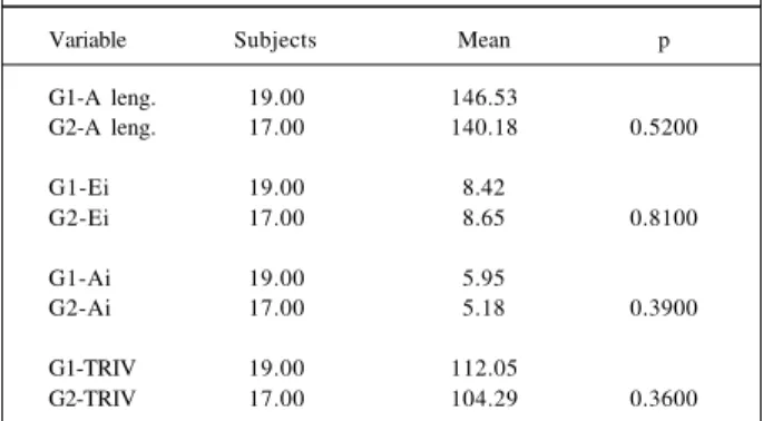
Table 1 shows ejection fraction and fractional shorte-ning. Tables II, III, and IV present the variables of the transmi-tral Doppler and the pulmonary vein. Table V contains the di-astolic patterns found in both groups.
Discussion
Based on the findings of the present study, we obser-ved the existence of an interrelation between systolic and diastolic functions. Deterioration in systolic function, jud-ged by the more advanced functional class, is accompanied by worsening in the left ventricular diastolic pattern. This mechanism has been previously identified and reflects a de-crease in ventricular compliance, as it occurs in advanced congestive heart failure 1.
stu-134
Kamel et al
Functional class and systolic and diastolic functions
Arq Bras Cardiol 2001; 76: 132-5.
dy, it was possible to correlate functional classes III and IV with pseudo-normal and restrictive diastolic patterns.
A correlation exists between left ventricular diastolic function and hemodynamic parameters. The restrictive pat-tern of transmitral flow and the short deceleration time are cor-related with elevated pulmonary capillary pressure 17,18. Like-wise, the increased maximum diastolic velocity of pulmo-nary venous flow (D) is associated with elevated pulmona-ry capillapulmona-ry pressure 19.
The elevated E/A ratio of the transmitral flow, the short deceleration time, the reduced S/D ratios of the pulmo-nary venous flow, the lower S pulmopulmo-nary venous velocity
and the pseudo-normal and restrictive diastolic patterns served to characterize functional classes III and IV. Similar-ly, the lower ejection fraction and fractional shortening to-gether with the more severe systolic dysfunction also cha-racterize functional classes III and IV. More recently, the tissue Doppler and color M-mode Doppler techniques of mitral flow propagation have facilitated the characterization of left ventricular diastolic filling patterns.
According to several studies 20-27, other factors, such as the use of correct medication, especially angiotensin converting enzyme inhibitors, may also influence functio-nal class. In the present study, 56% of the patients regularly used some type of angiotensin converting enzyme inhibitor, without differences between groups. It is interesting to note the low percentage of use of angiotensin converting enzyme inhibitors (which are well known to reduce morbidi-ty andmortalimorbidi-ty in cases of congestive heart failure) proba-bly due to low adherence to the treatment.
The question of drug treatment for congestive heart failure and the pattern of diastolic filling of the left ventricle was explored by Keren et al 22, who demonstrated that the maximum velocity of initial diastolic filling (E) decreases with treatment and is associated with a longer period of
Table I - Ejection fraction and fractional shortening
Variable Subjects Mean P
G1-FE 19.00 44.84
G2-FE 17.00 32.59 0.0007
G1-fr. short. 19.00 23.16
G2-fr. short. 17.00 16.00 0.0004
G1 - group 1; G2 - group 2; EF - ejection fraction; fr. short. - fractional shortening.
Table II - Variables of the transmitral Doppler study
Variable Subjects Mean P
G1- E 19.00 62.53
G2- E 17.00 76.47 0.0800
G1- A 19.00 66.21
G2- A 17.00 55.18 0.1500
G1-E/A ratio 19.00 1.07
G2-E/A ratio 17.00 1.98 0.0300
G1-TD 19.00 180.26
G2-TD 17.00 148.70 0.0800
G1- group 1; G2- group 2; E- maximum E velocity - cm/s; A - maximum A velocity - cm/s; E/A ratio; DT- E deceleration time - ms
Table III - Variables of transmitral Doppler and of IVRT
Variable Subjects Mean p
G1-A leng. 19.00 146.53
G2-A leng. 17.00 140.18 0.5200
G1-Ei 19.00 8.42
G2-Ei 17.00 8.65 0.8100
G1-Ai 19.00 5.95
G2-Ai 17.00 5.18 0.3900
G1-TRIV 19.00 112.05
G2-TRIV 17.00 104.29 0.3600
G1 group 1; G2 group 2; A leng. length of A ms; Ei and Ai -integral of E and A velocities, respectively; IVRT- isovolumetric rela-xation time - ms
Table IV - Variables of pulmonary venous Doppler
Variable Subjects Mean P
G1-S 19.00 53.53
G2-S 17.00 43.41 0.0200
G1-D 19.00 37.05
G2-D 17.00 42.88 0.1100
G1-S/D ratio 19.00 1.52
G2-S/D ratio 17.00 1.08 0.0100
G1-Ar leng. 19.00 169.53
G2-Ar leng. 17.00 176.70 0.6200
G1-A / A r 19.00 0.90
G2-A / A r 17.00 0.86 0.6900
G1- group 1; G2- group 2; S- maximum systolic velocity of pulmonary venous flow - cm/s; D- maximum diastolic velocity of pulmonary venous flow - cm/s; S/D ratio - ratio between maximum systolic velocity of pulmonary venous flow and maximum diastolic velocity of pulmonary venous flow; Ar leng. - length of retrograde A in pulmonary vein; A/Ar-A length/retrograde A/Ar-A length ratio in pulmonary vein.
Table V - Diastolic patterns
Group 1 Group 2
Normal 5 (26.32%) 0
Alt. Relax. 10 (52.63%) 7 (41.18%)
Pseudo 2 (10.53%) 3 (17.65%)
Restrictive 2 (10.53%) 7 (41.18%)
Arq Bras Cardiol 2001; 76: 132-5.
Kamel et al Functional class and systolic and diastolic functions
135 1. Nishmura RA, Tajik J. Evaluation of diastolic filling of left ventricle in health and
desease: doppler echocardiography is the clinician’s rosetta stone. J Am Coll Cardiol 1997; 30: 8-18.
2. Davies SW, Fussell AL, Jordan SL, et al. Abnormal diastolic filling patterns in chro-nic heart failure - relationship to exercise capacity. Eur Heart J 1992; 13: 749-57. 3. Miyagi K, Asanoi H, Ishizaka S, et al. Limited value of anaerobic threshold for assessing
functional capacity in patientes with heart failure. Clin Cardiol 1993; 16: 133-7. 4. Traversi E, Pozzoli M, Cioffi G, et al. Mitral flow velocity changes after 6 months
of optimized therapy provide important hemodynamic and prognostic informa-tion in patients with chronic heart failure. Am Heart J 1996; 132: 809-19. 5. Finkelhor RS, Sun JP, Castellanos M, et al. Predicting left heart failure after a
myo-cardial infarction: a preliminary study of the value of echocardiographic measures of left ventricular filling and wal motion. J Am Soc Echocardiogr 1991; 4: 215-23. 6. Van den Broek SA, van Veldhuisen DJ, de Graeff PA, et al. Comparison between New York Heart Association Classification and peak oxygen consumption in the assessment of functional status and prognosis in patients with mild to mode-rate chronic congestive heart failure secondary to either ischemic or idiopathic dilated cardiomyopathy. Am J Cardiol 1992; 70: 359-63.
7. Gianuzzi P, Temporelli PL, Bosimini E, et al. Independent and incremental prog-nostic value of doppler-derived mitral deceleration time of early filling in both symptomatic and asymptomatic patients with left ventricular dysfunction. J Am Coll Cardiol 1996; 28: 383-90.
8. Pozzoli M, Capomolla S, Sanarico M, et al. Doppler evaluation of left ventricular diastolic filling and pulmonary wedge pressure provide similar prognostic in-formation in patients with systolic dysfunction after myocardiol infarction. Am Heart J 1995; 129: 716-25.
9. Giannuzzi P, Temporelli PL, Bosimini E, et al. Independent and incremental prognostic value of doppler-derived mitral deceleration time of early filling in bo-th symptomatic and asymptomatic patients wibo-th left ventricular dysfunction. J Am Coll Cardiol 1996; 28: 383-90.
10. Nijland F, Kamp O, Karreman AJ, et al. Prognostic implications of restrictive left ventricular filling in acute myocardial infarction: a serial doppler echocar-diographic study. J Am Coll Cardiol 1997; 30: 1618-24.
11. Helmcke F, Nanda NC, Hsuing MC, et al. Color Doppler assessment of mitral re-gurgitation with orthogonal planes. Circulation 1987; 75: 175-83. 12. The CRITERIA Committee of the New York Heart Association: Diseases of the
Heart and Blood Vessels; Nomenclature and Criteria for Diagnosis. 6th edition, Boston: Little, Brown and Co., 1964. In: Braunwald E. Heart Disease: A Textbo-ok of Cardiovascular Medicine, 4th edition. Philadelphia: WB Saunders, 1992; cap I: p. 12.
13. Goldman L, Hashimoto B, Cook EF, et al. Comparative reproducibility and vali-dity of systems for assessing cardiovascular functional class: advantages of a new specific activity scale. Circulation 1981; 64: 1227.
14. Feigenbaum H. Echocardiography. Echocardiographic Evaluation of Cardiac Chambers. Williams and Wilkins. 5th Edition, 1994; Cap. 3.
References
15. Shen WF, Tribouilloy C, Rey JL, et al. Prognostic significance of Doppler-deri-ved left ventricular diastolic filling variables in dilated cardiomyopathy. Am Heart J 1992; 124: 1524-33.
16. Werner GS, Schaefer C, Dirk R, et al. Prognostic value of Doppler echocardiogra-phic assessment of left ventricular filling in idiopathic dilated cardiomyopathy. Am J Cardiol 1994; 73: 792-8.
17. Giannuzzi P, Shabetai R, Imparato A, et al. Effects of mental exercise in patients with dilated cardiomyopathy and congestive heart failure. An echocardiogra-phic Doppler study. Circulation 1991; 83: 4(suppl): II 155-II 165.
18. Masuyama T, Lee JM, Nagano R, et al. Doppler echocardiography pulmonary flow-velocity pattern for assessment of the hemodynamic profile in acute conges-tive heart failure. Am Heart J 1995; 129: 107-13.
19. Ogawa S, Oki T, Iuchi A, et al. Evaluation of pulmonary venous flow patterns in left heart failure: a study using transesophageal Doppler echocardiography. J Cardiol 1993; 23: 193-204.
20. The CONSENSUS Trial Study Group: Effects of Enalapril on Mortality in Severe Congestive Heart Failure. N Engl J Med 1987; 316: 1429-35.
21. The SOLVD Investigators: Effects of enalapril on survival in patients with redu-ced left ventricular ejection fraction and congestive heart failure. N Engl J Med 1991; 325: 293-302.
22. Keren G, Pardes A, Eschar Y, et al. Left ventricular filling dynamics by Doppler echocardiography in dilated cardiomyopathy: one-year follow-up in patients treated with captopril compared to placebo. Cardiology 1992; 81: 196-206. 23. Iga K, Hori K, Matsumura T, et al. Left ventricular filling pattern in congestive
heart failure patients with normal sinus rythm - a decreased ratio of peak mitral flow velocity in atrial systole relative to that in early diastole may reflect marke-dly increased left ventricular end-diastolic pressure. Jpn Circ J 1990; 54: 473-7. 24. Giunta A, Maione S, Arnese MR, et al. Effects of intravenous digoxin on pulmona-ry venous and transmitral flows in patients with chronic heart failure of different degrees. Clin Cardiol 1995; 18: 27-33.
25. Yamamuro A, Yoshida K, Akasaka T, et al. Prognostic value of serial Doppler echo-cardiography follow-up of transmitral flow patters in patients with congestive heart failure who presented with pulmonary edema. J Cardiol 1996; 27: 321-7. 26. Andersson B, Caidahl K, di Lenarda A, et al. Changes in early and late diastolic
filling patterns induced by long-term adrenergic beta-blockade in patients with idiopathic dilated cardiomyopathy. Circulation 1996; 94: 673-82. 27. Litwin SE, Katz SE, Morgan JP, et al. Long-term captopril treatment improves
di-astolic filling more than systolic performance in rats with large myocardial infarc-tion. J Am Coll Cardiol 1996; 28: 773-81.
28. Ito T, Suwa M, Kobashi A, et al. Reversible left atrial dysfunction possibly due to afterload mismatch in patients with left ventricular dysfunction. J Am Soc Echo-cardiogr 1998; 11: 274-9.
29. Magnusson G, Gordon A, Kaijser L, et al. High intensity knee extensor training, in patients with chronic heart failure. Major skeletal muscle improvement. Eur Heart J 1996; 17: 1048-55.
exercise and improvement in functional class. Similarly, other researchers have demonstrated that the patterns of di-astolic filling of the left ventricle might change from one type to the other, with the patients’ clinical improvement and the optimization of treatment 4,15,22-28. The restrictive pattern of the left ventricle, which remains unaltered despite the opti-mization of treatment for congestive heart failure, has been pointed out as an important marker of clinical deterioration and worsening in functional class in patients with dilated myocardiopathy, according to Shen et al 15. The clinical and functional class improvement in congestive heart failure pa-tients is accompanied by a reduction in the E/A transmitral flow ratio and by an augmentation of the deceleration time 15. Reports exist in the literature about functional class im-provement in patients who undergo physical training. It is known that one of the adaptive mechanisms of congestive heart failure consists of physical conditioning loss resulting
from a decrease in peripheral tissue perfusion. Magnusson et al 29 demonstrated that alterations in skeletal muscles in con-gestive heart failure are not entirely irreversible. Localized muscle training is efficient and may result in a marked aug-mentation of the local work capacity and in a small increase in the total work capacity. In trained patients, an increase in the quadriceps muscle transverse area, an increase in the ca-pillary/fiber index, and an increase in oxidative enzymatic ac-tivity have been demonstrated 29. These factors might explain some discrepancies found in the present study, such as the presence of 2 patients with pseudo-normal diastolic pattern and 2 others in group 1 with a restrictive pattern.