294
Canellas R et al. Spontaneous regression of pulmonary alveolar proteinosis
Radiol Bras. 2012 Set/Out;45(5):294–296
Spontaneous regression of pulmonary alveolar proteinosis:
a case report
*
Regressão espontânea de proteinose alveolar pulmonar: relato de caso
Rodrigo Canellas de Souza1, Daniel Kanaan2, Pedro Henrique Rodrigues Martins3, Guilherme
Abdalla Gavinho Vianna4, Viviane Brandão Amorim4, Edson Marchiori5
The authors report the case of a 21-year-old female patient with a six-month history of progressive dyspnea, dry cough, and weight loss. High-resolution computed tomography revealed a “crazy-paving” pattern with areas of focal sparing. The patient underwent transbronchial lung biopsy which confirmed the diagnosis of alveolar proteinosis. Two years later, without treatment, a marked improvement in pulmonary opacities was observed.
Keywords: Alveolar proteinosis.
Relatamos o caso de uma mulher de 21 anos com história de seis meses de dispneia progressiva, tosse seca e perda de peso. A tomografia computadorizada de alta resolução revelou padrão de pavimentação em mosaico com áreas focais poupadas. A paciente foi submetida a biópsia pulmonar transbrônquica, que confirmou o diagnóstico de protei-nose alveolar. Dois anos depois, sem tratamento, houve importante melhora das opacidades pulmonares.
Unitermos: Proteinose alveolar.
Abstract
Resumo
* Study developed at Hospital Universitário Clementino Fraga Filho – Universidade Federal do Rio de Janeiro (HUCFF-UFRJ), Rio de Janeiro, RJ, Brazil.
1. MD, Resident at Instituto do Câncer do Estado de São Paulo “Octavio Frias de Oliveira” (Icesp) – Faculdade de Medicina da Universidade de São Paulo (FMUSP), São Paulo, SP, Brazil.
2. MD, Resident of Interventional Vascular Radiology at Hos-pital das Clínicas da Faculdade de Medicina da Universidade de São Paulo (InRad/HC-FMUSP), São Paulo, SP, Brazil.
3. MD, Resident at Clínica de Diagnóstico por Imagem (CDPI), Rio de Janeiro, RJ, Brazil.
4. MDs., Residents at Clínica Radiológica Luiz Felippe Mat-toso, Rio de Janeiro, RJ, Brazil.
5. Full Professor, Department of Radiology, Universidade Fe-deral Fluminense (UFF), Niterói, RJ, Adjunct Coordinator of the Course of Post-graduation in Radiology, Universidade Federal do Rio de Janeiro (UFRJ), Rio de Janeiro, RJ, Brazil.
Mailing Address: Dr. Rodrigo Canellas de Souza. Rua João Moura, 1119, ap. 55, bloco B, Pinheiros. São Paulo, SP, Brazil, 05412-002. E-mail: rodcanellas@ig.com.br
Received October 26, 2011. Accepted after revision July 2, 2012.
Canellas R, Kanaan D, Martins PHR, Vianna GAG, Amorim VB, Marchiori E. Spontaneous regression of pulmonary alveolar proteinosis: a case report. Radiol Bras. 2012 Set/Out;45(5):294–296.
0100-3984 © Colégio Brasileiro de Radiologia e Diagnóstico por Imagem CASE REPORT
dry cough and weight loss of approxi-mately 6 kg. The patient did not report fe-ver or chest pain and did not present a his-tory of smoking. Physical examination demonstrated normal heart and lung aus-cultation, with no cyanosis or digital club-bing. Blood count and serum biochemistry test results were also normal.
Chest radiography revealed symmetric reticular perihilar infiltrate, with confluence areas in the right lower lobe (Figure 1). High-resolution computed tomography (HRCT) demonstrated patchy areas of nary surfactant by type II alveolar cells and
macrophages(2). Most PAP cases are idio-pathic, although in some cases the condi-tion results from exposure to silica (silico-proteinosis), association with hematologic diseases (for example: lymphoma and leu-kemia) or with infection by the human immunodeficiency virus.
CASE REPORT
A 21-year-old female presenting with a six-month history of progressive dyspnea,
INTRODUCTION
Pulmonary alveolar proteinosis (PAP) is characterized by alveolar filling with pro-tein material. Such material can be detected by periodic acid-Schiff staining (PAS) and has high lipid content(1). PAP is most com-monly diagnosed in adult individuals be-tween 20 and 50 years of age, but it is also reported in other age groups.
The most accepted hypothesis is that PAP results from an abnormal production, altered metabolism or depuration of
295
Canellas R et al. Spontaneous regression of pulmonary alveolar proteinosis
Radiol Bras. 2012 Set/Out;45(5):294–296 ground glass attenuation and inter and in-tralobular septal thickening, characterizing the crazy-paving pattern, with areas of fo-cal sparing (Figure 2). The patient was sub-mitted to transbronchial lung biopsy, and the histopathological analysis identified al-veolar filling with amorphous granular eosi-nophilic PAS-positive material, which pre-served the alveolar septal architecture. Such characteristics were compatible with PAP diagnosis. The patient was followed-up on an outpatient basis, with no specific treat-ment and two years later presented signifi-cant improvement of clinical symptoms and pulmonary opacities at HRCT (Figure 3).
DISCUSSION
PAP is a diffuse pulmonary disease characterized by accumulation of amor-phous lipoproteinaceous, PAS-positive
material in the distal air spaces(1,3). Typi-cally, there is little or no pulmonary inflam-mation and the adjacent pulmonary archi-tecture is preserved(1,3). The following three forms of this condition are recognized: congenital, secondary and acquired PAP.
The congenital form occurs in the neo-natal period and seems to be either a result of mutations in genes that encode the sur-factant, the macrophage colony stimulating factor receptor and granulocytes (GM-CSF), or the result of a defect in the plasma membrane of the cationic amino acid trans-porter (known as lysinuric protein intoler-ance)(4).
The secondary form of PAP develops in the adulthood, generally as a result of ex-posure to high dust levels (for example: silica, aluminum and titanium), malignant hematologic diseases or after allogeneic bone marrow transplant(4). In Brazil,
silico-proteinosis is the most common form of secondary alveolar proteinosis(5–7).
The acquired form is associated with a high prevalence of anti-GM-CSF auto-an-tibodies and is the most common form of PAP(4).
Patients with idiopathic or secondary PAP present with moderate and nonspecific respiratory symptoms such as progressive dyspnea which may occur for months or years, besides dry or minimally productive cough. Less common signs and symptoms include weight loss, fatigue, low fever, chest pain and hemoptysis(3,8). Physical examination may reveal crepitations, club-bing or cyanosis(8). The mean age at PAP diagnosis is 40 ± 13 years; and a strong association with smoking has been ob-served(3,8).
Patients with PAP present greater pre-disposition to pneumonic infection.
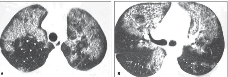
Infec-Figure 2. HRCT at two different levels showing the crazy paving pattern. Areas of ground-glass attenuation and intralobular septal thickening are observed in association with areas of focal sparing.
A B
Figure 3. HRCT at the same levels as those on Figure 2. Images acquired obtained two years after the diagnosis showing marked improvement of the lesions.
296
Canellas R et al. Spontaneous regression of pulmonary alveolar proteinosis
Radiol Bras. 2012 Set/Out;45(5):294–296 tious pneumonias in PAP patients are many
times opportunistic infections(9).
The diagnosis of PAP usually requires chest radiography, although it is not con-clusive for such diagnosis. Radiography can detect bilateral, symmetrical and cen-tral opacities(1,8). Additionally, many times there is a notable disparity between the clinical symptoms of PAP and radiographic changes (“clinical-radiological dissocia-tion”)(8). HRCT provides more anatomical details and information on the disease ex-tent. The crazy paving pattern, which cor-responds to the septal thickening overlap-ping ground-glass attenuation areas is the most frequent tomographic finding associ-ated with PAP(10,11). Regions with crazy paving pattern are typically generalized and bilateral, many times sparing well delim-ited areas or even an entire pulmonary lobe(12).
Although the crazy paving pattern is fre-quently detected at HRCT in PAP patients, such finding is also observed in several infectious, hemorrhagic, neoplastic,
inha-lation and idiopathic disorders, as well as in pulmonary edema(11,13).
In summary, the present report describes the case of a previously healthy, young, female patient with no history of heart problems or occupational disease, present-ing crazy pavpresent-ing pattern at HRCT. The di-agnosis of pulmonary alveolar proteinosis was confirmed by biopsy. Two years after diagnosis, the patient is in good clinical conditions, with marked improvement of the pulmonary opacities.
REFERENCES
1. Rosen SH, Castleman B, Liebow AA, et al. Pul-monary alveolar proteinosis. N Engl J Med. 1958;258:1123–42.
2. Wang BM, Stern EJ, Schmidt RA, et al. Diagnos-ing pulmonary alveolar proteinosis. A review and an update. Chest. 1997;111:460–6.
3. Presneill JJ, Nakata K, Inoue Y, et al. Pulmonary alveolar proteinosis. Clin Chest Med. 2004;25: 593–613.
4. Prakash UB, Barham SS, Carpenter HA, et al. Pul-monary alveolar phospholipoproteinosis: experi-ence with 34 cases and a review. Mayo Clin Proc. 1987;62:499–518.
5. Souza CA, Marchiori E, Gonçalves LP, et al.
Com-parative study of clinical, pathological and HRCT findings of primary alveolar proteinosis and silicoproteinosis. Eur J Radiol. 2012;81:371–8. 6. Marchiori E, Souza CA, Barbassa TG, et al. Silicoproteinosis: high-resolution CT findings in 13 patients. AJR Am J Roentgenol. 2007;189: 1402–6.
7. Marchiori E, Ferreira A, Müller NL. Silicoprotei-nosis: high-resolution CT and histologic findings. J Thorac Imaging. 2001;16:127–9.
8. Ioachimescu OC, Kavuru MS. Pulmonary alveo-lar proteinosis. Chron Respir Dis. 2006;3:149– 59.
9. Burkhalter A, Silverman JF, Hopkins MB 3rd, et al. Bronchoalveolar lavage cytology in pulmonary alveolar proteinosis. Am J Clin Pathol. 1996;106: 504–10.
10. Johkoh T, Itoh H, Müller NL, et al. Crazy-paving appearance at thin-section CT: spectrum of dis-ease and pathologic findings. Radiology. 1999; 211:155–60.
11. Vabo KA, Damato SD. Aspectos tomográficos e anatomopatológicos do padrão de pavimentação em mosaico. Radiol Bras. 2011;44:215–9. 12. Holbert JM, Costello P, Li W, et al. CT features
of pulmonary alveolar proteinosis. AJR Am J Roentgenol. 2001;176:1287–94.