Tomographic findings of gastric gastrointestinal stromal
tumor: a 14-case study*
Aspectos tomográficos do tumor estromal gastrintestinal de origem gástrica: estudo de 14 casos
Gustavo Lemos Pelandré1
, Maria Célia Djahjah2
, Luiz Felipe Nobre3
, Emerson Leandro Gasparetto4
, Edson Marchiori5
, Bruno Vilhena Pereira6
, Marcus Valadão7
, Eduardo Linhares8
OBJECTIVE: The purpose of this study was to describe the tomographic findings of gastric gastrointestinal stromal tumor. MATERIALS AND METHODS: Fourteen patients with histopathologically and immunohisto-chemically confirmed gastric gastrointestinal stromal tumors, who had already been submitted to computed tomography scans before the treatment, were evaluated in the period between January 1999 and December 2006. The following tomographic variables were analyzed: lesion topography, size/dimensions, homogeneity, contour, margins, morphology, pattern and intravenous contrast-enhancement intensity, growth pattern, invasion of adjacent organs, presence of ulceration, fistula, calcifications, mesenteric fat infiltration, lymphadenomegaly and presence of distant metastasis. RESULTS: Tumors were found in the body (57.1%) or in the gastric fundus (42.9%), with sizes ranging between 6.0 cm and 23.0 cm (mean, 11.5 cm). Pre-dominantly extraluminal growth was observed in 57.1% of cases and intra/extraluminal in 35.7%. Subtle contrast-enhancement was observed in 50%, moderate in 50%, and heterogeneous in 64.3% of cases. Additionally, central hypodensity was observed in 64.3%, invasion of adjacent organs in 42.9%, and he-patic metastasis in 7.2% of cases. CONCLUSION: In the present study, the majority of tumors were found in the gastric body, with an average size of 11.5 cm, presenting with central hypodensity, heterogeneous contrast-enhancement and predominantly extraluminal growth.
Keywords: Gastrointestinal stromal tumors; Stomach neoplasms; Spiral computed tomography.
OBJETIVO: Descrever os achados tomográficos do tumor estromal gastrintestinal de origem gástrica. MA-TERIAIS E MÉTODOS: No período de janeiro de 1999 a dezembro de 2006, foram selecionados 14 pacien-tes com diagnóstico histopatológico e imuno-histoquímico de tumor estromal gastrinpacien-testinal gástrico que apresentavam tomografia computadorizada realizada anteriormente ao tratamento. As variáveis tomográfi-cas analisadas foram: topografia da lesão, dimensões, homogeneidade, contornos, limites, morfologia, pa-drão e intensidade do realce pelo meio de contraste venoso, papa-drão de crescimento, invasão de órgãos adjacentes, presença de ulceração, fístula, calcificações, infiltração da gordura mesentérica, linfonodome-galias e metástases a distância. RESULTADOS: Os tumores foram localizados no corpo (57,1%) ou fundo gástrico (42,9%), com dimensões variando entre 6,0 e 23,0 cm (média de 11,5 cm). O crescimento foi predominantemente extraluminal (57,1%) ou intra/extraluminal (35,7%). O realce pelo contraste venoso foi discreto em 50% dos casos, moderado em 50% e heterogêneo em 64,3%. Foram ainda observadas hipo-densidade central em 64,3% dos casos, invasão de órgãos adjacentes em 42,9% e metástases hepáticas em 7,2%. CONCLUSÃO: No presente estudo, a maioria dos tumores localizava-se no corpo gástrico, com tamanho médio de 11,5 cm, apresentando área hipodensa central, realce heterogêneo pelo meio de con-traste e crescimento predominantemente extraluminal.
Unitermos: Tumores do estroma gastrintestinal; Neoplasias gástricas; Tomografia computadorizada helicoidal. Abstract
Resumo
* Study developed at Hospital do Câncer I, Instituto Nacional de Câncer (INCA), Rio de Janeiro, RJ, Brazil.
1. Resident in Radiology and Imaging Diagnosis at Instituto Nacional de Câncer (INCA), Fellow Master degree, Course of Post-graduation in Radiology at Universidade Federal do Rio de Janeiro (UFRJ), Rio de Janeiro, RJ, Brazil.
2. PhD in Radiology, Associate Professor, Department of Radiology, Universidade Federal do Rio de Janeiro (UFRJ), MD, Physician at Unit of Radiodiagnosis, Instituto Nacional de Cân-cer (INCA), Rio de Janeiro, RJ, Brazil.
3. PhD in Radiology, Associate Professor of Radiology, Head for the Unit of Radiology, Hospital Universitário da Universidade Federal de Santa Catarina (UFSC), Florianópolis, SC, Brazil.
4. PhD in Radiology, Associate Professor in the Department
Pelandré GL, Djahjah MC, Nobre LF, Gasparetto EL, Marchiori E, Pereira BV, Valadão M, Linhares E. Tomographic findings of gastric gastrointestinal stromal tumor: a 14-case study. Radiol Bras. 2008;41(5):297–303.
of Radiology, Universidade Federal do Rio de Janeiro (UFRJ), Rio de Janeiro, RJ, Brazil.
5. PhD in Radiology, Full Professor, Department of Radiology, Universidade Federal Fluminense (UFF), Niterói, RJ, Adjunct Coordinator for the Course of Post-Graduation in Radiology at Universidade Federal do Rio de Janeiro (UFRJ), Rio de Janeiro, RJ, Brazil.
6. MD, Oncologist at Instituto Nacional de Câncer (INCA), Rio de Janeiro, RJ, Brazil.
7. Master in Surgery, Oncological Surgeon at Instituto Nacio-nal de Câncer (INCA), Rio de Janeiro, RJ, Brazil.
8. PhD in Surgery, Oncological Surgeon at Instituto Nacional de Câncer (INCA), Rio de Janeiro, RJ, Brazil.
Mailing address: Dr. Gustavo Lemos Pelandré. Serviço de Radiologia, Hospital do Câncer I – Instituto Nacional de Câncer. Praça Cruz Vermelha, 23, Centro. Rio de Janeiro, RJ, Brazil, 20230-130. E-mail:[email protected]
Received September 23, 2007. Accepted after revision February 7, 2008.
INTRODUCTION
involving the digestive tract and 5% of all stromal tumors(1). Approximately 4,500–
6,000 cases of GIST are diagnosed every year in the United States of America, with a peak incidence in the sixth-seventh de-cade of life. The disease may affect any portion of the gastrointestinal tract, the stomach being most frequently involved (45–65%), followed by the small bowel (15–25%), colon (5–10%) and other re-gions of the abdominal cavity (5%)(2).
Although this mesenchymal neoplasm is known for decades, recent findings have allowed a deeper knowledge of its cellular origin, as well as of the molecular events involved in the development of this le-sion(2). Up to about 20 years, it was
be-lieved that most of gastrointestinal mesen-chymal tumors originated from the smooth musculature and were called “leiomyomas” and “leiomyosarcomas”. The use of elec-tronic microscopy and immunohistochem-istry, however, has demonstrated that very few of these tumors presented with smooth muscle characteristics(3). Later, some
au-thors have demonstrated that these tumors also presented characteristics of neuronal differentiation, calling them de “plexosarcomas” and “gastrointestinal au-tonomic nerve tumors(4). Only recently it
was suggested that this neoplasm is a well defined entity called GIST based on the finding that it originates from interstitial cells of Cajal(5) and from KIT proteins
ex-pression(6).
KIT is a membrane tyrosine kinase re-ceptor responsible for different cellular functions such as adhesion, apoptosis and cellular differentiation. In GIST, the muta-tion of the kit gen is responsible for the con-stitutive activation in the KIT protein caus-ing unopposed stimulation of cellular pro-liferation(7).
One of the most promising recent find-ings concerning cancer treatment has been the utilization of molecular target therapy, the GIST being the better example of ap-plication of this therapeutic modality. The discovery of imatinib mesylate has revolu-tionized the therapy for GIST, considering that this is the first drug that affects specifi-cally the molecular alteration responsible for the disease etiology(2). Imatinib
mesylate is an inhibitor of the tyrosine ki-nase of KIT receptors(8).
Computed tomography is the most sen-sitive imaging method for detecting and characterizing GIST, allowing the defini-tion of the tumor size, anatomic locadefini-tion, growth pattern, necrosis evidence, invasion of adjacent organs and metastases, besides the monitoring of the treatment and evalu-ation of the disease progression(2,9).
The authors seek to describe the tomo-graphic findings in a group of 14 patients with gastric GIST, emphasizing the rel-evance of this imaging method for the char-acterization of the lesion in its most fre-quent topography as well as for the defini-tion of differential diagnoses.
MATERIALS AND METHODS
The present study has followed an ob-servational-descriptive model for a series of cases including 41 patients admitted to the Hospital do Câncer I – Instituto Nacional de Câncer, Rio de Janeiro, RJ, Brazil, in the period between January 1999 and December 2006, with histopathologi-cal diagnosis of GIST. Out of these pa-tients, 9 were excluded for lack of immu-nohistochemical staining data, and 18 ei-ther because they had not undergone ab-dominal computed tomography before be-ing submitted to oncologic therapy, or their studies were incomplete or had not been found.
The study population included nine fe-male patients (64.3%) and five fe-male pa-tients (35.7%). Mean age was 59.4 ± 18 years (mean ± standard deviation).
Clinical manifestations observed were: abdominal pain or discomfort (n = 10), weight loss (n = 4), increased abdominal volume (n = 3), hematemesis (n = 1), mel-ena (n = 1), emesis (n = 1) and fever (n = 1). One patient was asymptomatic and the tumor was incidentally found during an abdominal ultrasonography.
Nine patients underwent computed to-mography in the author’s institution in a he-lical CT SCT 7000 TS Shimadzu equip-ment (Shimadzu Corp.; Kyoto, Japan), through axial images acquisition with 5 mm collimation and 7 mm reconstruction interval, pitch 1.5, 120 kV and 130 mA. Ionic iodinated contrast agent (1,000 ml) in a 2% solution was orally administered one hour before each examination (diatrizoate
meglumine). Two acquisitions were per-formed before and after intravenous injec-tion of 100 ml non-ionic iodinated contrast agent (ioexol 300 mgI/ml, 2 ml/s, 55 sec-onds after the infusion was initiated). Five patients had undergone computed tomog-raphy in other institutions, with different techniques, all of them utilizing oral con-trast agents, and three intravenous concon-trast. The tomographic images were indepen-dently analyzed by three radiologists. In cases of interobserver disagreement, the final decision was reached by consensus. The following characteristic were consid-ered: lesion topography, dimensions, ho-mogeneity, contour, limits, morphology, intravenous contrast-enhancement inten-sity and pattern, lesion growth pattern, ad-jacent organs invasion, presence of ulcer-ation, fistulas, calcifications, mesenteric fat infiltration, lymphadenomegaly and distant metastases.
As regards topography, the lesions were classified according to their site of origin. Lesion contours were classified as regular, lobular or irregular, and limits as well-de-fined, ill-defined or invasive. The lesion morphology was defined as round, ovoid or irregular. Intravenous contrast-enhance-ment pattern was described as heteroge-neous or homogeheteroge-neous, and the enhance-ment intensity was compared with the in-tensity of the liver and abdominal muscles: subtle enhancement if the density was equal or lower than the muscle density; mild enhancement, if the density was higher than the muscle and equal or lower than the liver density; marked enhancement if the density was higher than the liver. The lesion growth pattern was classified ac-cording to the predominant component: intra or extraluminal. Cases with the pres-ence of both components without predomi-nance of either of them, were classified as intra/extraluminal. For defining the lesion dimensions, the major measurements in the orthogonal plane were taken into consid-eration.
All of the patients were submitted to surgical treatment, as follows: wedge resec-tion in eight cases (57.1%), total tomy in four (28.6%) and subtotal gastrec-tomy in two (14.3%). Three patients (21.4%) underwent supplementary therapy with imatinib mesylate.
The present study was approved by the Committee for Ethics in Research with Humans of the institution.
RESULTS
The main tomographic findings in the 14 gastric GIST patients are shown on Table 1. The tumors were localized in the gastric body (n = 8; 57.1%) or gastric fun-dus (n = 6; 42.9%). Lesions dimensions ranged between 6.0 cm and 23.0 cm, with a mean size of 11.5 ± 4.4 cm (mean ± stan-dard deviation). The lesion growth pattern was predominantly extraluminal in eight patients (57.1%) (Figure 1), intraluminal in one (7.2%) patient (Figure 2), and intra/ extraluminal in five (35.7%) patients (Fig-ure 3).
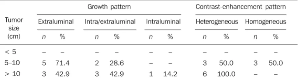
Lesion with extraluminal growth pre-sented dimensions ranging between 5.0 cm and 10.0 cm in five cases, and > 10.0 cm in three, with mean size of 11.4 ± 5.1 cm (Table 2). Lesions with intra/extraluminal growth pattern presented dimensions be-tween 5.0 cm and 10.0 cm in two cases, and > 10.0 cm in three, with mean size of 11.5 ± 4.1 cm. The only intraluminal lesion
measured 11.5 cm (Figure 2). No lesion < 5 cm was found in the present study.
As regards the intravenous contrast-en-hancement pattern, nine patients (64.3%) presented heterogeneous contrast-enhance-ment, and three (21.4%), homogeneous contrast-enhancement. Intravenous con-trast agent was not utilized in two patients (14.3%). Among the patients who received
Table 1 Main tomographic findings in 14 GIST patients.
Features
Location
Size
Growth
Morphology
Limits
Intravenous contrast-enhancement
Contrast-enhancement intensity
Central hypodensity
Gastric body Gastric fundus
< 5 cm 5–10 cm > 10 cm
Extraluminal Intra/extraluminal Intraluminal
Ovoid Irregular Round
Well-defined Ill-defined Invasive
Heterogeneous Homogeneous Non-evaluated
Subtle Mild Marked
Present Absent
n
8 6
– 7 7
8 5 1
7 4 3
10 2 2
9 3 2
6 6 –
9 5
%
57.1 42.9
– 50.0 50.0
57.1 35.7 7.2
50.0 28.6 21.4
71.4 14.3 14.3
64.3 21.4 14.3
50.0 50.0 –
64.3 35.7 Patients
n, number of patients. * Only for patients who received intravenous contrast agent.
Figure 2.A, B: Computed tomography image with both oral and intravenous contrast-enhancement showing an intraluminal vegetative lesion in the upper third of the great curvature, forming a fistulous tract with the gastric lumen (A). Presence of gas and oral contrast agent within the lesion (B). C: Intra-operative appearance of the tumor. D: Surgical specimen where an intraluminal lesion can be observed in the great curvature.
A B
C D
Figure 1. Computed tomography image with both oral and intravenous contrast-enhancement showing a large, expansile, solid and heterogeneous lesion with hypodense areas inside, and extraluminal growth (A), without a cleavage plane with the small gastric curvature (B).
contrast agent, six (50%) presented mild enhancement. Marked contrast-enhancement was not observed in any case of the present study.
Among the tumors measuring 5–10 cm, 50% presented heterogeneous contrast-en-hancement and 50% homogeneous con-trast-enhancement. All the tumors with > 10 cm presented heterogeneous contrast-enhancement (Table 2). Heterogeneous
tu-mors presented mean size of 12.9 ± 4.6 cm, while homogeneous tumors presented mean size of 8.4 ± 1.1 cm.
In the majority of cases, the lesions were ovoid (50%), with regular contour (71.4%) and well-defined limits (71.4%). A central area of hypodensity was observed in nine cases (64.3%), ulceration in six (42.9%), fistula in four (28.6%), mesenteric fat
in-filtration in four (28.6%) and intratumoral calcification in two (14.3%). Adjacent lymphadenomegaly was observed in only one patient (7.2%).
Adjacent organs invasion was observed in sis cases (42.9%), with diaphragm (n = 3; 21.4%), spleen (n = 2; 14.3%) and pan-creas (n = 2; 14.3%) being most frequently affected (Figures 4 and 5).
Hepatic metastases were identified in only one case (7.2%) (Figure 6).
DISCUSSION
Although rare, GISTs are the most com-mon mesenchymal neoplasms of the gas-trointestinal tract, representing 1% to 3% of all gastrointestinal tumors and 2.5% of Figure 3. Computed tomography image with both
oral and intravenous contrast-enhancement show-ing an expansile, heterogeneous lesion with a cen-tral hypodense area and intra/excen-traluminal growth, located on the anterior wall and great gastric cur-vature.
Figure 4. Computed tomography image with both oral and intravenous contrast-enhancement showing a large expansile, heterogeneous lesion with extraluminal growth, located in the gastric fundus and ex-tending towards the spleen (A) and left diaphragmatic pillar (B).
A B
Figure 6.A: Abdominal ultrasonography demonstrating expansile, nodular, heterogeneous lesions in the right hepatic lobe. B: Noncontrast-enhanced computed tomography image demonstrating solid lesions with a hypodense center, corresponding to the sonographic findings (liver metastasis).
A B
Figure 5. Computed tomography image with both oral and intravenous contrast-enhancement show-ing an expansile, hypodense formation, with irregu-lar margins, located in the great gastric curvature and extending towards the pancreatic body and tail.
Table 2 GIST growth pattern and contrast-enhancement*, according to the lesion dimensions.
Tumor size (cm)
< 5 5–10 > 10
Growth pattern Contrast-enhancement pattern
Extraluminal Intra/extraluminal Intraluminal Heterogeneous Homogeneous
n
– 2 3
%
– 28.6 42.9
n
– 3 –
%
– 50.0
–
n
– 5 3
%
– 71.4 42.9
n
– – 1
%
– – 14.2
n
– 3 6
%
– 50.0 100.0
gastric tumors(10). The stomach is the most
frequent site of this disease, accounting for 45–65% of cases, followed by the small bowel in 15–25% of cases(2). Most rarely
these tumors are found in the rectum, co-lon, esophagus, appendix, mesenterium, omentum, retroperitoneum and other sites not related to the gastrointestinal tract(11).
The incidence of this disease is higher in-dividuals with > 50 years, with mean age ranging between 55 and 67 years(9,12), and
is rarely found in individuals with < 40 years(11). Association between the disease
incidence and geographic location, race and occupation has not been established. Additionally, some authors have indicated a slight prevalence of the disease in men(13)
and increased incidence in patients with type-1 neurofibromatosis(14). The present
casuistic included nine female (64.3%) and five male patients (35.7%). Mean age was 59.4 years.
Clinical manifestations in GIST patients are non-specific and depend on the lesion location. Digestive hemorrhage is the most frequent symptom presenting as hematem-esis, melena, hematoquezia or signs of ane-mia due to occult bleeding(9–11,15,16). Other
common manifestations are non-specific abdominal pain, dysphagia, weight loss, nausea, emesis, abdominal mass or obstruc-tive symptoms. Approximately 20% of cases may present with asymptomatic lesions incidentally found and diagnosed during imaging evaluation or surgical proce-dures(2,11). In the present casuistic,
abdomi-nal pain was the most frequent symptom. Classification of mesenchymal tumors can be based on findings at optic micros-copy and immunohistochemistry. The his-topathological classification is based on the predominant cellular type: spindle cells, epithelioid cells or pleomorphic cells(17).
Gastric GISTs are characterized by the presence of spindle cells in 70–80% of cases. The diagnosis is achieved by means of immunohistochemical staining based on the expression of KIT protein (CD117), a product of c-Kit proto-oncogene (a growth factor receptor with tyrosine kinase activ-ity). GISTs are CD117-positive (95%) and CD34-positive (30–40%) tumors(17). Many
studies have demonstrated that about 4% of cases present clinical and pathological characteristics compatible with GIST
al-though KIT-protein expression is absent. Heinrich et al.(18) have demonstrated that
this group of tumors presents activating mutation in other tyrosine kinase receptor similar to KIT (platelet-derived growth fac-tor recepfac-tor, alpha polypeptide – PDGFRA), representing an alternative hy-pothesis in the pathogenesis of this neo-plasm. Differential diagnoses also include other mesenchymal neoplasms with a dif-ferent histochemical profile such as leiomyomas, leiomyosarcomas, schwann-omas, neurofibromas and neuroendocrine tumors(11,16). In the present study, the
ma-jority of patients presented spindle cell tu-mor (85.7%), while epithelioid cell tutu-mors (7.2%) or pleomorphic cell tumors (7.2%) were less frequently found.
In the literature, many factors are iden-tified as variables capable of predicting GIST progression: size, mitotic index, pres-ence of tumor necrosis, cell proliferation markers, tumor site(19). Findings, however,
are controversial and a consensus is still to be reached; so the biological behavior of the tumor can hardly be predicted. So, the terms “benign” or “malignant” have been avoided, and GIST is classified according to their potential for malignancy based on the most relevant prognostic factors de-scribed in the literature (tumor site, size and mitotic index)(20). So, gastric tumors may
be classified as follows: high risk, interme-diate risk, low risk or very low risk. High-risk tumors are those with > 10 cm in size, more than 10 mitoses per 50 high magnifi-cation fields (50 HMF), or even those with > 5 cm in size with more than 5 mitoses per 50 HMF; intermediate risk if less than 5 cm with 6 to 10 mitoses per 50 HMF or mea-suring 5–10 cm with less than 5 mitoses per 50 HMF; low-risk tumors are those with 2– 5 cm in size, and less than 5 mitoses per 50 HMF; and very low-risk tumors are those with < 2 cm in size and less than 5 mitoses per 50 HMF(2,17).
The tomographic characteristics of GISTs have been studied by some au-thors(9–12,15,16,21–27). Sandrasegaran et al.(10)
have found tumors measuring between 3 cm and 10 cm, with a predominantly exo-phytic growth and heterogeneous intrave-nous contrast-enhancement. On the other hand, Levy et al.(11) have found lesions
quite variable in size, presenting as
typi-cally circumscribed masses, some of them with focal areas of hemorrhage, cystic de-generation and necrosis. In the literature review, the authors observed that the gas-tric body was the segment most frequently affected by GIST (38–75%)(11,15,16),
pre-senting with mean size ranging between 5.4 cm and 13.0 cm(22,26). In the present study,
gastric tumors were predominantly located in the gastric body (57.1%) with a mean size of 11.5 cm.
Tumors with < 5 cm in size present in-traluminal growth and homogeneous intra-venous contrast-enhancement, while the majority of tumors with > 10 cm present extraluminal component and heteroge-neous contrast-enhancement(9,15,23). Kim et
al.(15) have found 57% of extraluminal
gas-tric tumors with > 10 cm and 89% of in-traluminal tumors with < 5 cm. On the other hand, Da Ronch et al.(23) have
ob-served 100% of tumors with > 5 cm in size with extraluminal growth, the intraluminal growth pattern being predominantly related to small lesions. Similar results have been demonstrated by Tateishi et al.(9), who have
observed larger sizes in lesions with extrin-sic growth, 91.3% of them above the mean size. In the present casuistic, no tumor < 5 cm in size was found, and only one patient presented a lesion with intraluminal growth. However, among the cases with tumors measuring between 5 cm and 10 cm and with > 10 cm in size, there was a pre-dominance of lesions with extraluminal growth in respectively 71.4% and 50%.
Also the pattern of contrast-enhance-ment is variable according to the lesions di-mensions(15,24,26,27). In the study developed
by Kim et al.(15), 49% of heterogeneous
tu-mors were > 10 cm in size, and 77% of the homogeneous tumors were < 5 cm. On the other hand, Lee et al.(26) have found a mean
size of 11.6 cm among heterogeneously contrast-enhanced tumors and 3.8 cm among homogeneously contrast-enhanced tumors. Similar results have also been re-ported by Horton et al.(24) and Nishida et
al.(27). In the present study, the authors
ob-served 66.7% of heterogeneous tumors with > 10 cm in size and 100% of homo-geneous tumors measuring between 5 cm and 10 cm.
mu-cosal ulceration, cavitation and central hypodense areas likely corresponding to cystic degeneration, hemorrhage or necro-sis. The necrotic cavities can also commu-nicate with the gastrointestinal lumen forming fistulas with gas, air-fluid level or oral contrast agent(25). In the present study,
central areas of hypodensity were found in 64.3% of patients, while other authors re-port this finding in 20–49% of cases(12,15).
The presence of gas or contrast agent within the lesion also may suggest the pres-ence of mucosal ulceration with fistula formation. Other studies report ulceration in 3–88% of cases, most of them with high histological degree(9,12,13,15,16). In the
present casuistic, signs of mucosal ulcer-ation were found in 42.9% of patients, and fistula in 28.6%.
Liver and peritoneum are the most fre-quent sites of GIST metastases which may occur in up to 38% of cases(12). Other less
common sites of metastasis are: lungs, mesenterium, omentum, ovaries and blad-der(12). Liver metastases may be visualized
at computed tomography as hypodense le-sions, isodense lesions in the portal phase, with intense contrast-enhancement in the arterial phase, or cystic lesions with periph-eral soft tissues density(15). Implants may
become hypovascular or cystic after che-motherapy, and this pattern should not be confused with a sign of disease progression of new lesions(10,12). In the present study,
only one patient (7.2%) presented liver metastasis at presentation, and six patients (42.9%) presented adjacent organs inva-sion with predominant involvement of the diaphragm, spleen and pancreas.
Computed tomography still remains as the imaging method of choice in the GIST characterization, as well as in the evalua-tion of adjacent organs involvement, ab-dominal metastases and treatment re-sponse(1). The addition of new technologies
to this imaging method, particularly with the utilization of multidetectors and multiplanar reconstruction, has allowed a better evaluation of great exophytic tumors and of the relationship between gastric le-sions and adjacent structures, besides al-lowing tumors characterization in specific circumstances, such as masses of unknown origin or originating from endoscopically hardly accessible regions(23,25). Parameters
such as progressive mass hypoattenuation, decrease in the nodular enhancement and vascularization indicate a good treatment response(28).
Complete surgical resection is the stan-dard treatment for GIST, considered as the sole modality capable of providing curative therapy, although about 20% to 50% of patients submitted to complete surgical resection present recurrence of the dis-ease(29). The post-procedural five-year
sur-vival rate in cases of GIST ranges between 30% and 65%(29). In cases of unresectable
or metastatic tumors, the treatment of choice is performed with imatinib mesylate (STI 571), a selective tyrosine kinase en-zyme inhibitor that has been utilized as an adjuvant or neoadjuvant therapy for large tumors initially unresectable or in cases where complete resection is unfeasible(28).
Many authors suggest that a disease invo-lution may occur, allowing the resection of previously unresectable tumors(30). Recent
studies have demonstrated that imatinib presents a significant activity in patients with advanced stages of GIST, achieving a partial response rate in 53.7% of cases and disease stabilization in 27.9%(8). In the
present casuistic, all the patients underwent surgical treatment, most of them with wedge resection (57.1%), and three pa-tients (21.4%) were also submitted to imatinib mesylate therapy.
CONCLUSION
In the present study the majority of GISTs were located in the gastric body, with a mean size of 11.5 cm, a central hypodense area, heterogeneous contrast-enhancement and predominantly extraluminal growth. The knowledge of typical findings and tomographic variations of this neoplasm in its most frequent topog-raphy allows the radiologist to establish the differential diagnosis for gastric tumors, as well as guiding the subsequent phases of the diagnostic investigation, aiding in the therapeutic planning for GIST patients.
REFERENCES
1. Blay JY, Bonvalot S, Casali P, et al. Consensus meeting for the management of gastrointestinal stromal tumors. Report of the GIST Consensus Conference of 20-21 March 2004, under the aus-pices of ESMO. Ann Oncol. 2005;16:566–78.
2. Efron DT, Lillemoe KD. The current management of gastrointestinal stromal tumors. Adv Surg. 2005;39:193–221.
3. Mazur MT, Clark HB. Gastric stromal tumors. Reappraisal of histogenesis. Am J Surg Pathol. 1983;7:507–19.
4. Lauwers GY, Erlandson RA, Casper ES, et al. Gastrointestinal autonomic nerve tumors. A clini-copathological, immunohistochemical, and ultra-structural study of 12 cases. Am J Surg Pathol. 1993;17:887–97.
5. Kindblom LG, Remotti HE, Aldenborg F, et al. Gastrointestinal pacemaker cell tumor (GIPACT): gastrointestinal stromal tumors show phenotypic characteristics of the interstitial cells of Cajal. Am J Pathol. 1998;152:1259–69.
6. Hirota S, Isozaki K, Moriyama Y, et al. Gain-of-function mutations of c-kit in human gastrointes-tinal stromal tumors. Science. 1998;279:577–80.
7. Huizinga JD, Thuneberg L, Klüppel M, et al. W/ kit gene required for interstitial cells of Cajal and for intestinal pacemaker activity. Nature. 1995; 373:347–9.
8. Demetri GD, von Mehren M, Blanke CD, et al. Efficacy and safety of imatinib mesylate in ad-vanced gastrointestinal stromal tumors. N Engl J Med. 2002;347:472–80.
9. Tateishi U, Hasegawa T, Satake M, et al. Gas-trointestinal stromal tumor. Correlation of com-puted tomography findings with tumor grade and mortality. J Comput Assist Tomogr. 2003;27: 792–8.
10. Sandrasegaran K, Rajesh A, Rydberg J, et al. Gastrointestinal stromal tumors: clinical, radio-logic, and pathologic features. AJR Am J Roentgenol. 2005;184:803–11.
11. Levy AD, Remotti HE, Thompson WM, et al. Gastrointestinal stromal tumors: radiologic fea-tures with pathologic correlation. Radiographics. 2003;23:283–304.
12. Sandrasegaran K, Rajesh A, Rushing DA, et al. Gastrointestinal stromal tumors: CT and MRI findings. Eur Radiol. 2005;15:1407–14.
13. Ghanem N, Altehoefer C, Furtwängler A, et al. Computed tomography in gastrointestinal stromal tumors. Eur Radiol. 2003;13:1669–78. 14. Boldorini R, Tosoni A, Leutner M, et al. Multiple
small intestinal stromal tumours in a patient with previously unrecognized neurofibromatosis type 1: immunohistochemical and ultrastructural evaluation. Pathology. 2001;33:390–5.
15. Kim HC, Lee JM, Kim KW, et al. Gastrointesti-nal stromal tumors of the stomach: CT findings and prediction of malignancy. AJR Am J Roentgenol. 2004;183:893–8.
16. Martín-Lorenzo JG, Aguayo-Albasini JL, Tor-ralba-Martínez JA, et al. Gastrointestinal stromal tumors. Diagnosis, prognosis and current surgi-cal treatment. Follow-up of 18 treated patients. Cir Esp. 2006;79:22–7.
17. Fletcher CD, Berman JJ, Corless C, et al. Diag-nosis of gastrointestinal stromal tumors: a consen-sus approach. Hum Pathol. 2002;33:459–65.
18. Heinrich MC, Corless CL, Duensing A, et al. PDGFRA activating mutations in gastrointestinal stromal tumors. Science. 2003;299:708–10.
with special reference to prognostic factors: analysis of results in 140 surgically resected pa-tients. Gastric Cancer. 2003;6:39–48.
20. Miettinen M, El-Rifai W, Sobin LH, et al. Evalu-ation of malignancy and prognosis of gastrointes-tinal stromal tumors: a review. Hum Pathol. 2002;33:478–83.
21. Buckley JA, Fishman EK. CT evaluation of small bowel neoplasms: spectrum of disease. Radiographics. 1998;18:379–92.
22. Burkill GJC, Badran M, Al-Muderis O, et al. Malignant gastrointestinal stromal tumor: distri-bution, imaging features, and pattern of metastatic spread. Radiology. 2003;226:527–32. 23. Da Ronch T, Modesto A, Bazzocchi M.
Gas-trointestinal stromal tumour: spiral computed to-mography features and pathologic correlation. Radiol Med (Torino). 2006;111:661–73.
24. Horton KM, Juluru K, Montogomery E, et al. Computed tomography imaging of gastrointesti-nal stromal tumors with pathology correlation. J Comput Assist Tomogr. 2004;28:811–7.
25. Lau S, Tam KF, Kam CK, et al. Imaging of gas-trointestinal stromal tumour (GIST). Clin Radiol. 2004;59:487–98.
26. Lee CM, Chen HC, Leung TK, et al. Gastrointes-tinal stromal tumor: computed tomographic fea-tures. World J Gastroenterol. 2004;10:2417–8. 27. Nishida T, Kumano S, Sugiura T, et al.
Multi-detector CT of high-risk patients with occult
gas-trointestinal stromal tumors. AJR Am J Roentgenol. 2003;180:185–9.
28. Cormier JN, Patel SR, Pisters PW. Gastrointesti-nal stromal tumors: ratioGastrointesti-nale for surgical adjuvant trials with imatinib. Curr Oncol Rep. 2002;4: 504–9.
29. DeMatteo RP, Lewis JJ, Leung D, et al. Two hun-dred gastrointestinal stromal tumors: recurrence patterns and prognostic factors for survival. Ann Surg. 2000;231:51–8.