Original Article
252 Rev Bras Hematol Hemoter. 2013;35(4):252-5
Platelet antibody detection by low cytometry: an effective method to evaluate and give
transfusional support in platelet refractoriness
Carolina Bonet Bub Beatriz Moraes Martinelli Thayná Mendonça Avelino Ana Cláudia Gonçalez Maria de Lourdes Barjas-Castro Vagner Castro
Universidade Estadual de Campinas – UNICAMP, Campinas, SP, Brazil
Conlict-of-interest disclosure:
The authors declare no competing inancial
interest
Submitted: 2/17/2013 Accepted: 4/5/2013
Corresponding author Vagner Castro
Universidade Estadual de Campinas –
UNICAMP, Hemocentro Rua Carlos Chagas, 480 Cidade Universitária Zeferino Vaz 13083-878 Campinas, SP, Brazil Phone: 55 19 3521 8606 [email protected]
www.rbhh.org or www.scielo.br/rbhh
DOI: 10.5581/1516-8484.20130062
Introduction
Refractoriness to platelet transfusions - platelet refractoriness (PR) - is deined as inappropriately low platelet count increments following two or more, preferably consecutive, transfusions(1). PR must be determined by objective data which determine platelet transfusion
outcomes, such as the corrected count increment (CCI) and the predicted percentage recovery (PPR)(2).
This condition may be caused by immune and non-immune factors. Non-immune causes, represent the main etiology (more than 80% of cases) of platelet refractoriness and include splenomegaly, fever/sepsis, antibiotics or disseminated intravascular coagulation(3). Immune
causes, occurring in less than 20% of the cases, involve alloimmunization against human leukocyte antigens (HLA) and, to a lesser extent, human platelet antigens (HPA) following exposure through transfusion, pregnancy or transplantation. Among immune causes, HLA antibodies are responsible for approximately 80-90% of PR cases and HPA antibodies for approximately 10-20% of cases, associated with HLA antibodies or not. It is important to note that the presence of antiplatelet antibodies does not mean PR, since in approximately 30% of cases, they occur in the absence of clinically detected PR(4).
The immune causes of PR can be diagnosed by laboratory tests and patients should receive compatible platelet transfusions(5). Testing for HPA antibodies is technically demanding and
few laboratories offer the exam in their routine. The platelet antibody detection tests available include microcytotoxicity using Amos modiication that detects both IgG and IgM antibodies, the platelet immunoluorescence test (PIFT) either by microscopy or low cytometry (Capture-P®) and monoclonal antibody immobilization of platelet antigens (MAIPA)(2,6-8). The
latter is the gold standard technique that permits the identiication and quantiication of platelet speciic antibodies however this technique is very laborious and time-consuming. Thus, a fast, effective and low cost antibody-screening method, which could detect both HLA and HPA platelet antibodies is essential for the recognition and for clinical support in immune PR.
The main aim of the study was to evaluate the eficiency of the low cytometry platelet immunoluorescence test (FC-PIFT) as a screening test to identify immune PR.
Background: Immune platelet refractoriness is mainly caused by human leukocyte antigen antibodies (80-90% of cases) and, to a lesser extent, by human platelet antigen antibodies. Refractoriness can be diagnosed by laboratory tests and patients should receive compatible platelet transfusions. A fast, effective and low cost antibody-screening method which detects platelet human leukocyte/platelet antigen antibodies is essential in the management of immune platelet refractoriness.
Objective: The aim of this study was to evaluate the eficiency of the low cytometry platelet immunoluorescence test to screen for immune platelet refractoriness.
Methods: A group of prospective hematologic patients with clinically suspected platelet refractoriness treated in a referral center in Campinas, SP during July 2006 and July 2011 was enrolled in this study. Platelet antibodies were screened using the low cytometry platelet immunoluorescence test. Anti-human leukocyte antigen antibodies were detected by commercially available methods. The sensitivity, speciicity and predictive values of the immunoluorescence test were determined taking into account that the majority of antiplatelet antibodies presented human leukocyte antigen speciicity.
Results: Seventy-six samples from 32 female and 38 male patients with a median age of 43.5 years (range: 5-84 years) were analyzed. The sensitivity of the test was 86.11% and speciicity 75.00% with a positive predictive value of 75.61% and a negative predictive value of 85.71%. The accuracy of the method was 80.26%. Conclusion: This study shows that the low cytometry platelet immunoluorescence test has a high correlation with the anti-human leukocyte antigen antibodies. Despite a few limitations, the method seems to be eficient, fast and feasible as the initial screening for platelet antibody detection and a useful tool to crossmatch platelets for the transfusional support of patients with immune platelet refractoriness.
253 Platelet antibody detection by low cytometry: an effective method to evaluate and give transfusional support in platelet refractoriness
Rev Bras Hematol Hemoter. 2013;35(4):252-5
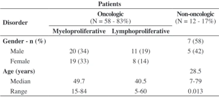
Table 1 - Clinical data of patients enrolled in the study
Table 2 - Distribution of the results of the low cytometry platelet immunoluorescence test (FC-PIFT) versus anti-human leukocyte antigen antibody (PRA)
Patients
Disorder (N = 58 - 83%)Oncologic (N = 12 - 17%)Non-oncologic
Myeloproliferative Lymphoproliferative
Gender - n (%) 7 (58)
Male 20 (34) 11 (19) 5 (42)
Female 19 (33) 8 (14)
Age (years) 28.5
Median 49.7 40.5 7-79
Range 15-84 5-60 0.013
FC-PIFT PRA
n % n %
Positive 39 51 36 47
Negative 35 46 40 53
Inconclusive 02 03
Methods
A group of hematologic patients with clinically suspected PR treated at the hospital complex of the Universidade Estadual de Campinas during the period July 2006 to July 2011 was prospectively enrolled in this study. Serum samples were collected before transfusion for direct platelet antibody screening and samples were then stored at -20°C until processing.
Platelet antibodies were screened by the FC-PIFT. Sera from 24 regular male blood donors with no history of previous transfusions were analyzed and a negative luorescence standard curve was deined. Pooled platelets from two O blood group male donors with no history of previous sensitization were buffer washed and re-suspended in 0.1% phosphate buffered saline (PBS)/ ethylenediaminetetraacetic acid (EDTA) (inal concentration 100,000 platelets/mL) and then incubated with patient serum (5 mL) for 30 minutes at 37°C. Negative and positive controls were added to each test batch. After three consecutive washes, cells were incubated for 50 minutes with luorescein isothiocyanate (FITC) goat anti-human immunoglobulin G [AfiniPure F(ab’) Fragment Goat Anti-Human 2 IgG, Fc Fragment Speciic - Jackson Immuno Research – Baltimore, USA] at 1:50 dilution. Samples were read after a second buffer wash in a FACScalibur low cytometer (Becton Dickinson, San Jose, CA, USA) using the CellQuestH software (Becton Dickinson). For data acquisition and analysis, 10,000 events were analyzed. The test was considered positive if the median luorescence (MF) obtained was greater than or equal to two standard deviations (SD) above the negative MF control and inconclusive if MF was between one and two SD above the negative MF control.
Anti-human leukocyte antigen antibody analysis
The detection of anti-HLA antibodies (PRA) was determined by commercially available methods: enzyme-linked immunosorbent assay (ELISA - LAT® One Lambda Inc, Canoga Park, CA, USA) and Luminex technology (LabScreen® and LabSingle Antigen® One Lambda Inc, Canoga Park, CA, USA) according to the manufacturer´s instructions.
Flow cytometry platelet immunoluorescence test versus anti-human leukocyte antigen antibody analysis
The results of FC-PIFT and PRA obtained from the samples collected were compared. The sensitivity and speciicity as well as predictive values of FC-PIFT were determined taking into account that the majority of antiplatelet antibodies present HLA speciicity.
Statistical analysis
The R software version 2.13.1 (2011-07-08) was used for statistical analysis. The Fisher exact test was applied for count data with the level of signiicance set at 0.5% (p-value ≤ 0.05). Since the great majority of antibodies involved in PR present HLA speciicity(3), PRA was used as a reference test to calculate
sensitivity, speciicity, and positive and negative predictive values of FC-PIFT(9,10).
Results
Population characteristics
This prospective study analyzed 76 blood samples from 32 female and 38 male patients with a median age 43.5 years (range: 5-84 years). The group characteristics are described in Table 1. Of the patients studied, 55.7% (n = 39) presented myeloproliferative oncologic disorders and 27.1% (n = 19) lymphoproliferative disorders. Non-oncologic disorders were detected in 17.1% (n = 12) (Bernard Soulier Syndrome, Glanzmann’s thrombasthenia and aplastic anemia).
Correlation Flow cytometry platelet immunoluorescence test versus anti-human leukocyte
antigen antibody analysis
There were correlations between positive FC-PIFT and positive PRA in 38.15% (n = 29) and negative FC-PIFT and negative PRA in 39.47% (n = 30) of the samples. On the other hand, the FC-PIFT was positive when the PRA was negative in 13.16% (n = 10) and the FC-PIFT was negative when the PRA was positive in 6.58% (n = 5) of the cases. Finally the FC-PIFT was inconclusive when the PRA was positive in 2.6% of the total samples (n = 2). Data are shown in Table 2.
Flow cytometry platelet immunoluorescence test - sensitivity and speciicity
254
Bub CB, Martinelli BM, Avelino TM, Gonçalez AC, Barjas-Castro ML, Castro V
Rev Bras Hematol Hemoter. 2013;35(4):252-5 Discussion
The present study demonstrates that FC-PIFT is an interesting tool to identify patients with immune PR. The test is fast, relatively simple and allows the selection of compatible platelet donors by crossmatching to support thrombocytopenic patients or those with platelet dysfunction bleeding.
The identiication of antibodies against antigens present on the platelet surface strongly suggests immune PR, nevertheless the majority of PR cases have non-immune causes. When immune factors are present, the identiication of the antibodies linked to the platelets, as well as the availability of compatible platelet components, may signiicantly enhance the response to platelet transfusion and improve patient outcome.
There is a signiicant association between platelet transfusion failure and patient survival; this increases the clinical impact of platelet refractoriness(11). Providing an adequate post-transfusion
platelet count increment in refractory patients is not an easy task and so transfusion of compatible platelets is crucial, particularly in immune PR(12). HLA is the most frequent cause of immune
PR and inding multiple HLA-compatible related donors for one individual is very dificult.
Successful transfusion of patients with platelet-refractory thrombocytopenia is extremely important(13,14). However, several
potential donors are needed to sustain HLA platelet matched transfusion programs considering not only HLA diversity but also the transfusional demand of these patients. Pool size calculations may provide essential data for a rational planning of platelet transfusion support programs and help guide different institutions that aim to build a platelet donor registry. Feasibility and costs should be taken into account when considering the donor pool size required(15).
The use of HLA platelet matching is not the only approach to manage alloimmune platelet transfusion refractoriness. Crossmatching and support with antigen negative platelet units allow rapid selection of donors, mainly for those patients with uncommon HLA types to whom it might be virtually impossible to ind HLA-compatible donors(16-18). Recently, the use of the
HLAMatchmaker algorithm has been reported as an emerging concept for the management of refractory patients(19). The
combination of matching compatible antigens and the application of mismatch acceptability determined by serum screening for HLA antibodies has offered an effective approach for HLA-based platelet transfusion support of refractory patients.
This study showed that the FC-PIFT has a high correlation with PRA, and also demonstrated good sensitivity, accuracy and a high positive predictive value. A larger number of patients
analyzed could improve the statistics of the study, reducing the conidence interval found (Table 3). However, due to the low frequency of hematological patients with suspected PR, even with cooperative studies, the ideal number of patients enrolled probably could not be achieved. FC-PIFT does not discriminate the speciicity of antiplatelet antibodies (anti-HLA antibodies or anti-HPA antibodies) and some limitations of the method should be discussed. The frequency of positive FC-PIFT with negative PRA (13%) suggests the presence of HPA antibodies (in agreement with the literature 10-20%). Moreover, the great polymorphic diversity of the HLA system imposes another limitation for FC-PIFT; the use of pooled platelets from two random donors may not ensure the ideal range of antigens. However, the use of more platelet donors could cause a ‘dilution’ of the HLA antigens.
Conclusions
Even with limitations, the FC-PIFT seems to be eficient, fast and feasible as an initial screening to detect platelet antibodies and a useful tool to crossmatch platelets for the transfusional support of patients with refractoriness. The use of additional techniques that identify anti-HPA antibodies, such as MAIPA, is essential for the appropriate clinical management of these cases.
Acknowledgements
The authors wish to thank Roberto Zulli for his assistance with the statistical analysis and Raquel Susana Foglio for corrections in the English version of the manuscript.
References
1. Rebulla P. Refractoriness to platelet transfusion. Curr Opin Hematol. 2002;9(6):516-20.
2. Vassallo RR. New paradigms in the management of alloimmune refractoriness to platelet transfusions. Curr Opin Hematol. 2007;14(6):655-63.
3. Pavenski K, Freedman J, Semple JW. HLA alloimmunization against platelet transfusions: pathophysiology, signiicance, prevention and management. Tissue Antigens. 2012;79(4):237-45.
4. Doughty HA, Murphy MF, Metcalfe P, Rohatiner AZ, Lister TA, Waters AH. Relative importance of immune and non-immune causes of platelet refractoriness. Vox Sang. 1994;66(3):200-5.
5. Legler TJ, Fischer I, Dittmann J, Simson G, Lynen R, Humpe A, et al. Frequency and causes of refractoriness in multiply transfused patients. Ann Hematol. 1997;74(4):185-9.
6. Ferreira AA, Zulli R, Soares S, Castro V, Moraes-Souza H. Identiication of platelet refractoriness in oncohematologic patients. Clinics (Sao Paulo). 2011;66(1):35-40.
7. He Y, Zhao YX, Zhu MQ, Wu Q, Ruan CG. Detection of autoantibodies against platelet glycoproteins in patients with immune thrombocytopenic purpura by low cytometric immunobead array. Clin Chim Acta. 2013;415:176-80.
8. van Velzen JF, Laros-van Gorkom BA, Pop GA, van Heerde WL. Multicolor low cytometry for evaluation of platelet surface antigens and activation markers. Thromb Res. 2012;130(1):92-8.
9. Armitage P, Berry G, Mathews JNS. Statistical methods in medical research. 2nd ed. Boston, MA: Blackwell Science; 1987. 559 p. *p-value ≤ 0.001; Odds Ratio = 16.53; 95% conidence interval = 4.72-70.34
Table 3 - Comparison of the results of the low cytometry platelet immunoluorescence test (FC-PIFT) versus anti-human leukocyte antigen antibody (PRA)
Positive PRA Negative PRA
n % n %
Positive FC-PIFT * 29 38.2 10 13.2
Negative FC-PIFT* 5 6.6 30 39.4
255 Platelet antibody detection by low cytometry: an effective method to evaluate and give transfusional support in platelet refractoriness
Rev Bras Hematol Hemoter. 2013;35(4):252-5
10. Altman D. Practical statistics for medical research. London: Chapman and Hall; 1990. 624 p.
11. Kerkhoffs JL, Eikenboom JC, Schipperus MS, van Wordragen-Vlaswinkel RJ, Brand R, Harvey MS, et al. A multicenter randomized study of the eficacy of transfusions with platelets stored in platelet additive solution II versus plasma. Blood. 2006;108(9):3210-5.
12. Sacher RA, Kickler TS, Schiffer CA, Sherman LA, Bracey AW, Shulman IA; College of American Pathologists. Transfusion Medicine Resource Committee. Management of patients refractory to platelet transfusion. Arch Pathol Lab Med. 2003;127(4):409-14.
13. Duquesnoy RJ. Structural epitope matching for HLA-alloimmunized thrombocytopenic patients: a new strategy to provide more effective platelet transfusion support? Transfusion. 2008;48(2):221-7. Comment in: Transfusion. 2008;48(2):204-6.
14. Heal JM, Blumberg N, Masel D. An evaluation of crossmatching, HLA, and ABO matching for platelet transfusions to refractory patients. Blood. 1987 Jul;70(1):23-30.
15. Mickey R. Donor pool sizes for HLA matching. Transfusion. 1989;29(4):285-6.
16. O’Connell BA, Lee EJ, Rothko K, Hussein MA, Schiffer CA. Selection of histocompatible apheresis platelet donors by cross-matching random donor platelet concentrates. Blood. 1992;79(2):527-31.
17. Petz LD, Garratty G, Calhoun L, Clark BD, Terasaki PI, Gresens C, et al. Selecting donors of platelets for refractory patients on the basis of HLA antibody speciicity. Transfusion. 2000;40(12):1446-56. Comment in: Transfusion. 2000; 40(12):1425-6.
18. Hod E, Schwartz J. Platelet transfusion refractoriness. Br J Haematol. 2008;142(3):348-60.
19. Brooks EG, MacPherson BR, Fung MK. Validation of HLAMatchmaker algorithm in identifying acceptable HLA mismatches for thrombocytopenic patients refractory to platelet transfusions. Transfusion. 2008;48(10):2159-66.