In studies about cerebral infarcts in children, despite many different methodologic approach, a common observation is the wide diversity of etiologies. According to the Pan American Health Organization, in the last decade the mortality ra-te for Brazilian children under one year was 6 ti-mes higher than the rates reported by developed
countries1. We hypothesized that the high
mor-bidity condition of the disease in Brazil is a con-sequence of distinctive characteristics in the dis-tribution of the causes of cerebral infarcts.
Thus, the objective of the present investigation was to identify cerebral infarcts in children aged zero to 15 years attended at a tertiary hospital,
CEREBRAL INFARCT IN CHILDREN AGED
ZERO TO FIFTEEN YEARS
Thelma Ribeiro Noce
1, Soraia Ramos Cabete Fábio
1, José Ibiapina Siqueira Neto
2,
Antônio Carlos dos Santos
1, Carolina Araújo Rodrigues Funayama
1ABSTRACT - Cerebral infarcts in children present peculiar characteristics either due to their diversity of causes or due to the unknown nature of the causes. The etiologies of cerebral infarct were reviewed in children from zero to 15 years old, attended at a tertiary hospital, in Ribeirão Preto (Brazil), from 1990 to 1997, adopting the modified Trial of ORG 10172 in Acute Stroke Treatment (TOAST) criteria of classifica-tion; 1 - Atherosclerosis in large arteries; 2 - Cardioembolic; 3 - Occlusion of small vessels; 4 - Other etiolo-gies; 5 - Undetermined cause. Thirty-nine children were included, 18 males and 21 females, aged 2 months to 15 years, mean age 5.67. The largest group, N=22 (56.4%), included children with “other eti-ologies”, 7 of them aged under two years. The most common etiology was dehydration and septic shock leading to brain hypoperfusion and watershed infarcts. Nine (23%) children had “Undetermined etiolo-gy”, 7 (17,9%) cardioembolic subtype and none had atherosclerosis. Laboratory improvement is needed for the large number of patients without a defined cause, and the high proportion of children with dehy-dration in the group with a determined cause emphasizes the need for preventive health actions among infants and children.
KEY WORDS: cerebrovascular disease, cerebral infarct, stroke, children, infants, etiology.
Infarto cerebral em crianças de zero a quinze anos de idade
RESUMO - Infartos cerebrais em crianças apresentam peculiaridades, como grande variedade de causas e alta freqüência sem etiologia definida. Foram revistos os diagnósticos etiológicos em crianças de zero a 15 anos, atendidas durante o ictus e com imagens cerebrais sugestivas de infarto, entre 1990 e 1997 em hospital terciário de Ribeirão Preto (SP). Adotou-se o critério de classificação modificado do Trial of ORG 10172 in Acute Stroke Treatment (TOAST): 1 - Arterioesclerose de grandes artérias, 2 - Cardioembólico, 3 - Oclusão de pequenos vasos, 4 - Outras etiologias, 5 - Não determinada. Trinta e nove crianças foram incluídas, 18 do sexo masculino e 21 do feminino, com idade variando entre 2 meses e 15 anos e média de 5,67. O maior grupo, com 22 crianças (56,4%), foi o de “Outras etiologias”, 7 das quais com idades entre 2 meses e um ano. A etiologia mais freqüente foi desidratação e choque séptico, levando a hipoperfusão cerebral e infarto em zonas limítrofes. Nove (23%) com etiologia não determinada constituiram o segun-do grupo, 7 (17,9%) apresentaram causas cardioembólicas e nenhum caso foi registrasegun-do com arterioscle-rose. Ressalta-se a necessidade de investimento laboratorial, considerando-se a alta freqüência de casos sem diagnóstico. O alto número de crianças com infarto decorrente de desidratação requer atenção para ações preventivas em saúde infantil.
PALAVRAS-CHAVE: doença vascular cerebral, infarto cerebral, criança, lactente, etiologia.
1Faculdade de Medicina de Ribeirão Preto da Universidade de São Paulo (FMRP/USP) Ribeirão Preto SP, Brasil; 2Faculdade de
Medicina da Universidade Federal do Ceará, Fortaleza CE, Brasil.
Received 6 June 2003, received in final form 28 August 2003. Accepted 6 October 2003.
studying the specific distribution of etiological groups according to age and reviewing some as-pects of each etiology identified.
METHOD
After approval by the Research Ethics Committee of the University Hospital of Ribeirão Preto, the me-dical records of all patients aged zero to 15 years with cerebral infarct, from 1990 to 1997, were reviewed. Those with a focal ictal history and vascular etiology confirmed by cerebral imaging were included. Chil-dren with perinatal or post-natal hypoxic-ischemic en-cephalopathy were excluded. The age limit of 15 years was considered according to Williams et al.2, who
stat-ed that infarcts at 15 to 18 years of age are similar to those of adults.
The sample was divided in groups according to the modified criteria of the TOAST3, which classifies
cere-bral infarcts as: (1) atherosclerosis of the large arteries, (2) cardioembolic (3) occlusion of small vessels or lacu-nae, (4) other etiologies, and (5) undetermined cause.
RESULTS
Thirty-nine patients were included, 18 (46.2%) males and 21 (53.8%) females. Mean age was 5.67 ± 5.02 years (range: 2 months to 15 years) (Fig.1).
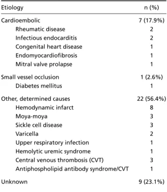
The causes of cerebral infarcts are listed in Table 1 and Fig. 2.
No child presented atherosclerosis.
Of the 7 (17,9%) children with cardioembolic infarcts, one had Down’s syndrome and presen-ted congenital cardiopathy of the complete atri-oventricular septal defect type. Two with a metal prosthesis had rheumatic disease. Prolapse of the mitral valve was considered to be the cause in one adolescent patient with extensive fruitless in-vestigation, and in view of the angiographic evi-dence of occlusion with subsequent flow reestab-lishment.
One diabetic child, age 8, presented classic mi-croangiopathic lacunar syndrome and signs of cap-sular lacunar lesion (smaller than 1.5 cm) on reso-nance imaging.
Of the 22 (56,4%) patients with infarct caused by other etiologies, 8 presented the cerebral in-farct during the course of dehydration due to acute diarrhea, evolving to septic shock; 7 of them aged below 2 years. Six had multiple bilateral border zone lesions, and 2 had a single unilater-al corticunilater-al lesion, one of them anterior and the other in the vertebral-basilar territory.
Three with moyamoya disease were females, one of them with Down’s syndrome.
Fig 1. Number of cases according to age.
In two children the cerebral infarct respective-ly occurred in the acute phase of chicken pox and 50 days the disease. Both presented subcortical infarcts of lacunar dimensions and normal venous studies.
Sickle cell disease was the cause of multiple bilateral border-zone infarct of the middle and anterior arteries in 3 patients aged 1, 3 and 7 years, two of them with recurrence.
One patient aged 18 months presented sud-den hemiparesis on the forth day of an upper respiratory infection. Amino acid analysis, deter-mination of C and S proteins and antithrombin III, cerebral angiography and echocardiography were normal. Magnetic resonance imaging sho-wed signs of infarct smaller than 1.5 cm in the lenticulostriate territory.
Hemolytic uremic syndrome was detected in another patient aged 45 months, whose cerebral computed tomography demonstrated widespread diffuse hypodensity of white matter associated with a small sign of lesion in the head of right cau-date nucleus, later evolving with resolution of hypodensity and greater delimitation of a lacu-nar infarct.
The etiologies of three cases that presented central venous thrombosis (CVT) were: dehydra-tion, otitis associated with a history of undefined
hypercoagulability and acute lymphatic leukemia, with the detection of empty delta signs in all of them. In another patient with CVT at the age of 1 year and 4 months, antiphospholipid antibody syndrome was diagnosed, since this patient pre-sented recurrent and persistently altered doses of anticardiolipin antibody. This patient presented slightly reduced protein S levels.
The cause of the infarct was not determined in nine patients (23%), although seven of them showed abnormal signs upon angiography.
DISCUSSION
Children can present stroke at any age. The in-cidence is higher under the age of 2 years and pro-gressively decreases throughout adolescence4-7.
This trend was also identified in our study. Alth-ough we found a slight predominance of female patients, the literature on the topic does not de-monstrate statistically significant differences2,4-7.
We did not find newborn infants in the present series, probably due to difficulties in performing imaging exams during the acute phase of the dis-ease at this time during the investigation. In new-born infants, when peri-intraventricular hemorr-hages are excluded, cerebrovascular disease mo-re fmo-requently pmo-resents convulsive seizumo-res as the initial manifestation8,9.
A system for categorization of cerebral infarct subtypes based principally on the etiology of in-farcts was developed by TOAST3, and is currently
the most frequently adopted classification for adults. Williams et al.2were the first to use TOAST
criteria in children series. Analysis of the distribu-tion of our patients demonstrated a predomi-nance of infarcts caused by other etiologies, fol-lowed by cardioembolic cases and those with an undetermined cause.
No patient had stroke secondary to atheroso-clerosis of the large arteries. Our results are in li-ne with the major series involving populations in the same age range2,5-7,9,10. The prevalence of
in-farcts resulting from atherosclerosis of the large arteries increases with age2. Infarcts of this type
are particularly common among the elderly, but represent, along with lacunar infarcts, less than 30% of infarcts in people aged under 50 years11.
Patients with occlusion of large arteries due to atherosclerosis have various well established asso-ciated risk factors such as hypertension, diabetes, smoking, ethylism and hyperlipidemia, which are
Table 1. Etiologies of cerebral infarct in 39 children.
Etiology n (%)
Cardioembolic 7 (17.9%)
Rheumatic disease 2
Infectious endocarditis 2
Congenital heart disease 1
Endomyocardiofibrosis 1
Mitral valve prolapse 1
Small vessel occlusion 1 (2.6%)
Diabetes mellitus 1
Other, determined causes 22 (56.4%)
Hemodynamic infarct 8
Moya-moya 3
Sickle cell disease 3
Varicella 2
Upper respiratory infection 1 Hemolytic uremic syndrome 1 Central venous thrombosis (CVT) 3 Antiphospholipid antibody syndrome/CVT 1
Unknown 9 (23.1%)
not usually found in the pediatric group4. In the
Williams et al.2series, among 92 patients aged 1
to 18 years no case was recorded, but 16% of 116 were 18 to 45 years old. Siqueira11found 2.6% of
patients in the 15 to 29 year range and 11.8% in the 30 to 45 year range.
Only one patient of school age who suffered from diabetes mellitus presented lacunar infarcts in our study. This reduced frequency is in line with most of the series in the literature, where no or few cases were identified2,5,6,8. Risk factors such as
chronic hypertension, atherosclerosis or diabetes mellitus, which are important in adults, are rarely present in children with lacunar infarcts, an age at which such lesions usually originate from em-bolic events or arteritis12.
Cardioembolic infarcts were responsible for 18% of our cases. In the major series, the preva-lence of cardioembolic infarcts varied from 3 to 65%2,5,6,7. This wide range in the prevalence of
infarcts was attributed to the differences in the methodologies used in each study. According to some authors, congenital13,14 and acquired
car-diopathies12-17 are the most common causes of
cardioembolic infarcts in children, with a special emphasis on complex cardiopathies15-17. Emboli
from the heart or from right - left shunt by para-doxical embolisms constitute usual mechanisms of cerebral embolism in these patients. Cerebral infarcts in association with congenital cardiopa-thy occur more frequently in children aged under 2 years14.
The only patient we detected with congenital cardiopathy had Down’s syndrome, with A - V shunt, a common congenital cardiac alteration for this chromosomal anomaly.
Two patients (28.6%) with cardioembolism presented bacterial endocarditis. Congenital or acquired valvar stenoses is usually responsible for the production of bacterial or aseptic growth16.
These defects were not present in either of our two cases. The mechanisms involved in the cere-bral infarct are septic embolization, formation of aneurysm and vasculitis and beside the increase of the power and spectrum of antibiotics, 15 to 20% of patients with endocarditis still develop a cerebral infarct17.
The relation between mitral valve prolapse (MVP) and strokes is controversial. The prevalen-ce of MVP in the general population is around 5
to 15%. We did not find any specific information regarding the risk and prevalence of MVP in the pediatric population, although it is believed to be minimal16. In the TOAST classification, MVP was
considered a source of cardioembolism with a medium risk3. The current belief is that MVP
should only be implied in the etiology of infarcts in the presence of a pathologic MVP, according to strict criteria of morphologic alterations of the valves and if the extensive search for other etio-logies provides no results17. Our patient with MVP
presented ictus during puberty and other possi-ble causes were excluded after an extensive in-vestigation. Furthermore, the angiography stron-gly suggested an embolic process demonstrating occlusion and subsequent reestablishment of the arterial flow.
Synthetic valve prostheses represent high risk of thromboembolism, particularly cerebral em-bolism, which is still one of the most devastating complicating factors in the appearance of this di-sease. The annual risk is estimated at 4% for mi-tral prostheses and 2% for prostheses located in the aorta17,19. In our patient, mechanic prostheses
represented 28.6% of the sources of cardioem-bolism.
“Other etiologies” accounted for 56.4% of our sample, slightly superior to Williams et al. 49%2
and lower than the more recent series of Wraige et al, 80%20. Systemic hypoperfusion accounted
for the highest number of cases in our sample, corresponding to 20.5% of the total. These in-farcts occur when there is a critical reduction in the blood supply, insufficient to maintain the in-tegrity of the nerve tissue.
The concept of hemodynamic disorders of ce-rebral perfusion as a cause of infarcts is not re-cent21, although it has been seldom covered or
discussed in major case studies. All the factors that promote a reduction in the volemia or glob-al circulatory insufficiency could be implied. It re-presents 8 to 10% of ischemic cerebrovascular le-sions, even though autopsy studies consider this number underestimated21,22. Of our 8 patients
found hematologic and cardiac diseases as main cause of cerebrovascular disease.
Moyamoya disease is an angiographic pattern similar to smoke puff associated with progressive arterial occlusion and colaterals telangiectasias distal to the occlusion. It has been reported in 8%2to 14%23of the cases. Our 3 patients were
females and two were diagnosed during the first decade of life. The literature emphasizes the pre-dominance of the disease in females and the bi-modal age distribution, with peaks of incidence under 10 and between 30 and 40 years23,24.
Fran-co et al.25provided an excellent review of the
dis-ease and reported 3 cases involving young males, with the youngest being 14 years old.
Sickle cell anemia corresponded to 7.7% of our sample, lower than 12.6%24or 19.5%2
repor-ted in series from the Unirepor-ted States, where 0.15% of the black population is SS homozygous. Many of the ischemic lesions in sickle cell patients are clinically silent and are revealed only by neuroi-maging26. The first ictus occurs mainly between
age 5 and 15 years2,22,24,26. Our 3 cases had it earlier.
Among the studies on cerebral infarct associa-ted with varicella-zoster, the great majority invol-ved ophthalmic herpes zoster27,28. Varicella was
responsible for 2 (5.1%) of our 39 cases. Higgins et al.24 detected only 1 case of varicella in their
study of 95 patients aged 1 month to 22 years. It has been suggested that infarcts are late compli-cations of varicella, occurring 3 weeks to 6 months after the infection4, and intrathecal production
of antibodies to varicella zoster has been detect-ed up to four years after the primary infection in children with stroke and cerebrovascular disea-se29. However, there are reports of acute
hemi-paresis as early as during the first week of infec-tion28, as was recorded in one of our patients.
Respiratory infections have been related to stroke ever since the dawning of the medical li-terature and still generate reports of sporadic cases14,30-32. Although in many of these patients
the infecting organism is not identified, many vi-ral infections attributed to the Influenza A, Coxsackie B4 and Coxsackie A9 viruses have been
documented31,32. The physiopathology of the
event was not well established. The inflammato-ry mechanisms are hypothetically capable of sti-mulating coagulation by different means, includ-ing alteration of lysis and platelet aggregation, or inhibition of the anticoagulant system by a
reduction of C and S protein levels and an increa-se in fibrinogen, among others30-32. All of these
factors suggest a possible association between ce-rebral infarcts, infection and prothrombotic con-ditions. In our patient we found normal C and S proteins and antithrombin III. Since a highly evi-dent temporal relation occurred between the ic-tus event and upper respiratory infection, and since we discarded all the other etiologies, we considered this association.
Hemolytic uremic syndrome is characterized by the triad microangiopathic hemolytic anemia, uremia and thrombocytopenia33. The basic lesion
consists of endothelial vascular damage, especial-ly renal, although other organs may be affected. Neurological malfunctions occur in 30 to 50% of the cases, and when present are related to a less optimistic prognosis33. Our patient presented a
good neurological evolution.
We detected 3 (7.7%) patients with CVT, all of them presenting prothrombotic conditions such as acute intense dehydration, acute lymphatic leu-kemia and acute otitis media with hypercoagula-bility state. Although the empty delta signal was viewed in all patients, the signal has a low gener-al rate of detection in approximately 30% of ca-ses34. It is attributed to two main factors: either
thrombosis does not involve the upper third sec-tion of the upper sagittal sinus, or when the tests are performed very early, during the first five days, when the clots are isodense with the paren-chyma, or very late, after two months, when the sinus flow has already been reestablished34.
We diagnosed one case of antiphospholipid antibody syndrome during the investigation of a CVT episode in a nursing male infant who did not demonstrate clinical or laboratory factors associ-ated with other prior or concomitant diseases, raising the total CVT in the case study to 4 (10.2%), i.e., 18.2% of infarcts caused by other etiologies. Antiphospholipid antibody syndrome presents clinical and laboratory manifestations of recur-rent arterial and/or venous thrombosis, recurrecur-rent abortions and thrombocytopenia35, besides
auto-immune hemolytic anemia and cardiac, cutane-ous and neurological alterations36.
25-47.6%4,7,12 of undetermined causes to values
such as 8.3%20.
In conclusion, in agreement with the literatu-re, the present sample of Brazilian children pre-sented a wide diversity of etiologies for cerebral infarcts, and therefore required more extensive investigation. We emphasized the importance of severe dehydration related to border zone in-farcts in the group aged under two, as well as the importance of preventive strategies in infan-tile health. In addition, we call attention to the need for progress in laboratory diagnosis in view of the high number of cases of undetermined etiology.
REFERENCES
1. OPAS. Organización Panamericana de la Salud. Estatisticas de la salud de las Americas. Washington: OPAS, OMS, 1997.
2. Williams LS, Garg BP, Cohen M, Fleck JD, Biller J. Subtypes of ischemic stroke in children and young adults. Neurology 1997;49:1541-1545. 3. Adams HP Jr, Bendixen BH, Kappelle LJ, et al. Classification of
sub-type of acute ischemic stroke, definitions for use in a multicenter clin-ical trial. Stroke 1993;24:35-41.
4. Mathews KD. Stroke in neonates and children: overview. In Biller J, Mathews KD, Love BB, (eds). Stroke in children and young adults. Boston: Butterworth - Heinemann, 1994:15-30.
5. Lanska MJ, Lanska DJ, Horwitz SJ, Aram, DM. Presentation, clinical course, and outcome of childhood stroke. Pediat Neurol 1991;7:333-341. 6. Schoenberg BS, Mellinger JF, Schoenberg DG. Cerebrovascular dis-ease in infants and children: a study of incidence, clinical features, and survival. Neurology 1978;28:763-768.
7. Moura-Ribeiro MVL, Ferreira LS, Montenegro MA, et al. Doença cere-brovascular na infância. Arq Neuropsiquiatr 1999;57:594-598. 8. Moura-Ribeiro MVL, Pessoto MA, Marba STM. Cerebrovascular
dis-ease in neonates. Arq Neuropsiquiatr 1999;57:84-87.
9. Rotta NT, Silva AR, Silva FLF, et al. Cerebrovascular disease in pedi-atric patients. Arq Neuropsiquiatr 2002;60:959-963.
10. Kirkham FJ. Stroke in childhood. Arch Dis Child 1999;81:85-89. 11. Siqueira JI Neto, Santos AC, Fabio SRC, Sakamoto AC. Cerebral
in-farction in patients aged 15 to 40 years. Stroke 1996;27:2016-2019. 12. Roach ES. Cerebrovascular disease in young patients. American
Aca-demy of Neurology. Annual Meeting: Seattle 1995:53-78.
13. Lasjaunias P, Brugge K. Arterial ischaemic stroke. In Lasjaunias P (ed). Vascular diseases in neonates, infants and children. Berlin: Springer - Verlag, 1997:393-418.
14. Trescher WH. Ischemic stroke syndromes in childhood. Ann Pediat 1992;21:374-383.
15. Roach ES, Riela AR. Introduction and overview. In Roach ES, Riela AR (eds). Pediatric cerebrovascular disorders. New York: Futura, 1995:1-12.
16. Roach ES, Riela AR. Cerebral embolism. In Roach ES, Riela AR (eds). Pediatric cerebrovascular disorders. New York: Futura, 1995:51-68. 17. Streifler JY, Furlen AJ, Barnett HJM. Cardiogenic brain embolism:
incidence, varieties, treatment. In Barnett HJM, Mohr JP, Stein BM, Yatsu FM (eds). Stroke: pathophysiology, diagnosis and management. New York: Churchill Livingstone, 1992:967-994.
18. Wolf PA, Sila CA. Cerebral ischemia with mitral valve prolapse. Am Heart J 1987;113:1308-1315.
19. Cerebral Embolism Task Force. Cardiogenic brain embolism. Arch Neurol 1986;43:71-84.
20. Wraige E, Hajat C, Jan W, Pohl KR, Wolfe CD, Ganesan V. Ischaemic stroke subtypes in children and adults. Dev Med Child Neurol 2003; 45:229-232.
21. Bladin CF, Chambers BR. Frequency and pathogenesis of hemody-namic stroke. Stroke 1994;25:2179-2182.
22. Biller J. Non-atherosclerotic vasculopathies. In Biller J, Mathews KD, Love BB (eds). Stroke in children and young adults. Boston: Butter-woth - Heinemann, 1994:57-82.
23. Yonekawa Y, Goto Y, Ogata N. Moyamoya disease: diagnosis, treat-ment, and recent achievement. In Barnett HJM, Stein BM, Mohr JP, Yatsu FM (eds). Stroke-pathophysiology, diagnosis and management. New York: Churchill Livingstone, 1992:721-748.
24. Higgins JJ, Kammerman LA, Fitz KC. Predictors of survival and char-acteristics of childhood stroke. Neuropediatrics 1990;22:190-193. 25. Franco CMR, Fukujima MM, Oliveira RMC, Gabai AA, Moyamoya
disease. Arq Neuropsiquiatr 1999;57:371-376.
26. Riela AR, Roach S. Etiology of stroke in children. J Child Neurol 1993;8:201-220.
27. Takeoka M, Takahashi T. Infectious and inflammatory disorders of the circulatory system and stroke in childhood . Curr Opin Neurol 2002;15:159-164.
28. Ganesan V, Kirkham FJ. Mechanism of ischaemic stroke after chicken-pox. Arch Dis Child 1997;76:522-525.
29. Häusler MG, Ramaekers VTh, Reul J, Meilicke R, Heimann G. Early and late onset manifestations of cerebral vasculitis related to varicella zoster. Neuropediatrics 1998;29:202-207.
30. Grau AJ, Buggle F, Heindl S, et al. Recent infection as a risk factor for cerebrovascular ischemia. Stroke 1995;26:373-379.
31. Roach ES, Riela AR. Inflamatory vascular disorders. In Roach ES, Riela AR (eds). Pediatric cerebrovascular disorders. New York: Futu-ra, 1995:121-140.
32. Zilkha A, Mendelsohn F, Borofsky LG. Acute hemiplegia in children complicating upper respiratory infections. Clin Pediatr 1976;15:1137-1142.
33. Sheth KJ, Swick HM, Haworth N. Neurological involvement in hemolytic-uremic syndrome. Ann Neurol 1986;19:90-93.
34. Virapongse C, Cazenave C, Quisling R, Sarwar M, Hunter S. The empty delta sign: frequency and significance in 76 cases of dural sinus thrombosis. Radiology 1987;162:779-785.
35. Louzada P Jr, Simon SM, Voltarelli JC, Donadi EA. Síndrome do anti-corpo antifosfolípide. Medicina (Ribeirão Preto) 1998;31:305-315. 36. Kenet G, Sadetzki S, Murad H, et al. Factor V Leiden and
antiphos-pholipid antibodies are significant risk factors for ischemic stroke in children. Stroke 2000,31:1283-1288.