UNIVERSIDADE DE LISBOA
Faculdade de Medicina Veterinária
VECTOR-BORNE PATHOGENS FOUND IN CARNIVORES IN WILD NAMIBIA
MARIA CAROLINA REGATEIRO MACHADO E COSTA
2019
LISBOA CONSTITUIÇÃO DO JÚRI:
Doutor Virgílio da Silva Almeida
Doutor Luís Manuel Madeira de Carvalho Doutora Solange Judite Roque Coelho Alves Gil
ORIENTADOR
Doctor Georg von Samson-Himmelstjerna
CO-ORIENTADOR
UNIVERSIDADE DE LISBOA
Faculdade de Medicina Veterinária
VECTOR-BORNE PATHOGENS FOUND IN CARNIVORES IN WILD NAMIBIA
MARIA CAROLINA REGATEIRO MACHADO E COSTA
DISSERTAÇÃO DE MESTRADO INTEGRADO EM MEDICINA VETERINÁRIA
2019 LISBOA CONSTITUIÇÃO DO JÚRI:
Doutor Virgílio da Silva Almeida
Doutor Luís Manuel Madeira de Carvalho Doutora Solange Judite Roque Coelho Alves Gil
ORIENTADOR
Doctor Georg von Samson-Himmelstjerna
CO-ORIENTADOR
vi
vii
Acknowledgements
I would like to thank first and foremost to Doctor Jürgen Krücken, my closest supervisor at the Institut für Parasitologie und Tropenveterinärmedizin in Berlin for all the patience and explanations and all the help he provided me with. His wisdom and guidance were crucial to this dissertation and my knowledge has grown immensely whilst under his wing.
To my supervisor Professor Doctor Georg von Samson-Himmelstjerna I am deeply grateful, for welcoming me to his incredible laboratory, giving me a chance to work alongside some incredible scientists, even though I didn’t even speak German. Thank you for investing in me.
I would also like to express my wholehearted appreciation for Professor Doctor Luís Madeira de Carvalho, for always believing in me and in my abilities. Thank you so much for all the support and friendship.
I also want to thank Flávia and Mariana, my Portuguese speaking “partners in crime” in Berlin, without whom my experience would definitely not have been the same. Flávia for not only mentoring me, but also for teaching me persistence and how to see the good things in life, but most importantly for all the special memories in this brand new really cold city. And Mariana, for bringing joy and light and always laughing with me, even when we were way out of our depth and didn’t know what we were doing. I am also really grateful for everyone at the Institute, especially Sabrina and all the PhD students, with their good disposition and patience for all of my questions and doubts and for teaching me and giving me some of their time without a second thought. Monika and Catarina as well, for being a part of this project and doing some of the work. And lastly I would like to thank Bettina Wachter, Gabor Czirják, Marion East and Ortwin Aschenborn, without them this project would have never existed.
To my girls from FMV, Bina, Tina, Lenka, Mer e Bea for putting up with me. I have grown, changed and been through a lot during this past few years but you have made everything better and easier, and I could not have asked for better company and friends. Words cannot express how lucky I am to have you in my life and to have shared this journey with you, forever as minhas linduxas.
To my incredible friends Rita, Bea, Mary, Ana, Mafaldas, Herberto, Miguel, Monty e Bernardo, for all the love and support and for accepting me exactly as I am, weirdness and all. And to everyone else who has crossed my life and contributed somehow to the person I am today, I am forever grateful. And lastly to my family, for your unconditional love. Thank you Avó, for not understanding what I was doing, but still supporting me through it. Thank you Isinha, Nena and Lu for becoming a part of my family, even though you were not in it initially. Thank you Pai for your constant support, interest and for showing me that constantly wanting to know more is perfectly normal. Thank you Henrique for shining a light on me from wherever you are. Thank you Maggie for being the best sister I could have asked for, and for being weird and random with me. And lastly, thank you Mãe, my biggest rock, for raising me and giving everything you had to your children, for being the most incredible person I know and one of my major inspirations. Love every single one of you with all my heart.
viii
This study was a partnership between the Institute for Parasitology and Tropical Veterinary Medicine of Freie Universität in Berlin, the Leibniz Institute for Zoo and Wildlife Research and the Research Training Group 2046 "Parasite Infections: From Experimental Models to Natural Systems"
ix
Abstract
Vector-borne pathogens found in carnivores in wild Namibia
This dissertation aimed to identify and molecularly characterize vector-borne pathogens from several parasite families, all possessing stages found in peripheral blood, from a wide variety of free-ranging carnivores living in Namibia, in the southern part of Africa.
Blood samples collected from 9 bat-eared foxes (Otocyon megalotis), 17 brown hyenas (Parahyaena
brunnea), 19 spotted hyenas (Crocuta crocuta) and 85 cheetahs (Acinonyx jubatus
) were screened
by Polymerase Chain Reactions (PCRs) and tested for pathogens of the Onchocercidae family, the order Piroplasmida, bacteria from the Anaplasmataceae and the Rickettsiaceae families and, lastly,
the Hepatozoidae family. The PCRs targeted both the ITS-2 and 12S, 18S, 16S, 18S and 18S rRNA
genes respectively and were followed by nucleotide sequencing.
In total, sampled animals showed a 43.1% rate of Onchocercidae infection, 67.7% of Piroplasmida, 60% of them were positive for Anaplasmataceae, 10% for Rickettsiaceae and Hepatozoidae were detected in 47.7% of them.
Obtained filaroid sequences showed high homologies with both Acanthocheilonema reconditum and Acanthocheilonema dracunculoides and further phylogenetic analysis were performed in both brown and spotted hyenas, with the construction of a phylogenetic tree. Piroplasmida results were not studied any further. For Anaplasmataceae, subsequent sequencing results indicated high similarity with both Anaplasma phagocytophilum and Anaplasma platys and varied PCR protocols were conducted in order to differentiate between these organisms, but no conclusions were reached. The Rickettsiaceae found displayed high homologies with Rickettsia raoultii. And finally, the
Hepatozoidae infection showed to be a mixed one with both Hepatozoon canis and Hepatozoon felis. These results are important not only on a conservation level for the infected host species, but are also relevant for domestic animals coexisting in the surrounding areas, as well as humans, especially since a few of the parasites found may have zoonotic potential. Future studies should focus on understanding vectors, transmission routes, infection dynamics and host specificity in order to better evaluate the possible danger these infections may withhold.
Key-words: Bat-eared fox; Brown hyena; Spotted hyena; Cheetah; Parasitology; Wildlife; Africa; Namibia; Acanthocheilonema; Anaplasma; Rickettsia; Hepatozoon.
x
Resumo
Agentes patogénicos transmitidos por vetores presentes em carnívoros na Namíbia
Esta dissertação teve como principal objetivo identificar e caracterizar molecularmente agentes patogénicos transmitidos por vetores de várias famílias parasitárias, com o aspeto em comum de todas possuírem fases do desenvolvimento encontradas no sangue, de espécies variadas de carnívoros selvagens que habitam na Namíbia, no Sul de África.
Foram testadas amostras sanguíneas de 9 raposas-orelhas-de-morcego (Otocyon megalotis), 17 hienas-castanhas (Parahyaena brunnea), 19 hienas-malhadas (Crocuta crocuta) e 85 chitas (Acinonyx jubatus) por PCR e analisadas para pesquisa de parasitas da família Onchocercidae, da ordem Piroplasmida, bactérias das famílias Anaplasmataceae e Rickettsiaceae e, finalmente, da família Hepatozoidae. Os PCRs foram direcionados aos genes do rRNA ITS-2 e 12S, 18S, 16S, 18S e 18S respetivamente e foram seguidos de sequenciação de nucleótidos.
Na totalidade, os animais testados mostraram uma taxa de infeção de 43.1% por Onchocercidae, de 67.7% de Piroplasmida, 60% deles tiveram resultados positivos para Anaplasmataceae, 10% para Rickettsiaceae e Hepatozoidae foram detetados em 47.7% da população.
As sequências obtidas de filarídeos, mostraram possuir elevadas homologias com Acanthocheilonema reconditum e Acanthocheilonema dracunculoides, e estudos filogenéticos mais intensivos foram realizados, nomeadamente uma árvore filogenética que inclui ambas as espécies de hienas. Os resultados relativos a Piroplasmida não foram aprofundados. Para as Anaplasmataceae, as sequenciações subsequentes indicaram elevada similaridade com Anaplasma phagocytophilum e Anaplasma platys e múltiplos protocolos de PCRs foram efetuados, com o intuito de diferenciar entre estas duas espécies, mas não foram retiradas quaisquer conclusões. As Rickettsiaceae presentes evidenciaram fortes semelhanças com Rickettsia raoultii. E finalmente, as infeções por Hepatozoidae mostraram ser uma infeção mista por ambos Hepatozoon canis e Hepatozoon felis.
A importância destes resultados não se limita apenas à conservação das espécies animais em causa, mas são também relevantes em termos dos animais domésticos coabitantes na mesma região, assim como humanos, especialmente tendo em conta o possível potencial zoonótico de algumas espécies parasitárias. Estudos futuros devem ter como principais objetivos o estudo dos vetores respetivos, tipo de transmissão, dinâmica da infeção e especificidade parasitária, para melhor avaliar os possíveis perigos que podem advir da presença destes parasitas.
Palavras-chave: Raposa-orelhas-de-morcego; Hiena-castanha; Hiena-malhada; Chita; Parasitologia; Vida selvagem; África; Namíbia; Acanthocheilonema; Anaplasma; Rickettsia;
1
Table of Contents
Acknowledgements ... vii Abstract ... ix Resumo ... x Table of Contents ... 1 List of Figures ... 2 List of Tables ... 3 List of Abbreviations ... 3 1 – Internship Activities ... 4 2 – Introduction ... 5 3 – Goals ... 7 4 – Bibliographic Review ... 8 4.1 – Bat-eared Fox ... 8 4.2 – Brown Hyena ... 10 4.3 – Spotted Hyena ... 13 4.4 – Cheetah ... 16 4.5 – Vector-borne Parasites ... 19 4.5.1 – Onchocercidae ... 21 4.5.2 – Piroplasmida ... 214.5.3 – Bacteria Rickettsiales (Anaplasma, Rickettsia)... 23
4.5.4 – Adeleorina (Hepatozoon) ... 23
4.6 – PCR in parasitology ... 24
4.7 – Phylogenetics ... 25
5 – Material and Methods ... 28
5.1 – Study area, sample collection and DNA extraction ... 28
5.2 – PCR amplifications and sequencing ... 28
5.3 – Sequence and Phylogenetic analyses ... 32
5.4 – Statistical analyses ... 32 6 – Results ... 33 6.1 – Frequency of pathogens ... 33 6.2 – Gel Electrophoresis ... 33 6.3 – Phylogenetic Analysis ... 35 7 – Discussion ... 49 7.1 – Onchocercidae ... 51 7.2 – Piroplasmida ... 52 7.3 – Anaplasma ... 53
2 7.4 – Rickettsia ... 54 7.5 – Hepatozoon ... 54 8 – Conclusion ... 56 9 – Bibliography ... 57
List of Figures
Figure 1 - Bat-eared fox by Yathin Krishnappa... 8Figure 2 – Distribution map of the two bat-eared fox subspecies. ... 9
Figure 3 - Brown Hyena by Tambako the Jaguar ... 10
Figure 4 – Brown hyena distribution map. ... 11
Figure 5 - Spotted Hyena by Bettina Watcher ... 13
Figure 6 – Spotted hyena distribution map ... 14
Figure 7 - Mehgan Murphy, Smithsonian's National Zoo ... 16
Figure 8 - Cheetahs' distribution map ... 18
Figure 9 - Distribution map of piroplasmid infection in wild carnivores worldwide from Alvarado-Rybak et al. (2016) ... 22
Figure 10 - Molecular structures of rRNA gene repeats (Álvarez & Wendel, 2003) ... 27
Figure 11 - Anaplasma PCR for bat-eared foxes’ samples ... 34
Figure 12 - Onchocercidae PCR from spotted hyenas’ samples. ... 34
Figure 13 - Anaplasma PCR for brown hyenas’ samples. ... 34
Figure 14 - Anaplasma PCR for spotted hyenas’ samples.. ... 35
Figure 15 – Phylogenetic analysis of the Acanthocheilonema isolates ... 36
Figure 16 –Phylogenetic analysis of the Acanthocheilonema isolates. ... 37
Figure 17 – BLASTn comparison for cheetahs’ Acanthocheilonema sequences ... 38
Figure 18 –BLASTn comparison between one cheetahs’ Acanthocheilonema sequence and two belonging to spotted and brown hyenas. ... 39
Figure 19 – BLASTn comparison for bat-eared foxes’ Anaplasma sequences. ... 40
Figure 20 – BLASTn comparison for brown hyenas’ Anaplasma sequences. ... 41
Figure 21 – BLASTn comparison for spotted hyenas’ Anaplasma sequences. ... 42
Figure 22 – BLASTn comparison between one brown hyena’s Anaplasma sequence and one belonging to a spotted hyena. ... 43
Figure 23 – BLASTn comparison between one bat-eared fox’s Anaplasma sequence and two belonging to both hyena species, brown hyena on the left and spotted on the right. ... 44
Figure 24 – BLASTn comparison between cheetahs’ Rickettsia sequences ... 45
Figure 25 – BLASTn comparison for brown hyenas’ Hepatozoon sequences. ... 46
3
Figure 27 – BLASTn comparison between one spotted hyena’s Hepatozoon felis sequence and one brown hyena’s Hepatozoon felis sequence. ... 48
List of Tables
Table 1 – PCR Protocol ... 29 Table 2 - PCR Primer ... 30 Table 3 - Frequency of pathogens ... 33
List of Abbreviations
µl – MicroliterµM – Micromole
BLAST – Basic Local Alignment Search Tool
BLASTn – Nucleotide-nucleotide Basic Local Alignment Search Tool Bp – Base pair
CIs – Confidence intervals
CITES – Convention on International Trade in Endangered Species of Wild Fauna and Flora DNA – Deoxyribonucleic nucleic acid
dsDNA – Double strand DNA ssDNA – Single strand DNA
dNTP – Deoxyribonucleotide triphosphate
IUCN – International Union for Conservation of Nature MgCl2 – Magnesium Chloride
mM – Milimole
PCR – Polymerase Chain Reaction RNA – Ribonucleic acid
rRNA – Ribosomal RNA Sp. – Species
Spp. – Species plural U/µl – Units per microlitre
4
1 – Internship Activities
This study resulted from the curricular internship that took place between the 18th September 2017 and the 27rd of March of 2018, at the Institut für Parasitologie und Tropenveterinärmedizin of Freie Universität, Berlin, Germany, under the direct supervision of Dr. Jürgen Krücken and Professor Georg von Samson-Himmelstjerna.
During the period mentioned above, the student performed a study of parasite infection in wild animals, based on PCR amplifications of specific gene fragments on blood samples from Bat-Eared Foxes, Brown and Spotted Hyenas and Cheetahs and directly supervised screenings performed on Cheetah samples. Then, she proceeded to clone the DNA obtained within several positive samples and sequenced them and with that information, she was able to analyse the different existent homologies and disparities.
Besides this project, the author still had the chance to learn other techniques being used by PhD colleagues at the Laboratory and actively participated on occurring events, such as the 28th Annual Meeting of the German Society for Parasitology, between the 21st and the 24th of March 2018, in Berlin.
5
2 – Introduction
Parasites are extremely prevalent worldwide (Kofer, Hofer, & Hartmann, 2017) and, since the pioneering work of Anderson & May (1978) in the late 1970’s, there has been an increasing interest on the impact of diseases and parasites of wild animals at a population level (McCallum & Dobson, 1995; Thompson & Polley, 2015). However, the focus has been primarily on emerging diseases with zoonotic potential or those affecting domestic animals, generally of viral or bacterial origin ( McCallum & Dobson, 1995; Holmes, 1996; Jones et al., 2008; Taylor, Latham, & Woolhouse, 2001; Thompson, Lymbery, & Smith, 2010; Rhyan & Spraker, 2010; Robertson, Utaaker, Goyal, & Sehgal, 2014), neglecting the biodiversity and ecology of wildlife parasites.
This means we have yet to gain a broader understanding of the normal parasite fauna of wildlife populations and how these emergent pathogens interact with the assemblage of cohabiting organisms in the ecosystem, since very little is known about their life cycles, transmission routes, host specificity, or pathogenic potential (Thompson et al., 2010; MacPhee & Greenwood, 2013; Kelly et al., 2014). Also, due to these close linkages between hosts, parasites, and ecosystem structure and function, host-parasite associations can be useful in the recognition and assessment of ecosystem and environmental disruption and instability, and this knowledge may help with optimizing global wildlife and overall planet Earth’s health (Thompson et al., 2010; Polley & Thompson, 2015). Without improved and ongoing surveillance of wildlife hosts, not only will we always lag behind in terms of predicting the possibility of reservoirs being established and/or outbreaks occurring, but also at a disadvantage in preventing declines of native fauna resulting from infectious disease, situations which justify the increasingly importance of understanding the impact and transmission of parasites in wildlife populations ( Polley, 2005; MacPhee & Greenwood, 2013; Watson, 2013; Seguel & Gottdenker, 2017).
Additionally, vector-borne pathogens of carnivores can be responsible for severe diseases in domestic animals, such as babesiosis (Matijatko, Torti, & Schetters, 2012; Solano-Gallego, Sainz, Roura, Estrada-Peña, & Miró, 2016) and heartworm disease (Dantas-Torres & Otranto, 2013; Hoch & Strickland, 2014). And whilst canine vector-borne diseases have received a lot of attention these past decades, felines are not very frequently investigated, are overall neglected (Otranto et al., 2009a, 2009b; Day, 2011; Otranto, 2018) and wild carnivores’ diseases are only addressed if there is the possibility of vector interaction with either domestic animals or humans. Hypervirulent canine babesiosis in sub-Saharan Africa caused by Babesia rossi, of which wild canids such as side-striped jackals and wild dogs are natural reservoirs, is an example of the former (Penzhorn, 2011).
While we have a relatively good notion regarding the current impact of wildlife in highly industrialised areas, such as Europe or the U.S.A, the knowledge regarding vector-borne pathogens of wild carnivores in tropical and subtropical areas and its impact on the surrounding humans and domestic animals is quite limited, as well as their effects on the health and fitness of endangered species.
6
Increasing contact between domestic animals and wildlife is always expanding (Junker, Horak, & Penzhorn, 2015; Espinaze, Hellard, Horak, & Cumming, 2018), especially due to decreasing habitats for wildlife, anthropogenic factors and changes in the environment. This can also be dangerous for threatened wildlife species, since domestic animals might serve as reservoirs and amplifiers for vector-borne pathogens transmitted to wildlife (Daszak, Cunningham, & Hyatt, 2001; Czupryna et al., 2016; Espinaze et al., 2018). Furthermore, these novel pathogens can then become responsible for high mortality, decline and even local extinctions (Cleaveland, Laurenson, & Taylor, 2001; Aguirre, 2016; Van der Weyde, Mbisana, & Klein, 2018).
7
3 – Goals
The main goal of the current study was the identification and molecular characterization of several vector-borne pathogens of the family Onchocercidae, the order Piroplasmida, the family Hepatozoidae and bacteria from the Rickettsiales order and to further study their phylogenetic relationship with other related organisms.
8
4 – Bibliographic Review
4.1 – Bat-eared Fox
4.1.1 – Taxonomy and Distribution
Bat-eared Fox (Otocyon megalotis) (Desmarest, 1822) Kingdom – Animalia Phylum – Chordata Class – Mammalia Order – Carnivora Family – Canidae Subfamily – Caninae
Genus – Otocyon (Müller, 1835)
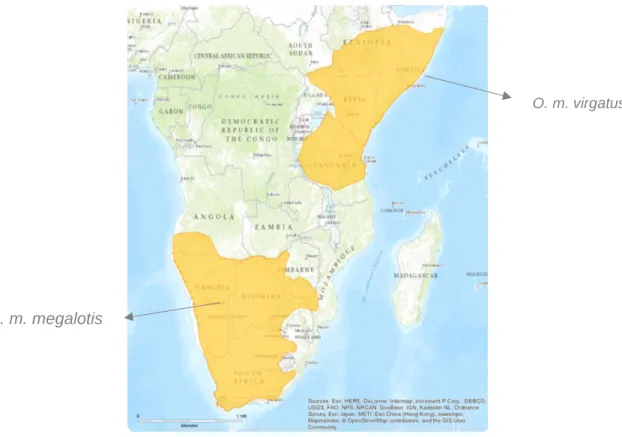
Bat-eared foxes are the only species in the Otocyon genus, being set apart from the rest of the Canidae family by their different morphological characteristics, more specifically their dentition. These animals are currently divided between two discrete subpopulations in eastern and southern Africa (representing each of the known subspecies) and they are both widespread and common in conservation areas (Dalerum, Roux, Vries, & Kamler, 2016). However, they are becoming uncommon in arid areas and on farms in South Africa where they are occasionally persecuted (Hoffmann, 2014). Subspecies O. m. virgatus ranges from southern Sudan, Ethiopia and Somalia down through Uganda and Kenya to southwestern Tanzania, whilst O. m. megalotis occurs from Angola through Namibia and Botswana to Mozambique and South Africa (Nel, J. A. J. and Maas, B. 2013; Hoffmann, 2014; Dalerum et al., 2016). They generally dwell on semi-arid and arid habitats (Nel & Mackie, 1990; Thomson & Meredith, 1993), especially open grasslands, open scrub, Acacia savanna and shrublands (Kuntzsch and Nel, 1992; Nel, J. A. J. and Maas, B. 2013; Dalerum et al., 2016).
According to the 2014 International Union for Conservation of Nature (IUCN) Red List of Threatened Species (http: //www.iucnredlist.org), they are regarded as “Least Concern” (Hoffmann, 2014) since
they are occasionally persecuted, but there seem to be no main threats that can result in any major range-wide declines, affecting the population trend overall (Dalerum et al., 2016). Their current population is considered as “stable” and within a circumscribed habitat, numbers can fluctuate from abundant to rare depending on rainfall, food, breeding stage and disease (Hoffmann, 2014; Dalerum et al., 2016).
Figure 1 - Bat-eared fox by Yathin Krishnappa. Image downloaded from https://en.wikipedia.org/wiki/Bat-eared_fox
9
3.1.2 – Group size, Diet, Ranging and Social Behaviour
Generally considered as group living species, bat-eared foxes are regarded as the most social of canids (Nel & Mackie, 1990). They generally form monogamous pairs (Wright, 2006; Nel, J. A. J. and Maas, B. 2013) with or without cubs, with possible extra-pair paternity (Nel, Mills and Vanaarde, 1984; Pauw, 2000), and were also found in family groups consisting of one male and up to three related females (Maas, 1993). The size of the groups and the way they geographically distribute themselves are not socially fixed parameters, and it might change according to various reasons, such as litter size, mortality and the time of year (Nel, Mills, & Vanaarde, 1984) and their foraging habits increase during the dry season due to changes in food resources (Dalerum et al., 2016). Also, they have been consistently described as not very territorial animals, different group ranges seemingly overlapping without generating any further conflict (Kamler, Gray, Oh, & Macdonald, 2013). However, in the Serengeti the majority of intergroup encounters were hostile, possibly due to the fact that these areas were lower in food availability (Maas, 1993).
Bat-eared foxes are considered the only truly insectivorous member of the canid family (Kuntzsch & Nel, 1992), their meals consisting primarily of harvester termites, as well as other insects, with the rare appearance of small vertebrates (Nel, J. A. J. and Maas, B. 2013) and sometimes berries (Kuntzsch & Nel, 1992; Nel & Mackie, 1990).
Figure 2 – Distribution map of the two bat-eared fox subspecies.
Otocyon megalotis. The IUCN RED List of Threatened Species
O. m. virgatus
10
Due to their unorthodox diet, these animals encounter different constraints when compared to other carnivores, especially because insect eaters rely on foraging time to collect a sufficient amount of food (Maas, 1993). In order to contradict this fact, male bat-eared foxes play a very important role in raising their cubs, taking over parental duties when they are about 2 weeks old and later teaching them how to forage (Nel, 1993; Wright, 2006), in this way also maximizing their own foraging time (Nel, J. A. J. and Maas, B. 2013).
4.2 – Brown Hyena
4.2.1 – Taxonomy and Distribution
Brown Hyena (Hyaena brunnea, formerly Parahyaena brunnea) (Thunberg, 1820) Kingdom – Animalia Phylum – Chordata Class – Mammalia Order – Carnivora Family – Hyaenidae Genus: Hyaena
The 2015 IUCN Red List of Threatened Species lists this species as “Near Threatened”, almost qualifying as “threatened” under criterion C1, being the rarest of all hyena species (Westbury et al., 2018). This is due to the low mean global population size (estimated to be below 10,000 mature individuals) and to the deliberate and incidental persecution of these animals, which number may come close to meeting a continuing decline of 10% over the next three generations (Wiesel, 2015). They are also listed as Class B under the African Convention on the Conservation of Nature and Natural Resources. Moreover, old studies (Rohland et al., 2005) have hinted towards very low genetic diversity within the species, and more recently (Westbury et al., 2018) proved it, but it is still unknown the true influence this may have on the survivability of the brown hyena. However, knowledge of the evolutionary processes affecting a species is critical to inform conservation plans aimed at the long-term management of its evolutionary potential, which justifies potential further investigations concerning these animals (Westbury et al., 2018).
This species is endemic to southern Africa and is widely spread throughout the south-western arid region including most of Botswana, Namibia, southern Angola, southern Zimbabwe, South Africa, Swaziland and southern Mozambique (Furstenburg, 2012; Wiesel, 2015). In Namibia, they are mainly found along the coast, in Etosha National Park and more sporadically over the rest of the
Figure 3 - Brown Hyena by Tambako the Jaguar, donwloaded from http://animalia.bio/brown-hyena
11
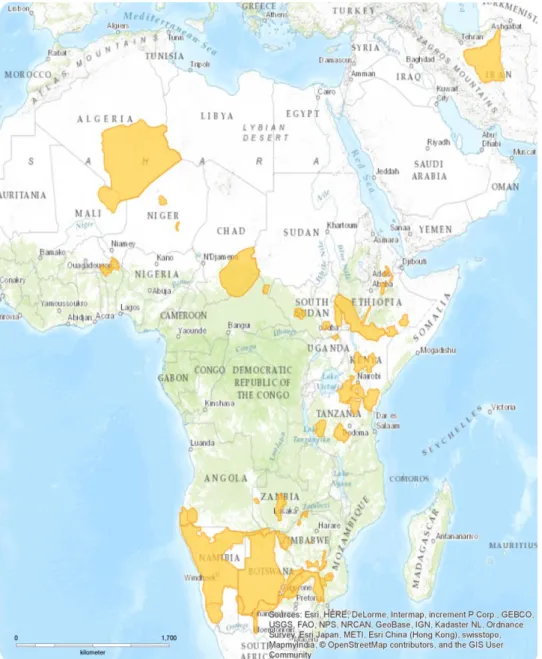
country (Hofer & Mills 1998a). Even though the range of the brown hyena has shrunk significantly since the end of the 18th century (Hofer and Mills 1998), they remain widespread in southern Africa, and in more recent years their distribution has been found larger than previously believed, particularly due to re-introductions (Slater & Muller, 2014) and range expansions (Thorn, Green, Bateman, Waite, & Scott, 2011). The total population size on the continent, has been estimated as a minimum of 5,000 to 8,000 individuals (Wiesel, 2015), with Botswana having the largest population (Hofer and Mills 1998; Kent and Hill, 2013) and Namibia the apparent second (Stein, Fuller, & Marker, 2013; Wiesel, 2015), even though Maude (2005) estimates that the numbers in Botswana may be a little higher than the ones stated before.
A significant proportion of the global population is now inhabiting non-protected areas (Thorn, Green, Bateman, et al., 2011), which may suggest the importance of these zones, when it comes to species conservation (Kent & Hill, 2013; Lindsey et al., 2013; Stein et al., 2013), but the hyenas still persist in areas of commercial farmland in a bigger number than previously thought (Thorn, Green, Keith, et al., 2011).
Figure 4 – Brown hyena distribution map.
12
3.2.2 – Group size, Diet, Ranging and Social Behaviour
This species shows an ability to survive close to urban areas and it is predominantly nocturnal in its activity (Mills, M.G.L. 2013), being most commonly found in deserts like the Namib (Mills, 1998) and other dry areas, particularly along the coast, and also semi-desert, open scrub and open woodland savanna (Hulsman et al., 2010; Mills, M.G.L. 2013). Although they are not dependent on the existence of drinking water (Furstenburg, 2012), they do require a covered space to lie on during the day, favouring rocky mountainous areas with bushes (Wiesel, 2015).
They are primarily scavengers of a wide range of vertebrate remains, scavenging larger carnivore kills, like leopard’s on Namibian farmlands (Stein et al., 2013), but their diet can be supplemented by wild fruit or other plants, insects, bird’s eggs (Owens & Owens, 1978; Siegfried, 1984; Stuart & Shaughnwssy, 1984; Burgener & Gusset, 2003; Kuhn, Skinner, & Wiesel, 2008; Maude & Mills, 2005; Furstenburg, 2012; Slater & Muller, 2014) and sometimes small animals, like the Cape Fur Seal pups they are able to hunt or scavenge along the Namib Desert (Siegfried, 1984; Stuart & Shaughnwssy, 1984; Kuhn et al., 2008; Wiesel, 2010), even though hunting is presumably opportunistic and largely unsuccessful (Furstenburg, 2012; Mills, M.G.L. 2013). The presence of other carnivores may be beneficial for this species due to the bigger amount of scavenging opportunities (Mills, M.G.L. 2013), scavenging being defined as feeding from prey that was killed and abandoned by another predator or that had died of a reason unrelated to predation (Höner, Wachter, East, & Hofer, 2002).
Although hunting comprises a relatively small proportion of their foraging behaviour, only sometimes killing sheep, goats, calves or poultry (Kent & Hill, 2013), when living in areas occupied by pastoralist herders, they are often persecuted, since the livestock owners believe them to be responsible for their losses ( G. Mills & Hofer, 1998; Kent & Hill, 2013; Lindsey et al., 2013).
The dietary benefit derived from the presence of subsistence pastoralists and the availability of livestock carcasses may be the primary reason that brown hyena populations are viable in cattle areas (Maude and Mills 2005), with the added bonus of lower levels of competition, which allows the brown hyenas to feed undisturbed (Kent & Hill, 2013). Increased efforts to educate farmers and pastoralists about the fact that brown hyenas pose very little risk to livestock may even have beneficial effects, as far as disease prevention and is thought to enhance conservation of these animals (Kent & Hill, 2013; Wiesel, 2015).
Although sightings of brown hyena give the idea of a solitary social structure, they are, actually a gregarious, socialized species (Furstenburg, 2012) that lives in clans ranging in size from a single female and her cubs to extended families, that include a female, her adult offspring of both sexes and an immigrant male (Owens & Owens, 1978; Westbury et al., 2018), however, they are strict solitary foragers (Skinner & van Aarde, 1981; Mills, M.G.L. 2013). Several individuals may come together at a large food source (Owens & Owens, 1978) and whilst the type of food determines clan
13
size, the way in which the food resources are distributed determines territory size (Furstenburg, 2012; Mills, M.G.L. 2013). They are not usually territorial animals, with each individual having a home range to which it generally adheres in its movements, even though there is great overlap between members of the group (Owens & Owens, 1978).
4.3 – Spotted Hyena
4.3.1 – Taxonomy and Distribution
Spotted Hyena (Crocuta crocuta)
(Erxleben, 1777) Kingdom - Animalia Phylum - Chordata Class - Mammalia Order - Carnivora Suborder - Feliformia Family - Hyaenidae
Genus – Crocuta (Kaup, 1828)
Considered by the 2015 IUCN Red List of Threatened Species as “Least Concern” as the species remains widespread in Africa, though with a continuing decline in populations outside, and even within protected areas, due to persecution and habitat loss. This, however, is not sufficient to warrant listing in a threatened category, with the total world population well exceeding 10,000 mature individuals (Kay E. Holekamp & Dloniak, 2010; Bohm & Höner, 2015) and their behavioural and ecological plasticity contributes majorly to this fact (J. M. Kolowski & Holekamp, 2009).
Spotted hyenas are relatively widely distributed in the south of the Sahara region of Africa (Bohm & Höner, 2015), with the largest known population occurring in the Serengeti ecosystem in Tanzania and Kenya and in South Africa at Kruger National Park (Hofer and Mills, 1998). They are, however, uncommon in South West Africa/ Namibia (Gasaway, Mossestad, & Stander, 1989). And even though the current population trend is decreasing, there may be recent evidence that a few populations have increased during the past years, more precisely in Eritrea (Bohm & Höner, 2015) and in Chad (Olléová and Dogringar 2013).
Spotted hyenas occupy an extraordinarily diverse array of habitats, including savannah, deserts, swamps, open woodlands, and montane forest (Kay E. Holekamp & Dloniak, 2010) and lower densities can be found in arid and semi-arid desert areas (Kay E. Holekamp & Dloniak, 2010; Bohm & Höner, 2015).
Figure 5 - Spotted Hyena by Bettina Watcher, downloaded from https://hyena-project.com/hyenas/
14
Some of the reasons for its decline in population number include their use by locals for tourism in Ethiopia and Nigeria, their persecution outside protected areas and even within the boundaries of conservation ones and the decline in densities of wildlife species on which these animals feed on (Bohm & Höner, 2015).
Figure 6 – Spotted hyena distribution map
15
3.3.2 – Group size, Diet, Ranging and Social Behaviour
Spotted hyenas are more commonly active at night and around dawn and dusk (Joseph M. Kolowski, Katan, Theis, & Holekamp, 2007; Matt W. Hayward & Hayward, 2007) and they can obtain food either by hunting live animals, or by scavenging on carrion, but they are very effective predators and mostly hunt, especially medium to large ungulates (Eloff, 1964; K. E. Holekamp, Smale, Berg, & Cooper, 1997; Kruuk, 1972; J. D. Skinner & van Aarde, 1981). However, they feed on a wide variety of animals, from insects to large herbivores (Höner et al., 2002), mostly on locally abundant prey species (S. M. Cooper, Holekamp, & Smale, 1999; Breuer, 2005), showcasing a remarkable plasticity when it comes to their prey preferences, which overall allows them to thrive in a large array of habitats (Kay E. Holekamp & Dloniak, 2010). Also, they are somewhat dependent on water when selecting an area to live and they commonly exist in areas within close contact with humans (Bohm & Höner, 2015).
They mainly form social groups called clans, generally female-dominated (Trinkel & Kastberger, 2005; Höner et al., 2010), that consist of societies in which individual members travel, rest and forage in subgroups (Kruuk, 1972; Smith, Kolowski, Graham, Dawes, & Holekamp, 2008) and they always include several closely related adult females and their offspring (Kay E. Holekamp & Dloniak, 2010) and one to several resident immigrant adult males (Boydston, Morelli, & Holekamp, 2001). These clans fluctuate a lot in size, ranging from small in the deserts of southern Africa (Gasaway et al., 1989; Tilson & Henschel, 1986) to the large groups found in eastern Africa (Kruuk, 1972), according to prey availability and stability. Under conditions where these are abundant, hyenas usually associate in large territorial clans, whilst in places where big fluctuations in prey number occurs, hyenas form smaller groups to hunt (Tilson & Henschel, 1986), needing to be more flexible with their range size in response to these fluctuations of migratory prey abundance (Trinkel & Kastberger, 2005)
The territorial behaviour is quite different among study populations, varying from territorial in high density locations (Kruuk, 1972; Boydston et al., 2001) to places with very low hyena density where both clan wars and border patrols tend to be rare, or are not observed at all (Tilson & Henschel, 1986).
Overall, a large variation has been documented throughout the range of spotted hyenas concerning their temporal patterning of activity, clan size, diet, territorial defence, patterns of space use and intrusion pressure. Nevertheless, like most other African carnivores, this species is facing encroachment, habitat loss and direct persecution from humans, as well as an increasingly uncertain future due to the potential effects of climate change (Kay E. Holekamp & Dloniak, 2010).
16
4.4 – Cheetah
4.4.1 – Taxonomy and Distribution
Cheetah (Acinonyx jubatus)
(Schreber, 1775) Kingdom - Animalia Phylum - Chordata Class - Mammalia Order - Carnivora Suborder - Feliformia Family - Felidae Genus – Acinonyx
Currently, 5 subspecies with different habitats are considered: A. j. jubatus jubatus (Southern Africa), A. j. hecki (Northwest Africa), A.j. raineyii (East Africa), A. j. soemmerringi (Central Africa) and A. j. venaticus (Iran). Out of these 5, only A. j. jubatus and A. j. raineyii have been genetically compared, and even though they were found to be extremely similar, the subspecies distinction was still maintained ( S. J. O’Brien et al., 1987; Menotti-Raymond & O’Brien, 1993).
Cheetahs are listed on Appendix I of Convention on International Trade in Endangered Species of Wild Fauna and Flora (CITES), as Class A under the African Convention on the Conservation of Nature and Natural Resources and considered as “Vulnerable” by the 2015 IUCN Red List of Threatened Species, predominantly due to the quite small world population that exists in present days (approximately 6,700 adult and adolescent animals distributed across 29 subpopulations). They are also protected under national legislation throughout most of its extant and some of its former range (IUCN SSC 2007a, b, 2012) and are considered as Africa’s most endangered large felid (Laurie L Marker, Muntifering, Dickman, Mills, & Macdonald, 2003).
Over the years, cheetahs have disappeared from vast tracts of their range. Ray, Hunter, & Zigouris (2005) estimated that the Cheetah has disappeared from at least 76% of its historical range in Africa and IUCN (2012) actually believes them to persist in only 10% of their historic range in Africa, whilst their distribution in Asia is limited to the central deserts of Iran (S. Durant, Mitchell, Ipavec, & Groom, 2015). Most of these animals currently reside in the northern part of Tanzania, Kenya, almost the entire southern boundary of Ethiopia, South Sudan and Uganda and in the eastern and southern part of Africa most of the animals residing belong to a single population that stretches across Namibia, Botswana, south-western Angola, northern South Africa, south-western Mozambique and southern Zambia (IUCN SSC 2007b). The entire species is considered as “Critically Endangered” in the region of North and West Africa (S. Durant et al., 2015).
Figure 7 - Mehgan Murphy, Smithsonian's National Zoo, downloaded from https://nationalzoo.si.edu/animals/cheetah
17
Furthermore, there are two cheetah subspecies currently listed as “Critically Endangered” in all its habitat – A. j. hecki, that can be found in northwest Africa and is currently largely confined to desert environments (Belbachir, 2008), having been extirpated from nearly all its range (S. Durant et al., 2015) and which is thought to number less than 250 individuals (IUCN, 2012; Belbachir, Pettorelli, Wacher, Belbachir-Bazi, & Durant, 2015), and the Asiatic cheetah – A. j. venaticus, which is known to reside only in Iran (Charruau et al., 2011; M. S. Farhadinia et al., 2017), with the possibility of a few individuals existing in Pakistan (B. M. S. Farhadinia, 2004) and Afghanistan (Manati & Nogge, 2008).
Their decline over time is mainly a result of habitat loss and fragmentation, killing and capture as a result of livestock depredation, conflict and loss of prey (Gros, 2002; L. Marker, 2005; Mallon, 2007; L. L. Marker, Dickman, Mills, Jeo, & Macdonald, 2008; S. Durant et al., 2015; M. S. Farhadinia et al., 2017), and in Iran there was also the habit of capturing live cheetahs that were then trained to hunt deer and gazelle as sport for the aristocracy (Mallon, 2007), even though in this country the key factor affecting cheetah numbers is the disappearance of prey (L. Hunter et al., 2007).
Overall, the current population trend is decreasing, since these animals are not doing well in protected wildlife reserves due to increased competition from other large predators such as lions and hyenas. Therefore, a large percentage of the remaining free-ranging cheetah populations are outside of protected reserves or conservation areas (L. Marker, 2005). Also, they are considered a genetically depauperate species (S. J. O’Brien et al., 1987; May R. M, 1995; Stephen J. O’Brien & Johnson, 2005), with their populations being extremely fragmented, which means conservation requires large scale land management planning as most existing protected areas are not large enough to ensure their long term survival (S. Durant et al., 2015).
In Africa, nearly all range states are actively involved with the Range Wild e Conservation Program for Cheetah and African Wild Dogs, developing regional strategies and national conservation action plans using the IUCN strategic planning process (IUCN, 2008). Apart from that, there are still other projects and/or non-governmental organizations established in southern and eastern Africa that are working towards the conservation of cheetahs specifically or of general large carnivores (S. Durant et al., 2015).
In Iran, the Asiatic Cheetah is completely protected since the United Nations Development Programme have established a program of work to support its conservation since 2008 (S. Durant et al., 2015).
3.4.2 – Group size, Diet, Ranging and Social Behaviour
Cheetahs are the fastest land mammals and they take advantage of that fact, especially when it comes to catching their prey, which, although it may vary, generally consists of the most available medium sized prey present (M. W. Hayward, Hofmeyr, O’Brien, & Kerley, 2006). It can range from
18
ground-dwelling birds, to small mammals, (M. G. L. Mills, 1984), medium sized ungulates and large herbivores (Purchase & du Toit, 2000; M. G. L. Mills, Broomhall, & Du Toit, 2004; M. W. Hayward et al., 2006; A. B. Cooper, Pettorelli, & Durant, 2007; L. Hunter et al., 2007). Also, contrary to many other African predators, they rarely scavenge (Sarah M. Durant, Bashir, Maddox, & Laurenson, 2007) and they also rarely prey on domestic stock, with apparent selection towards common, indigenous game species (Laurie L Marker et al., 2003). Even so, the cheetah has long been regarded as a significant threat to the interest of farmers of both game and livestock (Laurie L Marker et al., 2003).
Figure 8 - Cheetahs' distribution map
19
They tend to be active primarily during the day to minimize the competition (Sarah M. Durant, 1998; S. Durant et al., 2015) since they can lose up to 10% of their kills to lions and spotted hyenas, especially in areas where these live in higher densities (J. S. Hunter, Durant, & Caro, 2007a) and they also don’t usually stay with their kills for long, abandoning the carcasses as soon as they have eaten (J. S. Hunter, Durant, & Caro, 2007b). On the other hand, in areas on which prey is more abundant, cheetahs are mainly nocturnal (Belbachir et al., 2015), but this can also be a result of increased human activity in these zones.
Unique among felids (Gottelli, Wang, Bashir, & Durant, 2007), cheetahs’ social organization is generally comprised by solitary females, that can be accompanied by their offspring when these are still dependent (Caro & Collins, 1987 cited by Sarah M. Durant, Bashir, Maddox, & Laurenson, 2007), and these groups usually follow the herds in case of migratory prey. The males on the other hand can either live alone or in stable coalitions of two or three members (Caro & Collins, 1987 cited by Sarah M. Durant, Bashir, Maddox, & Laurenson, 2007), generally siblings (Broomhall, Mills, & du Toit, 2003), and they chose to establish on small areas attractive to females. In the cases of non-migratory prey both females and males may share their range (Broomhall et al., 2003), which can be quite extended.
Older (Sarah M. Durant, 1998, 2000) and more recent studies (Broekhuis, Cozzi, Valeix, Mcnutt, & Macdonald, 2013; Rostro-García, Kamler, & Hunter, 2015), defend that the natural evolution of cheetahs towards these different social systems and ranging patterns has its premise on risk avoidance by the animals, as this strategy to remain mobile in the presence of larger and stronger competitors enables them to avoid direct spatio-temporal competition.
Cheetahs appear to show relatively low habitat selectivity in comparison with other carnivores (Sarah M. Durant et al., 2010). In Africa, this species can be found in a wide array of habitats, such as woodland savannahs (J. Skinner & Smithers, 1990) as dry forest, thick scrub, open grassland plains (M. G. L. Mills et al., 2004) and even arid deserts like the Sahara (S. M. Durant et al., 2014) and in Iran their habitats consists of deserts.
4.5 – Vector-borne Parasites
Parasitism can be defined as an ecological association between species in which one organism, the parasite, lives on or in the body of another organism, the host. The parasite may spend the majority of its life in association with one or more host species, or alternatively, it may spend only short periods, adopting a free-living mode for the major part of its developmental cycle. But during the parasitic phase of its life cycle, the organism depends upon its host for the synthesis of one or more nutrients essential for its own metabolism. The relationship is usually regarded as obligatory for the parasite and harmful or damaging for the host. To classify an animal species as parasitic, therefore, three conditions must be satisfied: utilization of the host as a habitat, nutritional dependence, and
20
causing harm to its host (Anderson & May, 1978). Basically, a parasite is any small organism living at the expenses of another, by feeding, inhabiting on or in a bigger organism, the host, and exploiting its biological, ecological and metabolic patterns (Bowman, D., 2014; Otranto, 2018).
According to where they reside in the host’s body, parasites can then be divided into endoparasites, living within the body of hosts, and ectoparasites, inhabiting on the external surface of the host or in its skin. Parasites can also be vectors, if they transmit other parasites directly from host to host and the term vector-borne disease refers to any of a broad array of infectious diseases caused by pathogens that are transmitted by arthropods or other invertebrate as biologic intermediaries (Bowman, D., 2014).
Vector-borne pathogens have developed a close relationship with blood feeding arthropod ectoparasites, such as mosquitoes, ticks, phlebotomine sand flies, black flies, fleas, kissing bugs and lice (Otranto, 2018). And these parasitic arthropods are highly efficient vectors of several bacteria, viruses, protozoa and helminths affecting livestock, domestic and wild animals and even humans worldwide (Jongejan & Uilenberg, 2004; Otranto, Dantas-Torres, & Breitschwerdt, 2009; Colwell, Dantas-Torres, & Otranto, 2011; Kofer et al., 2017). Along the way, the life cycles of these pathogens turned into a long evolved balance with the respective arthropod biology, ecology and blood feeding habits, having taken advantage of the biology of blood feeders to ensure their transmission and distribution to receptive hosts (Otranto, 2018). Transmission of vector-borne pathogens usually occurs during blood feeding by an infected insect or acarine, but it can also happen when a vertebrate host ingests a vector, or on wound contaminations by infectious organisms in the faeces of the arthropod (Bowman, D., 2014).
Vector-borne pathogenic zoonoses are part of the constantly changing world and they are constantly adapting to their new circumstance, changing their vectors, hosts, distribution and also their virulence. Furthermore, they are also quite difficult to diagnose, posing a high amount of constraints, and also hard to control and to prevent, due to their complex transmission among host compartments, trophic levels, and the environment (Dantas-Torres, Chomel, & Otranto, 2012). Climate change, for example, can alter the geographic distribution of arthropod vectors, enhancing the risk of infectious disease transmission in wild species and the incidence of zoonoses in humans (Cumming & Van Vuuren, 2006). So, due to the lack of data on vectors for specific parasites, studies are needed to identify vectors as well as determine transmission routes, infection dynamics, and host specificity (Williams et al., 2014).
The incidence of tick-borne diseases, for example, is rapidly increasing worldwide (Piesman & Eisen, 2002; Dantas-Torres, 2007; Nicholson, Allen, McQuiston, Breitschwerdt, & Little, 2010; Estrada-Peña, Ayllón, & de la Fuente, 2012).
21
4.5.1 – Onchocercidae
In humans and domestic animals, nematodes and their deleterious effects are well documented at an individual and population level, being one of the most significant, though neglected, tropical parasite responsible for diseases in humans (Bartsch et al., 2016). However, these parasites are rarely studied in wild animals and despite the serious impact they can have in their hosts, there is no currently available summary on the number of nematodes described and the significance of their infection in free-ranging wild mammals (Seguel & Gottdenker, 2017).
Concerning their life cycle, sexually mature females found in the definitive host are viviparous and sexual reproduction occurs, with the production of microfilariae that are released onto the bloodstream. Then, microfilariae are ingested during the blood feeding of the intermediate host and they go through a series of transformations until reaching the infective stage and eventually accumulating in the mouthparts of the respective vector involved. Final hosts become infected during a new blood feeding (E. V Schwan & Schroter, 2006).
As for wildlife findings, two spotted hyenas in Kenya were reported to be positive for Acanthocheilonema dracuncoloides (Lightner & Reardon, 1983) and this parasite was also found in two dogs in the same area, as well as in dogs in Namibia (E. V Schwan & Schroter, 2006). However, parasites in both studies were either only identified by morphometry of microfilaria (Lightner & Reardon, 1983) or by acid phosphatase staining (E. V Schwan & Schroter, 2006) and therefore the exact species identification should be considered as doubtful in the absence of any morphological data on adult parasites or DNA sequence data.
4.5.2 – Piroplasmida
Piroplasmosis are among the most prevalent arthropod transmitted diseases of animals and in the last few years there has been a dramatic increase in the number of studies reporting infection with piroplasmids in wildlife (Yabsley & Shock, 2013; Alvarado-Rybak, Solano-Gallego, & Millán, 2016). Alvarado-Rybak et al. (2016) presented abundant evidence of piroplasmid infections in wild carnivores worldwide and whilst some of these species serve as reservoirs for piroplasmids, others are potential vectors, allowing these parasites to maintain endemic sylvatic lifecycles in their geographical distribution area. For example, wildlife species are known reservoirs for several Babesia spp. even though the vectors of many species are still unknown. Babesiosis is currently considered a worldwide emerging zoonosis (Yabsley & Shock, 2013; Zanet et al., 2014), with Babesia spp. being considered the second most commonly found parasite in the blood of mammals after trypanosomes (Schnittger, Rodriguez, Florin-Christensen, & Morrison, 2012).
Their importance spreads farther than just veterinary medicine, also demonstrating a great economic and medical impact worldwide (Anna Mari Bosman, Oosthuizen, Peirce, Venter, & Penzhorn, 2010; Giadinis et al., 2012; Schnittger et al., 2012).
22
Piroplasmosis are diseases caused by hemoprotozoan parasites of the phylum Apicomplexa belonging to four related genera: Babesia, Theileria, Cytauxzoon and Rangelia, some of which can occasionally cause severe disease in domestic animals, humans (Yabsley & Shock, 2013) and wild animals, even though piroplasmid infections in these last ones are typically subclinical (Banie L. Penzhorn, 2006; Williams et al., 2014). Their main vectors appear to be ticks (Chauvin, Moreau, Bonnet, Plantard, & Malandrin, 2009; Alvarado-Rybak et al., 2016).
Figure 9 - Distribution map of piroplasmid infection in wild carnivores worldwide from Alvarado-Rybak et al. (2016)
As for their life cycle, the sexual phase of reproduction occurs in the tick when the gametes fuse to form a zygote, followed by an asexual form of reproduction, sporogony, also in the tick. The resultant forms, ookinetes, invade either the salivary gland or the ovary of the tick, where they participate in transtadial and transovarial (only Babesia spp.) transmissions, respectively (Banie L. Penzhorn, 2006; Chauvin et al., 2009; Schnittger et al., 2012). In the case of transtadial transmission, the sporozoites are released from the tick’s salivary glands while it’s feeding and enter the blood stream of the vertebrate host. Then, once sporozoites are in the erythrocytes or leukocytes of the host, they undergo asexual reproduction and merogony, and the daughter cells can infect new cells (Banie L. Penzhorn, 2006; Chauvin et al., 2009).
Several Babesia sp. have been identified in wild carnivores, but the more relevant to this particular study were the findings of a novel species, Babesia lengau, thought to exist exclusively in cheetahs (Anna Mari Bosman et al., 2010). However, more recently, Williams et al., 2014 identified Babesia sp. in spotted hyenas that were very similar to Babesia lengau and Burroughs et al. (2017) confirmed this fact, extending it to brown hyenas as well.
23
4.5.3 – Bacteria Rickettsiales (Anaplasma, Rickettsia)
Members of the Rickettsiales order are obligate intracellular gram-negative bacteria and the survival of these organisms and transmission between animals are dependent on invertebrate vectors, ticks being by far the most common one (Bowman, D., 2014).
They are transmitted through the bite of an infected nymphal or adult tick vector that had been previously infected in the larval or nymphal stage while feeding on an infected animal (usually a wild animal species) which are known as being reservoir hosts (Nicholson et al., 2010).
So far, to our knowledge, there is no report of Anaplasma sp. infecting wild animal species, and for Rickettsia sp. there is a single old serological study describing that one of three investigated spotted hyaenas was positive for antibodies against Rickettsia akari (Heisch et al., 1962).
4.5.4 – Adeleorina (Hepatozoon)
Hepatozoon sp. constitute a group of apicomplexan parasites that primarily infect leukocytes of mammals and erythrocytes of amphibians, reptiles and birds (Gad Baneth, Samish, Alekseev, Aroch, & Shkap, 2001), involving arthropods such as ticks, mites, fleas and lice as intermediate hosts (McCully, Basson, Bigalke, De Vos, & Young, 1975).
Their life cycle typically involves gametogenesis, fertilization, and sporogony in a hematophagus invertebrate, and merogony followed by gametogony in a vertebrate intermediate host. Infection of the vertebrate host normally occurs by ingestion of an infected invertebrate host such as a tick (East et al., 2008).
They have been observed in a wide variety of wild carnivores, including hyenas, jackals, lions, leopards and cheetahs (McCully et al., 1975; Averbeck, Bjork, Packer, & Herbst, 1990; Lopez-Rebollar, Penzhorn, de Waal, & Lewis, 1999). Generally Hepatozoon infections in domestic and wild carnivore species have been attributed to Hepatozoon canis or closely related undetermined species (Brocklesby & Vidler, 1965; Conceição-Silva, Abranches, Silva-Pereira, & Janz, 1988).
Spotted hyenas have long been known to be infected with Hepatozoon sp. (McCully et al., 1975) and in the Serengeti it has been shown that, these parasites are highly similar or identical to Hepatozoon felis (East et al., 2008). In Zambia, however, spotted hyenas were found to be positive for both, Hepatozoon canis and H. felis (Williams et al., 2014).
Although generally regarded as being non-pathogenic in wild animals (Kocan et al., 2000; Rishniw et al., 2006; Williams et al., 2014), there is a report of Hepatozoon infection perhaps contributing to the death of spotted hyenas in Tanzania (East et al., 2008).
24
4.6 – PCR in parasitology
Molecular diagnostic assays, primarily those based on particular amplification by polymerase chain reaction (PCR) allow detection and diagnosis of pathogens with enhanced specificity and sensitivity compared to the traditional methods of microscopy and serology (Bosman, Venter, & Penzhorn, 2007; Piron et al., 2007; Abdul-Ghani, Al-Mekhlafi, & Karanis, 2012).
The deoxyribonucleic acid (DNA) molecule consists of two intertwined and complimentary strains, the 3’ end and the 5’ end. The bases of opposite strands pair up with one another, and are held together by hydrogen bonds and hydrophobic interactions and can be dissociated to produce two single strands, either by chemical means or by heating to a temperature of at least 94°C. Once denaturation has occurred, fragments or portions of DNA can be specificaIIy bound to complementary regions, in a process called hybridisation (Singh, 1997).
By using Taq DNA polymerase, a thermostable enzyme isolated from the thermophilic bacterium Thermophilus aquaticus, the problem of low sensitivity of DNA hybridisation assays, or low amounts of target DNA was solved, because it became possible to amplify original target DNA a million-fold with a method employing concurrent cycles DNA duplication, called polymerase chain reaction (Singh, 1997).
During PCR, specific regions of DNA are amplified enzymatically through successive cycles, each consisting of three steps:
1. Denaturation: double-strand DNA (dsDNA) is denatured to produce two single-strand DNA (ssDNA) strands;
2. Annealing: two different oligonucleotides (referred to as primers) hybridise to complementary DNA sequences on each of the target ssDNA strands;
3. Extension: the enzyme DNA polymerase catalyses the addition of deoxynucleotide triphosphates to the two oligonucleotides in a 5’ to 3’ direction.
Each cycle of PCR doubles the amount of specific DNA resulting in a million-fold amplification of the target sequence after a minimum of 30 cycles. Each of the three steps is undertaken at different temperatures: usually the denaturation step occurs at 94°C, extension occurs at 72°C and the annealing temperature depends on the length and the nucleotide content of the oligonucleotide used in the PCR. These temperatures and the duration of each step are attained using a thermocycling machine (Singh, 1997).
To put this into practice in parasite detection, it involves processing the specimen to produce parasite DNA template, several different protocols having been described for this step according to the parasite in question, identification of target DNA sequence, design of primers, optimisation of PCR parameters and detection of product. The next step involves the removal of inhibitors of PCR, like heparin for example, which is used in the collection of blood to purify the DNA and then, once the PCR product is produced following amplification, it can be detected by various means. The
25
commonest method is to analyse the product using agarose gel electrophoresis, in which case the DNA products are separated according to their respective sizes (Singh, 1997).
The epidemiology of parasitic diseases includes the study of host-parasite interactions, and for parasites transmitted by vectors it involves host-vector and vector-parasite interactions, and these DNA-based methods are also applicable for the detection of parasites in vectors (Singh, 1997). BLAST (Basic Local Alignment Search Tool) is a method that directly approximates alignments that optimize a measure of local similarity, the maximal segment pair (MSP) score, based on well-defined mutation scores. It allows the detection of weak, but biologically significant sequence similarities, and as DNA and amino acid sequence databases continue to grow in size, they become increasingly useful in the analysis of newly sequenced genes and proteins because of the greater chance of finding homologies (Altschul, Gish, Miller, Myers, & Lipman, 1990).
4.7 – Phylogenetics
Taxonomic characterization of organisms was originally based on morphological observations and certain general phenotypic characteristics, which enabled many parasites to be categorized into a particular genus. However, application of molecular genetic techniques, such as the PCR for gene amplification and DNA sequencing have revealed gross inconsistencies in the assignation of some parasite genetic variants.
Furthermore, recently gene sequencing has become more easily available and less costly, which means molecular phylogeny is emerging as a major tool, especially since it can be applied in a wide variety of areas. These include the analysis of gene or genome duplication events (Pfeil, Schlueter, Shoemaker, & Doyle, 2005), recombination (Chare & Holmes, 2006), horizontal gene transfer (Philippe & Douady, 2003), variation of selective pressures and adaptive evolution (Nielsen et al., 2005), divergence times between species (Ramírez, Gravendeel, Singer, Marshall, & Pierce, 2007), elucidate the origin of epidemics (Taubenberger, 2006), and host-parasite cospeciation events (Pompei, Loreto, & Tria, 2012). They can also be used as complementary tools for taxonomy (Hajibabaei, Singer, Hebert, & Hickey, 2007), have contributed to the formulation of strategies in conservation biology and have also been employed outside of the realm of biological sciences, in areas such as linguistics (Gray & Atkinson, 2003).
Phylogenetics can be described as the study of the evolutionary history and relationships among individuals or groups of organisms and it is used to classify sequences of unknown origin based on their evolutionary relationships to other considered sequences (Lemey, Salemi & Vandamme, 2009; Medlar, Aivelo, & Löytynoja, 2014). In the case of molecular phylogeny, it is based on the comparison of DNA or amino acid sequences (Baldauf, 2003; Whelan, Liò, & Goldman, 2001), and phylogenetic methods consider the similarity among the genes, since taxonomic comparisons show that the genes of closely related species usually only differ by a limited number of point mutations (Lemey, Salemi & Vandamme, 2009).
26
The idea of representing these evolutionary relationship hypotheses as trees probably dates back to Darwin and his theory of evolution (Darwin, 1809-1882), but their application to molecular data is relatively recent (Zuckerkandl & Pauling, 1965). A phylogenetic tree can be defined as a diagram that describes evolutionary relationships (Holder & Lewis, 2003) and it is composed of branches and nodes, being nodes the point at which two or more branches diverge, connecting these last ones. There are rooted and unrooted trees, and the latter position the individual taxa relative to each other without indicating the direction of the evolutionary process (Lemey, Salemi & Vandamme, 2009). The simplest test of a phylogenetic tree’s accuracy is the bootstrap that shows how strongly the dataset supports each of the relationships depicted in the tree (Baldauf, 2003; Holder & Lewis, 2003). This is done by taking random subsamples of the dataset, building trees from each of these and calculating the frequency with which the various parts of your tree are reproduced in each of these random subsamples (Baldauf, 2003; Holder & Lewis, 2003; Lemey, Salemi & Vandamme, 2009). Also, molecular phylogenetic trees are usually drawn with proportional branch lengths, which means the lengths of the branches roughly correspond to the amount of evolution between the two nodes they connect. Thus, the longer the branches, the more relatively divergent are the sequences attached to them (Baldauf, 2003; Whelan et al., 2001).
The use of molecular data for inferring phylogenetic trees has gained considerable interest (Lemey, Salemi & Vandamme, 2009), and a few genes have become reference markers. The small subunit ribosomal ribonucleic acid (rRNA) gene has been proven extremely useful for classification, because these genes are under tight structural and functional constraint, substitution rates are low and there is no evidence of lateral gene transfer across lineages (Allsopp & Allsopp, 2006), which means it holds a considerable degree of conservation across all organisms (Delsuc, Brinkmann, & Philippe, 2005). Therefore, it is the gene for which most sequence information is available for phylogenetic analysis, and its comparison has then become a powerful tool for deducing phylogenetic and evolutionary relationships among bacteria, archaebacteria, and eukaryotic organisms (Weisburg, Barns, Pelletier, & Lane, 1991).
Most eukaryotes possess hundreds of tandem copies of this gene, each consisting of the 18S, 5.8S, and 28S rRNA genes, two external transcribed spacers (ETS1 and ETS2), two internal transcribed spacers (ITS1 and ITS2), and an intergenic spacer (IGS) (Nei & Rooney, 2005), like it is shown in Figure 6.
Amongst these, the 18S rRNA gene has several features that have led it to being widely used for the assignation of organisms to a particular genus. It has both conserved and variable regions, the former allowing unequivocal sequence alignment and the latter providing phylogenetic discrimination, even though it remains difficult to establish how much gene sequence variation must exist for the source organism to be considered a different species or to be considered merely a variant and/or genotype of a species. Moreover, the 18S rRNA gene, consisting of both conserved and variable regions, has the practical advantage of allowing the design of primers for PCR
27
amplification of near full-length genes in the presence of mammalian DNA, making it a suitable marker for detection and genetic characterization of blood parasites (Allsopp & Allsopp, 2006). ITS-2 sequence comparisons are also really common, perhaps representing the most common source of phylogenetic reconstructions at the species, genus and family level among all eukaryotes (Álvarez & Wendel, 2003; Coleman, 2003, 2009; Keller et al., 2010; Song et al., 2012). And they can be the most informative for discrimination at the species and subspecies levels, due to their conserved secondary structures that can be used to facilitate alignments of higher taxonomic categories (from genus to order) because of its function in rRNA processing (Nei & Rooney, 2005).