Ana Luísa Pécurto Cartaxo
Dissertation presented to obtain the Ph.D. degree in Bioengineering
Instituto de Tecnologia Química e Biológica António Xavier | Universidade Nova de Lisboa
in silico approaches to address targeted therapies
Oeiras,
October, 2020
Insert here an image with
rounded corners
Tumor microenvironment
models: ex vivo, in vitro and
in silico approaches to address
targeted therapies
Ana Luísa Pécurto Cartaxo
Dissertation presented to obtain the Ph.D. degree in
Bioengineering
Instituto de Tecnologia Química e Biológica António Xavier | Universidade Nova de Lisboa
models: ex vivo, in vitro and
in silico approaches to address
targeted therapies
Ana Luísa Pécurto Cartaxo
The work developed in this thesis was supervised by:
- Doctor Catarina Brito, Instituto de Biologia Experimental e
Tecnológica (iBET) e Instituto de Tecnologia Química e
Biológica António Xavier, Universidade Nova de Lisboa
(ITQB-NOVA)
Fundação para a Ciência e Tecnologia (FCT), Ph.D grant
PD/BD/114047/2015
iNOVA4Health
(UID/Multi/04462/2013),
financially
supported by FCT/MEC, through national funds and
co-funded by FEDER under PT2020 Partnership Agreement
Tumor microenvironment models: ex vivo, in vitro and in silico approaches to address targeted therapies
Copyright © 2020 by Ana Luísa Pécurto Cartaxo
Instituto de Tecnologia Química e Biológica António Xavier Universidade Nova de Lisboa
iii
v
Acknowledgments
I would like to express my gratitude to all the people who have contributed directly or indirectly to this thesis.
To my supervisor, Catarina Brito, for all the guidance, the scientific discussions and to always remind me to be focused on my PhD. To the hosting institutions, iBET and ITQB, for the excellent working conditions, in particularly to Prof. Paula Alves for giving me the opportunity to work in such amazing conditions at the Animal Cell Technology Unit.
To my thesis committee members, Inês Pires da Silva, Inês Isidro and Ana Barbas, for the discussion and feedback during the several steps of my PhD work. Specially, I would like to thank Inês Isidro for her invaluable help in the in silico model project.
To the IPOLFG team, especially to Saudade André, for always believing in our research and for the amazing effort in providing the primary material for the ex vivo breast cancer model project. Also, I would like to thank Ruben Roque and Fernanda Silva for their help in the processing and analysis of samples at the hospital. More importantly, and because without them this work could not be performed, I would like to sincerely thank all patients that agreed in entering this study and give their tissues for research purposes. To ICFO, specifically to Pablo Loza-Alvarez, for accepted receiving me in his group institution, in Barcelona. To Emilio Gualda, Maria Marsal and Monica Marro for all your “teaching lessons”, support and help to make my PhD more complete and valuable.
vi
To the “3D team”, Teresa Mendes, Rita Mendes, Francisca Arez, Nuno Lopes, Sofia Batalha, Beatriz Painho, Rodrigo Eduardo; and all ex-members, Sofia Rebelo, Marta Estrada, Ana Paula Terrasso, Daniel Simão, Lara Silva, Raquel Moita, Tatiana Martins, João Sá and Catarina Pinto, thank you for all the help, discussions and suggestions. I would like to give a very special thanks to Giacomo, who was always available to help me in all tasks of my PhD: he was crucial in the ex vivo breast cancer model work. Thank you for all “Molecular Biology teaching moments”, for the fruitful discussions and above all for being after all a very good friend.
To all my colleagues at the Animal Cell Technology Unit for creating such a great working environment and a special thanks to Ana Raposo for her help in managing orders and make always an effort to maintain everything on date.
To the MIT-Portugal PhD Program, for accepting me in this new step of my life. Especially to everyone involved in the Bioengineering Systems PhD, professors and colleagues for the intense learning and friendship.
Aos meus amigos, por todo o apoio emocional e por me convidarem para sair quando precisava apanhar ar e esquecer o doutoramento por breves momentos.
Ao Jaime, pela sua capacidade de me levantar sempre a moral e de me dizer incessantemente “vai correr tudo bem”. Por a tua ajuda incansável com o modelo computacional e todas as lições nocturnas de Python. Obrigado por acreditares sempre nas minhas capacidades e estares sempre ao meu lado.
vii
À minha família pelo apoio incondicional durante estes 4 anos com fases de altos e baixos. Obrigada pelo vosso suporte e confiança; pelos momentos de motivação. Ao meu pai pelo seu forte scientific
background que sempre “puxou” por mim e pelas discussões
frutíferas a tenderem para infinito sobre o modelo computacional. À minha mãe que sempre me motivou a não desistir e ser perseverante.
ix
Abstract
Cancer is the second leading cause of mortality worldwide, despite the extraordinary advances in the last two decades due to the development of targeted therapies. These target particular molecules required for cell growth and tumorigenesis; nonetheless, de novo or acquired resistance to therapy often lead to patient relapse and disease progression. There is cumulating evidence supporting the importance of tumor microenvironment (TME)-driven mechanisms in cancer progression and drug resistance. Therefore, there is a need for cancer models in which critical components of the TME, such as the non-malignant cell types and the extracellular matrix (ECM), are represented and tissue architecture is maintained.
The overarching aim of the work presented in this thesis was the development of cancer models to address targeted therapies in TME-relevant contexts.
Chapter I reviews the state-of-art, in terms of breast cancer (BC) subtypes, available therapies, relevance of the TME for therapeutic response, and available experimental and computational models.
In Chapter II, an ex vivo approach was explored to develop a BC patient-derived 3D cell model. Our strategy was based on encapsulation in alginate, an inert biomaterial, to promote the retention of the original TME, combined with dynamic culture, to promote diffusion of macromolecules and oxygen. The original tissue architecture and microenvironment components, namely epithelial, mesenchymal, endothelial, and immune cells, as well as an ECM composed of collagen fibers, were retained. Importantly, in the case of estrogen receptor α (ERα)-positive breast tumors, the
x
retention of the TME sustained ERα expression, at gene and protein level. Response to ERα stimulation and inhibition was observed at the level of downstream targets, demonstrating active ERα signaling. Moreover, the challenge experiments with the ERα inhibitor fulvestrant, widely used in endocrine therapy, stands as a proof-of-concept for the application of the model in the study of anti-endocrine targeted therapies.
In Chapter III, we focused on the biochemical and mechanical properties of the alginate capsules, since mechanical cues have been reported to influence tumor progression. For this, we employed 3D in vitro co-cultures of tumor cells and fibroblasts, a model previously established by our group for the reconstruction of the microenvironment of solid tumors. We showed that alginate encapsulation sustains BT474 BC spheroids phenotype and proliferation. Moreover, we observed that the mechanical properties of the capsule were affected by its content: the presence of cancer cell spheroids reduced the stiffness relatively to empty capsules; fibroblasts contributed to a stiffening of the microenvironment, when compared to the mono-culture capsules. Overall, this study contributed for the characterization of alginate capsules, employed by many research groups.
Finally, in Chapter IV, we addressed antibody transport within the TME. We explored the in vitro model system characterized in Chapter III to implement an integrated experimental and computational framework. The aim of the framework was to unravel how the several TME components influence antibody distribution. Encapsulated co-cultures were challenged with a fluorescent antibody and its location within the alginate capsules was tracked using light sheet fluorescent microscopy. The obtained
xi
data was then used to benchmark a computational model, developed to simulate a digitized alginate capsule slice. The benchmarked model can also be used to generate other capsule configurations, according to user specifications.
In the present thesis, we created and characterized new tools to tackle the influence of TME in targeted cancer therapies. We explored experimental approaches to establish ex vivo and in vitro cancer models, in which key features of the TME could be retained or reconstructed, respectively. The in vitro model was complemented by in silico approaches to describe transport of therapeutics within the reconstructed TMEs. These distinct, yet complementary approaches, are tools that can contribute to unravel the mechanisms underlying therapeutic response of solid tumors and as drug discovery platforms to assess novel targeted therapies.
Key words: tumor microenvironment, ex vivo models, in vitro models, in silico models, targeted cancer therapy
xiii
Resumo
O cancro é a segunda principal causa de mortalidade a nível mundial, apesar dos avanços extraordinários nas últimas duas décadas devido ao desenvolvimento de terapias direccionadas. Estas têm como alvo moléculas específicas que são necessárias ao crescimento celular e à formação de tumores. No entanto, a resistência inata ou adquirida à terapia conduz frequentemente a reincidência e à progressão da doença. Actualmente, existem evidências que apoiam a importância dos mecanismos moleculares relacionados com o microambiente tumoral (TME) na progressão do cancro e na resistência aos fármacos. Assim, existe uma procura por modelos de cancro em que os componentes críticos do TME, tais como tipos de células não-malignas e a matriz extracelular (ECM), estejam representados e em que a arquitectura dos tecidos seja mantida.
O objectivo primário do trabalho aqui apresentado foi desenvolver modelos oncológicos para abordar terapias direccionadas no contexto do TME.
O Capítulo I revê o estado da arte em termos de subtipos de cancro da mama (BC), terapias existentes, a relevância do TME na resposta terapêutica, bem como modelos experimentais e computacionais já publicados.
No Capítulo II foi testada uma abordagem ex vivo de forma a desenvolver um modelo celular 3D, baseado em tecido tumoral mamário de pacientes. A nossa estratégia baseou-se no encapsulamento em alginato, um biomaterial inerte, de forma a promover a retenção do TME original. Recorreu-se a um sistema de
xiv
cultura dinâmico que facilita a difusão de macromoléculas e oxigénio. A aplicação desta estratégia resultou na retenção da arquitectura original do tecido, incluindo os componentes do microambiente tumoral (fibroblastos e células epiteliais, endoteliais e imunitárias, bem como a ECM composta por fibras de colagénio). É de salientar que, no caso de tumores mamários positivos para o receptor de estrogénio α (ERα), a manutenção do TME reteve a expressão ERα, tanto a nível transcricional e como proteico. A resposta à estimulação e inibição do ERα foi observada ao nível de genes-alvo a jusante, demonstrando assim que a sinalização ERα se encontrava activa. Além disso, as experiências com o inibidor de ERα fulvestrant (amplamente utilizado em terapia endócrina) representaram uma demonstração da aplicabilidade do modelo no estudo de terapias anti-endócrinas.
O enfoque do Capítulo III foi a caracterização bioquímica e mecânica das cápsulas de alginato, dado que alguns parâmetros mecânicos têm sido reportados como moduladores da progressão tumoral. Para tal, utilizámos co-culturas 3D in vitro de células tumorais e fibroblastos, um modelo previamente estabelecido pelo nosso grupo para reconstrução do microambiente de tumores sólidos. Com este modelo, demonstrámos que o encapsulamento com alginato sustenta o fenótipo e a proliferação de esferóides de BC, BT474. Observámos ainda que as propriedades mecânicas da cápsula foram afectadas pelo seu conteúdo: a presença de esferóides de células cancerígenas reduziu a rigidez relativamente à de cápsulas vazias; e os fibroblastos contribuíram para um endurecimento do microambiente, em relação à cápsulas de mono-cultura. Em suma, este estudo contribuiu para a caracterização de
xv
cápsulas de alginato, amplamente utilizado por muitos grupos de investigação.
No Capítulo IV abordámos o transporte de anticorpos no TME. Explorámos o modelo in vitro caracterizado no Capítulo III, de forma a implementar um framework experimental e computacional integrado. O framework teve como objectivo a representação dos diferentes componentes do TME e da sua influência no transporte de anticorpos. As co-culturas encapsuladas foram tratadas com um anticorpo fluorescente cuja localização dentro da cápsula foi rastreada utilizando light sheet fluorescence microscopy. Os dados obtidos foram então utilizados para treinar um modelo computacional, desenvolvido para simular uma fatia digitalizada da cápsula de alginato. Este modelo treinado pode ser utilizado para gerar novas configurações de cápsulas, de acordo com as especificações do utilizador.
Nesta tese, criámos e caracterizámos novas ferramentas para avaliar a influência do TME na reposta a terapias de cancro dirigidas. Explorámos abordagens experimentais para estabelecer modelos de cancro ex vivo e in vitro, nos quais características importantes do TME foram respectivamente retidas e reconstruídas. O modelo in vitro foi complementado por abordagens in silico, de forma a descrever o transporte de fármacos no TME reconstruído. Estas abordagens distintas mas complementares, são ferramentas que tanto podem contribuir para descobrir os mecanismos subjacentes à resposta de tumores sólidos à terapia; bem como podem funcionar como plataformas para teste de novas terapias.
xvi
Palavras-chave: micro-ambiente tumoral, modelos ex vivo, modelos
xvii
Thesis publications
Ana Luísa Cartaxo*, Marta F Estrada*, Giacomo Domenici, Ruben Roque, Fernanda Silva, Emilio J. Gualda, Pablo Loza-Alvarez, George Sflomos, Cathrin Brisken, Paula M. Alves, Saudade André, Catarina Brito; A novel culture method that sustains ERα signaling in human
breast cancer tissue microstructures, Journal of Experimental &
Clinical Cancer Research, 2020, doi: 10.21203/rs.3.rs-20405/v1, accepted for publication
Ana Luísa Cartaxo, Jaime Almeida, Emilio J. Gualda, Maria Marsal, Pablo Alvarez-Loza, Catarina Brito, Inês A. Isidro; A computational
diffusion model to study antibody transport within reconstructed tumor microenvironments, submitted to BMC Bioinformatics
Ana Luísa Cartaxo, Henrique Almeida, Tomás Calmeiro, Daniela Gomes, Elvira Fortunato, Catarina Brito; 3D cancer cell models in
alginate capsules: biochemical and mechanical characterization, in
preparation
xix
Other publications
Nuno Lopes, Ana Luísa Cartaxo, Giacomo Domenici, Catarina Pinto, Sofia Rebelo, Emilio J Gualda, Pablo Loza-Alvarez, Maria José Oliveira, Catarina Brito, Exploiting 3D cell models to recapitulate
macrophage modulation in the breast cancer microenvironment, in
xxi
Table of contents
Chapter I: Introduction ... 1 Chapter II: A novel culture method that sustains ERα signaling in human breast cancer tissue microstructures ... 79 Chapter III: 3D cancer cell models in alginate capsules: biochemical and mechanical characterization ... 121 Chapter IV: A computational diffusion model to study antibody transport within reconstructed tumor microenvironments ... 161 Chapter V: Discussion ... 213
xxiii
List of figures
Figure 1.1: Schematic of segment of breast lobe showing the TDLU, lobules, acini and the duct system. ... 3 Figure 1.2: Healthy breast and two BC histological subtypes (ductal and lobular). ... 6 Figure 1.3: ER structure.. ... 9 Figure 1.4: Schematic representation of the different types of estrogen receptor alpha (ERα) signaling. ... 10 Figure 1.5: Types of breast cancer hormonal therapy.. ... 16 Figure 1.6: Breast cancer tumor microenvironment contains tumor cells and other cellular and non-cellular components. ... 22 Figure 1.7: Schematic representation of the immune-tumor network in the “cancer-immunity cycle”. ... 30 Figure 1.8: Tumor models used in cancer research can be divided in 2 main groups: experimental (in vitro, in vivo or ex vivo) and computational, or in silico. ... 38 Figure 1.9: Different types of computational models exist to model tumors. ... 57 Figure 1.10: Schematic representation of drug transport barriers, within the tumor interstitium, that hinder drug delivery between the vasculature and the target cell.. ... 58 Figure 1.11: Schematic representation of the research objectives pursued in this thesis and the experimental approaches followed to attain them. BC: breast cancer; ERα: estrogen receptor α; TME: tumor microenvironment. ... 64 Figure 2.1: Alginate encapsulated tissue microstructures maintained parental tumor architecture. ... 87 Figure 2.2: Alginate encapsulated tissue microstructures maintained cell populations and contain proliferating cells. ... 100 Figure 2.3: Encapsulated tissue microstructures maintained collagen fibrillar structures. ... 100 Figure 2.4: Estrogen Receptor α (ER) expression and functionality are maintained in alginate encapsulated tissue microstructures up to 1 month of culture. ... 103 Figure S2.1: Sample weight. ... 116 Figure S2.2: Encapsulated BC tissue microstructures do not present myoepithelial markers and maintain high metabolic viability. ... 116 Figure S2.3: Hematoxylin and eosin staining and immunohistochemistry for ERα of MDA-MB-231 (ER-negative cell line) cells cultured in 2D. ERα gene (ESR1) expression in encapsulated microstructures cultured for one month relatively to MDAMB-231 cells. ... 117 Figure S2.4: Encapsulated tissue microstructures were cultured for three days in depleted medium before stimulation with 17β-estradiol 119
xxiv
Figure S2.5: Encapsulated tissue microstructures were cultured for three days in depleted medium before stimulation with 17β-estradiol.. ... 119 Figure S2.6: Encapsulated tissue microstructures were cultured for 3-5 days in complete medium, before challenge with fulvestrant for two weeks….………....120 Figure 3.1: Schematic representation of the experimental layout…...129 Figure 3.2: Scanning electron microscopy of alginate microcapsules. 137 Figure 3.3: Alginate capsule diameter along culture time, in mono- and co-cultures.. ... 138 Figure 3.4: Non-encapsulated and encapsulated BT474 cell spheroids, in mono-culture or in co-culture with fibroblasts.. ... 140 Figure 3.5: Phenotypic characterization of encapsulated and non-encapsulated BT474 spheroids. ... 143 Figure 3.6: Young modulus of alginate capsules: empty, from mono- and co-cultures. ... 144 Figure 3.7: BT474 mono- or co-culture capsules show low accumulation of collagen and GAGs after 2 weeks of culture.. ... 147 Figure S3.1: BT474 cell line forms spherical and compact spheroids after one day of culture in spinner vessel. ... 155 Figure S3.2: Spheroid distribution per capsule and spheroid concentration in non-encapsulated cultures.. ... 155 Figure S3.3: Immunofluorescence microscopy of BT474 cells cultured in 2D.. ... 157 Figure S3.4: GAG detection in encapsulated cultures of H157 tumor cells.. ... 158 Figure 4.1: Fluorescence after antibody challenge for a representative capsule section.. ... 169 Figure 4.2: Fluorescence profiles for selected cell clusters and fitted curves.. ... 172 Figure 4.3: Simulated antibody concentration profile throughout the digitized capsule, over time. ... 175 Figure 4.4: Computational antibody concentration profiles after fitting of the saturation parameters a, n and p to selected cell clusters. ... 176 Figure 4.5: Example of a tuned stochastic computational capsule with and without fibers.. ... 180 Figure 4.6: Experimental and computational workflow. ... 192 Figure S4.1: Comparison of profile features (delay time and slope) with cell cluster features (distance to capsule periphery and section area) obtained experimentally and for the computational model.. ... 199 Figure S4.2: Definition of the initial setup by application of Python Imaging Library (PIL) tool.. ... 200 Figure S4.3: Diffusivity coefficient on the computational model over time, for the digitized capsule.. ... 201
xxv Figure S4.4: Comparison of best fittings obtained considering only Fick’s law (blue curve) or Fick’s law combined with exponential saturation (orange curve) to the experimental data (red curve). ... 202 Figure S4.5: Tunable stochastic computational model examples.. ... 203 Figure S4.6: Exponential saturation equation for several different inputs. ... 204 Figure S4.7: Schematic representation of the LSFM acquisition portion with a photography showing a zoom on the FEP chamber. ... 205 Figure S4.8: Mesh convergence study: 100x100, 200x200 and 300x300. Initial and final time-points for the model run with the specific mesh size. ... 206 Figure S4.9: A flow diagram of the tunable stochastic computational framework………... ... 207 Figure 5.1: Aim and achievements of each research chapter of the thesis………... 215
xxvii
List of tables
Table 2.1: Clinico-pathological parameters of the breast cancer patient.. ... 89 Table S2.1: Immunohistochemistry analysis: reagents and conditions used for... 115 Table S2.2: RT-qPCR analysis: primer sequences. ... 115 Table S3.1: Antibodies used for immunodetection. ... 159 Table 4.1: Properties for selected cell clusters and parameters for the adjusted fluorescence profiles. ... 171 Table 4.2: Fitted saturation parameters for the computational model and RMSE. ... 177 Table S4.1: Fitted Dcell and Dmedium by application of the BFGS algorithm,
xxix
List of abbreviations
AB: alcian blue
ABC: ATP-binding cassette ADC: antibody-drug-conjugate AF: aldehyde fuschin
AFM: atomic force microscopy APC: antigen presenting cells AREG: amphiregulin
ATCC: American Type Culture Collection ATP: adenosine triphosphate
BC: breast cancer
BCRP: BC resistance protein
BFGS: Broyden-Fletcher-Goldfarb-Shanno CAA: cancer-associated adipocytes CAF: cancer-associated fibroblast CDK: cyclin-dependent kinase CDX: cell line-derived xenograft CK: cytokeratin
CSF-1: colony stimulating factor CTRL: control
CTGF: connective tissue growth factor
CTLA4: cytotoxic T-lymphocyte-associated protein 4 CXCR: CXC‐chemokine receptor
DBD: DNA binding domain DCIS: ductal carcinoma in situ
DMEM: Dulbecco's Modified Eagle Medium ECM: extracellular matrix
xxx
EMDR: environment-mediated drug resistance EMT: epithelial-to-mesenchymal transition FEP: fluorinated ethylene propylene
ERBF-1: estrogen receptor promoter B associated factor 1 ERα: estrogen receptor α
ERβ: estrogen receptor β
ER+ BC: estrogen receptor α-positive breast cancer FBS: fetal bovine serum
FDA: fluorescein diacetate FGF: fibroblast growth factor FSG: fish skin gelatin
GAG: glycosaminoglycan
GEMM: genetically engineered mouse model
GM-CSF: granulocyte-macrophage colony-stimulating factor HA: hyaluronic acid
H&E: hematoxylin and eosin HD: hanging-drop
hDF: human dermal fibroblast
HER2: human epidermal growth factor receptor 2 HGF: hepatocyte growth factor
Hsp: heat-shock protein IFN-γ: interferon gamma IHC: immunohistochemistry IL: interleukin
IMDM: Iscove's Modified Dulbecco's Medium LBD: ligand binding domain
LCIS: lobular carcinoma in situ LOT: liquid overlay technique
xxxi LSFM: light sheet fluorescence microscopy
MAPK: mitogen-activated protein kinase MDSC: myeloid-derived suppressor cell MIDN: intraductal mouse model
MMP: matrix metalloprotease NF-kβ: factor nuclear kappa B
nGEMM: non-germline genetically engineered mouse model NK: natural killer
NSCLC: Non-Small Cell Lung Carcinoma NST: no special type
PDGF: platelet-derived growth factor PD-1: programmed cell death protein 1 PDX: patient-derived xenograft
PFA: paraformaldehyde PI: propidium iodide
PIL: Python Imaging Library PI3K: phosphoinositide 3-kinase
PIP3: phosphatidylinositol (3,4,5) trisphosphate PGR: progesterone receptor gene
PR: progesterone receptor P/S: penicillin-streptomycin PSR: picrosirius red
PTEN: phosphatase and tensin homolog RMSE: root mean square error
RPMI: Roswell Park Memorial Institute RT: room temperature
SEM: scanning electron microscopy SERD: selective ER down-regulator
xxxii
SERM: selective ER modulator SHG: second harmonic generation SMI: small-molecule inhibitor ST: special type
TAM: tumor-associates macrophage TDLU: terminal duct-lobular unit TGFβ: transforming growth factor β TF: transcription factor
TFF1: trefoil factor-1
TME: tumor microenvironment
Timp: tissue inhibitor of metalloproteinase TKI: tyrosine kinase inhibitor
TPEF: two-photon-excited fluorescence VEGF: vascular endothelial growth factor
VEGFR: vascular endothelial growth factor receptor YM: Young modulus
2D: two-dimensional 3D: three-dimensional
1
CHAPTER I
2
Table of contents
1. Breast cancer ... 3 2. Breast cancer classification ... 5 3. ER signaling in breast cancer ... 9 4. Targeted BC therapies ... 15 4.1 Hormonal therapy ... 15 4.2 Antibody therapy ... 18 4.3 Small-molecule inhibitors ... 19 5. The role of tumor microenvironment in the response to anti-cancer therapy ... 20 5.1. Cellular components ... 22 5.1.1. Tumor cells ... 22 5.1.2. Fibroblasts ... 23 5.1.3. Endothelial cells ... 26 5.1.4. Immune cells ... 28 5.1.5. Other cell types ... 32 5.2. Non-cellular components ... 33 5.2.1. Extracellular Matrix ... 33 5.2.2. Cytokines ... 36 6. Breast cancer models ... 37 6.1. In vitro models ... 39 6.2. In vivo models ... 47 6.1. Ex vivo models ... 51 6.2. In silico models ... 55 7. Aims and scope of this thesis ... 62 8. Author contribution and acknowledgements ... 65 9. References ... 66
3
1. Breast cancer
Breast cancer (BC) is a pathology which affects the mammary gland, characterized by an abnormal growth of cancer cells that eventually evolve and invade healthy regions 1. The vast majority of
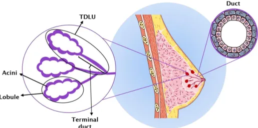
invasive BCs, and their in situ precursors, originate from the terminal duct-lobular unit (TDLU) 2. This is a structure in the breast
composed of a lobule, which is a small cluster of acini, and the terminal duct 3 (Figure 1.1). BC can spread to other tissues in the
body, giving origin to metastasis, mainly in lung, bone, lymph nodes and liver 4.
Figure 1.1: Schematic of segment of breast lobe showing the TDLU, lobules, acini and the duct system 2,3.
BC represents a major public health problem, since it is the cause of the greatest number of cancer-related deaths among women 5. Although organ-confined disease is mainly curable,
metastatic and recurrent disease has poor prognosis with a 5-year survival of only 27% 6. In the USA, about 1 in 8 women will develop
4
new BC cases are predicted to be diagnosed and more than 40,000 people are predicted to die due to BC, in 2020 7. In 2008, the
estimated total productivity loss as a result of premature mortality due to BC was $5.49 billion, for young women (aged 20–49) 8.
Metastatic BC represent a significant economic and social burden associated with high costs for healthcare systems, with direct costs alone accounting for as much as $4.2 billion per year 9. In Europe,
BC had an incidence estimated to be higher than 500,000 cases in 2018, accounting for €15 billion in 2009, which corresponded to 12% of the total cancer-related costs 10.
Fortunately, advances in the screening methods (such as digital mammography coupled with advanced computer-aid detection), early diagnosis, and breakthroughs in treatments have increased BC survival rates 11. Since 1990, a decline in BC mortality
started to be observed. This was due to the routine implementation of adjuvant therapy (that will be explained later on this chapter, section 4) and mammographic screening 12. BC research has
contributed significantly to the discovery of molecular pathways involved in tumorigenesis, that are the basis of the targeted therapies currently used in the clinics 13. Nonetheless, BC relapse is
still a relevant issue for a high number of patients: the high incidence of metastatic disease and drug resistance contribute to the high morbidity and mortality indexes 14–16. So, it is important to
uncover the molecular mechanisms which stand behind drug resistance and develop novel drugs and therapeutic regimens that overcome those mechanisms.
5
2. Breast cancer classification
BC is a genetically, histologically, biologically and clinically heterogeneous disease 17,18. Given such heterogeneity, different
response to therapy and outcome are reported 19,20 and various
classification at histological and molecular levels have been purposed to categorize BC 2,19.
Histologically, BC can be divided according with the invasiveness status of the disease into: in situ or invasive. In situ refers to tumors confined to the ducts (ductal carcinoma in situ, DCIS) and lobules (lobular carcinoma in situ, LCIS, Figure 1.2) 2.
DCIS can be further divided in several subgroups according to the tumor architecture features 2. Regarding the invasive BC group,
which refers to tumors that are not limited to the epithelial region but that have already penetrated into the surrounding stroma 2, it
includes the “no special type” (NST) and “special type” (ST). The ST represents 25% of the invasive BC cases and contains tumor with a predominant (i.e. >90% of the tumor) differentiation 2,21. On the
other hand, the NST represents the remaining 75% of the cases and contains tumor with heterogeneous features without any special differentiation patterns 20.
6
Figure 1.2: Healthy breast and two BC histological subtypes (ductal and lobular). Cancerous region is highlighted in dark purple in the mammary overall structure and the cell overgrowth highlighted in the region of the breast tissue where it occurs (orange region) 2.
It is difficult to establish a relationship between histological classification and patient outcome 22. The significant differences, in
terms of treatment and long-term survival, detected among patients having the same histological classification, support the belief that BC is an heterogeneous group of diseases 22. This
highlights the need for a BC classification based on tumor features that can be related with prognosis. In the past decade, microarray‐ based gene expression profiling has been extensively applied to the study of BC and led to a classification based on molecular signatures, reflecting differences on tumor cell biology rather than on morphology 20,22,23.
7
The molecular classification of BC was proposed for the first time in 2000 by Perou, Sorlie et al. 23. It was based on the gene
expression characterization patterns of a set of 65 surgical specimens of BC from 42 different individuals, using complementary DNA microarrays covering 8,102 human genes 23.
Patients were clustered into different groups according with the overall transcriptome differences. They identified four groups of samples: estrogen receptor α (ERα) positive/luminal-like, basal-like, Erb-B2+/human epidermal growth factor receptor 2 (HER2) positive and normal-like BC 23. In a work published one year later, gene
expression patterns were used as a prognostic marker with respect to overall and relapse-free survival 24. This work suggested to
further divide the “estrogen receptor positive” in two distinct groups (luminal subtype A and subtype B), with distinctive expression profile 23,24.
Currently, the standard molecular classification of BC divides tumors into five groups with unique biologic and prognostic features: luminal A, luminal B, HER2, basal-like and normal-like 20,22.
Luminal tumors display high expression levels of luminal cytokeratins (CK), such as CK8, CK18 and CK19 25. Luminal A
includes ERα-positive and HER2-negative cells with low levels of ki67 proliferation marker 20,22. Luminal B, with significantly worse
prognosis than luminal A, includes ERα-positive and HER2-positive cells, with higher proliferation rates 20,22. The HER2 subtype includes
cells with high expression of HER2 and low expression of ERα 20,22.
This highly proliferative and aggressive BC subtype represents ~15% of all the invasive BC cases 22 and usually has an unfavorable
prognosis. The basal-like BC subtype accounts for up to 15% of all BC 26. Basal BC cells typically express basal CKs, such as CK5, CK6
8
and CK14, have low or undetectable ERα and HER2 levels and are highly proliferative 22. Most basal-like BC have a triple-negative
phenotype (ERα-negative, progesterone receptor (PR)-negative and HER2-negative), but up to 20% express ERα or overexpress HER2 27.
Patients with this subtype of BC have the worst prognosis among all BC subtypes because of the intrinsic aggressiveness and high tendency to relapse rapidly. In addition, current therapeutic options for basal-like BC are limited to chemotherapy and relapse occurs frequently due to drug resistance 28.
A new molecular BC subtype, termed ‘claudin-low’, was proposed by Herschkowitz et al. 29. Claudin-low tumors are
characterized by the low expression of genes involved in tight junctions and cell-cell adhesion, including claudins 3, 4 and 7, Occludin, and E-cadherin 29. The “claudin-low” group is
characterized by inconsistent expression of basal keratins and low expression of HER2 and luminal markers, such as ERα and PR 30.
When compared with other BC subtypes, “claudin-low” highly express genes involved in immune response, cell communication, cell migration, angiogenesis, extracellular matrix and cell differentiation 30. The majority of “claudin-low” tumors show poor
prognosis 30.
Despite the high relevance of gene expression analysis, namely in grouping and stratifying BC patients, its use on clinical samples is resource and time intensive. Therefore, in the clinics, immunohistochemical detection of biomarkers of each subtype (evaluation of morphology, ERα, PR and HER2 expression status) is still the generalized methodology used in diagnosis stage and to select treatment options 31,32.
9
3. Estrogen receptor signaling in breast cancer
Estrogen receptor (ER) belongs to the steroid/nuclear receptor superfamily and has 2 isoforms in mammals: alpha and beta (ERα and estrogen receptor β (ERβ), respectively) 33. Theisoforms are encoded by two different genes 33, mapped on
chromosome 6 and 14, respectively 34,35. Although ERα role in BC
has been extensively studied, the role of ERβ is still under investigation 36. ERα is a ligand-modulated transcription factor,
responsible for the mediation of a plethora of cellular functions from development to carcinogenesis, whose structure is schematically represented on Figure 1.3 37,38. The ER protein is
composed of several functional domains, associated with specific roles. From the NH2 terminal, ER structure consists on an activation
function domain (AF)-1, followed by a DNA binding domain (DBD) and a hinge (H) region. Next to this region is the ligand binding domain (LBD) and the AF-2 37.
Figure 1.3: ER structure. From the N-terminal to the C-terminal: activation function 1 region (AF-1), DNA-binding domain (DBD), hinge (H) region, ligand-binding domain (LBD), activation function 2 domain (AF-2)
37.
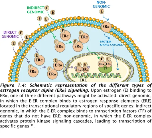
ERα can function both as signal transducer, activating various cell signaling pathways, and as transcription factor (TF), modulating the expression of several target genes. As signal transducer, ER is involved in non-genomic pathways, while as TF is involved in the direct and indirect genomic pathways 39 (Figure 1.4).
10
Figure 1.4: Schematic representation of the different types of estrogen receptor alpha (ERα) signaling. Upon estrogen (E) binding to ERα, one of three different pathways might be activated: direct genomic, in which the E-ER complex binds to estrogen response elements (ERE) located in the transcriptional regulatory regions of specific genes; indirect genomic, in which the E-ER complex binds to transcription factors (TF) of genes that do not have ERE; non-genomic, in which the E-ER complex activates protein kinase signaling cascades, leading to transcription of specific genes 39.
The activation/deactivation of any of the pathway types detailed on Figure 1.4 requires the presence of an ERα ligand, that interacts with the receptor by binding to the LBD region. In human, three different types of physiological estrogens are endogenously produced (endoestrogens): estrone (E1), estradiol (E2), and estriol (E3) 40. They are produced from cholesterol in the sex glands, such
as ovaries and testes, and in other organs, such as liver and brain. E2 is found both in females and males, while E3 is mainly found during pregnancy and E1 post menopause 40. In addition to
naturally produced estrogens within the body, a diverse array of small organic and inorganic molecules serve as ERα ligands 38. In
11
compounds naturally produced by plants), xenoestrogens (non-natural synthetic chemical compounds with estrogenic effects), metalloestrogens (small inorganic compounds in the form of heavy metal ions) and several molecules used in therapy, as described later in section 4 38. ERα ligands can have both stimulatory and
inhibitory effects 38. ERα inhibitors, such as tamoxifen and
fulvestrant, compete with the estrogen for the binding to ERα and block the downstream signaling. This way, they hamper cell growth and proliferation and reduce tumor progression 41,42. These
inhibitors will be further explained in the section 4 of this chapter. The inactive ERα exists in a molecular complex with: 1) chaperones – heat-shock proteins (hsp), namely hsp70 and hsp90, which bind to the ERα’s LBD region; 2) co-chaperons, such as immunophilin and p23, which bind to hsp 37,43,44. This complex
inactivates the transcriptional regulatory capabilities of ER but maintains its ability to bind to ligands 43. Upon estrogen binding,
receptor dimerization, dissociation of hsp and association of co-regulatory proteins occurs 33. In these conditions, ERα is able to
bind to estrogen responsive elements (EREs; which are 13 bp palindromic consensus sequence separated by a 3-base spacer 45)
present in the transcriptional regulatory regions of ERα-target genes. Here, ERα interacts directly with coactivator proteins and components of the RNA polymerase II transcription initiation complex, leading to enhanced transcription 33 (direct genomic
pathway, Figure 1.4). Around one third of all estrogen responsive genes do not bear an ERE region 46. In these cases, the regulation
12
(Figure 1.4). ERα interacts with other DNA-bound transcription factors and stabilizes their binding to the DNA and/or recruits coactivators 33,39.
Several genes have been identified as estrogen-responsive genes, due to the presence of functional ERE in their promoter 46.
Among them, one can find the trefoil factor-1 TFF1, also known as
pS2 47, and others reported by Lone et al. 46. pS2 is an
estrogen-specific response gene, since only estrogen but not progestins, glucocorticoids, and androgens, can induce it 48. pS2 role is
controversial, as it has been reported by different groups to have either the capacity to induce or inhibit tumorigenicity 49,50. Buache
et al. performed pS2 gain- and loss-of-function experiments in four
human mammary epithelial cell lines 49. They concluded that
constitutive expression of pS2 led to an increase in cell migration and invasion. Moreover, they observed that tumorigenicity capacity of MCF7-pS2 (MCF7 with pS2 overexpression) was the same as the parental MCF7. Additionally, they showed that cells with pS2 knock-down had similar proliferation but higher colony-forming ability. In an in vivo mice model, they showed that in pS2 knock-down, tumors appeared earlier and had higher incidence than in their control counterpart 49, suggesting that pS2 inhibits tumorigenesis.
In a different study, overexpression of pS2 in BC cell lines resulted in increased cell proliferation and survival 50. Additionally, it also
increased cell migration and invasion and led to an increase in tumor size, in xenograft models. Ablation of pS2 led to a reduction in cell viability in vitro and tumor regression in vivo. Then, they concluded that pS2 clearly possess oncogenic functions in mammary carcinoma cells 50.
13
Another relevant gene controlled by ER is the progesterone receptor gene (PGR). Progesterone receptor (PR) works closely and in a reciprocal manner with ERα. In fact, it is not only an ERα-induced gene target but also an ERα-associated protein that modulates its behavior 31. In one hand, PGR only has half ERE region
in its promoter. The binding of ER to that region revealed to work more as an inhibitor than a stimulator, as it is expected when full ERE is present 51. On the other hand, as described by Mohammed
et al., progesterone inhibited estrogen-mediated growth, both in
an in vivo mouse model and in primary ERα-positive BC explants. Moreover, they showed that PR boosts the anti-proliferative effect of tamoxifen, in a MCF7 BC cell line xenograft model 31. They
concluded that PR controls the chromatin binding and transcriptional activity of ERα 31, revealing the combined action
between ERα and PR.
In the group of genes regulated through indirect genomic pathways 46, one can find amphiregulin (AREG). Peterson et al.
showed that AREG is required for estrogen-dependent growth of xenografts generated from the ER-positive cell line, MCF7 52. AREG,
which is a ligand for the epidermal growth factor receptor (EGFR), is a critical mediator of the estrogen response in ER-positive BC 52.
ER is also involved in non-genomic signaling (Figure 1.4), in which estrogen binds to ER located in the cell membrane, leading to activation of several protein kinase cascade (e.g. ERK/MAPK, p38/MAPK, PI3K/AKT) 53–57. This eventually leads to indirect changes
in gene expression through phosphorylation of transcription factors and activation of several pathways 57. Mitogen-activated
14
family of kinase modules that work by transferring extracellular signals to the effectors that control diverse cellular processes, such as proliferation, differentiation, migration and apoptosis 56. MAPK
are involved in initiation of cancer and are activated by phosphorylation 56. p38/MAPK activity can suppress tumor
development and its signaling is important in cellular responses to conventional cancer therapies, including chemotherapy56.
ERK/MAPK has been associated with the ability of cancer cells to grow independently of normal proliferation signals and is deregulated in approximately 30% of human tumors 56. PI3K/AKT is
an important pathway regulating the signaling of multiple biological processes such as apoptosis, metabolism, cell proliferation and cell growth 53. The AKT signaling cascade, upon
activation, induce production of phosphatidylinositol (3,4,5) trisphosphates (PIP3) by phosphoinositide 3-kinase (PI3K). These lipids work as plasma membrane docking sites for proteins such as AKT. In turn, AKT, that needs then to be phosphorylated to become active, can be inhibited by tumor suppressor phosphatase and tensin homolog (PTEN) through dephosphorylation 53.
In addition to the above-mentioned pathways, ERα can be activated in the absence of ligand. This activation requires phosphorylation, in specific residues, that may be induced by growth factors, such as epidermal growth factor (EGF) and insulin-like growth factor. This involves MAPK phosphorylation cascades, mentioned above, and guanine nucleotide-binding protein p21ras
57,58.
ER expression itself is regulated by: TFs, DNA methylation, histone modification, RNA-binding proteins and microRNAs 59.
15
translation start site of human ER: estrogen receptor promoter B associated factor 1 (ERBF-1), AP2, forkhead box protein (FOXO3a), forkhead transcription factor (FOXM1), nuclear proteins recognize G-A-T-A nucleotide sequences (GATA-3), zinc finger repressor B-lymphocyte-induced maturation protein (BLIMP1) and factor nuclear kappa B (NF-kβ), which are reviewed in 59.
ERα is involved in the BC carcinogenesis by controlling cell proliferation and metastasis. Among the genes regulated by ERα are cyclin D1 and c-myc 59, which are proto-oncogenes involved in
cell proliferation and survival 60,61. Estrogen and ERα are also
involved in the BC metastization process by controlling the expression of Snail and e-cadherin 62,63. Loss of e-cadherin and
increased expression of snail is correlated with the epithelial-to-mesenchymal transition and consequently to BC metastasis 64.
4. Targeted breast cancer therapies
BC therapy is typically based on the combination of several types of treatments which include non-targeted therapies, such as chemotherapy, radiotherapy, surgery and immunotherapy, and targeted therapies, which include hormonal therapy, antibodies and small-molecule inhibitors 65,66.
4.1 Hormonal therapy
Hormonal therapy, also known as endocrine therapy, is included in the group of targeted therapy, since it acts only in cells carrying a specific cellular target. In the context of BC, hormone therapy targets the hormonal receptor ERα and is part of the recommended therapy for ER-positive BC patient, being applied
16
usually during 5-10 years or more 67,68. These therapies halt tumor
progression by blocking either estrogen synthesis or ER signaling pathways 67. This type of therapy has been shown to be
advantageous by reducing recurrence rates for almost 50%, when comparing with untreated patients 69. Hormone therapies are
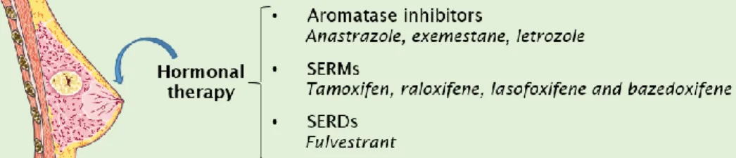
divided according to their mechanism of action: aromatase inhibitors, selective ER modulators (SERMs) and ER down-regulators (Figure 1.5) 70.
Figure 1.5: Types of breast cancer hormonal therapy. BC endocrine therapies include 3 different categories of molecules: aromatase inhibitors, which inhibit the production of estrogen from androgens; the selective ER modulators (SERMs) that agonizes/antagonize ER; selective ER down-regulators (SERDs), that fully antagonize ERα 70.
Aromatase inhibitors, such as the anastrozole, letrozole and exemestane, are chemical compounds that hamper aromatase activity. These enzymes are involved in the conversion of androgens, such as testosterone, into estrogen. By blocking this enzyme, it is possible to reduce the amount of endogenous ligand (estrogen) available to bind to the receptor (ERα) 70. This type of
hormonal therapy is only used in post-menopausal women, alone as adjuvant therapy or sequentially with SERM therapy, such as tamoxifen 68.
Another anti-endocrine therapy approach is based on the use of molecules that block specifically ERα signaling named SERMs. These are competitive inhibitors of estrogen binding to ERα,
17
such as tamoxifen and raloxifene 71. Tamoxifen, also known as ICI
46 474, is a non-steroidal anti-ER compound, belonging to the triphenylethylene chemical group of SERMs, that was developed to treat post-menopausal women carrying advanced disease 72.
Nowadays it is used in both pre- and post-menopausal women
68,70,72. SERMs have mixed agonist and antagonist activity, depending
on the target tissue 70. In the case of the BC tissue, tamoxifen works
as an antagonist, blocking transcription of estrogen-regulated genes, reducing tumor proliferation 70.
The third type of BC hormonal therapy is based on selective ER down-regulators (SERDs), that act similarly to ER modulators but have exclusively antagonist effect on the receptor 73. Fulvestrant,
also known as ICI 182 780, is a SERD. It binds to ER, inducing a structural change in the receptor that inactivates the 1 and AF-2 domains and inhibits the receptor dimerization. These changes lead to an increase in receptor surface hydrophobicity, reducing its translocation to the nucleus and promoting consequent faster proteasomal degradation 42,74–76. So, fulvestrant is both an ER
competitor and selective estrogen receptor degrader 77. Upon
fulvestrant binding, ER mRNA level is maintained and ER protein level is reduced 78. ICI 182 780 is currently indicated for the
treatment of postmenopausal women with metastatic ERα-positive BC, after non-steroidal aromatase inhibitor treatment failure 70.
Resistance to endocrine therapy might be intrinsic to the patient or acquired during treatment. In this last case are included: (1) mutations in amino acids in ERα, resulting in the constitutive transactivation of ERα in the absence of the ligand; (2) altered expression of ERα co-activators and co-repressors; (3) enhanced expression of transcription factors to which ERα binds in the
18
indirect genomic pathways; (4) microRNA action, for example, miRNA 221 and 222, that down-regulate the cell cycle inhibitor p27, which leads to continued cell division independent of ER blockers 79.
4.2 Antibody therapy
With the advances in cancer research, several cellular biomarkers have been proposed and their relationship with cancer development and progression has been reported. These discoveries boosted the development of more specific therapies, the so-called targeted anti-cancer agents, such as monoclonal antibodies and small-molecule inhibitors 80.
Antibodies have the capacity to selectively target cells expressing a specific antigen 80–82. Since they are designed to act on
a specific cellular target, they virtually present higher efficiency and less side-effects on non-targeted tissues, when comparing with non-targeted therapies 83. In the case of BC, examples are the
anti-HER2 antibodies. In the clinical setting, trastuzumab and pertuzumab antibodies have been used to target the HER2 receptor
84. Trastuzumab was, in fact, the first monoclonal antibody
approved for the treatment of a solid tumor by the Food and Drugs Administration, in 1998 81. It is used in both metastatic and
adjuvant settings 81. In clinical studies, combination of trastuzumab
with chemotherapy and hormonal therapy showed a benefit in terms of increased disease-free survival and overall survival 85,86.
Food and Drugs Administration also approved the use of an antibody-drug-conjugate (ADC) called trastuzumab-emtansine (Kadcyla®), which consists on Trastuzumab conjugated with the
19
chemotherapy agent maytansine 87,88, which blocks microtubule
polymerization, thus inducing apoptosis in target cells. This ADC was approved to be used as an adjuvant treatment of patients with HER2-positive early BC who have residual invasive disease after neoadjuvant taxane and trastuzumab-based treatment 87.
The major drawback related with anti-cancer antibodies is the development of resistance. In fact, the majority of patients who achieve an initial response to trastuzumab-based regimens develop resistance within one year 89. Another drawback is related with the
fact that several molecules and related pathways that are targeted by anti-cancer antibodies are also present in healthy cells, where they contribute to cell normal growth and homeostasis 90. One
example is the HER2 which is also present on healthy tissues, although at a much lower extent (to 100 vs 2 copies of the gene per cell, in cancer and healthy tissue, respectively) 91.
4.3 Small-molecule inhibitors
Small-molecule inhibitors (SMIs) are usually ≤500 Da in size, which allows them to translocate through the plasma membrane. Once inside the cell, they interact either with the cytoplasmic domain of cell-surface receptors, such as HER2 or EGFR, or intracellular signaling molecules, such as apoptotic proteins 80.
SMIs present several advantages over antibodies such as: (1) oral bioavailability, (2) generally good tolerance and (3) due to their small size, they can penetrate” sanctuary sites” in the human body
92.
Examples of SMIs approved for the treatment of BC include: lapatinib, palbociclib and ribociclib 93. Lapatinib is a tyrosine kinase
20
of HER2-positive BC 93,94. TKI are homologous of the adenosine
triphosphate (ATP), which allow them to compete for the ATP-binding domain of protein kinases (present on HER2, for example), preventing its phosphorylation and subsequent activation of the signal transduction pathways. This results in apoptosis and reduction of cellular proliferation 94. Lapatinib was approved by
Food and Drugs Administration as a combination treatment with the aromatase inhibitor letrozole in HER2-positive, advanced BC patients that have failed standard chemotherapeutic treatment 80.
Palbociclib and ribociclib are cyclin-dependent kinase (CDK) inhibitors that have been approved by Food and Drugs Administration for the treatment of advanced-stage hormone receptor-positive and HER2-negative BC, in combination with letrozole 93,95,96. Palbociclib inhibits specifically CDK4 and CDK6 96,
that play important role in tumorigenesis since they control the G1-S phase transition during cell cycle progression 95. So, inhibition of
CDK leads to reduced cell cycle progression and cell proliferation
95.
5. The role of tumor microenvironment in the
response to anti-cancer therapy
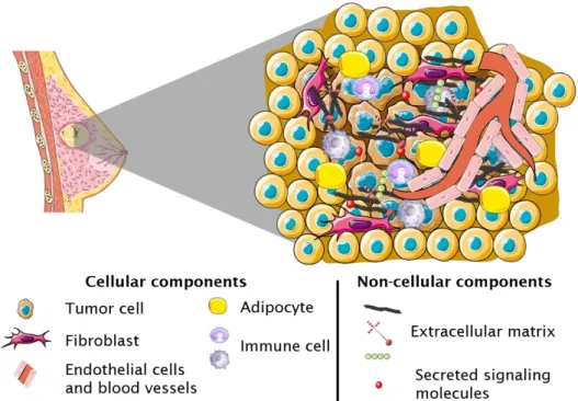
The tumor mass is not only composed by cancer cells; actually, other non-cancerous cells, can be found in the tumor environment (TME), such as fibroblasts, adipocytes, endothelial, immune cells, pericytes, myoepithelial cells and various progenitor cells 97. Besides them, several non-cellular components, such as
extracellular matrix (ECM) and secreted signaling molecules (e.g., cytokines and growth factors) are also a part of the TME (Figure 1.6). This intricate network of cellular and non-cellular components
21
has been reported to be a critical mediator of anti-cancer drug treatment outcome by playing important roles in tumor progression 97,98. While the TME of early-stage tumors confers
anti-malignancy functions, some cancer cells can tolerate the immune suppression and, in turn, reprogram the TME into one exerting pro-malignancy functions 99. So, the understanding of the TME changes
during this cancer progression is of high relevance when developing therapeutic strategies to tackle the tumor at a specific stage.
Some tumors present natural resistance to therapy, not responding to the drugs from the beginning (innate resistance)100.
In other cases, an initial response to the treatment is observed through cancer progression impairment, but cancer relapses due to acquired drug resistance 101. Tumor cell-driven mechanisms
behind this resistance include the activation of DNA-repair mechanisms, alterations in drug metabolism and drug transporters
101. The latter can be responsible for an increasing drug elimination
through the ejection of cytostatic therapeutic compounds to the extracellular space 100 and has a major influence in the failure of
chemotherapy strategies 100. Among those drug transporters are
ATbinding cassette (ABC) efflux transporters, such as P-glycoprotein, which are ubiquitously expressed and normally involved in transport of solutes 100.
In this thesis section, the TME components, highlighted in Figure 1.6, are presented in the perspective of therapy-related effects.
22
Figure 1.6: Breast cancer tumor microenvironment contains tumor cells and other cellular and non-cellular components. Non-tumor TME cellular components include fibroblasts, endothelial cells, immune cells, adipocytes and pericytes. Non-cellular TME components include extracellular matrix, (such as collagen fibers and glycosaminoglycans) and signaling molecules (such as cytokines and growth factors) 97,98.
5.1.
Cellular components
5.1.1.
Tumor cells
Tumor cells arise from healthy cells by a progressive series of transformations that lead to malignancy 102. Together with
uncontrolled growth, tumor cells are further characterized by genomic alteration, increased cell mobility, changes at the cellular surface, among others 103. However, tumor cells are a
heterogeneous population with variations at the morphological, genetic, epigenetic and phenotypic levels 104.
23
Tumor cells are the principal component of the TME and the
primum movens of tumorigenesis and metastasis so, for that
reason, they are the main target of anti-cancer therapies 80,105. A
multidrug resistance transporter of the ATP-binding cassette superfamily of transporters, termed BC resistance protein (BCRP), was proposed to be involved in drug resistance 106. Overexpression
of the full-length BCRP cDNA in MCF7 cells conferred resistance to mitoxantrone, doxorubicin, and daunorubicin and reduced daunorubicin accumulation and retention 106. Alternatively,
continuous exposure of tumor cells to anti-cancer drugs can lead to the development of acquired resistance, due to genetic and/or epigenetic changes leading to a proapoptotic pathway blockade, and/or constitutive expression of anti-apoptotic proteins, as well as increased efficiencies in cellular DNA damage repair mechanisms 107.
5.1.2.
Fibroblasts
Fibroblasts are mesenchymal cells derived from the embryonic mesoderm. They are the pillar of the connective tissue that holds the human body together. Fibroblasts produce ECM structural proteins (e.g., fibrous collagen and elastin), adhesive proteins (e.g., laminin and fibronectin), and ground substance (e.g., glycosaminoglycans (GAGs)) 108. In healthy tissue, in a wound
healing scenario, fibroblasts sense and respond to mechanical changes and damage signals in the tissue and differentiate into activated fibroblasts (myofibroblasts) 109. These cells are
responsible for tissue repair and wound healing through ECM production and remodeling and cross-talk with immune cells.
24
There are increasing evidence for the relevance of fibroblasts in the TME. In the “tumor is a wound that do not heal” theory it is hypothesized that the fibroblasts present on the tumor initially act in an anti-tumorigenic manner (by restraining growth and eliciting an anti-tumor immune response) 109. However,
fibroblasts are activated by cancer cells to become pro-tumorigenic cells, cancer-associated fibroblasts (CAFs) 110. CAFs secrete survival
cues that enhance cancer cell survival, remodel ECM to tumor invasion and reshape tumor immunity to generate an immunosuppressive environment, promoting tumor development
109,110. CAFs are a vastly heterogeneous stromal cell population,
representing one of the major components of TME. In BC setting, CAFs are the most prominent stromal cell type 109.
CAFs origin is controversial; in fact, not all CAFs derive from tumor-resident fibroblasts. CAFs have been shown to have diverse origins 110, including bone marrow-derived mesenchymal cells 111,
adipocytes 112 and endothelial cells 113. CAFs are multiple
subpopulations that have been divided in several CAFs subtypes: F1 tumor-restraining, F2 tumor-promoting, F3 secretory and F4 ECM-remodeling 114. CAFs secretome include, transforming growth
factor β (TGFβ), EGF, interleukins, fibroblast growth factors (FGFs), platelet-derived growth factors (PDGFs), protein ligands in the WNT signaling pathways, connective tissue growth factor (CTGF), prostaglandin E2 (PGE2), vascular endothelial growth factor (VEGF) and metabolites, such as lactate 110. Several reports have shown that
CAFs involvement in pro-tumorigenic functions occurs generally via modifications in their secretome 110.
CAFs are key players in therapy resistance and disease relapse 114,115. The mechanisms behind these include induction of
25
epithelial-to-mesenchymal transition (EMT), activation of survival pathways, immune reprogramming or stemness-related programs and metabolic reprogramming in tumor cells 114,115.
CAF-mediated drug resistance can be explained by environment-mediated drug resistance (EMDR): (1) based on soluble factors which include cytokines, chemokines and growth factors secreted by fibroblasts; (2) mediated by cell-adhesion between tumor cells and either fibroblasts or ECM components
116,117. Within group (1), a study by Straussman et al. used 23 stromal
cell types to study their ability to influence the innate resistance of 45 cancer cell lines to 35 anti-cancer drugs 118. They suggested that
anti-cancer drugs capable of killing tumor cells when cultured alone, frequently rendered ineffective when tumor cells were cultured in the presence of stroma. This effect was particularly pronounced with targeted agents compared with chemotherapy. They studied in detail the mechanism of stroma-mediated innate resistance to the RAF inhibitor PLX4720, in melanoma cells. The authors showed that hepatocyte growth factor (HGF) secreted by stromal cells induced the activation of the MET receptor tyrosine kinase. This lead to reactivation of the MAPK and PI3K/AKT pathways, and consequently resistance to RAF inhibition 118. Within
the group (2), it has was reported that adhesion of tumor cells to CAFs works as a drug-resistance mechanism, possibly via N-cadherin homotypic binding, which activates anti-apoptotic protein AKT/PKB, increasing pro-survival AKT signaling in melanoma cells