DOI: http://dx.doi.org/10.18363/rbo.v77.2020.e1827 Short Communication / Oral Radiology and Imaging
Magnetic Resonance Imaging of the
Temporomandibular Joint to Identify the
Discomaleolar Ligament: Case Study
Huyanne Cândida Souza e Silva Nascimento,1 Maria Rafaela dos Santos,2 Rebeca Pereira Espindola,2 Cláudia Borges Fontan Câmara,3 Fernando Augusto Pacífico,4 Lucas Carvalho Aragão Albuquerque,4 Gilberto Cunha de Sousa Filho5
1Health Family Health Residency Program, State Department of Health (SES), University of Pernambuco (UPE), Recife, PE, Brazil 2School of Dentistry, Federal University of Pernambuco (UFPE), Recife, PE, Brazil
3School of Medicine, Federal University of Pernambuco (UFPE), Recife, PE, Brazil 4School of Medicine of Olinda, Olinda, PE, Brazil
5Department of Anatomy, Federal University of Pernambuco (UFPE), Recife, PE, Brazil
• Conflicts of interest: none declared. AbstrAct
Objective: This article aims to identify and describe the path of the discomaleolar ligament (DML) using magnetic resonance imaging (MRI). Materials and methods: MRIs were acquired from the temporomandibular joint (TMJ) region of two research participants using a 1.5 T SIGNA Explorer device. Conclusion: based
on the images, we could observe a structure with an upward direction originating in the bilaminar zone towards the middle ear region. After analysis, the images obtained suggest that the observed structure is the DML.
Keywords: Magnetic resonance imaging; Ear-jaw articulation; Medium ear.
Introduction
T
he temporomandibular joint (TMJ) is one of the mostcomplex joints in the body, since it is the only one that allows rotational (hinge) and translational (slip) movements due to the double articulation of the condyle, being then classified as a ginglymus-arthrodial joint. Moreover, there are two joints connected to a single bone,
the jaw, which work simultaneously.1,2 TMJ bone components
are the mandible condyle, the glenoid fossa and, anteriorly to it, the joint eminence. There is an articular disc to avoid the direct contact between joint structures, in addition to the synovial membrane, retrodiscal tissue and TMJ ligaments, which play an important role in the protection of the joint,
acting as movement-restricting agents.1,3
Embryonic development of TMJ occurs around the eighth week of gestation. In this week, structures that will constitute the condyle, the glenoid fossa and the joint eminence are formed. Since the sixteenth week, the lateral pterygoid muscle is closely related to the joint capsule and the articular disc, sending its fibers to the peripheries of these structures.4 In the
twentieth week, the joint becomes functional; however, only mild mandibular movements occur until birth.5
Due to its complexity, TMJ is vulnerable to structural and pathological changes, such as Temporomandibular Dysfunction (TMD).6 TMD is a group of musculoskeletal and
neuromuscular conditions, involving temporomandibular joints, masticatory muscles and associated tissues. Joint noises, headaches, facial and cervical pain are among the
most common signs and symptoms. There are also otological symptoms such as otalgia, tinnitus and vertigo.7,8,9
A possible justification for the otological symptomatology is the presence of a ligament described by Pinto (1962), the discomalleolar ligament, which connects the anterior process of the malleus, through the petrotympanic fissure, to the medial, posterior and upper portion of the TMJ capsule, to the articular disc and to the sphenomandibular ligament.10,11
Magnetic resonance imaging is considered the gold standard for TMJ evaluation, since it allows a simultaneous evaluation of the position and morphology of the articular disc and adjacent bone structures, in addition to evaluating the functional relation of the condyle, disc, mandibular fossa and joint eminence.12 Therefore, our objective is to identify
and describe the path of the discomalleolar ligament using magnetic resonance imaging.
Materials and methods
Sample
For the development of this study, magnetic resonance imaging (MRI) acquisitions were performed in two patients at a diagnostic center in the city of Recife in 2018.
The inclusion criteria established sought to choose patients with a regular functioning of the stomatognathic system according the standards considered physiological, with absence of signs and symptoms in the components of the system. Patients had satisfactory occlusion, class I, all healthy teeth, absence of painful sensation that identified injuries in
TMJ and did not use or used orthodontic and or orthopedic devices. This study was conduct and approved according to the criteria required by the code of ethics in research of the Universidade Federal de Pernambuco (UFPE), under the number CAAE: 68317717.5.0000.5208.
Image acquisition
For the acquisition of the images, the SIGNA Explorer device (GE, HealthCare, Milwaukee, Wiscom, USA), 1.5T (magnetic field power) and a flex coil with 16 channels for magnetic resonance scanning were used. For the evaluation of the images obtained, we needed to work on the images in the Media Viewer program. (GE, HealthCare, Milwaukee, Wiscom, USA).
Based on the anatomical path of the discomalleolar ligament and its nature, the cut established for identification of this structure was the sagittal cut, in Proton Density (PD),
with 2mm thickness and 0.2 mm of distance between the cuts, totaling 84 images.
Results
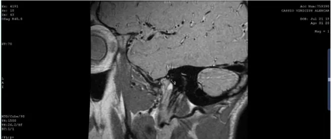
According to the study of the images obtained in the Media Viewer program (GE, HealthCare, Milwaukee, Wiscom, USA), we could determinate the region where we expected to observe the DML, which was marked by means of the 3D volumetric sequence in the first and second volunteers. In this follow-up, it is possible to analyze in Figures 1 and 2 the following anatomical structures: the neck of the malleus and the discomalleolar ligament.
In the first and second patient (Figures 1 and 2), we can see the structures of interest, in which the arrows listed indicate the location of the malleus present in the middle ear, and the discomalleolar ligament, from the middle ear to the bilaminar zone of the temporomandibular joint.
Figure 1. Nuclear Magnetic Resonance Image with the anatomical pathway of the discomalleolar ligament (DML) and its nature on sagital plane
axis, in Proton Density (PD), with 2 mm thickness and 0.2 mm of distance between the cuts. Arrow 1 indicates the neck of the malleus and arrow 2 the DML.
Figure 2. Nuclear Magnetic Resonance Image with the anatomical pathway of the discomalleolar ligament (DML) and its nature on sagital plane
axis, in Proton Density (PD), with 2 mm thickness and 0.2 mm of distance between the cuts. Arrow 1 indicates the neck of the malleus and arrow 2 the DML.
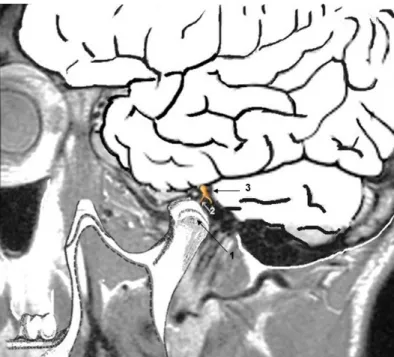
The schematic image shows the relation among the malleus, the discomalleolar ligament (DML), and the mandibular condyle (Figure 3).
Discussion
When evaluating the most indicated imaging tests for the analysis of TMJ, we chose the magnetic resonance imaging (MRI). According to Calderon et al.12 (2009), MRI
is advantageous because the examiner can easily identify the tissues with it, in addition to its non-invasive character. Westbrook et al.13 (2013) described the contrasts obtained
using the image, which are dependent on the variables of each tissue and the mechanisms that may interfere. These factors are divided into: intrinsic, which are unalterable, since they are inherent to organic tissues; and extrinsic, which may be modified.
According to Faria et al.14 (2010), there are several
parameters in each group of intrinsic factors: Relaxation time T1, which describes the exchange of energy between hydrogen nuclei and its environment; and T2, which presents the energy exchanges between adjacent hydrogen nuclei and the proton density, which is linked to discrepancies in signal intensity among tissues. According to Mazzola15 (2009), T1 relaxation,
also known as longitudinal relaxation, is relevant to fat mapping. On the other hand, relaxation in T2, or transverse relaxation, is used especially for water mapping.
In proton density (PD), differences in signal intensity occur between tissues and are directly related to their relative number of free hydrogen protons. To produce these differences by proton density, the transverse component needs to show these differences. According to Martins et al.16
Figure 3. Schematic diagram of the of the brain and body and neck
of the malleus. The mandibular condyle is shown by arrow 1, DML (discomalleolar ligament) by arrow 2 and neck of the malleus by arrow 3.
(2015), TMJ is composed of the head of the jaw (condyle); the mandibular fossa of the temporal bone; the articular cartilage and the articular disc, fibrous dense tissue, almost without blood vessels and nerve fibers. Loiola et al.17 (2015)
corroborated these findings describing the bilaminar zone as located in the region superior to the mandibular condyle.
Vasconcelos18 (2017) points the formation of the middle ear
by the malleus, incus and stapes ossicles, being surrounded by the eardrum tensor muscle and the stapes muscle, as well as the structures aforementioned and innervated by the trigeminal nerve. We also point that the tensor muscle of the eardrum is responsible for the passage of sound vibrations through the traction of the malleus.
The anterior discomalleolar and maleolar ligament belong to the middle ear, with embryonic origins common to the temporomadibular joint and mandible. The aforementioned ligaments begin in the malleus, follow different anterior routes towards the TMJ and the sphenoid, and then pass through the Huguier canal. The anterior discomalleolar and malleolar ligaments had similar lengths, which were 6.88 mm (SD 0.81) and 4.22 mm (SD 1.17), respectively.19
The discomalleolar ligament is an embryological remnant of the lateral pterigoid muscle. In this sense, the discomalleolar and the anterior malleolar ligaments, found in the Huguier canal, are separated by a bony crest, on the anterior board, in the canal.20 The anterior discomalleolar and
malleolar ligaments initiate a biomechanical communication between the mandible allied to the middle ear ossicles; consequently, they become overloaded due to disc dislocation or intra-articular edema, thus causing an instability in middle ear structures. With the movement of the capsular ligament, when mandibular movements occur, they promote the oscillation of the tympanic membrane and the ossicles, resulting in auditory disorders.21 In the images, we also
observe the presence of the malleus and a ligament compatible with the anatomical structure of the study by the anatomical description of its location allied to imaging examinations: the discomalleolar ligament.
In this aspect, we could perform the topographic description in our study with two volunteers, and the images chosen to compose the study were related to the two sequences – 3D volumetric. In a comparative analysis of the images and what was acquired to enrich our study, Testut and Latarjet22
(1975) mentions that the discomalleolar ligament has as characteristic its longitude due to its origin, at the base of the skull, near the spine of the sphenoid, tracing a path in which it passes in the external branch of the petrotympanic fissure, reaches the eardrum, and attaches to the head of the malleus at the base of its broad apophysis.
Araújo and Estevam23 (2018) performed an osteotomy on
the anterior wall of the petrous part of the temporal bone, in which the upper wall of the tympanic cavity was removed,
thus obtaining the visualization of the malleus bone and the discomalleolar ligament. With this concrete anatomical visualization on magnetic resonance imaging, we searched through the Sagittal cut with the help of a calibrated technical operator, presenting an image indicative of the discomalleolar ligament.
Thus, in the images studied, we observed a cut of MRI, PD, 3D, Sagittal, Open Mouth, in which we identified the malleus bone head and an ascending projection, showing that it begins in the bilaminar zone and indicating the discomalleolar ligament. We also noted that this projection goes against the ossicle structures of the ossicle in the middle ear, specifically the malleus bone, and that the analysis of the bone in its total anatomical contour is not feasible by the images acquired in our study.
Studies conducted by Pascoal et al.24 (2001) revealed
that 46.8% of patients that presented a possible temporomandibular disorder reported the presence of tinnitus. Parker & Chole25 (1995) could observe the presence
of otological symptoms such as tinnitus associated with dizziness in patients with temporomandibular dysfunction. Marasa and Ham26 (1988) showed that inflammations in
the TMJ region can spread through the petrotympanic fissure to the middle ear, causing otitis and tinnitus.
For Meira27 (2001), this is due to the intimate functional
and anatomical relation of the TMJ with the components of the ear, including innervation and vascularization in this proximity. Thus, despite the correlations with the otological symptoms presented by the researchers, in our study, we can analyze the presence of the ligament in the volunteers. Although it is not related to pathological factors, it is possible because the volunteers were selected in search of a pattern of anatomical normality of the DML.
Previous studies showed agreement among the authors, in which occlusion disorders, traumas and harmful oral habits were pointed out as the main triggering factors of temporomandibular dysfunction, since they overload the
TMJ.28,29,30,31
Conclusion
In our case study, we located the anatomical region of the aforementioned ligament and determined its probable path in a normality pattern, from the bilaminar zone, protruding to the middle ear and its insertion into the neck of the malleus. Studies with a larger sample, either correlated with TMD or not, may suggest greater accuracy in the identification by images of the discomalleolar ligament.
13. Westbrook C, Roth CK, Talbot J. Ressonância magnética: aplicações práticas. 4. ed. Rio de Janeiro: Guanabara Koogan; 2013.
14. Faria RF, Volkweis MR, Wagner JCB, Galeazzi S. Prevalência de patologias intracapsulares da ATM diagnosticadas por ressonância magnética. R. Cir. Traumatol Buco-Maxilo-fac. 2010;10(1):103-6.
15. Mazzola AA. Ressonância magnética: princípios de formação da imagem e aplicações em imagem funcional. RBFM. 2009;3(1):117-129.
16. Martins JS, Campos BM, Nahás-Scocate ACR, Fuziy A, Freitas CF, Costa ALF. Avaliação do volume do disco articular da ATM por meio de imagens de ressonância magnética usando um software de análise de imagem. Rev. Odontol. Univ. Cid. São Paulo. 2015;27(2):118-25.
17. Loiola M, Shibasaki W, Costa AL, Ferreira FC. Utilização da imagem de ressonância magnética no diagnóstico das alterações da ATM. Ortodontia SPO. 2015;48(2):179-184.
18. Vasconcelos MBN. Problemas auditivos associados a disfunção temporomandibular. 2017. Dissertação (Mestrado em Medicina Dentária) – Faculdade de Medicina Dentária, Universidade de Lisboa, Lisboa.
19. Ramírez LM, Ballesteros ALE & Sandoval OGP. A direct anatomical study of the morphology and functionality of disco-malleolar and anterior malleolar ligaments. Int. J. Morphol. 2009;27(2):367-379.
20. Rees LA. The structure and function of the mandibular joint. Br. Dent. J.1954;96:125-133.
21. Rodrigues VT, Bellato A, Moreira MA, Oliveira IB, Di Bernardo B, Pinto C. Relação entre distúrbios da articulação temporomandibular e alterações auditivas: revisão de literatura. Rev. Ulbra Torres. Rio Grande do Sul. 2017 22. Testut L, Latarjet A. Tratado de Anatomia Humana. VI, Barcelona, Salvat, 1959.
23. Estevam CS, Araújo PCA, Medeiros Junior MD, Pacífico FA, Cavalcante AB, Sousa Filho GB. Estudo anatômico do ligamento discomaleolar: contribuição descritiva topográfica para captura de imagens. An Fac Med Olinda. 2018;1(1):41-44.
24. Pascoal MIN, Rapoport A, Chagas JSF, Pascoal MBN, Costa CC, Magna LA. Prevalência dos sintomas otológicos na desordem temperomandibular: estudo de 126 casos. Rev. Bras. Otorrinolaringol. 2001;67(5):627-633 25. Parker WS, Chole RA. Tinnitus, vertigo, and temporomandibular References
1. Okerson JF. Tratamento das desordens temporomandibulares e oclusão. 6. ed. Rio de Janeiro: Elselvier; 2008.
2. Fuentes R, Cantin M, Ottone NE, Bucchi C. Characterization of Bone Components of the Temporomandibular Joint. Int J Morphol. 2020;33(4): 1569-1578.
3. Donnarumma MDC, Muzilli CA, Ferreira C, Nemr K. Disfunções Temporomandibulares: sinais, sintomas e abordagem multidisciplinar. Rev. CEFAC. 2010;12(5):788-794.
4. Alves N. Study About the Development of the Temporomandibular Joint in the Human Fetuses. Int J Morphol. 2008;26(2):309-312.
5. Katchburian E. Histologia e embriologia oral. 3. ed. Rio de Janeiro: Guanabara Koogan; 2012.
6. Pelicioli M, Myra RS, Florianovicz VC, Batista JS. Physiotherapeutic treatment in temporomandibular disorders. Rev Dor . 2017;18(4):355-361. 7. Sassi FC, Da Silva AP, Santos RKS, De Andrade CRF. Tratamento para disfunções temporomandibulares: uma revisão sistemática. Audiol Commun Res. 2018;23:1-13.
8. Cavalcanti MOA, Lima JMC, Batista AUD, De Oliveira LMC, De Lucena LBS. Grau de severidade da disfunção temporomandibular e hábitos parafuncionais em policiais militares. Rev Gaúcha Odontol. 2011;3(59): 351-56.
9. Maciejewska-szaniec Z, Maciejewska B, Mehr K, Piotrowski P, Michalac M, Wiskirska-Woznica B. et al. Incidence of Otologic Symptoms and Evaluation of the Organ of Hearing in Patients withTemporomandibular Disorders (TDM). Med Sci Monit.. 2017;23:5123-5129.
10. Salamanca C, Dias FJ, Fuentes R. Presencia y Relaciones Anatomofuncionales del Ligamento Discomaleolar. Una Revisión de la Literatura. Int J Morphol. 2018;36(4):1356-1360.
11. Poluha RL, Cunha CO, Bonjardim LR, Conti PCR. Temporomandibular joint morphology does not influence the presence of arthralgia in patients with disk displacement with reduction: a magnetic resonance imagingbased study. Oral Surg Oral Med Oral Pathol Oral Radiol. 2020;129(2):149-57. 12. Calderon PS, Reis KR, Araújo CRP, Rubo JH, Conti PCR. Ressonância magnética nos desarranjos internos da ATM: sensibilidade e especificidade. R. Dental Press Ortodon Ortop Facial. 2008;13(2):34-39.
Received: 06/08/2020 / Accepted for publication: 05/07/2020
Corresponding author Gilberto Cunha de Sousa Filho
E-mail: prof.gilbertodesousa@hotmail.com
Mini Curriculum and Author’s Contribution
1. Huyanne Cândida Souza e Silva Nascimento - BDS. Contribution: Bibliographic research, collection of the preparation of image data and writing of the manuscript. ORCID: 0000-0003-0365-6358
2. Maria Rafaela dos Santos - Dentistry student. Bibliographic research, collection of the preparation of image data and writing of the manuscript. ORCID: 0000-0002-8365-8320
3. Rebeca Pereira Espindola - Dentistry Student. Bibliographic research, collection of the preparation of image data and writing of the manuscript. ORCID: 0000-0002-0801-8577
4. Cláudia Borges Fontan Câmara - MD. Contribution: data collection and interpretation. ORCID: 0000-0002-9687-425X
5. Lucas Carvalho Aragão Albuquerque – DDS; PhD. Contribution: effective scientific and intellectual participation in the study. ORCID: 0000-0001-5069-6446 6. Fernando Augusto Pacífico – DDS; PhD. Contribution: effective scientific and intellectual participation in the study. ORCID: 0000-0002-5162-0694
7. Gilberto Cunha de Sousa Filho – DDS; PhD. Contribution: effective scientific and intellectual contribution in the study; data interpretation; preparation of the manuscript; writing of the manuscript; critical review and approval of the final text of the article. ORCID: 0000-0002-7419-8246
disorders. Am J Orthod Dentofacial Ortho. 1995;107(2):153-158.
26. Marasa FK, Ham BD. Case reports involving the treatment of children with chronic otitis media with effusion via craniomandibular methods. CRANIO. 1988;6(3):256-70.
27. Meira GSP. DTM x Problemas Otológicos. Rev. AONP Online. 2001;(7):1067-76.
28. Quinto CA. Classificação e tratamento das disfunções temporomandibulares: qual o papel do fonoaudiólogo no tratamento dessas disfunções? Rev CEFAC. 2000;2(2):15-22.
29. Pomeranc JMC. Distúrbios da articulação temporomandibular e dor miofascial – uma abordagem e tratamento fonoaudiológico. In: Marchesan IQ, coordenadora. Motricidade Orofacial – como atuam os especialistas. 2004;131-9. 30. Cauás M, Alves IF, Tenório K, HC Filho JB, Guerra CMF. Incidência de hábitos parafuncionais e posturais em pacientes portadores de disfunção da articulação craniomandibular. Rev Cir Traumatol Buco-Maxilo-Fac. 2004;4(2):121-129. 31. Rowicki T, Zakrzewska J. A study of the discomaleolar ligament in the adult human. Folia Modphol. 2006;65(2):121-5.