José Pedro Dias Cristóvão
Degree in BiochemistryThe Role of the Ral/Exocyst Pathway in Structural
Plasticity at the Drosophila Neuromuscular Junction
Dissertation to obtain the Master Degree in Biochemistry for Health
Supervisor: Doctor Rita Teodoro, Principal Investigator, CEDOC
José Pedro Dias Cristóvão
Degree in BiochemistryThe Role of the Ral/Exocyst Pathway in Structural
Plasticity at the Drosophila Neuromuscular Junction
Dissertation to obtain the Master Degree in Biochemistry for Health
Supervisor: Doctor Rita Teodoro, Principal Investigator, CEDOC
Jury:
President: Prof. Doctor António Sebastião Rodrigues Opponent: Doctor Maria Luísa Vasconcelos
Members of the jury: Doctor Rita Teodoro
Prof. Doctor Maria Teresa Nunes Mangas Catarino
NOVA Medical School | Faculdade de Ciências Médicas
v
Acknowledgments
I would like to thank to all the people that have helped me directly or indirectly during the development of this project
Firstly, I would like to thank to my supervisor, Dr. Rita Teodoro, for guiding and aiding me during this past year. I am especially grateful for her patience and motivation during the difficult steps and for helping me grow during this year, both professionally and as a person.
Next I would like to thank to my lab colleagues, João, Miguel, Cátia and Joana, for their help, support and companionship during the development of this thesis and especially during the long hours spent at the fly room.
I also have to extend my thanks to Dr. António Jacinto’s lab, for their help and patience and for sharing their knowledge with me. I would like to thank individually to Susana Ponte and Lara Carvalho, for having the courage to for putting up with me every morning.
To all my colleagues and friends from my Masters. Thank you for the friendship and the good times.
To Miguel Larguinho, without whom I wouldn’t be here today.
To my friends from back home, Carlos and Miguel, for the long years, for all the good moments and all the good stories.
To Joana, for the help and the support during this year and for bringing so much joy and happiness to my life.
Finally, I would like to thank to my parents, not only for making the person that I am today, but for helping me reach where I am today, for all the sacrifice and patience. Thank you
vii
Abstract
Defects in synaptic morphology and activity-dependent plasticity are a hallmark of neurodevelopmental and neurodegenerative disorders. Neuronal structure is critical for determining the properties of neurons, yet very little is known about the membrane dynamics that controls synaptic morphology. It is therefore critical to know the basic mechanisms by which neurons acquire their shape and change it in response to activity. This capacity of response is called synaptic plasticity, and allows modifications to be made in both pre- and post- synaptic elements of the synaptic terminal and their synapses. Given that synaptic plasticity is key for neurons to adapt to stimuli, it is important to study and understand the mechanisms by which it occurs and how defects can affect function.
In this study, using the Drosophila neuromuscular junction as model, we show that activity-dependent formation of new presynaptic boutons is compromised when Ral and exocyst function is impaired, suggesting that this pathway plays a central role in structural plasticity. Ral GTPase is a small GTPase from the Ras superfamily and the exocyst is a conserved protein complex that is an effector for several GTPases, which, collectively might serve to control where, when and how, are vesicles targeted to a specific exocytic place. Dissecting the signaling cascade triggered by the Ral/Exocyst pathway will be key to understand how intracellular trafficking participates in this form of plasticity.
ix
Resumo
Alterações na morfologia sináptica e na plasticidade dependente de actividade têm sido um ponto crucial no estudo das perturbações no desenvolvimento neuronal e nas doenças neurodegenerativas. A estrutura neuronal é importante para definir as propriedades neuronais, no entanto pouco é sabido acerca de como a dinâmica membranar controla a morfologia sináptica. Deste modo, é necessário perceber os mecanismos básicos através dos quais os neurónios adquirem forma e de como a mudam em resposta a actividade. Esta capacidade de resposta é denominada de plasticidade sináptica e permite que sejam feitas modificações nos elementos pré- e pós- sinápticos dos terminais sinápticos e nas sinapses neles contidas. Sabendo que a plasticidade sináptica é um elemento chave na resposta dos neurónios a um estímulo, é importante estudar e perceber que mecanismos estão envolvidos e de que forma defeitos nesses mecanismos podem afectar a sua função.
Neste estudo, recorrendo à junção neuromuscular de Drosophila melanogaster como modelo, é demonstrado que a formação de novos botões pré-sinápticos duma forma dependente de actividade é afectada quando existem defeitos na Ral ou no exocisto, sugerindo que a interacção entre estas proteínas é importante para a plasticidade estrutural. A Ral GTPase é uma pequena GTPase da superfamília das Ras GTPases, enquanto que o exocisto é um complexo proteico conservado que é um efetor de várias GTPases que pode controlar a maneira como as vesículas são exocitadas. Compreender a cascata de sinalização iniciada pela interacção entre a Ral e o exocisto poderá ser a chave para perceber como o tráfego intracelular participa neste tipo de plasticidade.
xi
Table of Contents
1. Introduction ... 1
1.1 Neurodegenerative Diseases and Neurodevelopmental Disorders ... 1
1.2 Synapses and Synaptic Plasticity ... 5
1.3 Membrane Trafficking and Synaptic Plasticity ... 9
1.3.1 Functional Synaptic Plasticity ... 9
1.3.1.1 Plasticity at the Presynaptic Terminals ... 9
1.3.1.2 Plasticity at the Postsynaptic Terminals ... 12
1.3.2 Structural Synaptic Plasticity ... 13
1.4 Ral/Exocyst Pathway: Membrane Trafficking and Neuronal Developing ... 15
1.4.1 The Exocyst Complex... 15
1.4.2 Ral GTPase (Ras-like GTPase) and the Ral/Exocyst Pathway ... 17
1.5 Drosophila Neuromuscular Junction as Model of Study... 21
1.6 The UAS-GAL4 system ... 23
1.7 Aim of the Thesis ... 25
2. Materials and Methods ... 27
2.1 Fly Stock and Maintenance ... 27
2.2 Fly Crossing and Selection... 29
2.2.1 Exocyst Impairment: Exo84 mutant ... 29
2.2.2 Exocyst Impairment: Sec5 depletion ... 29
2.2.3 Exocyst Overexpression: HA-Sec3 experiments ... 30
2.2.4 RalA involvment in Structural Synaptic Plasticity ... 31
2.2.5 Exocyst Role in Microtubule Regulation: Sec8 experiments ... 31
2.3 High K+ Depolarization Paradigm ... 33
2.4 Immunostaining Assay ... 35
2.5 Mounting and Imaging ... 37
2.6 Ghost Bouton Count and Analysis ... 37
3. Results and Discussion ... 39
3.1 Ral GTPase Involvement in Activity-dependent Structural Plasticity ... 39
3.1.1 Role of Ral GTPase in Structural Plasticity ... 39
3.1.2 Ral Rescue of Wild Type Phenotype... 43
xii
3.1.4 Ral Mutants Appear to have Aberrant Microtubules ... 47
3.2 Exocyst Involvement in Activity-dependent Structural Plasticity ... 49
3.2.1 Exocyst Role in Structural Plasticity ... 49
3.2.2 Exocyst Overexpression Effect on Presynaptic Plasticity ... 53
3.2.3 Sec8 Role in Presynaptic Plasticity ... 55
4. Conclusions ... 59
xiii
Figure Index
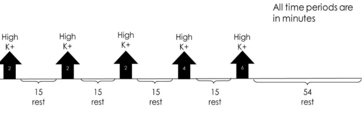
Figure 1.1 – Worldwide dementia prevalence (data frim 2015) with projections for 2030 and 2050. Taken from World Alzheimer Report 2015 (ADI, [s.d.]) ... 2 Figure 1.2 – Scheme of a mature glutamatergic synapse. The presynaptic compartment possesses the synaptic vesicles (SV) necessary for neurotransmission. When triggered by the opening of the voltage-gated calcium channels (VGCC), the SVs in the readily releasable pool are fused with the membrane with help of SNARE complexes. Glutamate travels through the synaptic cleft, where it binds to both NMDA or AMPA ionotropic receptors in the postsynaptic side. These receptors are controlled by scaffolding proteins. Postsynaptic VGCCs are important for potentiation of synaptic transmission (Michalak and Biala, 2016) and adhesion molecules allow for proper location of both pre- and postsynaptic compartments. Adapted from (Volk et al., 2015). ... 5 Figure 1.3 – Synaptogenesis model. (A) The formation of a synapse starts with proper contact between the presynaptic terminal and the postsynaptic structure. (B) After contact is established, vesicles containing active zone proteins (yellow) and vesicles containing synaptic vesicle-associated proteins (blue) are transported to their target location, while adhesion proteins (red) stabilize cell contact. (C) After the presynaptic terminal is assembled, postsynaptic terminal assembly starts, with glutamate receptors and scaffold proteins being trafficked to their target location. (D) When postsynaptic maturation finishes, the synapse is fully formed and neurotransmission can occur. Figure taken from (Goda and Davis, 2003). ... 10 Figure 1.4 – Ghost boutons appear in response to a stimulation paradigm. (A) A Drosophila melanogaster NMJ without being submitted to a stimulation paradigm possesses no ghost boutons. (B) After being submitted to a stimulation protocol, several ghost boutons appear around the Drosophila melanogaster NMJ. White arrows point to ghost boutons. HRP (red) marks presynaptic membrane and DLG (green) marks postsynaptic compartment. Adapted from (Ataman et al., 2008). ... 14 Figure 1.5 – Exocyst components individual interactions obtained via different studies. Interaction in red were obtained using yeast two hybrid assays, interactions in yellow were obtained in protein binding assays with recombinant proteins from E.coli and red interaction were obtained in protein assays using in vitro translation. Figure taken from (Liu and Guo, 2012). ... 15 Figure 1.6 – (A) Representative scheme of a Drosophila melanogaster NMJ on two arbitrary muscles, designated muscle 1 and muscle 2. Actives zones represent the synaptic bouton (presynaptic compartment) and the SSR represents the postsynaptic compartment. (B) The NMJ possesses two types of glutamatergic boutons, type Ib (big) and type Is (small) bouton, which distinguished by the size of both pre- and postsynaptic. Boutons of type Ib have both larger pre- and postsynaptic compartments when compared to type Is boutons. Adapted from (Menon, Carrillo and Zinn, 2013) ... 21 Figure 2.1 – Schematic representation of the stimulation protocol. The black arrows indicate the stimulation periods, while the brackets indicate the resting periods. The protocol total time is of 130 minutes. ... 34
xiv
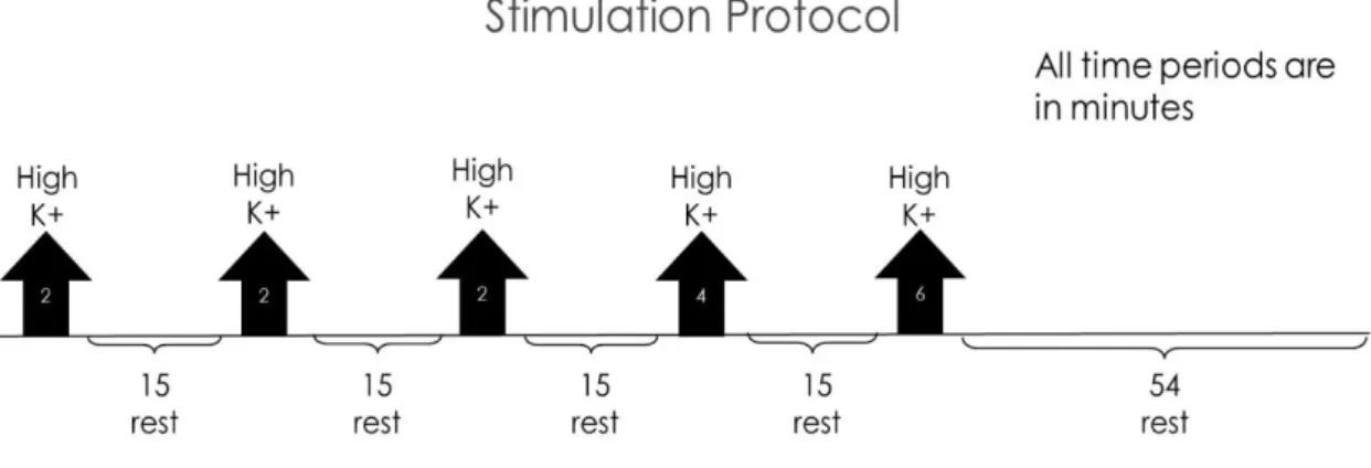
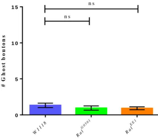
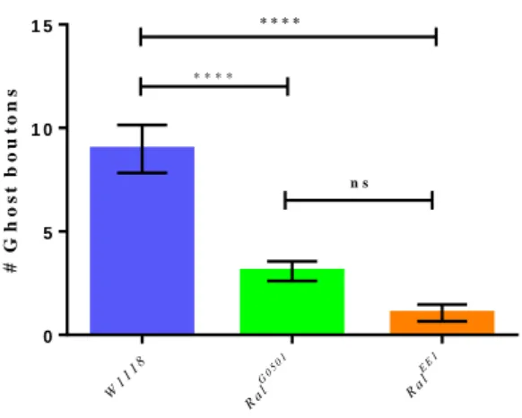
Figure 3.1 – Schematic representation of the stimulation protocol. The black arrows indicate the stimulation periods, while the brackets indicate the resting periods. The protocol total time is of 130 minutes. ... 39 Figure 3.2 – Number of ghost boutons obtained per NMJ without using the stimulation paradigm for W1118 (control) RalG0501 and RalEE1. Results are expressed as means ± SEM. NW1118 = 34; NRalG0501 = 37; NRalEE1 = 36. No significant differences from control were found (ns P > 0.05) by one-way ANOVA. ... 40 Figure 3.3 – Unstimulated NMJ of W1118 (control) and both RalG0501 and RalEE1 mutants. White arrows point to the existent ghost boutons. HRP (red) marks presynaptic membrane and DLG (green) marks postsynaptic compartment. ... 41 Figure 3.4 – Number of ghost boutons obtained per NMJ using the stimulation paradigm for W1118 (control), RalG0501 and RalEE1. Results are expressed as means ± SEM. N
W1118 = 57; NRalG0501 = 78; NRalEE1 = 17. Significant differences from control are expressed with asterisks (ns P > 0.05 and ****P < 0.0001) by one-way ANOVA. ... 42 Figure 3.5 – Stimulated NMJs of W1118 (control) and both RalG0501 and RalEE1 mutants.
White arrows indicate the location of ghost boutons. HRP (red) marks presynaptic membrane and DLG (green) marks postsynaptic compartment. ... 43 Figure 3.6 – Number of ghost boutons obtained per NMJ using the stimulation paradigm for RalG0501 rescue. Results are expressed as means ± SEM. NW1118 = 57; NRalG0501 = 78; NOK6/+ = 68; NRal neuronal rescue = 40. No significant differences between the rescue and the control, but there is a significant difference between the Ral rescue and the RalG0501 mutants (ns P > 0.05 and ****P < 0.0001) by one-way ANOVA. ... 44 Figure 3.7 – Stimulated NMJs of OK6/+ (control) and RalG0501 rescue. White arrows indicate the location of ghost boutons. HRP (red) marks presynaptic membrane and DLG (green) marks postsynaptic compartment. ... 44 Figure 3.8 – Number of ghost boutons obtained per NMJ using the stimulation paradigm for Ralwt overexpression. Results are expressed as means ± SEM. N
OK6/+ = 68; NRalWT Overexpression = 49. No significant differences from control were found (ns P > 0.05) by Mann-Whitney test. ... 46 Figure 3.9 – Stimulated NMJs of OK6/+ (control) and both RalWT overexpression. White arrows indicate the location of ghost boutons ... 46 Figure 3.10 – (A) Muscle 4 nerve of W1118 (control) and both RalG0501 and RalEE1 mutants. (B) Average thickness of muscle 4 nerve of W1118 (control) and both RalG0501 and RalEE1 mutants. Results are expressed as means ± SEM. NW1118 = 32; NRalG0501 = 31; NRalEE1 = 22. Significant differences from control are expressed with asterisks (****P < 0.0001) by one-way ANOVA. HRP (green) marks presynaptic membrane. ... 47 Figure 3.11 – Muscle 4 nerve of the W1118 (control) and both RalG0501 and RalEE1 mutants. White arrows indicate aberrant Futsch accumulations. HRP (red) marks presynaptic membrane and DLG (green) marks postsynaptic compartment. ... 48 Figure 3.12 – Number of ghost boutons obtained per NMJ without using stimulation protocol of controls and both strains with exocyst impairments (Exo84/Df and Sec5 RNAi). Results are expressed as means ± SEM. N W1118 =9; NExo84/Df= 14; NC155;Dicer2/+ = 12; N Sec5 RNAi/ C155;Dicer2 = 19. No significant differences from control were found (ns P > 0.05) by one-way ANOVA. Even though there was no significant difference between the two controls, they were separated due to the different genetic background. ... 50
xv
Figure 3.13 – Unstimulated NMJ of controls and both strains with exocyst impairments (Exo84 Df and Sec5 RNAi). White arrows point to the existent ghost boutons. HRP (red) marks presynaptic membrane and DLG (green) marks postsynaptic compartment. ... 50 Figure 3.14 – Ghost bouton count of stimulated NMJ of W1118 and C155;D2 (controls) and both strains with exocyst impairments (Exo84 Df and Sec5 RNAi). NW1118 = 17; NExo84/Df = 39; NC155;Dicer2/+ = 18; NSec5 RNAi/ C155;Dicer2 = 32. Results are expressed as means ± SEM. Significant differences from control are expressed with asterisks (*P < 0.05 and ****P < 0.0001) by one-way ANOVA. ... 51 Figure 3.15 – Images of stimulated NMJs of controls and both strains with exocyst impairments (Exo84/Df and Sec5 RNAi). White arrows point to the existent ghost boutons. HRP (red) marks presynaptic membrane and DLG (green) marks postsynaptic compartment. ... 52 Figure 3.16 – Number of ghost boutons per NMJ using the stimulation paradigm for W1118 (control) and HA-Sec3 (exocyst overexpression). NW1118 = 23; NHA-Sec3 = 29. Results are expressed as means ± SEM and significant differences from control are expressed as asterisks (*P < 0.05) by Mann-Whitney test. ... 53 Figure 3.17 – Stimulated NMJs of both W1118 (control) and HA-Sec3 (exocyst overexpression). White arrows point to the existent ghost boutons. HRP (red) marks presynaptic membrane and DLG (green) marks postsynaptic compartment. ... 54 Figure 3.18 – The two images above represent the same NMJ. (A) Staining for pre- and postsynaptic compartments allow the visualization of ghost bouton. (B) Staining for presynaptic compartment and inserted Sec3 gene, shows co-localization of exocyst and ghost boutons. The white arrows indicate ghost boutons. HRP (red) marks presynaptic membrane, DLG (green) marks postsynaptic compartment and HA tag (blue) marks the inserted Sec3 gene containing the HA tag. ... 54 Figure 3.19 – Number of ghost boutons obtained per NMJ for W1118 (control) and Sec8 Pi (Sec8 mutant) with and without being submitted to the submitted to the stimulation paradigm. NW1118 = 23; NHA-Sec3 = 29 NW1118 = 23; NHA-Sec3 = 29. Results are expressed as means ± SEM and significant differences from control are expressed as asterisks (ns P > 0.05 and ****P < 0.0001) by one-way ANOVA. ... 56 Figure 3.20 – Unstimulated and stimulated NMJs of both W1118 (control) and Sec8Pi. White arrows point to the existent ghost boutons. ... 56
xvi
Table Index
Table 2.1 – Stocks of Drosophila melanogaster utilized. ... 28 Table 2.2 – Antibodies used in the immunostaining assays. ... 36
xvii
Abbreviations
AMPA – α-amino-3-hydroxy-5-methyl-4-isoxazolepropionic acid ASD – Autism Spectrum Disorders
AZ – Active Zone
BDSC – Bloomington Drosophila Stock Center;
ChR2 – Channelrhodopsin2
DABCO – 1,4-diazabicyclo[2.2.2]octane DLG – Disc Large
EMS – ethyl methanesulfonate HL – Hemolymph
HRP – Horseradish Peroxidase
MAP1B – Microtubule Associated Protein 1B NGS – Normal Goat Serum
NMDA – N-methyl-D-aspartate NMJ – Neuromuscular junction
PBT – Phosphate Buffered Saline with Triton PSD – Postsynaptic Density
PTV – Piccolo-bassoon Transport Vesicles SEM – Standard Error of the Mean
STV – Synaptic Vesicle Protein Transport Vesicles SV – Synaptic Vesicle
UAS – Upstream Activating Sequence VDRC – Vienna Drosophila Resource Center
xviii WT – Wild Type
1
1. Introduction
1.1 Neurodegenerative Diseases and Neurodevelopmental Disorders
Neurons are the cell type responsible for the core functions of the brain. They are responsible for processing and transmitting information through electrical and chemical signals transmitted between them through structures existent in the synaptic terminal, the synapses. Neurons assemble into neuronal networks, responsible for coordinating several functions of the human body (Lodish et al., 2013). Neuronal communication occurs at a specialized structure called the synapse. Synapses are therefore the interface and the site of communication between two neurons and any damage to these structures can compromise the neuronal network function. Two of the main factors that can cause such damage are neurodegenerative diseases and neurodevelopmental disorders.
Neurodegenerative disease is a term that commonly defines a condition that affects neurons (Przedborski, Vila and Jackson-Lewis, 2003) and other brain cells (Skovronsky, Lee and Trojanowski, 2006), causing progressive degeneration and eventually the death of this type of cells (Skovronsky, Lee and Trojanowski, 2006). These type of conditions are common of developed countries due to a longer life expectancy, and their incidence has been increasing over the years (Brown, Lockwood and Sonawane, 2005). The great majority of these diseases are characterized by an accumulation of insoluble aggregates, which can be composed of different proteins (Skovronsky, Lee and Trojanowski, 2006). Thus, many of the therapies that exist or that are in development, focus on this aspect of the disease, mainly trying to correct the processes that lead to these aggregates (Skovronsky, Lee and Trojanowski, 2006).
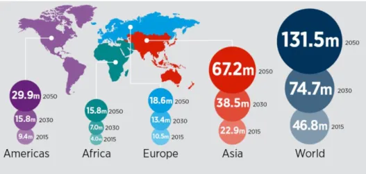
One of the most well-known and studied neurodegenerative diseases is Alzheimer’s disease. Alzheimer’s disease is the most common neurodegenerative disease worldwide and also the most common form of dementia (Alzheimer’s Association, 2014). Dementia affected 46.8 million people worldwide in 2015 and it is predicted to increase to 131.5 million in 2050, with Alzheimer’s disease accounting for 50% to 75% of the cases ( Figure 1.1) (Prince et al., 2015).
2
Figure 1.1 – Worldwide dementia prevalence (data from 2015) with projections for 2030 and 2050. Taken from World Alzheimer Report 2015 (ADI, [s.d.])
This disease is characterized by the formation of β-amyloid plaques outside the neurons and of tau protein tangles inside them. This leads to neuronal cell death, neuronal inflammation, cortical atrophy and synapse loss (Koffie, Hyman and Spires-Jones, 2011). The loss of synapses in the brain is the cause of the first symptoms of this disease. This process starts about 20 years before the first symptoms, as the brain is able to compensate the loss of synapses. From there, the symptoms start to appear and the individual will start experiencing decay of cognitive functions, such as memory loss, difficulty in decision making and forgetting how to perform daily tasks (Alzheimer’s Association, 2014).
Neurodevelopmental disorders affect neurons in a different way. These disorders usually affect brain growth and development. They can be caused either by defective genes, brain lesions or by environmental factors (such as, diseases or malnutrition) (Cioni, Inguaggiato and Sgandurra, 2016) affecting cognitive development, socioemotional development or sometimes both (Boivin et al., 2015). The disorders caused by genetic defects can have multiple genes involved in different pathways, originating several different disorders, such as epilepsy, intellect disabilities and autism spectrum disorders (ASDs) (Sahin and Sur, 2015). ASDs usually converge onto a few major signaling pathways, such as protein synthesis, cellular metabolism, transcriptional control and synapse development and function (Sahin and Sur, 2015). In fact, many of the candidate genes in ASD are located at the pre- or postsynaptic compartments or can regulate synaptic functions in neurons, affecting synaptic processing and plasticity (Meredith,
3
2015). An example of this is PSD95, which is a scaffolding protein that anchors NMDA/AMPA receptors in glutamatergic synapses. Mutations that affect signaling pathways or proteins that regulate PSD95 can lead to synaptic deficits (Sahin and Sur, 2015). Unlike neurodegenerative diseases, which can appear at different stages of life, neurodevelopmental disorders can usually be diagnosed in the first weeks or months of life (Cioni, Inguaggiato and Sgandurra, 2016).
The current therapies that are being developed to treat these two major types of brain conditions address them separately. Regarding neurodegenerative diseases, the focus has been on the pathway that leads the formation of the aggregates, with different drugs targeting different steps (Skovronsky, Lee and Trojanowski, 2006). However, for neurodevelopmental disorders, due to the high number of candidate genes that exist for the several disorders, research has been focused on possible common molecular pathways that can simplify the number of interventions needed (Sahin and Sur, 2015).
New approaches that simultaneously address both ailments can be of great interest. Although the origin and development of neurodegenerative and neurodevelopmental diseases are different, both affect synapses. This can be caused by either the synapses being destroyed (neurodegenerative diseases) or the synapses not being able to be properly formed (neurodevelopmental disorders). Therefore, synaptic terminals, where synapses are formed, can potentially be viewed as a potential new target for the creation of treatments and therapies for these two different neuronal ailments.
In many neurodegenerative diseases, the formation of aggregates leads to neurodegeneration, including the elimination of synaptic terminals and their associated synapses (Skovronsky, Lee and Trojanowski, 2006). However, this process is gradual, with several synaptic terminals remaining in the brain. The adult brain does not possess the same potential for synaptic plasticity as a young brain does, but does retain some of it, as adults are able to form memories and learn. Therefore, by studying synaptic plasticity, new therapies can be created that use the existing synapses to form new ones and thus delaying the symptoms of neurodegeneration. In neurodevelopmental disorders, understanding the pathway that leads to synapse formation is important, as a defect in any gene involved may lead to a disorder. However, it seems that there are complementing pathways in the genetic network that, while compensating for the mutated gene, do lead
4
to a developmental delay of the neurons (Meredith, 2015). Therefore, by studying the genes that are involved in synapse formation, it may be possible to find novel ways to overcome this delay by, for example, accelerating these compensating pathways.
Overall, synaptic plasticity is a good mechanism to be explored in order to develop a new treatment for several different diseases.
5
1.2 Synapses and Synaptic Plasticity
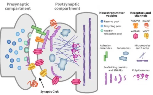
A synapse is a structure that mediates communication between two adjacent neurons or between a neuron and another type of cell, which comprehends a presynaptic and a postsynaptic specialization (Figure 1.2) (Hormuzdi et al., 2004).
There are two types of synapses, electrical and chemical. Electrical synapses are the least common type of synapses, being the signal transmitted directly through gap junctions. This allows almost instantaneous signaling between cells at the cost of signal modulation. These characteristics are useful, as the main role of this type of synapses is the electrical synchronization of populations of synapses (Purves et al., 2004). Chemical synapses are the most common type of synapse. Chemical synapses have a presynaptic and postsynaptic specialization, with the neurotransmitter travelling between the two specializations through the synaptic cleft (Figure 1.2) (Bito, 2010). This type of neurotransmission has minor delay in signaling transmission when compared to the one that occurs at electrical synapses, however it allows for signal modulation (Niswender and Conn, 2010; Purves et al., 2004).
Figure 1.2 – Scheme of a mature glutamatergic synapse. The presynaptic compartment possesses the synaptic vesicles (SV) necessary for neurotransmission. When triggered by the opening of the voltage-gated calcium channels (VGCC), the SVs in the readily releasable pool are fused with the membrane with help of SNARE complexes. Glutamate travels through the synaptic cleft, where it binds to both NMDA or AMPA ionotropic receptors in the postsynaptic side. These receptors are controlled by scaffolding proteins. Postsynaptic VGCCs are important for potentiation of synaptic transmission (Michalak and Biala, 2016) and adhesion molecules allow for proper location of both pre- and postsynaptic compartments. Adapted from (Volk et al., 2015).
6
The signaling process starts with an action potential being propagated through the axon. When it arrives at the presynaptic compartment, it triggers the opening of voltage-gated calcium channels, resulting in a rapid increase of calcium concentration inside the presynaptic membrane, which will bind to synaptotagmin-1 (Südhof, 2013). The binding of calcium then triggers synaptic vesicle fusion in the active zones, therefore releasing the neurotransmitters into the synaptic cleft (Südhof, 2013). In the case of excitatory synapses, this neurotransmitter will be glutamate (Lamprecht and LeDoux, 2004). Glutamate binds to glutamate receptors, NMDA receptors and AMPA receptors present in the postsynaptic density (Figure 1.2) (Li and Sheng, 2003; Michel et al., 2015).
As mentioned above, synapses form the bridge between two neurons or a neuron and a target cell. The process by which synapses are formed is called synaptogenesis. For synaptogenesis to occur, the axon must first make contact with its target location, which requires the growing axon to be properly guided to the appropriate location, so that the right connection can be made, and only after that can the synapses begin to form (Robichaux and Cowan, 2013). Sometimes, the axon extends beyond the region where synapses will form, triggering an axon pruning process. Once an axon reaches its terminal zone, transient chemical synapses are formed with their target postsynaptic cells (which can be another neuron or another cell type, like muscle). Some of these synapses will mature and form stable, functional synapses while other will be removed and lost (Robichaux and Cowan, 2013). The creation and removal of synapses is common during the development of organisms and it is guided by experience, allowing not only for growth but also the refinement of the neuronal network of the brain (Stoneham et al., 2010). However, this process is not restricted to early development. In adult mammalian brain it has been found that synapse formation and elimination is associated with long-term memory formation (Holtmaat and Svoboda, 2009).
Synaptic plasticity is defined as an activity-dependent process in which synaptic terminals can modify the strength and efficacy of the signal transmission (Citri and Malenka, 2008) or their whole structure (Holtmaat and Svoboda, 2009) in response to a stimulus. Functional plasticity is the type of synaptic plasticity that involves modifications in strength and efficacy of neurotransmission. This plasticity occurs at synapse level and does not cause heavy structural modifications of synaptic terminals. (Griffith and Budnik, 2006). Structural plasticity is the process by which synaptic
7
terminals change their structure in response to different levels of activity. This process occurs through the addition or removal of synapses, requiring protein synthesis and trafficking. Structural plasticity occurs over longer time periods, allowing the formation of long-time memories (Lamprecht and LeDoux, 2004).
9
1.3 Membrane Trafficking and Synaptic Plasticity
In the brain, membrane trafficking rules are similar to other cells in the body, being necessary to maintain viability and functionality of all cells, including glia, neurons and supporting cells. However, in neurons, there are more specialized or regulated forms of membrane traffic that regulate intercellular signaling. This more specialized type of trafficking can be seen in intracellular transport of synaptic vesicles that compose the basis of synaptic vesicle exocytosis and neurotransmitter release (Südhof, 1999).
Membrane trafficking can be described as the transport of proteins between endomembrane compartments and the cell membrane (Cheung and Vries, de, 2008). It occurs in both endocytic and secretory manner and relies on a series of processes, starting with the generation of vesicles loaded with a specific cargo. These vesicles are then transported to the proper location, where they will bind and fuse with their target membrane (Derby and Gleeson, 2007).
Even though neurons possess the same fundamental eukaryotic trafficking mechanisms as other cells, their unique morphology made those mechanisms evolve in different ways. Neurons are highly polarized cells, with the axon having the molecular machinery required for propagation of action potential and neurotransmitter release, while the dendritic filopodia carry the correspondent receptors and signaling components that respond to the neurotransmitter (Kennedy and Ehlers, 2006). At excitatory synapses, the assembly of presynaptic terminals and of the postsynaptic compartments requires several trafficking steps (Harris and Littleton, 2015).
1.3.1 Functional Synaptic Plasticity
1.3.1.1 Plasticity at the Presynaptic Terminals
Synaptic boutons are round presynaptic specializations where active zones form and synaptic transmission occurs. Upon leaving the cell body of the neuron, the axon must grow and extend in order to reach its correct region, where it will form connections with the target cells, with this process being mediated by the growth cone (Gallo, 2013).
10
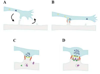
After the axon is properly positioned, it undergoes a morphological transformation, and from the growth cone are formed axonal varicosities (Harris and Littleton, 2015). After this process gives origin to a synaptic terminal (Collins and DiAntonio, 2007), a new process starts, in which new synaptic branches and varicosities are added, being the last denominated synaptic boutons (Harris and Littleton, 2015). Located in the synaptic bouton are the active zones, where synaptic vesicles dock and fuse, which allows the release of neurotransmitters to the synaptic cleft, where the neurotransmitters will travel until reaching the neurotransmitter receptors in the postsynaptic region (Figure 1.3) (Wichmann and Sigrist, 2010).
Figure 1.3 – Synaptogenesis model. (A) The formation of a synapse starts with proper contact between the presynaptic terminal and the postsynaptic structure. (B) After contact is established, vesicles containing active zone proteins (yellow) and vesicles containing synaptic vesicle-associated proteins (blue) are transported to their target location, while adhesion proteins (red) stabilize cell contact. (C) After the presynaptic terminal is assembled, postsynaptic terminal assembly starts, with glutamate receptors and scaffold proteins being trafficked to their target location. (D) When postsynaptic maturation finishes, the synapse is fully formed and neurotransmission can occur. Figure taken from (Goda and Davis, 2003).
For the synaptic bouton to be formed, the components of both synaptic vesicles and active zones must be recruited to the site of contact with the dendritic filopodia (Figure 1.3 A). The neuron achieves this by creating different vesicles that contain the presynaptic components: the active zone proteins are transported via piccolo-bassoon transport vesicles (PTV), while the synaptic vesicle-associated proteins are transported in synaptic vesicle protein transport vesicles (STV) (Figure 1.3 B). Both kinds of vesicles possess the proteins necessary for rapid assembly in presynaptic terminals and provide
11
components for this process. After presynaptic terminals are mature, glutamate receptors and structural proteins are recruited to the postsynaptic terminal and, after proper assembly, a fully mature synapse is formed (Figure 1.3 C-D) (Bury and Sabo, 2011).
The active zones (AZ) are target of presynaptic plasticity. AZs are the sites in the synaptic bouton in which the synaptic vesicles fuse in a Ca2+-dependent process to release the neurotransmitters necessary to propagate the signal. The AZ scaffolding proteins are essential to the localization of synaptic vesicle fusion site, positioning correctly the voltage-gated Ca2+ channels in order to achieve an efficient synaptic vesicle recruitment. The structure of AZ correlates directly with its function, as its size and complexity correlate directly with the synaptic output. More specifically, the AZ scaffold size has a relation with the probability to display evoked synaptic vesicle release in response to an action potential. Evoked release per AZ scales with the presence of several scaffolding proteins and with the presence of Ca2+ channels. The presence of these scaffold proteins favors Ca2+ channel clustering, which in turn favors the presence of SV fusion sites in the AZ. This process demonstrates that plasticity exists in presynaptic terminals and that it can modulate the neurotransmission process (Petzoldt, Lützkendorf and Sigrist, 2016). In both synaptic vesicles and active zones, there has been observed processes of synaptic plasticity. In response to an increase of activity, there is a modulation of the synaptic vesicles size, with larger vesicles being formed, recruited and released at the release site (Steinert et al., 2006). It has also been shown that, in response to an high-frequency stimulus, the presynaptic specialization undergoes structural changes, increasing the number and size of active zones, which is accompanied by an increase in the number of release-ready vesicles (Weyhersmuller et al., 2011). All these activity-dependent reactions show that there is presynaptic plasticity and that it requires trafficking not only for the changes in synaptic vesicles, but also for the structural changes in the active zone and in the synaptic bouton.
12 1.3.1.2 Plasticity at the Postsynaptic Terminals
Synaptic plasticity also exists in the synapse’s postsynaptic terminal. The postsynaptic terminals are present at dendritic structures called spines, which are protrusions located all along the dendritic shaft, and harbor the postsynaptic density (PSD). The PSD is an array of proteins that organize and stabilize the components necessary for synaptic transmission and function in postsynaptic terminal, such as synaptic receptors, ion channels, structural proteins and signaling molecules (Vallejo, Codocedo and Inestrosa, 2016). The PSD is an important structure for postsynaptic plasticity, as it regulates the synaptic transmission by modifying the number of neurotransmitter receptors present at the postsynaptic terminal (Vallejo, Codocedo and Inestrosa, 2016). Among all the proteins that compose the PSD, PSD-95 is one of the most important. PSD-95 is the most abundant scaffolding protein of dendritic spines and is enriched in excitatory synapses. Its scaffolding function derives mainly from its PDZ domains, which allows PSD-95 to bind to C-terminals, internal motifs and PDZ domains of other proteins, and also to lipids, allowing to form several large molecular complexes. Additionally, the PDZ domain interacts with transmembrane cell adhesion molecules and signaling molecules, making PSD-95 a linker between membrane proteins and cytoplasmic signaling pathways (Lardi-Studler and Fritschy, 2007).
In the postsynaptic structure, functional plasticity is mostly related with the neurotransmitter receptors. The PSD is the main structure present at the postsynaptic compartment and has the capability of regulate not only the number of neurotransmitter receptors present, but also their location. Since synaptic transmission strength relies on the contact between neurotransmitter and receptors, this ability allows for synaptic transmission modulation. With the existence of both binding slots and receptor-confining domains, the PSD possesses a pool of readily available receptor pockets that can be used either to strengthen or to weaken the synaptic transmission (Newpher and Ehlers, 2009).
13
1.3.2 Structural Synaptic Plasticity
The notion that modulation of synaptic weight alone could be responsible for learning and memory formation process came from classic interpretation of Hebbian plasticity, which consists in modulating excitatory synaptic strength (Caroni, Donato and Muller, 2012; ZHEN, 2007), with many studies being focused mainly in functional plasticity. Decades later, it is understood that modifying synaptic strength alone does not account for memory formation, and that synapse formation, stabilization and elimination are critical learning and memory formation (Caroni, Chowdhury and Lahr, 2014).
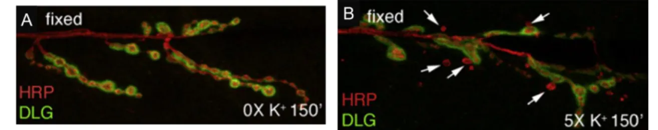
Structural synaptic plasticity is the capacity by which synaptic terminal and their postsynaptic counterpart suffer significant structural changes not only to allow neurodevelopment, but also to answer and adapt to external stimuli (Caroni, Donato and Muller, 2012). This ability is thought to be important for the formation of long term memory and learning processes (Holtmaat and Svoboda, 2009), as recent study demonstrated that formation of new synapses is directly connected to learning process (Hayashi-Takagi et al., 2015). New synapse formation has been demonstrated to be an activity-dependent process, as studies done in Drosophila melanogaster NMJ (a glutamatergic synaptic terminal) have demonstrated that, when submitted to stimulation protocol, new immature synaptic boutons appear (Figure 1.4) (Ataman et al., 2008). In fact, these immature boutons were already characterized, with the lack of postsynaptic structure being their main difference from normal synapses. Due to this fact, they were named ghost boutons (Ataman et al., 2006).
14
Figure 1.4 – Ghost boutons appear in response to a stimulation paradigm. (A) A Drosophila melanogaster NMJ without being submitted to a stimulation paradigm possesses no ghost boutons. (B) After being submitted to a stimulation protocol, several ghost boutons appear around the Drosophila melanogaster NMJ. White arrows point to ghost boutons. HRP (red) marks presynaptic membrane and DLG (green) marks postsynaptic compartment. Adapted from (Ataman et al., 2008).
These ghost boutons were formed de novo and did not arise from existing bouton retraction, which led to the hypothesis that these boutons are actually undifferentiated synaptic boutons and that can acquire overtime both presynaptic components and the postsynaptic structure characteristic of a fully mature synaptic bouton. Studies in live Drosophila larva using live imaging were done in order to determine whether ghost boutons could indeed mature into fully grown synaptic boutons. Several of the examined ghost boutons acquired glutamate receptors or presynaptic scaffold protein BRP and GluR over time until pupariation starts (Ataman et al., 2008). Since these boutons are a transient stage for mature boutons, there are also mechanisms by which they can be eliminated. It was shown that Draper/Ce-6 pathway, which functions in the muscle and glial cells that surround the synaptic boutons, clears the ghost boutons that fail to maturate (Menon, Carrillo and Zinn, 2013).
Since this type of plasticity possibly plays a central role in learning and memory formation and it is crucial in early neuronal circuit development, studying its mechanism may allow the discovery of new therapies to neurodegenerative diseases such as Alzheimer’s disease (Pilato et al., 2012). Since that in this disease a loss of synapses occurs (Koffie, Hyman and Spires-Jones, 2011), studying a neuronal property that allows for the creation of new synapses can lead to the development of new therapies to delay the progression of Alzheimer’s disease.
15
1.4 Ral/Exocyst Pathway: Membrane Trafficking and Neuronal
Developing
1.4.1 The Exocyst Complex
There are several proteins that are important to maintain membrane trafficking in cells, being the exocyst complex one of them (Wu and Guo, 2015). The exocyst is an octameric protein complex composed of Sec3, Sec5, Sec6, Sec8, Sec10, Sec15, Exo70 and Exo 84 proteins and was first identified in S. cerevisiae (Figure 1.4). In mammals, the exocyst was identified in rat brains, being found in every examined tissue (Figure 1.5) (Wu and Guo, 2015).
Figure 1.5 – Exocyst components individual interactions obtained via different studies. Interaction in red were obtained using yeast two hybrid assays, interactions in yellow were obtained in protein binding assays with recombinant proteins from E.coli and red interaction were obtained in protein assays using in vitro translation. Figure taken from (Liu and Guo, 2012).
The crystal structure of several of the exocyst subunits has been resolved and it shows that many of them have the same rod-like structure composed of α-helical bundles
16
common in other tethering complexes. These rod-shape units are likely to pack themselves side by side and interact with each other (Liu and Guo, 2012). Despite many studies, the organization of the complex is not fully known. However, it has been shown that the exocyst is a dynamic complex, where its assembly can occur via recruitment from pools of subunits and sub-complexes. Studies have shown that the absence of one of the subunits does not affect the assembly of the remaining seven subunits into a holo-complex, indicating the existence of individual interactions between them (Wu and Guo, 2015). The individual molecular interactions between the subunits were first demonstrated in a study using yeast Sec10 and Sec5 mutants, showing that Sec15 binds to the exocyst solely through interaction with Sec10 (Guo et al., 1999). Further studies have identified in both yeast and mammalian exocyst other individual interactions between the components, including Sec3–Sec5, Sec5–Sec6, Sec5–Exo84, Sec6–Sec8, Sec6–Secl0, Sec6–Exo70, Sec8–Exo70, Sec10– Secl5, and Sec10–Exo70, suggesting the presence of conserved interactions amongst different components of the complex and that Sec5 apparently is a core member to the exocyst (Figure 1.4) (Hsu et al., 2004).
The exocyst functions in several neuronal processes. The complex is involved in polarized exocytosis, an essential process for neuronal growth (Hsu et al., 2004). Polarized exocytosis occurs in three steps: targeting of the vesicles, interaction between vesicles and proteins and their fusion. The first one involves the targeting of Golgi-derived secretory vesicles to the designated plasma membrane domains through microtubule or actin based transport systems. The final step is the interaction between vesicle and integral plasma membrane proteins, termed v-SNARE and t-SNARE respectively, leading to the fusion of the secretory vesicle with the plasma membrane. This fusion allows the secretion of the vesicle contents to extracellular milieu and the incorporation of vesicle proteins at specific plasma membrane domains (Hsu et al., 2004). In Drosophila, this process participates in neurite outgrowth and synaptogenesis (section 1.3.1.1, Figure 1.3), whereas in mammal neurons polarized exocytosis is involved in axon outgrowth and receptor positioning and knockdown of several members of the exocyst resulted in defects in axon outgrowth, more specifically in fewer and shorter terminals (Jones et al., 2014). Several studies in Drosophila using different exocyst subunit mutants have shown the importance of this complex for neuronal growth. In Sec5 mutants, impairment of neurite growth has been shown to be caused by neuronal membrane
17
trafficking arrest, indicating a dependence of the exocyst in this process. However, there was no disruption of synaptic transmission, demonstrating that neuronal membrane trafficking and neurotransmission require different processes of exocytosis (Murthy et al., 2003). In another study, featuring Sec15 homozygous mutants, it was shown that the loss of Sec15 caused a defect in the proteins responsible for synaptic specificity. Normal synaptic development occurred, although neuronal targeting failed causing neurons to be connected to the wrong partners, thus affecting neuronal development (Mehta et al., 2005). It is also shown that Sec10 has an effect on neurite growth and synaptogenesis. Overexpression of a dominant negative form of Sec10 caused a blockage of neurite growth in neuronal cells, even in cultures supplied with nerve growth factor, thus showing that Sec10 is required for normal neurite growth (Liu and Guo, 2012)
There is a strong correlation between the exocyst and neuronal development and the exocyst role in membrane trafficking is essential for normal neuronal growth and synaptogenesis (Liu and Guo, 2012)(Wu and Guo, 2015). The molecular mechanisms by which the exocyst regulates neuronal development are not fully understood. However, studies demonstrated that Ral GTPase binds to Sec5 and Exo84, initiating the exocyst assembling. This suggests that Ral GTPase is a regulator of the exocyst function (Moskalenko et al., 2003) (Moskalenko et al., 2003).
1.4.2 Ral GTPase (Ras-like GTPase) and the Ral/Exocyst Pathway
Ral GTPases are members of the Ras branch of the Ras superfamily, sharing 46%-51% sequence identity and structure domain with Ras proteins (Gentry et al., 2014). In humans, Ral family only has two isoforms: RalA and RalB, which share 82% of homology. Unlike mammals, invertebrates, such as Drosophila melanogaster and the nematode C. elegans, only possess one Ral gene. This gene arose in multicellular organisms during evolution since there are no Ral orthologs in yeast (Shirakawa and Horiuchi, 2015). The Ral ortholog present in Drosophila melanogaster has a higher sequence identity with the human RalA isoform (72%) than with the RalB isoform (71%) (Gentry et al., 2014).
18
The Ral GTPase is constituted by a N-terminal, a G domain and a C-terminal. The N-terminal of this enzyme possesses a 11 amino acid extension which is not present in the N-terminal of the remaining Ras proteins. The G domain is responsible for the GTP binding and hydrolysis. It also possesses two specific amino acid sequences, Switch I (SI) and Switch II (SII), which are responsible for the interaction of Ral with its modulators (RalGAP and RalGEF) and effectors (such as Sec5 and Exo84), being this domain the interaction site between Ral and the exocyst. The C-terminal is a membrane targeting domain and is where the majority of sequence divergence occurs between the two isoforms. This divergence results in distinct subcellular locations and, therefore, contribute to the different functions attributed to RalA and RalB (Gentry et al., 2014).
Ral GTPase is expressed in the nervous system and can be modulated by specific GTPase-activating proteins (GAPs), and guanine nucleotide exchange factors (GEFs) (Gentry et al., 2014). Ral-GAPs were first identified in 1991 (Emkey, Freedman and Feig, 1991), and their main function is to catalyze the hydrolysis of GTP bound to Ral, changing it to its inactivate conformation (Personnic et al., 2014). By contrast, Ral-GEFs catalyze the exchange of the GDP bound to Ral to GTP. This is important, as the Ral-GTP is the active conformation and Ral can only interact with its effectors in this conformation (Saito et al., 2012). Humans possess one of RalGDS and three RalGDS-like (RGL) proteins and two copies of RalGPS, which are all RalGEF proteins. D. melanogaster has two orthologs of RGL proteins and one ortholog of RalGPS. When accounting for RalGAPs, humans have two α subunits and one β subunit of this protein, while D. melanogaster only have one copy of the α subunit and one β subunit of this protein (Gentry et al., 2014).
Due to its connection with Ras oncogenic superfamiliy, many studies of Ral focus on its role in tumorigenesis (Chien and White, 2003; Gentry et al., 2014; Kashatus, 2013). However, with the discovery of the association of Ral GTPase to the exocyst, studies on Ral have started to focus on other areas besides cancer. Two of the most well-understood effectors of Ral are the exocyst subunits Sec5 and Exo84 (Kashatus, 2013). Exo84 and Sec5 have been found to bind competitively to Ral, having both Ral-Sec5 and Ral-Exo84 been crystalized and their structure studied (Shirakawa and Horiuchi, 2015; Wu and Guo, 2015). Also, through interactions with these exocyst subunits, it has been demonstrated the role of Ral GTPase in the exocyst assembling (Moskalenko et al., 2003). Together,
19
Ral-Sec5 and Ral-Exo84 have been implied in tumor cell invasion, polarized membrane trafficking, cytokinesis and tight junction formation (Kashatus, 2013).
In neurons, the Ral/exocyst pathway has a determinant role in neuronal development, being involved in neuronal polarity regulation and neurite branching, with both tasks requiring membrane trafficking (Das et al., 2014; Lalli, 2009; Lalli and Hall, 2005). At the synaptic level, this pathway modulates the readily releasable pool of synaptic vesicles, possibly influencing synaptic strength (Polzin et al., 2002). Recently, it has been shown that the Ral/exocyst pathway mediates activity-dependent growth of the postsynaptic membrane (Teodoro et al., 2013). However, there is no data of the role of this pathway in synaptic bouton development.
21
1.5 Drosophila Neuromuscular Junction as Model of Study
Drosophila melanogaster, commonly known as fruit fly, has been a model for the study of molecular mechanisms involved in synaptic development and function (Menon, Carrillo and Zinn, 2013), as well as to understand the molecular mechanisms of several human diseases (Pandey and Nichols, 2011), including neurodegenerative diseases (Chan and Bonini, 2000) and neurodevelopmental disorders (Gatto and Broadie, 2011). Drosophila melanogaster is a good model of study because many of its biological, physiological and neurological properties are conserved to mammals and nearly 75% of known human disease-causing genes are believed to possess a functional homolog in the fly (Pandey and Nichols, 2011). Also, the existent powerful and elegant genetic tools allow not only the obtainment of fly mutant strains that model for some human diseases (Bier, 2005; Duffy, 2002), but also the manipulation of gene expression in a temporal and tissue-specific manner (Collins and DiAntonio, 2007).
Figure 1.6 – (A) Representative scheme of a Drosophila melanogaster NMJ on two arbitrary muscles, designated muscle 1 and muscle 2. Actives zones represent the synaptic bouton (presynaptic compartment) and the SSR represents the postsynaptic compartment. (B) The NMJ possesses two types of glutamatergic boutons, type Ib (big) and type Is (small) bouton, which distinguished by the size of both pre- and postsynaptic. Boutons of type Ib have both larger pre- and postsynaptic compartments when compared to type Is boutons. Adapted from (Menon, Carrillo and Zinn, 2013)
Another important characteristic of this model for the study of the human central nervous system (CNS) is its neuromuscular junction (NMJ) (Figure 1.5, A). Human NMJ synapses are cholinergic, meaning they use acetylcholine as neurotransmitter. Drosophila
22
larval NMJ synapses, however, use glutamate, the same neurotransmitter used in the human CNS, making the Drosophila NMJ a good model of the human synaptic terminals (Figure 1.5, B). Also, the ionotropic glutamate receptor (iGluR) present in the NMJ are homologous to both NMDA and AMPA-type GluRs present in the mammalian brain and their postsynaptic scaffolds resemble those found in mammalian postsynaptic densities. Another advantage of using the NMJ is its accessibility, which allows the usage of several different experimental techniques, such as electrophysiology and immunochemistry, to the analysis of physiological and structural characteristics (Collins and DiAntonio, 2007). Drosophila NMJ are highly stereotyped, which allows to identify defects in NMJ function and development from one animal to another. Finally, Drosophila NMJ possess the same processes and molecules involved in synaptic development and plasticity that in mammalian synapses, allowing the study of these synaptic characteristics (Featherstone and Broadie, 2000). All these features make the Drosophila NMJ a perfect model for the study of excitatory synapses and their synaptic plasticity (Menon, Carrillo and Zinn, 2013).
In Drosophila, homologs of all the subunits of the exocyst have been found. The exocyst is involved in important processes such as cell elongation and cytokinesis (Giansanti et al., 2015), membrane trafficking in branch outgrowth of trachea terminal cells (Jones et al., 2014) and neuronal membrane traffic (Murthy et al., 2003). Ral GTPase, that is responsible for the exocyst assemble, has been demonstrated to be involved in Notch signaling required for activity-dependent synaptic plasticity at the Drosophila NMJ (Bivort, de, Guo and Zhong, 2009; Cho and Fischer, 2011). In mammalian neurons, the Ral/exocyst pathway has been shown to be involved in neuron polarity regulation (Das et al., 2014; Lalli, 2009), neurite branching (Lalli and Hall, 2005) and it mediates the growth of postsynaptic membrane in a activity-dependent way in Drosophila NMJ synapses (Teodoro et al., 2013).
23
1.6 The UAS-GAL4 system
The UAS-GAL4 system is a binary system of gene expression (Duffy, 2002), firstly identified in yeast, where it was involved in the transcription of the genes important for galactose metabolism (Duffy, 2002)(Traven, Jelicic and Sopta, 2006). This system is comprised two parts: the UAS (Upstream Activator Sequences), an enhancer sequence to which the GAL4, a yeast transcription activator factor, will bind, activating gene transcription.
In 1988, it was demonstrated that this system was able to stimulate the transcription of a reporter gene in D. melanogaster (Fischer et al., 1988), paving the way for the development of the bipartite UAS-GAL4 system for targeted gene expression in Drosophila (Brand and Perrimon, 1993). In this system, the expression of the gene of interest (the responder) is controlled by the presence of the UAS element. Since the transcription of this gene will occur only if GAL4 binds to the UAS element, the absence of GAL4 in the responder line keeps the UAS-controlled gene transcriptionally silent. The transcription is therefore achieved when the responder line is mated with a line expressing GAL4 (driver), with different GAL4 lines having different expressing patterns. The resulting progeny, having present both GAL4 and the UAS element, will have the gene of interest being expressed according to GAL4 pattern of the driver line (Duffy, 2002).
25
1.7 Aim of the Thesis
In both neurodegenerative diseases and neurodevelopmental disorders, defects in both synaptic structure and plasticity are a common factor and can affect normal neuronal development and structure. However, little is known on how membrane dynamics can affect synaptic morphology. Activity-dependent synaptic plasticity is a characteristic that allows synaptic terminals and their correspondent synapses to change their structure in order to adapt to stimuli applied. Since these adaptations require heavy membrane trafficking, studies focusing in proteins involved in membrane trafficking may prove to be crucial in understanding how synaptic plasticity occurs. The exocyst is an important protein complex for membrane trafficking, as it is involved in several processes such as polarized membrane addition, axon growth and formation and, in Drosophila melanogaster, this complex participates in neurite growth and synapse formation. Ral GTPase was recently found to interact directly with the exocyst, being responsible for the assembly of this complex. Therefore, studying how the Ral/exocyst pathway regulates membrane trafficking will be important to understand how intracellular trafficking maybe important for synaptic plasticity.
The aim of this thesis was to study the role of the Ral/exocyst pathway in the activity-dependent formation new synaptic boutons. Using the Drosophila NMJ as a model, we studied how this pathway affects the structural synaptic plasticity of the synaptic terminal, using the ghost bouton count as an indicator of how plasticity is negatively or positively affected by impairments in Ral GTPase or exocyst.
27
2. Materials and Methods
2.1 Fly Stock and Maintenance
The fly stocks were maintained at 18°C and were raised instandard cornmeal-agar medium. Flies used in experiments were maintained at 25°C and 70% humidity, in order to achieve a controlled life cycle of 10 days and to potentiate mating and fertility. Virgin females obtainment and Drosophila stocks maintenance were performed according to Ashburner et al. (Ashburner and Roote, 2007). The Drosophila stocks used are described in Table 2.1. These stocks were obtained either from Bloomington Drosophila Stock Center (BDSC), Vienna Drosophila Resource Center (VDRC), generated at the lab or were given as a gift from other laboratories.
28 Table 2.1 – Stocks of Drosophila melanogaster utilized.
Name Genotype Stock Source
w1118 w[1118] 5905 BDSC HA-Sec3/CAG; elavG4/TAG HA-Sec3/CyO-GFP; P{w[+mW.hs]=GawB}elav[C155]/ TM3, P{ActGFP}JMR2, Ser1 NA Schwarz Lab OK6-Gal4 P{GawB}OK6 64199 BDSC RalEE1/FAG Rala[EE1]/FM7c 25095 BDSC RalG0501/FAG w67c23 P{lacW}RalaG0501/FM7c 12283 BDSC C1555-Gal4; UAS-Dicer2 P{w[+mW.hs]=GawB}elav[C155] w[1118]; P{w[+mC]=UAS-Dcr-2.D}2 25750 BDSC
w;;onr142-5/TAG w;;onr142.5
/ TM3 NA Blakenship
Lab
Df(3R) Espl3/TM6c,
Sb,Tb
Df(3R)Espl3/TM6C, cu1 Sb1 Tb1 ca1 5601 BDSC
UAS- Sec5 RNAi w1118; P{GD13789}v28873 28873 VDRC
UAS-Ral wt w; P{w1, UAS-Rala1} NA Gift Maria Balakireva
Sec8Δ1 w*; Sec8Δ1/TM3, P{ActGFP}JMR2,
Ser1
9555 BDSC
TM3, Sb1 Ser1/TM6B, Tb1
w[*]; TM3, Sb[1] Ser[1]/TM6B, Tb[1] 2537 BDSC
29
2.2 Fly Crossing and Selection
2.2.1 Exocyst Impairment: Exo84 mutant
In the exocyst assembly process, Ral GTPase binds competitively to either Sec5 or Exo84 exocyst subunits. Therefore, to study the role of the Ral/exocyst pathway in new ghost bouton formation, the importance of the Exo84 subunit was firstly examined. Males from a stock containing a mutation in the onr gene, which encodes the Exo84 subunit, described as a hypomorphic mutation (Blankenship, Fuller and Zallen, 2007; Giansanti et al., 2015) were crossed with female virgins collected from a stock containing a deletion of the region in which the onr is located. Being an hypomorphic mutant, an
onr142.5/onr142.5 crossing could still express enough protein, with enough function to give
origin to a mutant phenotype (Blankenship, Fuller and Zallen, 2007). By crossing with a deletion, it was ensured the exacerbation of the defective exocyst by having less Exo84 present in the larvae. Also second point mutations can be present and affect the final result due to the method used to obtain the mutant (Giansanti, 2004). By crossing with a deletion, the effect of these possible second point mutations was eliminated given that the two lines have different origins. The control used for this experiment was the w1118 wild type strain.
2.2.2 Exocyst Impairment: Sec5 depletion
During the exocyst assembly, Ral GTPase can also bind to Sec5 exocyst subunit. Thus its importance was also assessed. Since Sec5 mutants available cannot survive up to third instar, a RNAi was used in order to interfere with Sec5 function. Males from a UAS-Sec5 RNAi stock were crossed female virgins collected from a C155;D2 containing stock, with the resulting embryos being grown at 30 °C to augment the efficiency of the RNAi. The C1555;D2 and the Sec5 RNAi function as UAS-Gal4 system. The C1555 expresses Gal4 only in the neurons, thus resulting in neuronal expression of both D2 and Sec5 RNAi. This results in a decreased expression and presence of the exocyst subunit Sec5 only in the synaptic terminal. The control used was a cross between wildtype and C155;D2, in order to reduce the difference between genetic backgrounds.
30
Regarding progeny selection, only males were used, due to the discovery of a difference between the ghost bouton count between males and females, with the later having a phenotype similar to the control when compared to males. When the first depolarization paradigm was made, no selection was done, being the Sec5 larvae either male or female. The phenotype observed was different between larvae, with an inconsistent ghost boutons count observed.
A second experiment was conducted in order to test if the difference in the RNAi expression levels was related with the larva gender, since there were no other variables that could originate the observed difference. The experiment was done with one set of males and one set of females, using the same control described above. After analyzing the obtained results, a significant difference was observed between male and female larvae. Males had fewer visible ghost boutons than the females and this was consistent in all tested larvae, being conclude that the RNAi was more effective in males than in females.
In the following Sec5 experiments, only male larvae were used. The selection was performed using the male testes as a discriminating factor, being these appendixes located on both sides of the posterior section of the larva.
2.2.3 Exocyst Overexpression: HA-Sec3 experiments
In order to analyzed if the overexpression of the exocyst would have an effect in structural synaptic plasticity, a fly strain containing a wildtype and an inserted copy of the exocyst subunit Sec3 gene was used. The effect was analyzed by counting the ghost boutons that appeared due to the stimulation paradigm. Additionally, the inserted copy of the Sec3 gene codes for a protein expressing a HA tag, which allowed to observe the location of the exocyst and if there was any co-localization of the exocyst with the ghost bouton obtained, thus allowing to study if there was a direct interaction between the exocyst and the newly formed ghost boutons. The inserted HA-Sec3 gene was expressed via the UAS-Gal4 system.
No crossing was made and the selection of the larvae for this experiment was done by negative selection of GFP, having the chosen larvae two copies of the HA-Sec3 gene. The control used for this experiment was the w1118 wildtype strain.
31
2.2.4 RalA involvment in Structural Synaptic Plasticity
For the RalA impairment experiments, two different mutants were used: RalG0501 and RalEE1. By using these two mutants, it was ensured that the obtained results were caused only by the mutated protein and not from second point mutations.
The selection of the larvae for this experiment was done by negative selection of GFP and all selected GFP-negative larvae were unintentionally males. The RalA gene is located in the X chromosome, which results in the females having two copies of the gene and the males only having one. The heterozygotic mutant females always have a GFP marker, making them indistinguishable under blue light from homozygotic GFP females. Since RalA mutant homozygoty is lethal at birth, there are no GFP-negative females. Therefore, all selected larvae were males. The control used for this experiment was the w1118 wildtype strain.
For the rescue experiments only RalG0501 mutant was analyzed. The strain RalG0501/FAG;;UAS-Ralwt/TAG possesses a phenotype to a normal RalG0501 mutant. The rescue occurs when males from this stock were crossed with females collected from a OK6 stock, activating the expression of RalA wildtype gene. This crossing allowed to assess if the insertion of the wildtype gene could revert the mutant phenotype to wildtype. The selected larvae were all GFP negative to ensure the presence of the OK6 gene. The control used was a cross between wildtype males and OK6 virgin females, in order to reduce the difference between genetic backgrounds.
2.2.5 Exocyst Role in Microtubule Regulation: Sec8 experiments
In order to investigate if the exocyst was the link between the Ral mutants and the increase of the axon thickness, a Sec8 mutant was studied. This subunit was chosen due to previous reports that showed that a deletion in the Sec8 gene lead to an increase in synaptic microtubule density (Liebl et al., 2005).
For this experiment, firstly a mutant with a balancer chromosome with a marker visible in larvae was obtained. Males from the Sec8 mutant stock (Sec8Δ1/TM3, Ser1) was crossed with TM3, Sb1 Ser1/TM6B, Tb1 virgins. The resultant progeny was selected against Sb1 (shorten back hairs), with the reaming Sec8/TM6B, Tb1 being stored for
32
stock growing. After obtaining this stock, the progeny for the experiment was negatively selected for Tb1 (tubby larvae), ensuring that all Sec8 subunits expressed were mutant.