D
Epiphytic
Planctomycetes from
macroalgae: insights
of their morphology,
physiology and
ecology
Joana Isabel Correia Bondoso
Programa Doutoral em Biologia
Departamento Biologia, 2013 2013
Orientador
Olga Maria Lage, Professora Auxiliar, Faculdade de Ciências
Coorientador
macroalgae: insights of
their morphology,
physiology and
ecology
Joana Isabel Correia Bondoso
Programa Doutoral em Biologia
Departamento Biologia, 2013 2013
her support, it would not be possible to reach so far. Thank you for all, especially for your friendship and encouragement during all this time.
To Professor Jens Harder, my co-supervisor at the MPI-Bremen, for allowing me to be part of the “Microbiology group” during my PhD thesis project. Without your supervision I would not have had the opportunity to undertake this study. I value the experience of working in such facilities with such a team as you lead. I am also grateful for the time, advice, effort, and direction you have provided me with and for teaching me all the technical aspects of the experimental work.
To Doctor Pep Gasol, that supervised me during my stay at the Institute de Ciències del Mar. Thank you for all the guidance during my thesis and the trust deposited, especially to let me use the brand new cytometer for the first time! Thank you for all the ideas provided during my research and for the time spent in supervising me.
To the Microbiology group at the MPI, Christina Probian, Thomas and the PhD students that I’ ve crossed with during my stays there, thank you for all the help and support.
To the Biology group at the Institute de Ciències del Mar, that made my stays there a wonderful experience, thank you. Thanks to Clara Cardelús that has helped me in conceiving the experiments and in the financial management, to Vanessa Balagué for being a good “teacher” helping me with DGGE technique, to Irene Forn, many thanks for helping me with the CARD-FISH and for all the ideas and advices during this experiment. ¡Muchas gracias guapisímas! I would also like to thank to Rafel Simó, for all the supervision and guidance through the DMSP experiments. Special thanks to Célia Marrasé, for helping me and taking care of me when Pep was not around.
To Filipa Godoy-Vitorino, for all the help with the clone libraries treatment and analyses.
To my research group at the Faculty of Sciences, LEMUP, that has made all these years of thesis a little bit funnier. To all the people that have been here, especially those that have been there in the last year, Patrícia, Carlos, Inês, Ricardo and Mafalda, thank you for making my life easier and funnier during this year.
I would especially like to thank to all my friends, especially to Manuela, for their on-going support and encouragement.
Most important, I am eternally grateful to my parents and to my grandmother. Thank you for all the support and forbearance during this time, allowing me to reach so far and for always encouraging me. Especially to my mammy, thanks for always been there for me, without you it wouldn’t be possible to get there! To Carlos, thank you for all the patience during this time and support during the hardest times of these last years.
The work presented in this thesis was finantially supported by the European Regional Development Fund (ERDF) through the COMPETE - Operational Competitiveness Programme and national funds through FCT – Foundation for Science and Technology, under the individual PhD grant SFRH/BD/35933/2007 and the projects PEst-C/MAR/LA0015/2013 and PTDC/QUI-QUI/098053/2008 and by a Marie Curie Early Stage Training Site MarMic EST MEST-CT-2004-007776.
Summary
Marine macroalgae are widely colonized by a variety of macro and micro-organisms like invertebrates, diatoms, fungi and bacteria. Of these, the association bacteria-macroalgae is the one most studied and it has been described for over a century. Research in this area has mainly been focused on reproductive and nutritional aspects and on the antimicrobial properties of the macroalgae polymers that protect them from biofouling microorganisms. However, studies to investigate the microbial community associated with macroalgae and its dynamics are still reduced. In the last 5 years, we have assisted to a high increase in molecular studies addressing the diversity of bacteria associated with macroalgae. These showed that Planctomycetes, a very particular phylum of Bacteria, has a widespread distribution among macroalgae. Furthermore, these bacteria were reported to be the dominant group in the kelp
Laminaria hyperborea which suggests an important potential ecological role in these
habitats. The present study, focused on the Planctomycetes-macroalgae association, aims to enlarge our knowledge on this ecosystem by characterizing new planctomycetes previously isolated from the macroalgae biofilm community and also their geographic and temporal distribution on several macroalgae.
A previous study based on culture dependent methods showed a high diversity of unknown planctomycetes associated with different macroalgae from the north coast of Portugal. Based on 16S rRNA gene analysis, 39 strains, representing five novel taxa of planctomycetes, were selected for further characterization. The strains were phylogenetically related to the genus Rhodopirellula that only comprised one species validly described, R. baltica SH1T. The taxa were taxonomically characterized through
a polyphasic approach, i.e., by combining a wide range of morphological, metabolic, chemotaxonomic and genetic characteristics that allowed separating them from the type species R. baltica SH1T. Rhodopirellula lusitana, a novel species with 97.6 %
similarity in the 16S rRNA gene to R. baltica SH1T
, was one of the most abundant taxa
recovered from all the macroalgae sampled and appeared to be a specific group found only in macroalgae. Rhodopirellula rubra shares ~98 % similarity in the 16S rRNA gene to R. baltica SH1T and was also found in association with other macroalgae in Sweden
and with marine sponges. Rhodopirellula formosa was isolated from the surface of
Fucus spiralis and Ulva sp. and shares 96.6 % 16S rRNA sequence gene similarity
with R. baltica SH1T. The two novel genera, Roseimaritima and Rubripirellula, shared
less than 94 % similarity in the 16S rRNA sequence gene to R. baltica SH1T and also
distinguish all the taxa between them and from R. baltica SH1T supporting the creation
of novel species and genera. The physiological and biochemical characterization of these organisms showed their high capacity to metabolize a wide range of carbohydrates, many of them found in the sulphated polymers produced by macroalgae. This result suggests a specialization of planctomycetes as macroalgae colonizers.
In order to determine in more detail the relationships between the isolates belonging to the novel taxa under study, the application of a different molecular marker was applied to the Planctomycetes group as a complement of the 16S rRNA gene and as a possible substitute of DNA-DNA hybridization (DDH). DDH is required for species description when the 16S rRNA gene similarity between two strains is higher than 97 % as it is the case of R. rubra and R. lusitana. The gene encoding for the beta subunit of the RNA polymerase (rpoB), a conserved and widespread gene among Bacteria, was analysed and applied to the order Planctomycetales. Based on a partial sequencing of this gene, it was determined that novel planctomycetes species are delimitated by a 96.3 % threshold value in the rpoB gene sequence similarity and that intraspecific relations among strains are defined by a value higher than 98.2 %. These threshold values were applied to the isolates under taxonomic identification and allowed to determine their phylogenetic position and their taxonomic affiliation to R. baltica SH1T
supporting the creation of the novel taxa without the need of DDH evaluation.
The analysis of the planctomycetes communities associated with six different macroalgae (Chondrus crispus, Fucus spiralis, Mastocarpus stellatus, Porphyra dioica,
Sargassum muticum and Ulva sp.) through Denaturing Gradient Gel Electrophoresis
(DGGE) and 16S rRNA gene clone libraries showed that these communities are host-specific and do not present significant geographical and temporal variations. Furthermore, results evidenced that specific species of planctomycetes are associated with each macroalgae.
The 16S rRNA gene clone libraries constructed for the planctomycetes communities of C. crispus, F. spiralis and Ulva sp., revealed the presence of 51 unique Operational Taxonomic Units (OTU) at a 97 % cut-off value of which 31 % were found to be exclusively associated with macroalgae. The diversity obtained through molecular methods was much higher than the one obtained from culturing methods. The obtained clones were related to the Pirellula-Rhodopirellula-Blastopirellula genera (74.1 %) and to the uncultured class OM190 (16.6 %). The genus Planctomyces represented only 5.7 % of the total OTUs. The highest richness of planctomycetes was found in the macroalgae C. crispus, which also had the highest number of unique OTUs (14). F.
spiralis had the lowest richness. The community of planctomycetes harboured by C. crispus is more closely related to the one existing in Ulva sp. as shown by DGGE
fingerprinting and taxonomic composition.
The results presented in this thesis showed that the application of molecular techniques is fundamental for an in deep characterization of microbial communities. DGGE fingerprinting and 16S rRNA gene clone libraries provided new insights into the diversity and ecology of planctomycetes associated with macroalgae and gave further support to a host-specific association of bacteria with macroalgae. Macroalgae revealed to be a promising source of novel planctomycetes that remains to be isolated in pure culture.
Resumo
As macroalgas são amplamente colonizadas por uma grande variedade de macro e microorganismos, tais como invertebrados, diatomáceas, fungos e bactérias. Destas, a associação mais estudada é a existente entre bactérias e macroalgas, a qual foi descrita há mais de um século. Os estudos efectuados até à data têm-se focado essencialmente em aspectos reprodutivos e nutricionais e nas propriedades antimicrobianas de polímeros produzidos pelas macroalgas para sua proteção contra
biofouling por microrganismos. Contudo, estudos realizados para determinar a
comunidade microbiana na superfície das macroalgas e sua dinâmica são ainda escassos. Nos últimos 5 anos, tem-se assistido a um aumento considerável de estudos moleculares que abordam a diversidade de bactérias associadas às macroalgas. Estes estudos mostraram que os planctomycetes, um filo muito peculiar do reino Bacteria, estão amplamente distribuídos em macroalgas. Para além disso, estas bactérias são o grupo dominante na alga Laminaria hyperborea, o que sugere um importante papel ecológico nestes habitats. O presente estudo, focado na associação planctomycetes-macroalgas, tem como objectivo alargar o nosso conhecimento neste ecossistema particular através da caracterização de novos planctomycetes previamente isolados da comunidade presente nos biofilmes de macroalgae e, também, da sua distribuição geográfica e temporal em várias macroalgas.
Um estudo anterior, baseado em métodos dependentes de cultivo, revelou uma grande diversidade de novos planctomycetes associados com diferente macroalgas da costa norte de Portugal. Com base no gene 16S rRNA, 39 estirpes, que representam cinco novos grupos de planctomycetes, foram seleccionadas para caracterização. As estirpes são filogeneticamente relacionadas com o género Rhodopirellula, que contém apenas uma espécie descrita, R. baltica SH1T. Os novos grupos foram caracterizados
taxonomicamente através de uma abordagem polifásica, i.e., combinando diversas características morfológicas, metabólicas, quimiotaxonómicas e genéticas que permitiram a sua separação da espécie R. baltica SH1T. Rhodopirellula lusitana, uma
nova espécie de Rhodopirellula com 97.6 % de semelhança no 16S rRNA com R.
baltica SH1T, foi um dos grupos mais abundantes isolados a partir de todas as
macroalgas amostradas e aparenta ser exclusivo deste habitat. Rhodopirellula rubra apresenta uma semelhança de cerca de 98 % no gene do 16S rRNA com R. baltica SH1T, e foi observada associada a macroalgas amostradas na Suécia e também a
Ulva sp., e tem uma semelhança de 96.6 % no gene do 16S rRNA com R. baltica
SH1T. Os dois géneros novos, Roseimaritima e Rubripirellula, possuem uma
semelhança inferior a 94 % com o gene do 16S rRNA de R. baltica SH1T e também
parecem ser específicos de macroalgas. A caracterização polifásica permitiu a distinção dos grupos entre si e de R. baltica SH1T, apoiando a criação dos novos
géneros e espécies. A caracterização fisiológica destes novos planctomycetes mostrou que estes são capazes de utilizar uma grande variedade de carbohidratos, muitos dos quais podem ser encontrados nos polissacarídeos sulfatados produzidos pelas macroalgas. Estes resultados sugerem uma especialização dos planctomycetes como colonizadores de macroalgas.
De modo a determinar com mais detalhe as relações entre os isolados pertencentes aos novos taxa em estudo, aplicou-se um marcador molecular diferente como complemento do gene do 16S rRNA e como possível substituto da hibridização DNA-DNA. A realização de hibridização DNA-DNA é necessária para a descrição de espécies quando a semelhança do gene 16S rRNA entre duas estirpes é maior que 97 %, como é o caso de R. rubra e R. lusitana. O gene que codifica a subunidade beta da RNA polimerase (rpoB), um gene conservado e presente nas Bacteria, foi analisado e aplicado à ordem Planctomycetales. Com na sequenciação parcial deste gene, determinou-se que uma espécie nova de Planctomycetes é delimitada pelo valor de 96.3 % de semelhança na sequência do gene do rpoB e as relações intra-específicas são definidas por valores superiores a 98.2 %. Estes valores limites foram aplicados às estirpes a serem identificadas taxonomicamente, permitindo determinar a sua posição filogenética e afiliação taxonómica em relação a R. baltica SH1T e validando a criação
dos novos taxa sem a necessidade de realização de hibridização DNA-DNA.
A análise das comunidades de Planctomycetes associadas com seis macroalgas diferentes (Chondrus crispus, Fucus spiralis, Mastocarpus stellatus,
Porphyra dioica, Sargassum muticum e Ulva sp.) através de DGGE (Electroforese em
Gel de Gradiente Desnaturante) e bibliotecas genómicas do gene do 16S rRNA mostrou que estas comunidades apresentam especificidade no hospedeiro e não variam significativamente com o tempo ou o espaço geográfico. Para além disso, os resultados evidenciaram que existem determinadas espécies de planctomycetes associadas com cada macroalga.
As bibliotecas genómicas construídas a partir das comunidades de planctomycetes associadas com C. crispus, F. spiralis e Ulva sp. mostraram a existência de 51 unidades taxonómicas operacionais (OTU) definidas com base no valor de 97 %. Destas, 31 % são encontrados exclusivamente associados a
macroalgas. A diversidade obtida através deste método molecular foi bastante superior à obtida através de métodos de cultura. Os clones obtidos estão relacionados com os géneros Pirellula, Rhodopirellula e Blastopirellula (74.1 %) e com o grupo OM190 (16.6 %) que não possui nenhum organismo cultivado. O género Planctomyces representa apenas 5.7 % dos clones obtidos. C. crispus foi a macroalga com maior riqueza de planctomycetes e com o maior número de OTUs únicos (14). F. spiralis apresentou a menor riqueza em OTUs. A comunidade de planctomycetes presente em C. crispus é mais relacionada com a comunidade existente em Ulva sp., como demostrado pelos perfis de DGGE e composição taxonómica.
Os resultados apresentados nesta tese demonstraram que a aplicação de técnicas moleculares é fundamental para uma caracterização mais aprofundada de comunidades microbianas. Os perfis de DGGE e as bibliotecas genómicas do gene do 16S rRNA revelaram novos conhecimentos acerca da diversidade e ecologia de planctomycetes associados com macroalgas e suportam a existência de uma associação específica de bactérias com macroalgas. As macroalgas mostraram ser uma fonte promissora para isolamento de novos planctomycetes que, até à data, não existem em culturas puras.
Table of Contents
Acknowledgements ... v
Summary ... vii
Resumo ... xi
Table of Contents ... xv
List of Tables ... xviii
List of Figures ... xx
List of publications ... xxiii
Chapter 1. Literature Review. ... 25
1.1 The Phylum Planctomycetes ... 27
1.1.1 Taxonomy and Phylogeny ... 28
1.1.2 Morphology and cellular biology ... 33
1.1.3 Physiology and ecology of Planctomycetes ... 37
1.2 Microbial systematics ... 40
1.2.1 Description and characterization of novel species ... 41
1.3 Bacterial diversity and community structure ... 44
1.3.1 Methods to assess bacterial community structure ... 45
1.3.2 Bacterial communities on macroalgae ... 48
References ... 52
Chapter 2. Aims and Thesis Outline ... 79
Chapter 3. ... 83
Roseimaritima ulvae gen. nov., sp. nov. and Rubripirellula obstinata gen. nov., sp. nov. two novel planctomycetes isolated from the epiphytic community of macroalgae Abstract ... 85
Introduction ... 86
Methods ... 86
Results and discussion ... 89
References ... 101
Supplementary material ... 104
Chapter 4. ... 105
Rhodopirellula lusitana sp. nov. and Rhodopirellula rubra sp. nov., isolated from the surface of macroalgae Abstract ... 107
Introduction ... 108
Material and methods ... 108
Results and discussion ... 111
Conclusions ... 118
References ... 122
Supplementary material ... 125
Chapter 5. ... 131
Rhodopirellula formosa sp. nov., a novel species of Rhodopirellula isolated from macroalgae surfaces Abstract ... 133
Introduction ... 134
Material and Methods ... 134
Results and Discussion... 137
Conclusions ... 142
References ... 146
Chapter 6. ... 149
rpoB gene as a novel molecular marker to infer phylogeny in Planctomycetales Abstract ... 151
Introduction ... 152
Material and Methods ... 154
Results ... 158
Discussion ... 165
Chapter 7. ... 173
Community composition of the Planctomycetes associated with different macroalgae Abstract ... 175
Introduction ... 176
Material and methods ... 177
Results ... 179
Discussion ... 186
References ... 190
Chapter 8. ... 197
Seasonal and geographical variation of epiphytic Planctomycetes associated with three main lineages of macroalgae Abstract ... 199
Introduction ... 200
Material and methods ... 201
Results ... 204
Discussion ... 213
Conclusions ... 218
References ... 219
Supplementary material ... 224
Chapter 9. General Discussion ... 237
Novel taxa isolated from the surface of macroalgae ... 239
Population dynamics of planctomyces associated with macroalgae ... 245
The unknown epiphytic planctomycetes community ... 249
References ... 251
List of Tables
Table 3. 1 Differential characteristics of the two novel genera Roseimaritima,
Rubripirellula and the closest genus Rhodopirellula. ... 92
Table 3. 2 Differential characteristics of strains UC8T, UF3, UF42, LF1T and R. baltica
SH1T in API 50CH, API ZYM and Biolog GN2. ... 93
Table 3. 3 Fatty acid composition of strains UC8 T, UF3, UF42, LF1 T and type strain of
Rhodopirellula baltica SH1T grown in liquid M13 medium at 26 ºC. ... 95
Table 4. 1 Differential characteristics between the two novel species Rhodopirellula
lusitana and Rhodopirellula rubra and Rhodopirellula baltica. ... 115
Table 5. 1 Differential features of strains FF4T, FC92, UF2 and R. baltica SH1T in API
50CH, API ZYM and Biolog GN2. ... 139 Table 5. 2 Fatty acid composition of strains FF4 T, FC92, UF2 and type strain of R.
baltica grown in liquid M13 medium at 26 ºC. ... 141
Table 5. 3 Differential characteristics of the novel species Rhodopirellula formosa and
Rhodopirellula baltica. genera Roseimaritima, Rubripirellula and Rhodopirellula.
... 143 Table 6. 1 Planctomycetes used in the study of 16S rRNA and rpoB genes. ... 157 Table 6. 2 Pairwise similarities in the 16S rRNA and rpoB genes of strains of genus
Rhodopirellula to R. baltica SH1T. DDH and ANI values are also shown. ... 159
Table 7. 1 Oligonucleotides used for PCR-DGGE ... 178 Table 7. 2 Number of bands (S) observed in each macroalga and respective Shannon
diversity index (H’). ... 181 Table 8. 1 Oligonucleotides used for PCR-DGGE and 16S rRNA clone libraries ... 203 Table 8. 2 Comparative analysis of similarity (ANOSIM) of planctomycetes
communities on the surfaces of F. spiralis, Ulva sp. and C. crispus. DGGE profiles were grouped by “Site”, “Season” and “macroalgae species” as factors. ... 207 Table 8. 3 Clone libraries diversity and richness estimates grouped by algae, site and
season ... 208 Table 8. 4 Taxonomic classification of the 13 OTUs that contributed to the differences
Supplementary Table S 1 Strains under study designation and details of the isolation ... 126 Supplementary Table S 2 Differential features of group B and C strains and R. baltica
SH1 in API 50CH, API ZYM and Biolog GN2. ... 127 Supplementary Table S 3 Fatty acid composition of strains grown in liquid M13 medium at 26 ºC. ... 129 Supplementary Table S 4 Pairwise similarities of rpoB gene sequences (lower left) and
16S rRNA gene (bottom right) betwen the strains under study and Rhodopirellula spp. ... 171 Supplementary Table S 5 Taxonomic affiliation of the DGGE bands sequences with the closest uncultured and isolated organisms ... 195 Supplementary Table S 6 Clone libraries description and estimates of diversity and
richness ... 224 Supplementary Table S 7 Taxonomic affiliation of the representative OTUs sequences
with the closest uncultured and isolated organisms ... 225 Supplementary Table S 8 Differential characteristics between the novel taxa described
List of Figures
Fig. 1. 1 Publications and citations records on planctomycetes ... 27 Fig. 1. 2 Microphotographs of several morphotypes of Planctomycetes observed in environmental samples ... 29 Fig. 1. 3 Diagram of the taxonomic outline of the phylum Planctomycetes ... 30 Fig. 1. 4 23S rRNA gene phylogenetic tree showing the phylogenetic position of members of the (PVC) superphylum ... 31 Fig. 1. 5 Cell plan of planctomycetes ... 34 Fig. 1. 6 Non-prosthecate appendages present in planctomycetes ... 36 Fig. 1. 7 Schematic representation of the techniques used in polyphasic taxonomy ... 43 Fig. 3. 1 Morphological characteristics of strains UC8T and LF1T. ... 90
Fig. 3. 2 Transmission electron microscopy of strains UC8T and LF1T ... 90
Fig. 3. 3 Two-dimensional thin-layer chromatography of polar lipids of Rhodopirellula
baltica SH1T, strain UC8T and strain LF1T ... 91
Fig. 3. 4 Maximum likelihood 16S rRNA gene phylogenetic tree showing the relationships between strains UC8T, UF3, UF42 and LF1T and other
representatives of the phylum Planctomycetes ... 96 Fig. 4. 1 Maximum likelihood 16S rRNA gene phylogenetic tree showing the relationships of strains belonging to groups B and C with other representatives of the phylum Planctomycetes ... 111 Fig. 4. 2 Morphological characteristics of strains UC17T and LF2T ... 113
Fig. 4. 3 Transmission electron microscopy of strains LF2T and UC17T . ... 114
Fig. 4. 4 Two-dimensional thin-layer chromatography of polar lipids of Rhodopirellula
baltica SH1T, strain LF2T and strain UC17T ... 117
Fig. 5. 1 Morphological characteristics of strain FF4T ... 137
Fig. 5. 2 Scanning and transmission electron micrographs of cells of strain FF4T. ... 138
Fig. 5. 3 Two-dimensional thin-layer chromatography of polar lipids of Rhodopirellula
baltica SH1T and strain FF4T ... 140
Fig. 5. 4 Maximum likelihood 16S rRNA gene phylogenetic tree showing the relationships between strains FF4T, FC92 and UF2 and other representatives of
Fig. 6. 1 Mean variability for successive windows of 25 nucleotide position along the complete rpoB gene ... 155 Fig. 6. 2 Comparison between the 16S rRNA gene and rpoB gene phylogenetic trees for the order Planctomycetales. ... 158 Fig. 6. 3 Correlation between the pairwise similarity values of the complete rpoB gene sequence and the 1200-bp gene fragment. ... 160 Fig. 6. 4 Scatter plot representing the correlation between the partial rpoB gene sequence (~1200bp) similarity and the 16S rRNA gene sequence similarity ... 160 Fig. 6. 5 Scatter plots representing the correlation between the partial rpoB gene sequence (~1200bp) similarity and the ANI. ... 161 Fig. 6. 6 Maximum-Likelihood tree of members of the order Planctomycetales based on the 16S rRNA and rpoB genes sequences... 163 Fig. 6. 7 ERIC-PCR fingerprinting profiles of strains of groups A , B, and C. ... 164 Fig. 7. 1 DGGE profiles of 16S rDNA amplified from different algae with the direct PCR protocol or the nested approach ... 180 Fig. 7. 2 DGGE fingerprinting profiles of the planctomycetes community and respective dendrogram based on Bray-Curtis similarity ... 182 Fig. 7. 3 nMDS plots ... 183 Fig. 7. 4 Maximum-Likelihood tree of 16S rRNA gene sequences extracted from DGGE bands ... 185 Fig. 8. 1 Dendograms of DGGE profiles of the planctomycetes communities associated with Fucus spiralis, Ulva sp. and Chondrus crispus from Porto and Carreço ... 205 Fig. 8. 2 Dendograms of DGGE profiles of planctomycetes communities associated with Fucus spiralis, Ulva sp. and Chondrus crispus in Autumn, Winter, Spring and Summer from Porto and Carreço ... 206 Fig. 8. 3 Taxonomic distribution of the OTUs in the 16S rRNA gene clone libraries .. 209 Fig. 8. 4 OTU network map showing the shared OTUs among the clone libraries. .... 211 Fig. 8. 5 UPGMA dendrogram with weighted UniFrac and respective Principal coordinate analysis (PCoA) ... 212
Supplementary Fig. S 1 ERIC-PCR and BOX-PCR profiles of strains UC8T, UF3 and
UF42. ... 104 Supplementary Fig. S 2 ERIC-PCR profiles of strains from groups B and C. ... 125 Supplementary Fig. S 3 DGGE fingerprinting profiles of planctomycetes community associated with Fucus spiralis, Ulva sp. and Chondrus crispus from Porto and Carreço ... 229 Supplementary Fig. S 4 DGGE fingerprinting profiles of planctomycetes communities associated with Fucus spiralis, Ulva sp. and Chondrus crispus in Autumn, Winter, Spring and Summer from Porto and Carreço ... 229 Supplementary Fig. S 5 Observed and estimated clone library-based rarefaction curves at 97 % sequence homology ... 230 Supplementary Fig. S 6 OTU heatmap displaying raw OTU counts per sample ... 231 Supplementary Fig. S 7 Maximum-Likelihood tree of 16S rRNA gene OTUs sequences found associated with Fucus spiralis , Ulva sp. and Chondrus crispus ... 236
List of publications
This thesis is based on the following original articles
Chapter 3 - Joana Bondoso, Luciana Albuquerque, M. Fernanda Nobre, Alexandre Lobo-da-Cunha, Milton S. da Costa and Olga Maria Lage. Roseimaritima ulvae gen. nov., sp. nov. and Rubripirellula obstinata gen. nov., sp. nov. two novel planctomycetes isolated from the epiphytic community of macroalgae. Accepted for publication in Systematic and Applied Microbiology
Chapter 4 - Joana Bondoso, Luciana Albuquerque, Alexandre Lobo-da-Cunha, Milton S. da Costa, Jens Harder and Olga Maria Lage. Rhodopirellula lusitana sp. nov. and Rhodopirellula rubra sp. nov., isolated from the surface of macroalgae. Accepted for publication in Systematic and Applied Microbiology
Chapter 5 – Joana Bondoso, Luciana Albuquerque, Alexandre Lobo-da-Cunha, Milton S. da Costa, Jens Harder and Olga Maria Lage. Rhodopirellula formosa sp. nov.. a novel Rhodopirellula associated with macroalgae. Manuscript in preparation
Chapter 6 - Joana Bondoso, Jens Harder and Olga Maria Lage. (2013). rpoB gene as a novel molecular marker to infer phylogeny in Planctomycetales. Antonie van Leeuwenhoek 104: 477-488.
Chapter 7 - Joana Bondoso, Vanessa Balagué, Josep M. Gasol and Olga Maria Lage. Community composition of the Planctomycetes associated with different macroalgae. Submitted for publication to FEMS Microbial Ecology
Chapter 8 - Joana Bondoso, Filipa Godoy-Vitorino, Vanessa Balagué, Josep M. Gasol, Jens Harder and Olga Maria Lage. Epiphytic Planctomycetes associated with three main lineages of macroalgae. Manuscript in preparation.
1.1 The Phylum Planctomycetes
The Planctomycetes is an unusual and intriguing phylum of the Domain Bacteria that possesses a unique combination of physiological, morphological and genetic features that sets them apart from the common bacteria. They are a widespread group usually found in aquatic and terrestrial habitats, as well in association with a number of diverse eukaryotic organisms. In the last decade, special importance has been given to this group in the field of evolutionary biology because of the unusual presence of characteristics usually found in eukaryotic cells only (Devos and Reynaud, 2010; Fuerst and Sagulenko, 2012). This subject has been widely debated and the acceptance of planctomycetes as a transition from the prokaryote to the eukaryote form has not been achieved so far (Forterre and Gribaldo, 2010; Forterre, 2011; McInerney et al., 2011; Vesteg and Krajcovic, 2011).
Phylogenetically, the phylum Planctomycetes is placed in the large monophyletic PVC super phylum, together with the Chlamydiae, Verrucomicrobia, Lentisphaerae, and the Candidate Poribacteria, Candidate phylum OP3 and Candidate division WWE (Wagner and Horn, 2006)
In the last decade there has been a great increase in Planctomycetes studies (Fig. 1.1) mainly focused on their phylogenetic position, evolution, nutritional role in the ecosystem and unusual cellular organization which indicate the increasing relevance of this group in actual science. Albeit their recognized importance in several fields of research, only a reduced number of species are isolated in pure culture, which limits the knowledge on this interesting group. Furthermore, molecular ecology studies have reported the existence of an unknown diversity of Planctomycetes that can reveal new physiological, morphological and genetic aspects of these organisms.
Fig. 1. 1 Publications and citations records on planctomycetes. Results were extracted from the ISI Web of Knowledge by searching keywords related to planctomycetes.
1.1.1 Taxonomy and Phylogeny
The history of Planctomycetes started back in 1924, when they were observed, for the first time, by Gimesi in the Lake Lágymányos, Budapest, Hungary (Gimesi, 1924). Originally, Gimesi described the observed organism as a fungus because of its morphological resemblances to this group (Fig. 1.2a) and named it as a novel Fungi genus, Planctomyces (Planc.to.my’ces. Gr. adj. planktos wandering, floating; Gr. masc. n. mukês fungus; N.L. masc. n. Planctomyces floating fungus) and Planctomyces
bekefii was designated the type species. Albeit the wide distribution of this organism in
several aquatic habitats (Hirsch, 1974) it was never isolated in pure culture, which hampered its correct identification. Thus, it was considered a fungus for almost fifty years. Only in 1972 Hirsch assigned Pl. bekefii to the Bacteria (Hirsch, 1972) after realized its similarities to Blastocaulis sphaerica, a freshwater budding bacterium described in 1935 by Henrici and Johnson (1935). Several other budding bacteria resembling Pl. bekefii were observed and assigned as novel species to the genus
Planctomyces but they were also never isolated. Examples are Pl. condensatus, Pl. stranskae, Pl. subulatus, Pl. ferrimorula, Pl. crassus (Fig. 1.2b) and Pl. guttaeformis
(Wawrik, 1952; Skuja, 1964; Hortobágyi, 1965). It was only in 1972 that the first planctomycete was isolated from a creek and a lake (Staley, 1973). However, it was incorrectly identified as Pasteuria ramosa, which is a parasite of Daphnia and it was only assigned to the genus Planctomyces ten years after (Starr et al., 1983). In 1987 its name was changed to Pirellula staley (Schlesner and Hirsch, 1987), its current taxonomic name (Fig. 1.2c). The first validly described planctomycete was a marine organism, Planctomyces maris (Fig. 1.2d), isolated in 1976 by Bauld and Staley (1976). With the introduction of the 16S rRNA sequencing technique, Schlesner and Stackebrandt (Schlesner and Stackebrandt, 1986) described the family
Planctomycetaceae and the order Planctomycetales, but planctomycetes were only
recognized as an independent phylum within the Bacteria in 2001 (Garrity and Holt, 2001).
Fig. 1. 2 Microphotographs of several morphotypes of Planctomycetes observed in environmental samples. (a) Rosettes of Pl. bekefii observed in a pond in Kiwanis Park, Arizona (Schmidt et al., 1981); (b) Pl. crassus from the railroad-bridge pond, Budapest, Hungary (Schmidt et al., 1981); (c) Optical microscopy images of rosettes of P. staleyi, the first isolated planctomycete (Staley 1973); (d) Shadowed electron micrograph of cells of Pl.
maris, the first described planctomycete (Ward et al., 2006).
Presently, the phylum Planctomycetes is formed by two classes:
Planctomycetacia, with the order Planctomycetales that contains the majority of the
taxa isolated and the order “Candidatus Brocadiales” which includes the anaerobic ammonium-oxidizing (anammox) bacteria; and the class Phycisphaerae with the order
Phycisphaerales (Fig. 1.3). Of the twelve described genera, the majority includes only
one species, in a total of fifteen validly described species. The majority of the species are aquatic and freshwater organisms. The order “Candidatus Brocadiales” has no cultured representatives because, so far, the efforts to isolate strains in pure culture have been unsuccessful and so these bacteria only exist in mixed cultures in bio-reactors.
In spite of the relative low number of described species in comparison with other groups of Bacteria, the isolation and description of novel planctomycetes has recently increased. In the last 4 years, one new class and order, five new genera, and six species have been described (Fig. 1.3, indicated by *). The isolation of novel planctomycetes has also increased in the last years through the use of selective isolation media with relatively low levels of nutrients and supplemented with antibiotics
(Winkelmann and Harder, 2009; Bondoso et al., 2011; Lage and Bondoso, 2011; Lage et al., 2012). Although this increase, the number of isolated strains does not yet reflect the diversity evidenced by culture independent studies. In August 2013, an analysis performed in the Ribosomal Database Project (RDP; Cole et al., 2009) showed the presence of 11045 clone sequences belonging to Planctomycetes of which only 291 (2 %) have been isolated in pure culture. The isolation of novel strains would contribute for a better understanding of the cellular biology of this group and to reveal its ecological role in the wide variety of habitats where they are found.
Fig. 1. 3 Diagram of the taxonomic outline of the phylum Planctomycetes showing the genera and species current and validly described.
The isolated and monophyletic position of the Planctomycetes is clear and it is confirmed by the 16S and 23S rRNA gene sequence analyses (Ward et al., 2000), the sequencing of the -subunit of the ATPase (Rönner et al., 1985), the heat shock protein (HSP70) (Ward-Rainey et al., 1997), the elongation factor Tu (Jenkins and Fuerst, 2001), concatenated protein-coding gene sets (Strous et al., 2006) and whole-genome analyses (Jun et al., 2010). These studies suggest an early evolutionary origin
of Planctomycetes albeit their still controversial and uncertain phylogenetic history. Based on the 16S rRNA gene, they were recently grouped in the PVC superphylum together with phyla Verrucomicrobia, Chlamydiae, Lentisphaerae and the Candidate phyla Poribacteria, OP3 and Candidate division WWE (Wagner and Horn, 2006). Although bearing huge differences in terms of their lifestyles, which range from free-living organisms to obligate pathogens, their relationships are supported by phylogenetic analyses with the 23S rRNA gene (Pilhofer et al., 2008; Glockner et al., 2010), concatenated sets of protein-coding genes (Kamneva et al., 2010; Gupta et al., 2012) and by whole genome analysis (Kamneva et al., 2012). However some studies based on 16S and 23S rRNA genes (Ward et al., 2000) and concatenated dataset of proteins (Ciccarelli et al., 2006; Griffiths and Gupta, 2007) do not confirm the PVC phylogenetic relations, probably because of a restricted number of sequences analysed. In Figure 1.4, it is possible to visualize the grouping of the phyla in the PVC super-phylum and its separation from the remnant bacteria.
Fig. 1.4 23S rRNA gene phylogenetic tree showing the phylogenetic position of members of the Planctomycetes–Verrucomicrobia–Chlamydiae (PVC) super phylum. Adapted from Glockner et al. (2010).
Despite their differences, members of PVC super-phylum share some eukaryotic-like features such as compartmentalization of the cell, absence of peptidoglycan and an FtsZ-less cell division (Devos and Reynaud, 2010; Reynaud and Devos, 2011) which places them as candidates in the prokaryote/eukaryote transition. Since the discovery of a complex cell organization, resembling that of eukaryotes, that the
Planctomycetes have been involved in the search of the last universal common
ancestor (LUCA) in order to understand the origin and evolution of Eukarya, one of the main gaps in Life history. However, the Planctomycetes position in the tree of Life and its evolutionary relationship has always been debated and remains a controversial subject. While some authors consider that planctomycetes and other PVC bacteria are intermediates in the transition from prokaryote to eukaryote (Devos and Reynaud, 2010) others claim that the eukaryotic-like features found in planctomycetes and in other PVC members are analogous to the ones found in eukaryotes, rejecting any homology and evolutionary relationships between them (McInerney et al., 2011). Phylogenetic reconstructions have not been helpful solving this issue. On Brochier and Philippe’s reconstruction of the Bacteria phylogeny based on conserved positions in ribosomal RNA identified by a “slow-fast” method, Planctomycetes emerged at the base of Bacteria (Brochier and Philippe, 2002). Latter, di Gulio (Di Giulio, 2003), with the same sequence data set used by Brochier and Philippe but considering different conserved positions, showed that the phylum Planctomycetes was not the first line of divergence. Another phylogenetic analysis, with a set of 20 concatenated protein confirmed this result (Barion et al., 2007). A whole genome based comparison with four planctomycetes genomes was done to evaluate their phylogenetic position (Fuchsman and Rocap, 2006) and showed that Planctomycetes do not share an unusually large number of genes with the Archaea or Eukarya as it was thought, comparatively to other bacteria. More recently, two studies based on proteome analysis using two different approaches, have reached two different opposed conclusions. One, conducted by Nasir and collaborators (Nasir et al., 2011) based on annotation of protein domains concluded that the PVC superphylum appeared not different from other bacteria. The other compared feature frequency profiles of whole proteomes and showed that
Planctomycetes are placed at the basal position of the Bacteria domain (Jun et al.,
2010).
Whether Planctomycetes are the ancestors of bacteria or not, and it is evident that results varied according to the method used, one must agree that this phylum should be studied more deeply because of its particularities, in order to resolve its position in the Tree of Life.
1.1.2 Morphology and cellular biology
Planctomycetes combine a number of morpho-physiological features that are unusual in the bacterial domain.
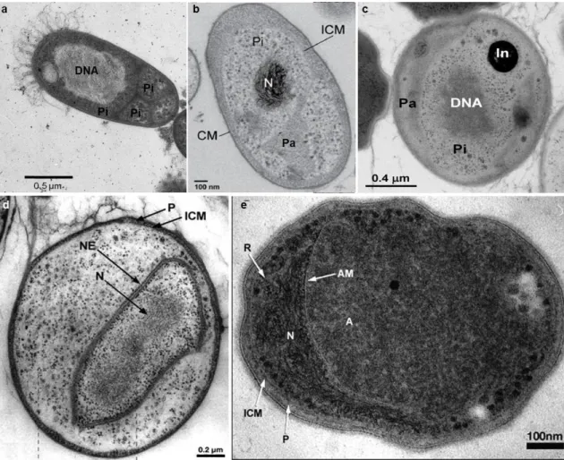
One of the most striking features of Planctomycetes is the presence of an internal membrane system comparable to the membrane configuration in the eukaryotes that contributes to their compartmentalized cell plan (Fuerst, 2005) and is also present in members of the phylum Verrucomicrobia (Lee et al., 2009). This feature contributed to the alternative theory that planctomycetes are key organisms in the eukaryogenesis (Forterre and Gribaldo, 2010; Fuerst and Sagulenko, 2011, 2012). Although some bacteria display internal membrane bound structures where specific metabolic processes take place (Kerfeld et al., 2010), bacteria are usually seen as non-compartmentalised organisms, without organization in organelles and with the DNA dispersed in the cytoplasm. This organization separates them from the eukaryotes whose cells contains a nucleus (consisting of an envelope and structured chromosomes), endoplasmic reticulum, peroxisomes, mitochondria and plastids in phototrophic organisms. According to Lindsay et al., (2001), planctomycetes possess an internal organization due to an intracytoplasmic membrane (ICM) that divides the cell in two compartments, the paryphoplasm, and the riboplasm or pirellulosome. The cell plan organization varies among different taxonomic groups (Fig. 1.5). The paryphoplasm, a ribosome-free space, can vary in size and invagination. It is surrounded on one side by the cytoplasmic membrane that is closely apposed to the cell wall. It surrounds the pirellulosome, an organelle that contains the highly condensed DNA and is rich in ribosome like particles. This is the most basic organization. Furthermore, the anammox bacteria and the genus Gemmata have a more complex organization that includes, respectively, the presence of an anammoxosome (where the anammox process takes place) or a nucleoid surrounded by a double membrane containing the condensed DNA, which resembles the nucleus of a eukaryotic cell.
Fig. 1. 5 Cell plan organization of planctomycetes. Thin sections obtained by transmission electron microscopy from a) Rhodopirellula sp. Cor3, courtesy of Olga Lage; b) Planctomyces limnophilus (Jogler et al., 2011); c)
Aquisphaera giovanonni, (Bondoso et al., 2011); d) Gemmata obscuriglobus, (Fuerst, 2005); e) “Candidatus
Brocadia anammoxidans” (Fuerst, 2006),. Pi/R, Pirellulosome or riboplasm, Pa, paryphoplasm, CM, cytoplasmic membrane, ICM, intra cytoplasmic membrane, N, nucleoid, NE, nuclear envelope.
The presence of a complex cellular organization in planctomycetes has been widely accepted for more than ten years, but recent studies in the “nucleated” Gemmata
obscuriglobus suggest that the cell plan in Planctomycetes might be similar to the one
of a Gram negative bacterium. Using electron tomography, Santarella-Mellwig et al. (2013) showed that Gemmata obscuriglobus cells are not compartmentalized and do not possess a nucleoid. Instead, they present a complex endomembrane system with invaginations of the cell membrane that are interconnected and do not form any close compartment. Thus, planctomycetes probably possess an outer membrane and an inner membrane (cytoplasmic membrane), the so-called intracytoplasmic membrane, which can be more or less invaginated. The space between those two membranes is the bacterial periplasm and not the paryphoplasm. The presence of several marker genes associated with the outer membrane in several planctomycetes genomes corroborates this hypothesis (Speth et al. 2012).
Recently, it was demonstrated via green fluorescent protein (GFP) that endocytosis is present in planctomycetes, particularly in Gemmata obscuriglobus (Lonhienne et al., 2010). Endocytosis is a universal eukaryotic process not identified in Bacteria or
Archaea by which cells incorporate molecules such as proteins through a
membrane-trafficking system and recycle them back to the surface or sort them to lysosomes for degradation. This feature is consistent with the presence of membrane coat (MC) – like proteins in planctomycetes and other compartmentalized members of the PVC phylum. These proteins are homologous to the MC proteins of eukaryotes that are known to induce coated vesicle formation associated with endocytosis (Santarella-Mellwig et al., 2010). Both endocytosis and the presence of MC-like proteins have evolutionary implications and suggest that Planctomycetes and other PVC members are indeed involved in eukaryogenesis.
Planctomycetes do not possess the universal peptidoglycan layer in the cell wall, a
feature also shared by Chlamydiae, archaea and eukaryotes. Planctomycetes possess a proteinaceous cell wall composed by amino-acids that vary among species. The cell wall can be rich in glutamate (König et al., 1984) or in proline and cystine/cysteine (Liesack et al., 1986). These particular characteristics of the cell wall allow strains to be resistant to ampicillin and other -lactamic antibiotics that target peptidoglycan synthesis like penicillin-G, D-cycloserine, cephalotin and vancomycin (Schmidt, 1978; König et al., 1984), which have been successfully used in specific isolation experiments of planctomycetes (Schlesner, 1994; Winkelmann and Harder, 2009; Bondoso et al., 2011; Lage and Bondoso, 2011).
The majority of planctomycetes divide by budding, a way of reproduction rare in bacteria. These normally divide by binary fission with the intervention of GTPase FtsZ protein. Planctomycetes, as well as the sister phylum Chlamydiae, lack the tubulin like protein FtsZ (Pilhofer et al., 2008) which is also absent in eukaryotes and in the archaeal group Crenachaeota (Vaughan et al., 2004). The FtsZ protein is also absent in planctomycetes that do not divide by budding as, for example, Phycisphaera
mikurensis that reproduces by binary fission (Fukunaga et al., 2009) and anammox
bacteria, which display ‘constrictive’ binary fission (van Niftrik et al., 2009). The mechanism underneath the reproduction in planctomycetes and chlamydia has not been so far discovered.
Another unusual trait characteristic of all planctomycetes is the presence of crateriform structures on the cell surface, whose function is still unknown (Fig. 1.6 a and b). These structures appear as electron-dense circular depressions after negative staining when viewed in transmission electron microscopy (Fig. 1.6a). They can be
uniformly distributed on the entire cell surface or only located on the reproductive pole. Most probably associated with the crateriform pits is the presence of fimbriae that emerge to the outside of the cell.
Presence of non-prosthecate stalks or appendages can be present in planctomycetes although this is not a universal feature among all the members. In some strains, the stalks lead to the formation of spherical rosettes (Fig. 1.6 c and e). In the Pirellula-Blastopirellula-Rhodopirellula (PRB) group, the cells secrete a fibrillar material, the holdfast, that groups the cells in the rosette (Fig. 1.6 c and d) and, most possibly, also facilitates the attachment to surfaces in the natural habitat. The holdfast is of glycoproteic nature (Lage, 2013).
Fig. 1. 6 Non-prosthecate appendages present in planctomycetes. a) – scanning electron photograph of a
Pirellula spp. cell evidencing the crateriform pits (courtesy of Olga Lage); b) Ultrathin section of the cell wall of Aquisphaera giovannonii evidencing the crateriform pits (arrow) (Bondoso et al., 2011); c and d) rosette and
individual cell of Pirellula spp. cell evidencing the holdfast (H) (courtesy of Olga Lage). e) Rosette of
Planctomyces bekefii, consisting of many spherical cells joined together at the distal tips of their stalks (Ward
et al., 2006). f) Fibrillar stalk of Pl. bekefii (Ward et al., 2006)
Cellular shape and arrangement are very diverse in planctomycetes. Cells can be pear-shaped, ovoid or spherical. Cells are normally arranged in rosettes (Fig. 1.6 c and e), that is a characteristic feature of many planctomycetes and is common among members of the PRB group. Some species are unicellular but filaments also exist (Ward et al., 2006). Planctomycetes may or may not display motility. They can be motile by means of a flagellum or by gliding motility, as Isosphaera pallida (Giovannoni et al., 1987a).
1.1.3 Physiology and ecology of Planctomycetes
Planctomycetes present some metabolic diversity. With the exception of the order
Candidatus Brocadiales that contains bacteria responsible for the ANaerobic
AMMonium OXidation (anammox), all planctomycetes described so far are chemoheterotrophs, with carbohydrates serving as prime sources of carbon. The anammox are chemoautotrophic planctomycetes that form a deep branching group within the Planctomycetes and were only identified in 1999 (Strous et al., 1999). They play an important role in the nitrogen cycle, converting ammonium and nitrite to dinitrogen gas with nitric oxide and hydrazine as intermediates (Strous et al., 2002). The majority of planctomycetes existing in pure culture are mesophilic and neutrophilic but some extremophile planctomycetes were also isolated. Isosphaera pallida is the only thermophilic planctomycete, isolated from a hot spring (Giovannoni et al., 1987b). Several acidophilic strains isolated from acidic peatlands were described in the last six years (Kulichevskaya et al., 2007; Kulichevskaya et al., 2008; Kulichevskaya et al., 2009; Kulichevskaya et al., 2012b; Kulichevskaya et al., 2012a; Kulichevskaya et al., 2012c). A halotolerant planctomycete, Pl. brasiliensis, was isolated from a hypersaline water in a salt pit in Brazil (Schlesner, 1989). Most of the members of this group are aerobes or facultative anaerobes, while the anammox are anaerobic.
However, the ecophysiological diversity of cultured planctomycetes is not at all representative of the one revealed by molecular studies. This group has a widespread distribution as they can be found in all kinds of habitats including marine (Vergin et al., 1998; Kirkpatrick et al., 2006; Martín-Cuadrado et al., 2007; Shu and Jiao, 2008; Pizzetti et al., 2011b; Shu et al., 2011), brackish (Schlesner, 1994; Kan et al., 2006; Zeng et al., 2013a) and freshwaters (Pizzetti et al., 2011a; Pollet et al., 2011; Steven et al., 2011; Wu et al., 2012) and terrestrial ecosystems (Wang et al., 2002; Buckley et al., 2006; Tsai et al., 2009; Zhou et al., 2009; Faoro et al., 2010; Michel and Williams, 2011; Chen et al., 2012; Miyashita et al., 2013) which suggests that they can adapt and colonize a diverse range of ecological niches. Planctomycetes can be found in upwelling systems (Woebken et al., 2007; Allen et al., 2012), which are rich in nutrients and have a high abundance of phytoplankton. Other studies reported higher densities of planctomycetes after diatom (Morris et al., 2006; Tadonleke, 2007; Pizzetti et al., 2011b) or cyanobacterial blooms (Eiler and Bertilsson, 2004) suggesting a possible association of planctomycetes with phytoplankton. Planctomycetes are reported to prefer an attached lifestyle rather than occurring as free-living organisms. In the marine environment, DeLong and co-workers (DeLong et al., 1993) found that planctomycetes
were more abundant in clone libraries from marine aggregate (marine snow) attached bacteria than the ones from bacterioplankton. Allgaier and Grossart (2006) reported that planctomycetes were absent on the free-living bacteria clone libraries but were present in the clone libraries of particle-associated bacteria. Similar results were obtained in deep sea enviroment (Eloe et al., 2011) and in the Black Sea (Fuchsman et al., 2011). Thus, it is not surprising that they are usually found in sediments (Kim et al., 2004; Mu et al., 2005; Inagaki et al., 2006; Liang et al., 2007; Dang et al., 2009; Li et al., 2009a; Polymenakou et al., 2009; Ghosh et al., 2010; Divya et al., 2011; Du et al., 2011; Durbin and Teske, 2011; Liao et al., 2011; Diaz et al., 2013; Li and Wang, 2013; Qiu et al., 2013) and are part of several biofilm communities (Baumgartner et al., 2009b; Boomer et al., 2009; Pašić et al., β010; Kriwy and Uthicke, 2011; Bartrons et al., 2012; Borsodi et al., 2012; Liu et al., 2012; Tang et al., 2012; Huang et al., 2013; Kostanjšek et al., β01γ).
They can be found in several extreme environments such as desert soils (Abed et al., 2010; Andrew et al., 2012), hypersaline environments (Burns et al., 2004; Lefebvre et al., 2006; Baumgartner et al., 2009a; Schneider et al., 2013), hot springs (Kanokratana et al., 2004; Portillo et al., 2009; Tekere et al., 2011; Bohorquez et al., 2012), acidophilic habitats (Hao et al., 2007; Xie et al., 2011; Ivanova and Dedysh, 2012; Lucheta et al., 2013), glacial waters (Liu et al., 2006; Zeng et al., 2013b) and Antarctic soils and waters (Christner et al., 2003; Newsham et al., 2010; Piquet et al., 2010; Huang et al., 2013). They were found to be one of the dominant groups on hydrocarbon polluted environments (Abed et al., 2011) accounting for more than 20 % of the total bacterial community. Several other studies reports of the presence of planctomycetes on other polluted habitats (Miskin et al., 1999; Reed et al., 2002; Chouari et al., 2003; Caracciolo et al., 2005; Abed et al., 2007; Akob et al., 2007; Hao et al., 2009; Halter et al., 2011; Yu et al., 2011; Han et al., 2012), suggesting a possible role on the degradation of hydrocarbons and other pollutants.
Several molecular studies showed that planctomycetes are associated with eukaryotic hosts, like ants (Eilmus and Heil, 2009), invertebrates (Fuerst et al., 1997), sponges (Webster et al., 2001; Pimentel-Elardo et al., 2003; Mohamed et al., 2008; Zhu et al., 2008; Ouyang et al., 2010; Sun et al., 2010; Webster et al., 2011; Costa et al., 2013), ascidians (Oliveira et al., 2013) and corals (Yakimov et al., 2006; Webster and Bourne, 2007; Duque-Alarcón et al., 2012). They can be also in association with macrophytes (Hempel et al., 2008; He et al., 2012), lichens (Bjelland et al., 2011; Grube et al., 2012), sphagnum peat bogs (Kulichevskaia et al., 2006) and the rizosphere of several plants (Jackson et al., 2006; Jenkins et al., 2006; Jensen et al.,
2007; Zhao et al., 2010; Zhang et al., 2011; Zhang et al., 2013). They are also frequent in the microbiota of several organisms like the human gut (Cayrou et al., 2013), intestinal tract of the Black tiger shrimp (Fuerst et al., 1997; Chaiyapechara et al., 2012), fecal samples of wild gorilla (Frey et al., 2006), stomach of oysters (King et al., 2012), intestinal track of termites (Shinzato et al., 2005; Mackenzie et al., 2007; Köhler et al., 2008; Makonde et al., 2013), intestinal tract of the polychaete Neanthes
glandicincta (Li et al., 2009b), foregut of the dromedary camel (Samsudin et al., 2011),
gastrointestinal tract of carp (van Kessel et al., 2011). Recently, molecular studies revealed that planctomycetes are widespread in the biofilm community of several species of macroalgae and present a high diversity. They are the dominant group in the biofilm community of the kelp Laminaria hyperborea with values that can reach 51 % of the total bacterial community (Bengtsson and Ovreas, 2010). Planctomycetes are also frequent colonizers of Ulvacean algae including Ulva compressa (Hengst et al., 2010), Ulva intestinalis (Hengst et al., 2010; Lachnit et al., 2011), Ulva australis (Longford et al., 2007; Burke et al., 2011b) and Ulva profilera (Liu et al., 2010). Several isolates have also been obtained from Ulva sp. and Ulva intestinalis (Lage and Bondoso, 2011). This group was also reported to be present in the green macroalgae
Chara aspera (Hempel et al., 2008) and Caulerpa taxifolia (Meusnier et al., 2001).
Epiphytic planctomycetes were also found in the red algae Porphyra umbilicalis (Miranda et al., 2013), Laurencia dendroidea (de Oliveira et al., 2012), Delisea pulchra (Longford et al., 2007) and Gracilaria vermiculophylla (Lachnit et al., 2011). Isolates were retrived from C. crispus, Mastocarpus stellatus, Gracilaria bursa-pastoris,
Gelidium pulchellum, Grateloupia turuturu and Porphyra dioica (Lage and Bondoso,
2011). A novel order of planctomycetes containing one species isolated from Porphyra sp. has been described (Fukunaga et al., 2009) 16S rRNA clone libraries from the brown algae Fucus vesiculosus releaved a great diversity of planctomycetes (Lachnit et al., 2011). Isolated planctomycetes were obtained from other brown algae like Fucus
spiralis, Sargassum muticum, Laminaria sp. (Lage and Bondoso, 2011) and Laminaria hyperborea (Bengtsson and Ovreas, 2010).
1.2 Microbial systematics
Systematics is “the scientific study of the kinds and diversity of organisms and of any and all relationships among them” (Emerson, 1961) and the ultimate goal of this scientific field is the definition of a species. Taxonomy is generally taken as a synonym of systematics and it consists in the classification, naming (nomenclature) and identification of organisms (Cowan, 1968), which are interdependent areas. Classification is the organization of organisms into groups or taxa; nomenclature is the assignment of names to taxa and identification consists in determining the identity of an isolate and its consequent allocation to a taxon. The first classification system was introduced by Linnaeus, in 1735 and divided the natural world into the animal kingdom, the plant kingdom, and the mineral kingdom (Linnaeus, 1735). Bacterial classification started in the end of the 18th century with the work of Otto Müller which created two
genera based on morphological differences, Monas and Vibrio. However, it was only in 1866 that bacteria were separated from plants and included in the Kingdom Protista, phylum Moneres (Haeckel, 1867). In 1872, Ferdinand Cohn recognized that bacteria were highly diverse and arranged them in six form genera creating the first system for classifying bacteria according to their morphology (Cohn, 1872). For several years, the classification systems were based solely on morphological characteristics, the so-called artificial classification. In the 70s, Carl Woese introduced the ribosomal RNA molecule analysis which revolutionized the microbial systematics field and in 1990 he proposed a three-domain classification system based on phylogeny and evolution, in which organisms were divided in the domains Archaea, Bacteria, and Eucarya (Woese, 1990). Prokaryotes are then classified into the domains Archaea or Bacteria and assigned to the lower hierarchical ranks ‘phylum’, ‘class’, ‘order’, ‘family’, ‘genus’ and ‘species’. The most widely accepted classification system for prokaryotes is the
Bergey's Manual of Systematic Bacteriology, which provides the basis for
nomenclature, classification, and descriptions of bacteria. Nomenclature has been called the ‘handmaid of taxonomy’ (Sneath, 1989) and in prokaryote systematic it is regulated by a system called International Code of Nomenclature of Bacteria, which is also known as the Bacteriological Revised Code published in 1975 (Lapage et al., 1976). This system started in 1980 with the publication of the Approved Lists of Bacterial Names (Skerman et al., 1980) which contained all bacterial names to date and it marked a new starting point in bacterial nomenclature. The list contained approximately 2000 bacterial names and, since then, more than 13000 species were validly published (http://www.bacterio.net/number.html#AL). Bacterial species are
named according to the binomial nomenclature established by Carl Linnaeus (Linnaeus, 1735) and names are Latinised. Species names consist of two parts, the first name is the genus and the second is the specific epithet. For the correct identification of an organism this must be compared to the most closely related strains already classified using phenotypic and genotypic properties in order to place it into particular taxa. When it is not possible to achieve an accurate identification, the unknown organism should be characterized in order to describe the new taxon based on the existing nomenclature. The characterization and nomenclature of a novel bacterium should follow several criteria in order to be validly published (discussed below), which are regulated by the ad hoc committee of the International Committee for
the Systematics of Prokaryotes, (Tindall et al., 2010).
1.2.1 Description and characterization of novel species
When a strain or a group of strains are shown not to belong to any other taxa already described in the literature, it must be extensively characterized in order to the new name to be validly published. The first problem that arises when describing a new organism is the definition of what is a species. This concept has been under debate for several years but, so far, there is no consensus on this definition. Several concepts have been suggested for microbial species but none is widely accepted. The most pragmatic and widely accepted definition states that a species can be defined as ‘a category that circumscribes a (preferably) genomically coherent group of individual isolate strains sharing a high degree of similarity in (many) independent features, comparatively tested under highly standardized conditions’ (Rossello-Mora and Amann, 2001). For taxonomists, however, it is important to define how to delineate a species. In that way, the ad hoc committee of the International Committee for the Systematics of
Prokaryote proposed that a species is a group of strains sharing 70 % or greater
DNA-DNA reassociation values and 5 °C or less ∆Tm (the difference in melting temperature between the heterologous and homologous DNA hybrids) and more than 97 % identity in 16S rRNA gene (Stackebrandt, 2002). Phenotypic and chemotaxonomic features should agree with this definition. For several years, prokaryotes were described and identified based only on their morphological characteristics, such as cellular shape and colony colour and morphology. Latter, due to the work of Robert Koch that allowed a larger number of bacteria to be isolated in pure culture, the physiological features were also included in the characterization of microorganisms and several tests were developed to rapidly identify them (Tindall et al., 2007). In the late 1950s, numerical taxonomy was introduced as an objective identification and classification system of
prokaryotes that was possible through the use of computers (Sneath and Sokal, 1973). The system established phenotypic relationships between the organisms based on similarity matrixes constructed based on a set of characters. At the same time, chemotaxonomy and analysis of lipids, cell wall amino acids and whole protein content were also introduced in bacterial systematics. In the 1960s, genome-based methods were developed to determine intraspecies relationships like DNA base composition (mol % G+C) and DNA-DNA hybridization (DDH). This technique allows determining the whole genome similarity of two strains based on the re-association of the single strands of DNA from each strain and has been used in the last 50 years as a criterion for species delineation based on a 70 % DNA-DNA binding value (Wayne et al., 1987). In the late 1970s, the work of Carl Woese and colleagues on the small subunit rRNA sequences (Woese and Fox, 1977) has revolutionized the microbial taxonomy. It was shown that the 16S rRNA gene could be used to infer phylogenies between microorganisms and could be used as a molecular chronometer due to its ubiquity and conservation among organisms (Woese, 1987). With the advances in DNA sequencing, that has made this technique a rapid and cost-effective method, the analysis of the 16S rRNA gene became a gold standard for species delineation in substitution of DDH, which is a much more laborious and expensive technique. It was shown that when two isolates have less than 97 % similarity in the 16S rRNA gene usually they share less than 70 % DDH and should be different species (Stackebrandt and Goebel, 1994). Nowadays, sequencing of the almost complete 16S rRNA gene is required for the classification and identification of a new species (Stackebrandt, 2002). Nevertheless, the 16S rRNA gene has been shown to lack enough discriminatory power to delineate bacterial species (Fox, 1992). DDH remains the best method for the correct species delineation but it is only necessary when the 16S rRNA gene sequence between two isolates is higher than 97 % (Tindall et al., 2010). A recent study where Stackebrandt and Ebers (2006) tried to correlate published DDH values with 16S rRNA gene showed that when two organisms share less than 98.5 % similarity in this gene, it is unlikely that they share more than 60 to 70 % DNA similarity. However this value has not been applied so far. Nevertheless, and because DDH is a very laborious and time-consuming technique, the current recommendations for the taxonomic characterization of prokaryotes allows DDH substitution by the sequencing and analysis of other genes with high resolution power (Tindall et al., 2010). Currently, a novel species of bacteria is described through a polyphasic approach that combines morphological, biochemical, chemotaxonomic and genetic characteristics (Fig. 1.7) in order to retrieve as much information as possible (Vandamme et al., 1996).