STUDY OF POPULATION DYNAMICS OF BACTERIA ASSOCIATED WITH PINE WOOD NEMATODE AFTER INOCULATION WITH DIFFERENT STRAINS OF BURSAPHELENCHUS
XYLOPHILUS IN MARITIME PINE (PINUS PINASTER)
Thesis presented to Escola Superior de Biotecnologia of the Universidade Católica Portuguesa to fulfill the requirements of Master of Science degree in Microbiology
by
Mariana Roriz Lemos Costa
under the supervision of
Dr. Marta Wilton Pereira Leite de Vasconcelos
R
ESUMO
Durante algum tempo pensou-se que o Bursaphelenchus xylophilus era o único agente etiológico da doença do nemátode da madeira do pinheiro. Recentemente, descobriu-se que existem bactérias associadas ao nemátode que contribuem para a patogénese desta doença, sobretudo através da libertação de toxinas que promovem a morte dos pinheiros. De entre as espécies mais comummente encontradas estão bactérias pertencentes aos géneros Bacillus, Pantoea, Pseudomonas e Xanthomonas.
Este trabalho teve como principal objectivo o estudo do efeito da inoculação de pinheiro bravo (Pinus pinaster) com quatro isolados diferentes de nemátodes, na população bacteriana dos nemátodes e das árvores, em diferentes fases da progressão da doença. A monitorização da progressão dos sintomas da doença foi igualmente registada. Pretendeu-se também identificar as bactérias isoladas do xilema das árvores e da superfície dos nemátodes através de métodos de identificação clássicos, do sistema de identificação API20E e da sequenciação de ADN bacteriano.
Os resultados obtidos demonstraram que, relativamente à progressão dos sintomas da doença, a diferença mais marcante foi verificada para os pinheiros inoculados com o isolado não virulento C14-5, onde se visualizou um agravamento de sintomas mais lento e menos severo do que nos pinheiros inoculados com os isolados virulentos. Verificou-se que numa fase inicial da doença, no geral, a população bacteriana dos ramos inoculados foi menor do que a que foi verificada passados 7 e passados 14 dias de inoculação. Num dos métodos de quantificação foi recuperado um maior número de bactérias nos pinheiros inoculados do que nos controlos aos 7º e ao 14º dias de inoculação. Os isolados que levaram à obtenção de maior quantidade bacteriana foram o HF e o 20, sendo que, comparativamente, o isolado não virulento levou à obtenção de menor quantidade bacteriana. Relativamente à identificação bacteriana, concluiu-se que o sistema API20E não foi suficiente na identificação das espécies bacterianas isoladas, mostrando-se pouco discriminatório, tendo sido identificada a espécie Enterobacter cloacae em 79% dos isolados e não sendo possível a identificação de sete colónias bacterianas. Assim, a adopção de métodos moleculares de identificação, através da sequenciação do ADN bacteriano, permitiu uma identificação mais fiável, tendo sido identificadas onze espécies bacterianas diferentes dentro dos géneros Klebsiella, Bacillus, Enterobacter, Paenibacillus, Terribacillus, Citrobacter, Pantoea e Escherichia. No geral, a diversidade bacteriana aumentou ao longo da progressão da doença. A espécie Bacillus spp. predominou na fase mais precoce da doença e a espécie Klebsiella oxytoca nas fases mais tardias. As espécies bacterianas isoladas da superfície dos nemátodes não diferiram muito das isoladas do xilema dos pinheiros.
Neste trabalho foram identificadas espécies bacterianas nunca antes reportadas neste tipo de estudo, podendo estas estar associadas a Portugal. A espécie de pinheiro utilizada neste estudo foi diferente das que são usualmente utilizadas no Japão e na China. Foi a primeira vez que foram isoladas e identificadas bactérias de um isolado não virulento do nemátode da madeira do pinheiro.
A
BSTRACT
For a long time it was thought that Bursaphelenchus xylophilus was the only agent of the pine wilt disease. Recently, it was discovered that there are bacteria associated with the nematodes that contribute to the pathogenesis of this disease, mainly through the release of toxins that promote the death of the pines. Among the species most commonly found, are bacteria belonging to the Bacillus, Pantoea, Pseudomonas and Xanthomonas genera.
The main objective of this work was to study the effect of inoculation of maritime pine (Pinus pinaster) with four different nematode isolates, in the bacterial population of nematodes and trees, at different stages of disease progression. The monitoring of progression of disease symptoms was also recorded. Also, the identification of bacteria isolated from the xylem of trees and the surface of nematodes was performed by classical identification methods, by identification system API20E and by sequencing of bacterial DNA.
The results showed that for the symptoms progression, the most striking difference was observed for the pines inoculated with the avirulent isolate, C14-5, which led to a slower and less severe aggravation of symptoms than in pines inoculated with the virulent isolates. It was found that at an earlier stage of the disease, in general, bacterial population of inoculated twigs was lower than what was observed 7 and 14 days after inoculation. In one of the quantification methods more bacteria were recovered from the inoculated pines than from the control pines on the 7th and 14th days after inoculation. A bigger bacterial quantity was isolated from pines inoculated with the nematode isolates HF and 20, and, comparatively, few bacteria were isolated from pines inoculated with the avirulent isolate. The identification system API20E proved to be insufficient and poorly discriminatory in the identification of bacterial species; Enterobacter cloacae species was identified in 79% of the isolated bacterial colonies and seven of these colonies couldn’t be identified by this method. Thus, the adoption of identification molecular methods, through bacterial DNA sequencing, allowed a more reliable identification: eleven different bacterial species within the Bacillus, Citrobacter, Enterobacter, Escherichia, Klebsiella, Paenibacillus, Pantoea and Terribacillus genera were identified. General bacterial diversity increased with the progression of the disease. Bacillus spp. species were predominant at the earlier stage of disease progression and Klebsiella oxytoca species at the later stages. Bacterial species isolated from the surface of nematodes were similar to those isolated from the xylem of pines.
In the present work new bacterial species were identified which have never been reported before in this type of study and may be associated with Portugal. P. pinaster, the pine species used in this study, was different from those commonly grown in Japan and China. Furthermore, it was the first time that bacteria were isolated and identified from an avirulent pine wood nematode isolate.
A
CKNOWLEDGMENTS
I would like to express my thanks to the Escola Superior de Biotecnologia of the Universidade Católica Portuguesa for receiving me as a Master Student, providing me the necessary conditions to carry out my work.
My deepest gratitude to Dr. Marta Vasconcelos for the encouragement, guidance, support and constructive advice throughout the entire investigation. I would like to thank the opportunity of making part of Plantech research group and for believing in my capacities to perform this work.
To Dr. José António Couto for his willingness and advice.
To my research group Plantech for the support inside the laboratory and for the fellowship that made this investigation always pleasant.
To Dr. Célia Manaia research group for their willingness and provided help. To Guida for her help in the work.
To all other friends for, directly or indirectly, supporting and motivating me. To the memory of my grandmother Angela, always interested in my work.
Finally, to my parents and brother, that always supported and helped me, and without whom the conclusion of my thesis would not be possible.
I
NDEX
Resumo... i Abstract ... ii Acknowledgments ... iii Index ... iv Index of figures ... viIndex of tables ... viii
List of abbreviations and symbols ... ix
Introduction ...1
Pine Wilt Disease, Bursaphelenchus xylophilus and Monochamus galloprovincialis ...1
Control/prevention of PWD ...4
Eradication ...4
Treatment of dead pine trees ...5
Traps for the insect vector ...6
Biocontrol strategies ...6
Growth of Resistant Pine Species ...6
Tree vaccination ...6
Plant innate immunity system ...7
The role of bacteria in the infection mechanism ...7
Methods for identifying bacteria ... 10
Main objectives ... 12
Specific objectives ... 12
Materials and Methods ... 13
Source and culture of nematodes ... 13
Source, culture and inoculation of pines ... 14
Development of symptoms by the diseased pines ... 15
Determination of the presence of nematodes at different experimental time points ... 15
Calculating the colony-forming units (CFU) on inoculated chips ... 16
Isolation of bacteria from the surface of nematodes ... 18
Identification of bacteria by classical methods ... 18
Identification of bacteria by molecular methods ... 19
Statistical analysis ... 19
Results and Discussion ... 20
Stage symptoms and oleoresin flow ... 21
Determination of the presence/absence of nematodes at different stages of disease progression ... 23
Quantification of bacterial colonies on inoculated chips ... 23
Colony-forming units (CFU) from inoculated chips calculated by dish count ... 28
Identification of bacteria by classical methods ... 30
Identification of bacteria by molecular methods ... 37
Classical vs. molecular identification methods ... 42
General conclusions ... 44
Future work ... 46
I
NDEX OF FIGURES
Figure 1: Portuguese PWN vector beetle, Monochamus galloprovincialis ...1
Figure 2: P. thunbergii old needles discoloration, 3 weeks after inoculation ...2
Figure 3: P. pinaster distribution in Portugal ...4
Figure 4: Fumigation technique for the treatment of wood infected with PWN ...5
Figure 5: HF virulent pine wood nematode isolate ... 13
Figure 6(A-B): A - Barley seeds with Botrytis cinerea; B - Nematodes growing on the walls of the tube and feeding on B. cinerea on barley seeds ... 13
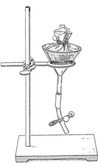
Figure 7: Baermann funnel technique ... 14
Figure 8: Nematode counting dish ... 14
Figure 9: Inoculation of P. pinaster trees ... 15
Figure 10: Schematic illustration of the method used for the calculation of inoculated chips with bacterial colonies performed at each experimental time point, with each treatment ... 16
Figure 11: Schematic illustration of the method used for the calculation of CFU on inoculated chips performed at each experimental point, with each treatment ... 17
Figure 12(A-E): Bacterial colonies in the chips inoculated with virulent nematode isolate 8A (A and B), with virulent nematode isolate 20 (C and D), and bacterial colonies along the trails of avirulent nematode isolate C14-5 (E), 3hai ... 23
Figure 13 (A-F): Bacterial colonies along the trails of virulent nematode isolate 20 (A and D), HF (B), 8A(C) and bacterial colonies in the chips inoculated with virulent nematode isolate 20 (E) and with avirulent isolate C14-5(F), 7dai ... 24
Figure 14 (A-E): Bacterial colonies along the trails of virulent nematode isolate 8A (A-C) and HF (D and E), 14dai ... 24
Figure 15: Number of chips with bacterial colonies, non inoculated (control) or inoculated with 4 nematode isolates, at the different experimental time points ... 27
Figure 17 (A-F): Example of bacterial colonies isolated from inoculated twigs and grown on NA
medium showing different morphological characteristics ... 35
Figure 18 (A-B): Example of a positive (A) and a negative (B) result for starch hydrolysis test
(bacterial colony 28 on the left and 29 on the right) ... 35
Figure 19: Agarose gel stained with SYBR® Safe DNA gel stain showing the amplification of the 1500
bp fragment corresponding to the 16S rRNA gene (M indicates the marker ladder; columns 1 to 38 represent the isolated colonies) ... 37
Figure 20: Bacterial genus isolated from trees inoculated with the four nematode isolates in the three
I
NDEX OF TABLES
Table 1: External symptoms of trees infected with PWN ...3
Table 2: General appearance and oleoresin flow study of pines after inoculation with the four
nematode isolates at three experimental time points: 3 hours after inoculation (3hai), 7 days after inoculation (7dai) and 14 days after inoculation (14dai)
...
21Table 3: Number of inoculated chips with bacterial colonies, 3hai, with different treatments (values
represent an average of eight samples ± standard deviation) ... 25
Table 4: Number of inoculated chips with bacterial colonies, 7dai, with different treatments (values
represent an average of eight samples ± standard deviation) ... 25
Table 5: Number of inoculated chips with bacterial colonies, 14dai, with different treatments (values
represent an average of eight samples ± standard deviation) ... 26
Table 6: Colony-forming units (CFU) average, 3hai, in different treatments (values represent an
average of five specimens ± standard deviation) ... 28
Table 7: Colony-forming units (CFU) average, 7dai, in different treatments (values represent an
average of five specimens ± standard deviation) ... 29
Table 8: Colony-forming units (CFU) average, 14dai, in different treatments (values represent an
average of five specimens ± standard deviation) ... 29
Table 9: Example of results of identification tests performed to bacteria isolated directly from the four
nematode isolates, from chips inoculated with different nematode isolates, 3hai, 7dai and 14dai .. 31-33
Table 10: Summary of the results obtained using the API20E identification system ... 36
Table 11: Bacterial species isolated from trees inoculated with the four nematode isolates in the three
experimental time points and bacteria isolated from non-inoculated nematodes identified after blastN of the amplified 16S rRNA gene fragment ... 38
Figure 20: Bacterial genus isolated from trees inoculated with the four nematode isolates in the three
L
IST OF ABBREVIATIONS AND SYMBOLS
ANOVA Analysis of variance
ATB Automatic Testing Bacteriology
blastN Nucleotide Basic Local
Alignment Search Tool
bp Base pair(s)
CFU Colony-Forming Unit(s)
DNA Deoxyribonucleic Acid
dNTP Deoxynucleotide
Triphosphate(s)
EDTA Ethylenediaminetetraacetic
Acid
ETI Effector-Triggered Immunity
H2O2 Hydrogen Peroxide
IN Isolated from the surface of nematodes
MAMP Microbe-Associated Molecular
Pattern(s)
NA Nutrient Agar
NaCl Sodium Chloride
NB-LRR Nucleotide-Binding
Leucine-Rich Repeat(s)
PAMP Pathogen-Associated
Molecular Pattern(s)
PCR Polymerase Chain Reaction
PROLUNP Programa Nacional de Luta
Contra o Nemátodo da Madeira do Pinheiro
PRR Pattern Recognition
Receptor(s)
PTI PAMP-Triggered Immunity
PWD Pine Wilt Disease
PWN Pine Wood Nematode(s)
rRNA Ribosomal Ribonucleic Acid
sd Standard deviation
TAE Tris-Acetate-EDTA
(W × D × H) (Width × Depth × Height)
Ø Control pine(s) (non-treatment)
# Number
3hai Three hours after inoculation
7dai Seven days after inoculation
14dai Fourteen days after inoculation
I
NTRODUCTION
Pine Wilt Disease, Bursaphelenchus xylophilus and Monochamus galloprovincialis
The pine wilt disease (PWD), as its name implies, is a disease found in pine species (Pinus spp.) whose main and best known etiologic agent is Bursaphelenchus xylophilus (Steiner & Buhrer) Nickle, the pine wood nematode (PWN) (Please see Figure 5 on page 13 of the Materials and Methods section).
The endoparasitic nematodes, unlike other nematodes, are able to overcome plant defenses, survive and feed on the plant (Jones et al., 2008). Particularly, the migratory endoparasitic nematodes, which include B. xylophilus, move within the plant, causing serious damage as they move and feed (Duncan and Moens, 2006; Jones et al., 2008). These nematodes feed mainly on fungi, appearing also in dead or dying trees (Jones et al., 2008). B. xylophilus has the particularity to parasitize aboveground parts of trees, not entering the soil and migrating through the tissues of the plant (Kikuchi, 2008).
One of the stages of Bursaphelenchus’s life cycle occurs at the same time as the formation of the late instar larvae or pupae of Cerambycidae beetles, that are the vectors for this nematode, being transported in the body of the insect (Aikawa, 2008; Jones et al., 2008). These longhorn beetles belong to the genus Monochamus spp. and exist in the same place of the tree occupied by the nematodes (Jones et al., 2008). In Portugal, the vector of PWN is Monochamus galloprovincialis Olivier 1795 (Sousa et al., 2001, 2002) (Figure 1). The PWN is transmitted to a diseased tree by the beetle during its oviposition, starting a new life cycle, and the infection period occurs between May and September (Jones et al., 2008). Once infected, trees can die in less than a year, if environmental conditions are favorable (Yoshimura et al., 1999; Jones et al., 2008). However, the main and foremost responsible agent for the rapid spread of the disease to other countries is the human (Jones et al., 2008), and there is no treatment for a susceptible tree infected with PWN (Zhao et al., 2007).
After infection, PWN feeds on parenchymal cells (Jones et al., 2008) and moves through the resin canals of the xylem and cortex, where he nourishes and reproduces itself, and also through the cambium cells (Ichihara et al., 2000). This process blocks the tree vascular system due to the appearance of secondary resin as a result of damage to the radial parenchyma cells by the nematodes; moreover, a cavitation phenomenon occurs, which affects water transportation (Jones et al., 2008) to the shoots and leads to rapid needle discoloration.
The severity of the symptoms depends on host species and the season of the year in which infection occurs, mainly due to temperature values (Jones et al., 2008). In the summer there is a rapid death of trees, and in the winter symptoms may not even manifest (Kiyohara and Tokushige, 1971). So, environmental factors (high air temperature and water stress) influence PWD incidence and disease development (Kiyohara, 1973; Suzuki and Kiyohara, 1978). The much talked about climate change adds concerns about this problem worldwide (Mota and Vieira, 2008a). It was also reported that PWN invasion depends on the age of the tree, mainly due to the absence of resin ducts in the stems of young trees (Mamiya 1975, 1980; Ichihara et al., 2000); however there are some authors that defend that PWD appears apart from the age of the host tree (Toda, 1997).
As mentioned above, one of the earliest symptoms of infection is the reduction or cessation of resin’s exudation on tree trunks, followed by the discoloration of pine needles (Figure 2) and death of the tree (Jones et al., 2008). PWN can survive in the host tree for long periods without causing any symptoms (Takeuchi and Futai, 2007).
Figure 2: Pinus thunbergii old needles discoloration, 3 weeks after inoculation (adapted from Kuroda,
Table 1 lists the appearance of external symptoms of trees infected with PWN, over time.
Table 1: External symptoms of trees infected with PWN (adapted from Zhao et al., 2008)
The PWN is native from North America; here, the autochthonous conifers are naturally resistant or tolerant to this agent, whereas the exotic tree species (non-native ones) are affected (Mamiya and Tamura, 1983; Wang et al., 2010). The disease firstly emerged in Canada, USA and Mexico, then spread to Japan, Korea and China, where it affected native species such as P. thunbergii and Pinus densiflora. When the disease arrived in Portugal, it was the first report of this disease in Europe (Yano, 1913; Cheng, 1983; Mamiya and Tamura, 1983; Tzean and Jan, 1985; Guiran and Bruguir, 1989; Yi et al., 1989; Dwinell, 1993; Mota et al., 1999). It is thought that Portuguese B. xylophilus’s populations derive from those from North America (Webster, 2004; Takemoto, 2008).
Thus, the PWD is known as the most severe threat to pine forests worldwide, representing a major economic problem for all affected countries (Mota and Vieira, 2008a; Wang et al., 2010).
In Portugal, after the discovery of the nematode in 1999, a national program, named "PROLUNP", for the control of PWN, was created, and a national survey to determine the affected area was undertaken, creating a zone with about 30 km of radius in south-east Lisbon, where all the symptomatic trees were razed, creating later a called “buffer zone” with about 5 km wide around the affected area, where all Pinus pinaster trees were killed, even if they weren’t symptomatic (Jones et al., 2008; Mota and Vieira, 2008b).
Unfortunately, Portugal assembles all the necessary conditions to the spread of this disease, in particular the fact that the most important pine species are susceptible to the pest (Braasch, 1997), nematodes are present and there is a compatible vector. The pine species Pinus sylvestris and Pinus halepensis are also favorable hosts for the PWN, but their distribution and abundance is limited in Portugal (Mota and Vieira, 2008b). The maritime pine, P. pinaster, is the most popular host as the insect vector feeds from it during its maturation (Mota and Vieira, 2008b). Stone pine, Pinus pinea, is one of the species considered "resistant" or less susceptible to the disease, not being consumed or colonized by the vector M. galloprovincialis; however, it is known that PWN is able to infect and kill this species, but more slowly than for P. pinaster (Mota and Vieira, 2008b). The reasons behind this phenomenon are still unclear.
P. pinaster trees play an important role in pine production, in the wood and resin industry, as well as coastal protection, being distributed throughout most of the country (Figure 3) and so the appearance of a problem like this is very serious for the Portuguese economy and environment (Mota and Vieira, 2008a).
Figure 3: P. pinaster distribution in Portugal (DFG, 2001). Control/prevention of PWD
Various approaches to control or prevent the disease have been studied in Japan and China: eradication, preventive sprays, host tree resistance to PWN, forest sanitation, trapping of the insect vector, biocontrol methods, breeding of natural parasites of the pine sawyer, cultivation of resistance pine species, among others.
Eradication
This method is effective because it kills vector beetles when they are in the larval galleries and pupal chambers and when the PWN transmission has not yet begun (Kamata, 2008).
One example of this approach is the physical controls. These include three techniques: cut and crush, cut and burn and cut and burry. The cut and crush technique consists of cutting and crushing the infected wood without any other treatment. The cut and burn technique is based on carbonizing the wood into charcoal; the complete mortality of vector beetles can be achieved. In the cut and burry technique the logs are buried in the ground or soaking in the water. The first one is efficient for coastal pine stands; the second one is not too useful because it takes too long to kill vector beetles (Kamata, 2008).
A second approach, the chemical control, is useful for diseased trees present in accessible and inaccessible zones. The cut and chemical application encompasses spray and fumigation techniques. After being cut into sections a chemical insecticide is applied to affected trees in the spray technique. These chemicals are composed of one or more chlorpyriphos-methyl, pyridaphenthion, prothiophos, or BPMC (2-sec-butylphenyl methylcarbamate). If this approach is applied at the end of October (fall treatment) about total mortality of pine sawyer can be achieved because that’s when larvae live beneath the host bark or are constructing pupal chambers. This treatment is not to efficient if applied from November to March (winter treatment) or after April (spring treatment) because it’s impossible for the chemical to reach the pupal chambers, unless an oil solution of chemicals is used. When the diseased trees are in inaccessible areas chemical insecticide can be sprayed from a helicopter; however this approach raises issues such as the adverse impact upon ecosystems. In the fumigation technique the concerns of the application seasons are overcome. The main components used are metam-ammonium carbam NCS or carbam sodium. Also, about 100% of pine sawyer mortality can be achieved and in addition to killing the vector beetle it also kills the nematodes. However, this technique is more expensive than the spray one. The procedure involves treating diseased stacked branches and logs with the chemicals which are then sealed with soil over a PVC (polyvinyl chloride) sheet (Figure 4) (Kamata, 2008).
Figure 4: Fumigation technique for the treatment of wood infected with PWN (picture taken in a pine
affected area in Tsukuba, Japan, kindly provided by Dr. Marta Vasconcelos).
Treatment of dead pine trees
The clearance of dead pine trees from forests is one of the main strategies to be adopted for PWD control. Even one infected pine tree left can spread the disease to other pines that are near. China adopted five treatments to wood to stop PWN reproduction and insect vector propagation: clear-cutting of pine trees to chipping and processing chipboard, heat treatment, heating and hydraulic pressing treatment, hot-water treatment and bag-fumigation and microwave treatment (Xu, 2008).
Traps for the insect vector
The choice of the host by the Japanese and Chinese vector beetle, Monochamus alternatus, is related to the chemical composition of pines. The terpene (+)-α-pinene is the main attractive compound to the beetle (Xu, 2008). Ethanol acts as a synergistic compound increasing the activity of attraction (Yang et al., 2003). An experiment conducted by Siegfried (1987) proved that traps with α-pinene, a β-phellandrene-limonene mixture, limonene and β-pinene are effective to catch weevils. There is still no available, effective trap of this type for M. galloprovincialis.
Biocontrol strategies
Pine sawyer can also be controlled by biological methods: bird and insect predators, entomophatogenic fungi and entomophilic nematodes (Kamata, 2008). There are several natural enemies for M. alternatus: insect parasites (Scleroderma guani and Dastarcus helophoroides), parasitic fungi (Beauveria bassiana, B. brongniartii, Metarhizium anisopliae, among others), parasitic bacteria (Serratia marcescens) and parasitic nematodes (Steinernema feltiae). C. massonianus is used to release B. bassiana in the larva canal of the beetle (Xu et al., 2000); this approach can achieve 95% of pine mortality. B. bassiana can also be transferred by S. guani however an undesirable effect arises because B. bassiana also infects S. guani (Xu et al., 2000). A good solution is to propagate S. guani alone on the the larvae of M. alternatus, not harming the environment (Xu, 2008). Again, no data is available regarding possible biocontrol strategies for M. galloprovincialis.
Growth of Resistant Pine Species
Cleared areas previously affected by PWD need to be cultivated again. In these areas resistant pine species, previously examined for their resistance, should be preferred for cultivation (Xu, 2008). It is thought that this resistance has a genetic nature (Nose and Shiraishi, 2008). Thus the study of gene expression (for example genes of the peroxidase and ethylene biosynthesis pathway that are important in the disease response (Miller et al., 2005; Shin et al., 2009)) of susceptible pine species is crucial and will help in the understanding of molecular mechanisms of tree resistance and susceptibility. It is required that this resistance is long lasting; however this is not always possible. The PWN isolate and virulence in each region should be taken into account to make this possible.
Tree vaccination
The injection of a nematicide or a chemical solution into a tree, which stops PWN reproduction or potentiates its death, is known as preventive vaccination. This control approach is very efficient because it’s long lasting (reinoculation once every 3-4 years) and does not depend on environmental conditions; however it cannot be applied in large scale due to costs and can lead to problems due to the inoculation (Kamata, 2008).
The content of the vaccine formulations can also be an avirulent nematode; it was found that pre-inoculation of trees with avirulent nematode isolates induces resistance on P. thunbergii to a
subsequent post-inoculation with virulent nematodes (Kosaka et al., 2001). Also in the experiment of Takeuchi et al. (2006) they concluded that pre-inoculated seedlings with C14-5 showed lower mortality than seedlings without pre-inoculation. In these cases the cambial zone is determinant for survival or death of pines (Fukuda, 1997). Resistance degree of pre-inoculated trees increases with avirulent nematode concentration and multiple pre-inoculations with C14-5, at different times, induces the resistance more effectively, than a single pre-inoculation, to post-inoculation with a virulent nematode. It has also been demonstrated that climate conditions influences the effect of inoculation with avirulent nematodes (Kosaka et al., 2001).
Plant innate immunity system
Plants possess a response to pathogen/microbe-associated molecular patterns (PAMPs or MAMPs), called PAMP-triggered immunity (PTI) (Boller and Felix, 2009; Boller and He, 2009). However it’s difficult to understand the role of this PTI because pathogen virulence effectors inhibit it (Boller and He, 2009). This innate immunity system present in plants detects and moves pathogens away (Chisholm et al., 2006; Jones and Dangl, 2006; Boller and Felix, 2009). The first step of this system is the recognition, in the plant’s cell surface, of MAMPs by pattern recognition receptors (PRRs); these are extremely sensitive and specific, able to detect all classes of microbes and with a nonself recognition characteristic (Boller and Felix, 2009; Boller and He, 2009). Although pathogen effectors try to suppress PTI, as stated earlier, plants possess other receptors (nucleotide-binding leucine-rich repeat (NB-LRR) proteins) which constitute an effector-triggered immunity (ETI) or R-gene-based or vertical resistance, as a second line of defense (Boller and Felix, 2009; Boller and He, 2009).
Thus, as plant pathogenic bacteria, fungi and nematodes excrete a lot of virulence effectors into plant cells, the recognition of the plant targets of these effectors will bring a new vision of plant immunity, pathogenesis and plant biology and will help us to better control plant diseases, such as PWD.
The role of bacteria in the infection mechanism
The pathogenic mechanism of PWD has not been well elucidated. For several years it was thought that the PWN was the only etiologic agent of the disease (Mamiya, 1975; Nickle et al., 1981; Mamiya and Tamura, 1983;; Nobuchi et al., 1984; Myers 1988; Fukuda et al., 1992; Yang, 2002). In fact, it was found that this agent produces phytotoxins and cellulases and that its invasion stimulates the production of ethylene and terpenoids by pine trees which could lead to their death (Wang et al., 2010). Inoculation studies with PWN leading to the death of pine also supported this viewpoint, noting that nematodes of different pine trees had different pathogenicity (Kiyohara and Bolla, 1990; Fukuda et al., 1992; Kojima et al., 1994; Hu et al., 1995). However, the rapid increase in the number of nematodes was posterior to the histological and physiological changes in an infected tree, raising the suspicion that there may be other microorganisms involved in the pathological process (Xie and Zhao,
2008). In addition, sterilization of the surface of the nematode leads to loss of pathogenicity (Cao, 1997; Kawazu and Kaneko, 1997).
More recent approaches report the existence of bacteria in symbiosis with nematodes that somehow have a crucial role in the pathogenesis of the disease; however, this hypothesis is still controversial (Oku et al. 1979; Kawazu et al., 1996b, 1998; Cao, 1997; Kawazu and Kaneko, 1997; Han et al., 2003; Zhao et al., 2003, 2005; Xie and Zhao, 2008). It is thought that the ability of the nematode to carry bacteria is nothing more than a natural phenomenon (Zhao et al., 2003, 2007, Guo et al., 2007). However, some authors think that these bacteria are not accidental contaminants but exist as symbionts of nematodes, and co-evoluted with them for a long period (Zhao et al., 2005, 2006). Others think they were obtained by the nematodes from the environment, possibly through the inoculation wounds, by mechanical injury, or via the scars left by the insects when they feed on trees (Zhao and Li, 2008). In fact bacteria are present in many environments, including soil and water, and are associated with some plant diseases. Species of Acidovorax, Afrobacterium, Burkholderia, Clavibacter, Erwinia, Pantoea, Pectobacterium, Phytoplasma, Pseudomonas, Ralstonia, Spiroplasma, Streptomyces, Xanthomonas and Xylella genera are involved in different kinds of symptoms: galls, overgrowths, wilts, leaf spots, specks and blights, soft rots, scrabs and cankers (Ellis et al., 2008). Some of them produce toxins, inject proteins to kill cells or synthesize enzymes that break down structural components of cells and walls (Ellis et al., 2008). An example is the bacterial wetwood or slime flux disease, affecting many shade and ornamental trees, including pines, caused by species of Bacillus megaterium, Enterobacter agglomerans (also called Pantoea agglomerans), Enterobacter cloacae, Klebsiella oxytoca and Pseudomonas fluorescens (Anonymous, 1999). This chronic disease usually does not manifest in trees with less than 10-years-old and is affected by environmental factors (Anonymous, 1999).
Several studies report the existence of a symbiosis between bacteria and entomopathogenic nematodes and promotion of development and reproduction of the nematode by the bacteria (Marainede et al., 1993; Forst and Nealson, 1996; Han and Ehlers, 2000). In the studies of Zhao et al. (2000) and Guo et al. (2002) it was observed under electronic microscopy, the presence of bacteria on the surface of PWN in an amount of 2.9×10² bacteria per nematode. No bacteria were found within the body of the nematode (Zhao et al., 2000). Kusunoki (1987) also found the presence of bacterial cells in resin ducts and parenchymal tissue of pine trees. However, several studies indicate that no bacteria were detected in healthy trees (Zhao et al., 2003; Zhang et al., 2004). Bacteria were also found in the insect vector, M. alternatus, and were similar to bacteria from PWN (Xie et al., 2005).
Among the main species of bacteria that are associated with PWN are the genus Pantoea, Pseudomonas and Xanthomonas (Higgins et al. 1999; Han et al., 2003). It was also found that bacteria in different geographic zones may differ (Han et al., 2003; Zhao et al., 2003; Wang et al., 2010). The main species found in China belong to Pseudomonas genus, in Japan to Bacillus genus and in Korea both are present (Zhao, 2008). Such variations cause differences in the susceptibility of plant species (for example Cedrus deodara); this plant dies after infection with PWN in Japan, while in
China it remains healthy (Jiao et al., 1996). It is thought that new bacteria from the local flora are acquired by PWN when a new local region is achieved (Zhao, 2008). Zhao et al. (2006, 2007) concluded that bacteria such as Pantoea sp. and Peptostreptococcus asaccharalyticus are not beneficial for the nematodes and even have an inhibitory role on their growth and development. The species Escherichia coli also exerts an inhibitory effect on the PWN and vice-versa (Zhao et al., 2005). Pseudomonas and Pantoea were found in trees infected with B. xylophilus but not in uninfected trees (Han et al., 2003). Xie and Zhao (2008) concluded that at later stages of the disease, when the number of nematodes increases rapidly, the bacteria population increases in volume and variety of species, indicating that the environment created due to wilt was good for the nematode and the bacteria.
It is also known that trees infected with bacteria alone or only with aseptic nematodes did not develop the disease, but the combination of nematodes and bacteria leads to the manifestation of the disease symptoms (Oku et al., 1980; Zhao et al., 2000; Han et al., 2003; Zhao et al., 2003). Thus, axenic PWN is not pathogenic compared to the wild PWN (Kawazu and Kaneko, 1997; Cao et al., 2001), and they cannot cause disease even if they survive in the xylem of the pine tree (Chi et al., 2006).
Several studies report the existence of toxins that play an important role in the process of pine wilt (Mamiya, 1980; Oku, 1988, 1990; Zhang et al., 1997), which cannot be produced by the nematode alone (Cao and Shen, 1996; Cao et al., 2001). Experiments in callus showed that the wilting observed after inoculation of the liquid where the bacteria grew, was due to the existence of toxins produced by bacteria (Han et al., 2003; Zhao et al., 2003, 2005). Kawazu et al. (1996a) and Kawazu (1998) confirmed the presence of three toxin producing species of Bacillus spp. that cause PWD and identified the toxic substance as phenylacetic acid. Oku et al. (1979, 1980) and Zhao et al. (2003) reported that the wilt toxin is associated with bacteria, namely to a bacteria belonging to the Pseudomonas genus associated with the nematode.
As in any symbiotic relationship both species benefit from each other; in this particular case alive or dead nematodes promote reproduction and pathogenicity of bacteria by the supply of essential metabolites and nutrients and in some way protect bacteria within the host tree, and bacteria also increase the reproductive rates of B. xylophilus in trees, serving as a power source and/or providing essential nutrients (Zhao et al., 2003, 2005, 2006, 2007; Wang, 2004; Guo et al., 2006). Living nematodes have a stronger stimulatory effect than the dead ones (Zhao, 2008). It was also found that phytotoxin-producing bacteria associated with the nematodes also increase egg production and accelerate the growth and development of B. xylophilus in cultured callus (Zao et al., 2007). This symbiotic relationship was classified as an ectosymbiosis (Zhao et al., 2007). The species Escherichia coli is reported as inhibitory of PWN reproduction (Zhao and Lin, 2005).
Thus, the PWD is a complex process that involves the PWN and the phytotoxin-producing bacteria associated with it (Zhao et al., 2003; Xie and Zhao, 2008; Kwon et al., 2010; Wang et al., 2010) whereas bacteria alone are not capable of causing disease (Zhao et al., 2003; Jones et al.,
2008). Zhao et al. (2003) reported that bacteria cannot invade a healthy tree alone; Xie (2003) and Lindberg et al. (2004) attribute this fact to the presence of certain metabolites in pine tree extracts that have antibacterial activity. Bacteria must be transported by nematodes to overcome and survive the defenses of the host tree, gaining protection from nematodes (Zhao et al., 2005). These and PWN then collaboratively invade and kill the host tree (Zhao et al., 2007).
Controversial opinions do not support this approach, since in trees inoculated with B. xylophilus the blocking of water in the xylem was found before the presence of fungi and bacteria were detected (Kuroda and Ito, 1992) and some think that because bacteria exist inside and outside of the tree, they are contaminants and are not pathogenic (Yang, 2002). All the different experimental data indicate that there is no consensus about the actual role of bacteria in disease progression, which supports the use of sterilization procedures in all experiments during the inoculation process to guarantee that the bacteria found are not mere contaminants (Zhao et al., 2003).
Methods for identifying bacteria
The identification and phenotypic characterization of microorganisms began in 1677 with Antony van Leeuwenhoek. Later, the need for isolation in pure culture lead to the advent of solid culture media by Robert Koch and analysis of physiological characteristics, metabolic and biochemical abnormalities (Ferreira and Sousa, 2000). However, the simplicity of this analysis did not make it suitable for a for proper microorganism identification (Ferreira and Sousa, 2000).
A disadvantage of traditional methods of microbial identification is the fact that they only analyze the phenotypic characteristics of organisms, such as cultural and morphological characteristics, biochemical and metabolic utilization of different substrates, etc (Ferreira and Sousa, 2000). These features often do not allow the differentiation of microbial species (Ferreira and Sousa, 2000).
An example is the Gram stain that allows separating the bacteria according to their staining properties, being the most widely used staining in bacteriology. It is a differential staining because it separates bacteria into two groups: gram-positive and gram-negative ones. The second dye used, the iodine solution, acts as a mordant promoting a stronger stain. Alcohol or acetone is used to decolorize the cells: gram-positive bacteria retain the first stain (crystal violet) and gram-negative bacteria lose the stain, becoming colorless. At the final step of the Gram staining procedure, safranin allows gram-negative bacteria to stain pink or red while the gram-positive bacteria remain with the first dye, appearing dark purple (Prescott, 2002).
The known oxidase test allows detecting the presence of the enzyme cytochrome c oxidase, which is able to reduce O2 and artificial electron acceptors. The catalase test serves to detect the presence of the enzyme catalase, which converts hydrogen peroxide to water and O2. Another test is the starch hydrolysis test that detects the presence of the enzyme α-amylase, which hydrolyzes starch, produced by certain bacteria (Prescott, 2002).
Some bacteria have structures of locomotion, the flagella, with 15-20 µm long and 14-20 nm thick, which can only be viewed directly under an electronic microscope or under an optical microscope with special stains (Ferreira and Sousa, 2000). The use of a mordant like tannic acid or potassium alum allows increasing the thickness of flagella being stained with pararosaniline (Leifson method) or basic fuchsin (Gray method) (Prescott, 2002).
The advent of molecular biology techniques allowed the fast, easy, inexpensive and accurate identification of microorganisms, allowing to overcome some limitations of classical methods. One example is the sequencing of nucleic acids that involves determining the sequence of genes with phylogenetic and taxonomic information, such as the 23S and 16S rRNA genes. This technique thus provides information about the identity and/or relationship of the new sequences obtained with those existing in a database (Ferreira and Sousa, 2000).
Main objectives
As it has already been described, bacteria play an important role in the pathogenicity of PWN. However, little is known regarding the bacterial population in Portuguese nematode isolates. Also, nothing is known regarding the bacteria associated with the Japanese avirulent isolate C14-5, and how it compares to virulent isolates. This work is therefore intended to study the effect of inoculation of healthy P. pinaster trees, with different nematode isolates, in the bacterial population of either the nematode or the tree itself, at different stages of disease progression (3 hours, 7 days and 14 days after inoculation). It is also proposed to know the bacterial species isolated from the xylem of the trees and from the surface of the nematodes.
Specific objectives
Thus, one year old P. pinaster trees will be inoculated with four different nematode isolates: three virulent isolates (HF and 20, isolated from Setúbal region and 8A isolated from Portuguese central region) and an avirulent one (C14-5 isolated from Japan). At the three experimental points several samples will be monitored for symptoms of disease progression such as resin exudation and discoloration of needles. The same samples will be subjected to nematode extraction by the Baermann funnel technique to confirm the efficiency of the inoculation process.
After incubation of twigs from inoculated samples collected at the three experimental points in Nutrient Agar (NA) medium, the quantity of inoculated twigs with bacterial colonies will be registered. Also, colony-forming units (CFU) will be studied after dilution of solutions of bacteria extracted from inoculated twigs and incorporation on NA medium. Bacteria will also be isolated from the four nematode isolates.
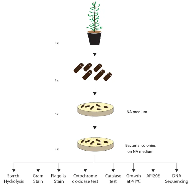
Bacterial colonies grown on NA medium from inoculated twigs and from the four nematodes will be successively purified and subjected to a series of identification tests such as Gram and flagella stain, cytochrome c oxidase and catalase tests, starch hydrolysis, growth at 41ºC and API20E identification system.
Finally total genomic bacterial DNA will be extracted from all isolated colonies and a 1500 bp fragment of 16S rRNA gene will be PCR amplified with bacterial universal primers. PCR products will be purified and sequenced at Macrogen Ltd (Seoul, Korea).
M
ATERIALS AND
M
ETHODS
Source and culture of nematodes
Three virulent isolates of B. xylophilus (HF (Figure 5) and 20, geographical isolates from Setúbal region and 8A from Portuguese central region), and an avirulent isolate (C14-5, from Japan), were used in the experiments.
Figure 5: HF virulent pine wood nematode isolate.
All B. xylophilus cultures were grown (Figure 6 B) on barley seeds with Botrytis cinerea Pers. mycelium (Figure 6 A) at 26ºC, in the dark, for 7 days. Juveniles and adult nematodes were extracted using the Baermann funnel technique (van Bezooijen, 2006) (Figure 7) during 24h at 25ºC.
Figure 6 (A-B): A - Barley seeds with Botrytis cinerea; B - Nematodes growing on the walls of the
tube and feeding on B. cinerea on barley seeds.
Figure 7: Baermann funnel technique (van Bezooijen, 2006).
The total number of nematodes was determined as follows: 20 µl of the nematode suspension obtained from the Baermann funnel were placed in a nematode counting dish (Figure 8) and living nematodes were counted and estimated for the initial solution. The solution was then adjusted to a final concentration of approximately 2000 nematodes/ml sterile deionized water and used in the inoculation experiments.
Figure 8: Nematode counting dish.
Source, culture and inoculation of pines
The experiments were carried out on one year old Portuguese Maritime pine specie P. pinaster at Escola Superior de Biotecnologia. Pines were provided by Sociedade Agrícola Pecuária Melo & Cancela Lda. and kept in a plant growth chamber (Fitoclima S600, Aralab Portugal) scheduled for 80% humidity, photoperiod of 8 hours light and 16 hours darkness and temperatures of 26ºC and 24ºC for periods of dark and light, respectively.
Nematodes were inoculated in pines according to Futai (1980) method (Figure 9): at about 3 cm from the top of the tree the pine needles were removed and three vertical cuts were made in the stem, with the help of a blade; after inoculation with the nematode suspension, the cutting area was sealed with absorbent paper and parafilm. 144 pines were inoculated with the different PWN isolates and 27 others without treatment (non-inoculated) were used as controls (Ø). Thus, a total of 171 pines were used in the experiments.
Figure 9: Inoculation of P. pinaster trees.
Three experimental points were considered: an early response of three hours, a mid response of seven days and a later response of fourteen days after inoculation.
Development of symptoms by the diseased pines
One of the earliest symptoms of pine wilt disease is the cessation of resin exudation. For this reason, in order to confirm disease progression, resin exudation was monitored. After every experimental point, a 5 mm hole in the trunk was made using a sterile blade for each of the inoculated and control pines to monitor the resin flow. Also, a visual scale of symptoms that consists of four levels of disease progression was used, ranging from healthy to dead plant (Please see Table 1 on page 3 of the Introduction section).
Determination of the presence of nematodes at different experimental time points
In all experimental time points, the whole pine stem was cut into small pieces and PWN were extracted with the Baermann funnel technique to check if the inoculation process was effective and if the nematodes had survived.
Calculating the quantity of inoculated chips with bacterial colonies
The quantity of inoculated chips with bacterial colonies was determined according to Xie and Zao (2008) (Figure 10): a 20 cm long, 1 year old inoculated stem was sterilized with 75% ethanol and both ends were cut; the central wood was then cut into 2 mm × 2 mm × 5 mm pieces (W × D × H). Eight pieces of the stem of a sample from inoculated and control pines were placed into a Petri dish containing nutrient agar (NA) medium (Frilabo, Portugal) and incubated at 26ºC for 3 days. Bacteria appeared in the site were the chip was placed in NA and in the marks left by the nematodes. These bacteria were also selected for identification (as described later).
Five stem samples were collected from the different experimental points and five replicate dishes, for each sample, were used to calculate the amount of bacterial colonies on the inoculated chips over all the chips from a treatment.
Figure 10: Schematic illustration of the method used for the calculation of inoculated chips with
bacterial colonies performed at each experimental time point, with each treatment.
Calculating the colony-forming units (CFU) on inoculated chips
Colony-forming units (CFU) were also calculated in the stem wood using the dish count method, based on dishes with colonies between 25 and 250, according to Xie and Zao (2008) (Figure 11): 1 gram of the stems described above were placed in a glass tube with 4.5 ml of a 0.85% NaCl solution. The tubes were then shaken vigorously for 5 minutes and left standing for 10 minutes. 0.5 ml of the previous solution was diluted in a series of concentrations (100, 10-1, 10-2, 10-3 e 10-4) and 0.5 ml of each concentration was placed on a sterile Petri dish. Ten milliliter of NA medium at 45°C was then added to the plate, which was incubated at 26°C for 72 hours. Three replicates from each treatment were used to calculate the CFU.
NA medium
Bacterial colonies on NA medium
Figure 11: Schematic illustration of the method used for the calculation of CFU on inoculated chips
performed at each experimental point, with each treatment.
1 gram
Bacterial colonies on NA medium
Isolation of bacteria from the surface of nematodes
Bacteria were also isolated from the surface of the four nematode isolates as described by Han et al. (2003): the solution of PWN obtained from the Baermann funnel was centrifuged at 17 g for 6 minutes and the supernatant was discarded; the remaining PWN were disinfected with 3% H2O2 for 5 minutes; finally, a single nematode was removed with a thin metal needle and placed on a plate with NA media which was incubated at 26ºC. Bacterial colonies which appeared in the track left by the nematodes were successively transferred to NA for colony purification.
Identification of bacteria by classical methods
Both isolated bacterial colonies from xylem and the surface of nematodes were selected for identification based on macroscopic differences. Morphologic characteristics (size, color, shape, transparency, prominence, edge and viscosity) of the purified isolated bacteria from the trees and nematodes were registered. Each isolate was tested for Gram stain: the smear was first stained with crystal violet for 30 seconds, water rinsed for 2 seconds, stained with Gram’s iodine for 1 minute, water rinsed, washed with 95% ethanol for 10-30 seconds, stained with safranin for 30-60 seconds, water rinsed and finally dried (Prescott, 2002).
Flagella stain was also performed as described by BD Flagella Stain Droppers manufacturer’s instructions (Difco, BBL).
Cytochrome c oxidase and catalase tests were carry out: for the oxidase test - fresh growth from the culture plate was scraped with an inoculation loop, rubbed on filter paper and examined for blue color (positive result) within 10 seconds (NHS, Oxidase Test; Oxidase Test Sticks – Frilabo, Portugal); for the catalase test - a drop of 3% hydrogen peroxide was placed on a glass slide and a colony from the culture plate was placed on the drop. The formation of bubbles indicated a positive result (Murray et al., 1998).
Starch hydrolysis was also studied according to manufacturer’s instructions: the surface of a 48 hour culture, grown in DifcoTM Starch agar was flooded with Gram’s Iodine; a positive result is indicated by the presence of a colorless zone surrounding the colonies.
The study of bacterial growth at 41ºC was also performed.
All identification tests described above were performed five times to confirm results.
The bacterial species were finally subjected to the identification systems API20E (bioMérieux Company, Craponne, France). After obtaining the numerical profile, isolated bacterial colonies were analyzed using the analytical catalog of the API20E (API20E Analytical Catalog, 1999).
Identification of bacteria by molecular methods
Total genomic bacterial DNA was successfully extracted for all 38 bacterial colonies (except colony 10) according to Wiedmann-Al-Ahmad et al. (1994): one bacterial colony was resuspended in 70 µl pure water, heated 5 min at 95ºC and sedimented at 16,000 x g for 5 min in a microcentrifuge (Thermo Scientific Heraeus Pico 17). The extracted DNA was quantified spectrophotometrically using a NanoPhotometerTM UV/Vis spectrophotometer (Implen GmbH, Germany). 16S rRNA genes were then PCR amplified: the mixture contained 25 mM MgCl2 (Fermentas, USA), 10 × Taq Buffer with KCl (500 mM KCl, 100 mM Tris-HCl (pH 8.8), 0.8% (v/v) Nonidet P40) (Fermentas, USA), 25 mM of each primer 27F (5'-GAGTTTGATCCTGGCTCA-3´) and 1492R (5'-TACCTTGTTACGACTT-3´), 500 U Taq DNA polymerase (Fermentas, USA) and 10 mM dNTPs (Bioron, Frilabo, Portugal). The amplification was performed in a thermocycler DOPPIO (VWR, USA) with the following parameters: an initial denaturation step at 95°C for 5 minutes, followed by 25 cycles at 95°C for 30 seconds, 54°C for 30 seconds and 72°C for 1 minute and a final extension at 72°C for 5 minutes. The amplified products were analyzed by electrophoresis in a 1.5% agarose gel in Tris-acetate-EDTA (TAE) buffer, with SYBR® Safe DNA gel stain (Invitrogen, UK) for 45 minutes at 120 V.
PCR products from 34 of the total 38 bacterial colonies were sent for purification and sequencing by Macrogen Korea. The obtained sequences were finally subjected to a blastN.
Statistical analysis
Obtained data were analyzed using GraphPad InStat for Windows (Version 3.05, 16 bit, GraphPad Sotware, Inc.). Treatment differences were tested by one-way ANOVA – Tukey comparison (p < 0.05).
R
ESULTS AND
D
ISCUSSION
The important role of bacteria in the pathogenicity of pine wilt disease (PWD) has already been demonstrated in several works.
Han et al. (2003), in order to examine the role of bacteria in the pathogenicity of the pine wood nematode (PWN), first isolated Bursaphelenchus xylophilus from naturally infected black pine wood and Bursaphelenchus mucronatus from naturally wilted Pinus massoniana; bacteria were then isolated from xylem and from the surface of nematodes and identified. Two species of Pseudomonas genus and one of Pantoea genus were isolated. Inoculations on aseptic black pine seedlings and callus were performed, as well as the study of toxic effects of bacteria on callus cultures. They concluded that the mixture of aseptic nematodes with bacteria led to the disease and that the two isolated species of Pseudomonas fluorescens produced toxic substances. Also in 2003, a geographical survey of host distribution of bacteria carried by PWN in China and the role of bacteria to pines were studied in the work of Zhao et al. (2003); bacteria were isolated from naturally infected black pine and Masson pine and identified. The production of phytotoxins by bacteria was also studied (17 of the 24 identified species produced phytotoxins and 11 of the 17 phytotoxin-producers belonged to Pseudomonas genus). Zhao and Lin (2005) studied the interactions between PWN and its associated bacteria by cultivating axenic nematodes and bacteria using callus of Pinus thunbergii. The effect of bacteria on reproduction of PWN and vice-versa were studied: 10 of the 29 bacterial species tested (all belonging to Pseudomonas genus) increased nematode reproduction rate and also the presence of the nematodes improved bacterial growth. One year later, Zhao et al. (2006) studied the interactions between PWN and three bacterial species (P. fluorescens, P. putida and Pantoea sp.) isolated from B. xylophilus. The effect of bacteria on fecundity, reproduction rate and body volume of PWN and the effect of PWN on reproduction of bacteria were studied: the two first isolated bacteria increased the three components studied and nematodes also promoted reproduction of these two bacteria; Pantoea sp., on the other hand, completely inhibited reproduction of nematodes. The same authors, in the following year, conducted a study on the effects of bacteria carried on the surface of the PWN on its egg hatch, development rate and egg production. Pseudomonas spp. species were strong phytotoxin producers and promoted egg production, developmental rate, body length and diameter growth in nematodes, while Pantoea sp. and Peptostreptococcus asaccharalyticus did not produce phytotoxins and completely inhibited egg production. Xie and Zhao (2008) observed the population dynamics of the PWN and its accompanying bacteria after inoculation of P. thunbergii. At the early stages of the disease only a few bacterial species were detected on non-inoculated twigs and only when a few needles became yellow; as the disease progressed the populations of both nematodes and bacteria started to increase rapidly (also bacterial species variety increased). The dominant species found were Pseudomonas spp., Pantoea sp. and Sphingomonas paucimobilis.
The nematode C14-5 is an avirulent isolate used in programs of tree “vaccination” (Kosaka et al., 2001; Takeuchi et al., 2006), whose bacterial population has never been investigated.
This work has compiled some of the techniques used in previous experiments, in order to study the effect of inoculation of four different nematode isolates (three virulent and the avirulent one, C14-5) in the bacterial population of the xylem of Pinus pinaster, in three different experimental time points. First, the appearance of disease symptoms was studied; also the presence of nematodes on inoculated trees was investigated. Bacteria were then isolated from the xylem of the trees by to different methods and the number of chips with bacterial colonies as well as CFU values, were registered. Bacteria were also isolated from the surface of the nematodes. Finally, all isolated bacterial colonies were identified.
Stage symptoms and oleoresin flow
The two most common symptoms of PWD are the reduction or cessation of oleoresin’s exudation (the earliest one) and discoloration of pine needles (Jones et al., 2008). In the present work, these two symptoms were studied in order to confirm if the inoculation procedure was effective and to compare these results with those obtained in the study of bacterial populations isolated from xylem and surface of B. xylophilus (discussed ahead), as it is thought that as the disease progresses, bacterial quantity increases in number and species (Xie and Zhao, 2008).
After inoculation of pines with the four different nematode isolates, two pines for each treatment were studied for the presence/absence of oleoresin flow and the general appearance of the plant was also registered according to Table 1 (Please see page 3 of the Introduction section). The results are shown in Table 2.
Table 2: General appearance and oleoresin flow study of pines after inoculation with the four
nematode isolates at three experimental time points: 3 hours after inoculation (3hai), 7 days after inoculation (7dai) and 14 days after inoculation (14dai)
Experimental time point Treatment Symptom Oleoresin
flow Stage 3hai Ø None Normal 1 8A None Normal 1 HF None Normal 1 20 None Normal 1 C14-5 None Normal 1 7dai Ø None Normal 1 8A None Normal
3
HF Discoloration of old needles Decreasing
3
20 Discoloration of old needles Decreasing3
C14-5 Discoloration of old needles Decreasing3
14dai
Ø None Normal 1
8A Discoloration of young needles None 4
HF Discoloration of young needles None 4
20 Discoloration of young needles None 4
Three hours after inoculation (3hai), the pines showed a normal oleoresin flow and had no external symptoms, as expected, and so were included in stage 1 (Please see Table 1 on page 3 of the Introduction section); control pines (Ø – non-inoculated) were also healthy.
Seven days after inoculation (7day), pines inoculated with the nematode isolate 8A showed a normal oleoresin flow and had no symptoms; those inoculated with the other nematode isolates (HF, C14-5 and 20) had decreasing oleoresin flow and a yellowing of the older needles, and so these last ones were included in stage 3 of symptoms; controls showed a normal oleoresin flow, as expected.
Finally, 14 days after inoculation (14dai), oleoresin flow in the control pines was normal and pines had no external symptoms; in the pines inoculated with the virulent isolates HF, 20 and 8A, oleoresin secretion totally ceased and almost all needles were brown (Stage 4); pines inoculated with the avirulent isolate C14-5 remained in the Stage 3 of the symptoms.
The progression of symptoms was as expected – healthy pines in the early infection (3hai) and severely diseased pines at 14dai. For pines inoculated with the avirulent isolate C14-5 a slower aggravation of symptoms was observed, consistent with what would be expected; this avirulent isolate, although causing disease symptoms, they were not as fast and severe as with pines inoculated with the virulent isolates. Other groups have inoculated P. thunbergii with this avirulent isolate, in order to study if this inoculation led to the emergence of resistance with post-inoculation with a virulent nematode isolate (Kosaka et al., 2001). They concluded that the mortality level of trees inoculated with the avirulent isolate was less than 10%, compared to the 90% obtained with inoculation with virulent nematodes. Takeuchi et al. (2006) found in a preliminary experiment, that the nematode C14-5 survived for a long time in the host tree without causing any symptoms. Twenty percent of 4-year-old seedlings of P. thunbergii inoculated with the avirulent nematode isolate died within 5 weeks of inoculation; also, 7 months after inoculation, C14-5 nematodes were present in the plants and these still exuded oleoresin (Takeuchi and Futai, 2007). In the present work, pines inoculated with C14-5 started to show some discoloration of old seedlings and decreased oleoresin exsudation, 7dai (1 week after inoculation) differing with the previous experiments. However, 1-year-old P. pinaster trees were used, which may justify this discrepancy, as younger trees are more sensitive to the infection (Kuroda, 2008).
Comparing the time to symptom development (Table 2) with those obtained in previous studies with P. thunbergii (Zhao et al., 2008), at 14dai all the plants from these authors reached stage 2 of symptom progression, whereas in our experiments, at the same time point, a subset of trees reached stage 4 of disease progression. As explained before, this can be explained by the fact that the pines used in the current experiment were young (1-year-old). In previous experiments authors reported data on 5- to 10-year-old samples of P. thunbergii and Pinus densiflora, and observed a stop in xylem sap ascent in the stem 4-6 weeks after inoculation (Kuroda, 2008). This suggests that tree’s age and size are factors that affect the duration of symptom appearance. It is possible that pine specie is also a variation factor.