ESCOLA UNIVERSITÁRIA VASCO DA GAMA
MESTRADO INTEGRADO EM MEDICINA VETERINÁRIA
LEFT VENTRICULAR DEFORMATION: ECHOCARDIOGRAPHIC ASSESSMENT AND CLINICAL IMPLICATIONS IN SMALL ANIMALS
Fátima Catarina Gomes Alves
ii ESCOLA UNIVERSITÁRIA VASCO DA GAMA
MESTRADO INTEGRADO EM MEDICINA VETERINÁRIA
LEFT VENTRICULAR DEFORMATION: ECHOCARDIOGRAPHIC ASSESSMENT AND CLINICAL IMPLICATIONS IN SMALL ANIMALS
Autor
Fátima Catarina Gomes Alves
Aluna de Mestrado Integrado em Medicina Veterinária Orientador
Professora Doutora Maria João Nobre de Matos Pereira Vieira Co-Orientador
Dr. João Manuel Pimenta Ferreira de Oliveira Médico Veterinário
iii “Dissertação do estágio curricular dos ciclos de estudo conducentes ao Grau de Mestre em Medicina Veterinária EUVG”
iv RESUMO
Em Medicina Veterinária, a ecocardiografia tornou-se um exame complementar de extrema importância no diagnóstico definitivo de várias cardiopatias, sendo considerada como o método diagnóstico de eleição. Recentemente, novas modalidades ecocardiográficas têm sido estudadas com o intuito de melhorar a capacidade de diagnóstico e introduzir novos parâmetros no exame ecocardiográfico de rotina.
Em Medicina Humana, as referidas ténicas de ecocardiografia mais avançadas, como o Doppler tecidular e o Speckle Tracking são já utilizadas na rotina da prática clínica, existindo diversos estudos que comprovam o seu valor e eficácia. Durante a última década estas ténicas têm vindo também a ser estudadas em Medicina Veterinária, sendo o Speckle tracking bi-dimensional considerado o mais recente. Tanto o Doppler Tecidular como o Speckle Tracking fornecem novos parâmetros de avaliação da performance do miocárdio, como a avaliação dos parâmetros de deformação (Strain e Strain Rate), torsão ventricular e sincronia mecânica do miocárdio.
Nesta revisão será dada relevância às alterações destes parâmetros no estudo do ventrículo esquerdo. Desta forma, esta revisão tem como objectivo reunir os estudos feitos recentemente em diversas cardiopatias, assim como em animais saudáveis, podendo estes servir como referência na validação futura dos parâmetros de deformação. Será dado maior relevo à capacidade destas técnicas detectarem alterações precocemente, possibilitanto ao clínico uma conduta terapêutica adequada, antes dos parâmetros obtidos na ecocardiografia convencional se apresentarem alterados. Este factor é de extrema importância para o desenvolvimento da abordagem terapêutica quer no prognóstico quer no diagnóstico definitivo das cardiopatias em Medicina Veterinária.
Palavras-chave: Strain, Strain rate, Tissue Doppler Imaging, Bi-dimensional Speckel Tracking, Deformação Ventricular Esquerda
v
ABSTRACT
Echocardiography has become a very important diagnostic exam in veterinary cardiology, being consider as the exam of choice in many of the small animals cardiopathies. Recently, new echocardiographic techniques have been studied with the intent of improving diagnostic accuracy, introducing new parameters on the routine echocardiographic exam.
These techniques, namely Tissue Doppler imaging and Specke Tracking, are already well established in human medicine cardiology and many studies have proven their value and effectiveness. During the last decade, these techniques have also been studied in veterinary medicine. Tissue Doppler imaging and Bi-dimensional Speckle Tracking deliver new parameters in evaluating the myocardium performance, as the evaluation of the deformation indices (Strain and Strain Rate), ventricular torsion and synchrony.
In this review, relevance will be given to these parameters and how they affect changes on the left ventricle. This will be achieved by gathering studies recently made in various cardiopathies, as well as the normal values for myocardial deformation in healthy animals. This review will also focus on these techniques’ ability to detect early changes, allowing the clinicians to use proper and timely therapeutic decisions. This is a very important factor, since it could have impact on definitive diagnosis and future prognosis of several diseases.
Key words: Strain, Strain rate, Tissue Doppler Imaging, Bi-dimensional Speckel Tracking, Left Ventricular Deformation
vi LIST OF CONTENTS
ABSTRACT ... v
LIST OF ABREVIATIONS ... ix
LIST OF FIGURES ... vii
LIST OF TABLES ... viii
RESUMO ... iv
1. Introduction ... 1
2. Left Ventricular Mechanics ... 2
3. Left Ventricular Deformation analysis ... 2
3.1.Deformation Parameters ... 3
3.2. Tissue Doppler Imaging ... 5
3.3. Speckle tracking echocardiography ... 8
4. Reference Values of Deformation ... 12
5. Clinical Implications of Left Ventricular deformation assessment ... 12
5.1. Cardiomyopathies ... 13
5.1.1. Dilated Cardiomyopathy ... 13
5.1.2. Hyperthrophic Cardiomyopathy ... 15
5.1.3. Duchenne Muscular Dystrophy ... 16
5.2. Valvular Disease ... 17
5.2.1. Mitral Valve Disease ... 17
5.3. Cardiac Involvment in Systemic Diseases ... 19
5.3.1. Hyperadrenocorticism ... 19
5.4 Congenital heart diseases ... 20
5.4.1. Patent Ductus Arteriosus ... 20
6. Conclusions and Future Perspectives ... 24
7. Bibliography ... 25
vii LIST OF FIGURES
Figure 1 - Calculation of deformation (strain) and rate of deformation (strain rate) ... 3 Figure 2 - Orthogonal Axes of the Left Ventricle. ... 4 Figure 3 - Examples of normal regional radial strain and strain rate profiles recorded within the left ventricular free wall in a healthy dog (right parasternal transventricular short-axis view) ... 6 Figure 4 - Examples of radial and longitudinal strain profiles obtained from 2 dogs with dilated cardiomyopathy during 3 cardiac cycles ... 7 Figure 5 - Examples of normal left ventricular radial strain and strain rate profiles recorded in 6 myocardial segments using 2D-STE in a dog (right parasternal transventricular short-axis view). ... 11
Note: All of the figures from this paper are originals and its reproduction was properly authorized by its authors.
viii LIST OF TABLES
Table 1 - Major advantages and disadvantages of 2D-STE imaging in comparison to TDI-derived St and SR imaging ... 9
Table 2 - Strain imaging clinical studies ... 21
ix LIST OF ABBREVIATIONS
2D - bi-dimensional
2D-STE – bi-dimensional speckle tracking echocardiography CHF – congestive heart failure
CMVI - chronic mitral valve insufficiency DCM – dilated cardiomyopathy
DMD – duchenne muscular dystrophy EF – ejection fraction
FS – fractional shortening
GRMD – golden retrievers muscular dystrophy HAC – hyperadrenocorticism
HCM – hypertrophic cardiomyopathy HF – heart failure
LV – left ventricle
LVFW – left ventricle free wall LVH – left ventricular hypertrophy MVD – mitral valve disease MR – mitral regurgitation ROI – region of interest RV – right ventricle SR – strain rate St – strain
1
1. Introduction
Echocardiography has become an essential diagnostic tool in the definitive diagnosis of many cardiopathies, decisively affecting the management of heart diseases both in human medicine and in veterinary medicine.
Veterinary cardiologists have an ever-increasing array of imaging modalities available, and the clinical applications of these techniques are rapidly expanding (Artis, Oxborough, Williams, Pepper, & Tan, 2008).
In small animal patients, standard transthoracic echocardiography has also become the most powerfull tool for the diagnosis and management of cardiovascular diseases (Valerie Chetboul, 2010).
Cardiac ultrasound enables visualization of the heart, including the ventricles and atria, the auricular appendages, all the cardiac valves and main vascular structures (Boon, 2011). This modality also enables the evaluation of cardiac kinetics (Madron, Chetboul, & Bussadori, 2015), with real-time visualization of the myocardium (Takano, Fujii, Yugeta, Takeda, & Wakao, 2011). For several reasons, standard transthoracic echocardiography is an attractive imaging tool, which is easily available and relatively inexpensive to perform by the operator (Artis et al., 2008).
Noninvasive assessment of regional myocardial function is getting increasing attention, due to its ability to predict clinical outcomes and suggest proper therapeutic interventions (Gorcsan III & Tanaka, 2011).
The assessment of left ventricular function is one of the main reasons for using echocardiography (Artis et al., 2008). In this review we are going to approach the function of the left ventricle making use of the most recent advances in ultrasound technology.
The need for quantitative assessment of regional myocardial function is increasing and there are emerging techniques that allow this kind of approach (Artis et al., 2008). Tissue doppler imaging (TDI) and bi-dimensional speckle tracking echocardiography (2D-STE) are techniques that allow the quantitative assessment of myocardial function by calculating regional velocities in real time, as well as myocardial segmental deformation (strain), and rate of deformation (strain rate) (Valerie Chetboul, 2010). TDI was the first method enabling the evaluation of strain, but 2D-STE is an even more recent ultrasound modality enabling not only strain (St) and strain rate (SR) assessment but also new parameters to assess myocardial performance, like ventricular twist, and mechanical synchrony (Valerie Chetboul, 2010). 2D-STE is based on 2D gray-scale echocardiographic images and is based on a non-Doppler assessment of regional myocardial motion (Valerie Chetboul, 2010).
2
After the introduction and study of these new echocardiographic techniques in human medicine, some veterinary studies have also been published, either using TDI or 2D-STE for left ventricle (LV) functional evaluation (Pedro, 2013).
The aim of this review is to describe these new echocardiographic techniques and their actual clinical implications.
2. Left Ventricular Mechanics
LV assessment is one of the major goals of an echocardiographic examination (Madron et al., 2015). A multimodal approach allows us to take into account the complexity of the left ventricular geometry (Madron et al., 2015).
First, it is important to understand that the distribution of stresses in the left ventricular wall is not only a function of the fiber tension but also of their spatial orientation (Steeter, Spotnitz, Patel, Ross, & Sonnenblick, 1969). LV anatomic studies reveal the coexistence of myocardial fibers oriented in a radial and longitudinal way, with a clear predominance of radially oriented fibers compared to longitudinal fibers at the base of the LV (Madron et al., 2015). Radial fibers occupy the middle layer of the ventricular wall, while longitudinal fibers are in the subendocardial and subepicardial regions (Streeter et al. 1969). It is well known that myocardial fibers have a relatively complex layout (Madron et al., 2015), in which they wrap around the LV chamber as spirals around the major axis (Lunkenheimer et al., 2006); they are “relatively” parallel, but gradually their orientation changes from one layer to the next layer, from a helix layout in the subendocardial layer, to a circular layout in the midwall layer and then back to a helix layout in the subepicardial layer, but in the opposite direction from the previous one (Madron et al., 2015). This results in ventricular torsion with a basal clockwise rotation and an apical counterclockwise rotation when the heart is observed from the apex (Streeter et al. 1969).
3. Left Ventricular Deformation analysis
In order to fully understand the different echocardiographic modalities available to assess myocardial contractile function, it is important to make a distinction between myocardial wall motion and wall deformation (Sutherland et al., 1994). Velocity and displacement characterize wall motion, St and SR are parameters of wall deformation (Dandel & Hetzer, 2009).
Concepts of motion (displacement and velocity) and deformation (St and SR), are inter related, but St and SR deformation analysis is more useful than wall motion analysis (displacement and velocity)
3
for assessing myocardial dysfunction (Urheim, Edvardsen, Torp, Angelsen, & Smiseth, 2000).
Wall motion measurements alone (like velocity and displacement) can not differentiate active and passive movement of a myocardial segment, while deformation parameters (St and SR) can (Dandel & Hetzer, 2009).
3.1.Deformation Parameters
Recently, complementing the indices obtained by conventional ultrasound imaging, new indices of myocardial function emerged. St and SR are measurements of deformation that are descriptors of both nature and function of cardiac tissue motion (Marwick, 2006).
The deformation parameters can be calculated from the instantaneous velocities which are the relative deformation (St) and deformation velocity (SR) (Madron et al., 2015). Specifying this, St represents the deformation of a myocardial segment over time and is expressed as the percentage (%) of change from its original size (Pellerin, Sharma, Elliott, & Veyrat, 2003), while SR is the temporal derivate from St and it describes the rate of deformation, i.e., how quickly a myocardial segment experiments shortening and lenghthening, and is expressed in s-1 (Valérie Chetboul, Serres, Gouni, Tissier, & Pouchelon, 2007).
SR is described as the velocity at which deformation (Strain) occurs and is equal to the St divided by D/T . Thus, SR is the temporal derivate of the St (Madron et al., 2015).
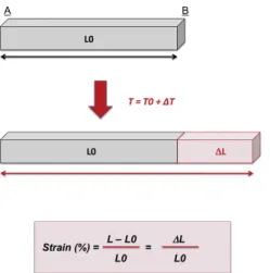
Figure 1 - Calculation of deformation (strain) and rate of deformation (strain rate). The initial myocardial length (at T0) is L0 (gray bar). One time interval (DT) later (at T0 + D/T ), the myocardial length increased from L0 to L0 + DT (gray bar added to red bar). The strain (St, expressed in %) undergone by the myocardial segment is DT /L0 and is positive. Rate at which length changes occur is strain rate (St/D/T , expressed in s-1). Extracted from Chetboul 2010.
4
As the ventricle contracts the muscle shortens in the longitudinal and circumferential dimensions and this is negative St; in the radial direction the muscle shortens or lenghts and this is a positive St (Marwick, 2006). Thus, radial St is positive during myocardial tickening and negative during ventricular relaxation (Valerie Chetboul, 2010).
Three types of deformation can be analysed:
Radial - Correspond to myocardial thinning and thickening. Contraction in the short axis is perpendicular to both long axis and the epicardium. If the myocardial thickness development during the incoming motion in systole, the value is positive, during diastole it is negative.
Circumferential - Defined as the change of the radius in the short axis, perpendicular to the radial and long axis. If the diameter of the left ventricular perimeter declines during systole the value is negative, the incoming in diastole have a positive value.
Longitudinal - Designates the change in the length of the myocardium directed from the base to the apex. During systole the base prevail moves toward the apex, the length declines and this is expressed as a negative value; the incoming during diastole are expressed as a positive value (Pedro, 2013)
Figure 2 - Orthogonal Axes of the Left Ventricle. Diagram demonstrating the orientation of the 3 types of myocardial segmental deformation most commonly reported with strain imaging. Adapted from D’Hooge et al. 2000.
5
3.2. Tissue Doppler Imaging
TDI is a special doppler technique that quantifies the regional myocardial function by measuring myocardial motion velocities throughout the entire cardiac cycle (Valérie Chetboul, 2002).
During the last two decades, thanks to the improvement of computer technology, TDI has been more intensively studied in human cardiology, giving not only sensitive information about myocardial function but helping to improve the early diagnosis of many heart diseases (Valérie Chetboul, 2002). Recently this tool has been increasingly investigated in veterinary medicine and the first small animal TDI studies appeared a few years after the human ones (Valérie Chetboul, Bussadori, & Madron, 2015).
To understand how TDI can provide information, it is important to understand the physical basics. The physical basics are similar to conventional doppler imaging, the main difference being that TDI is based on the ability to eliminate doppler information coming from the blood flow and keep what is coming from the myocardial wall (Valérie Chetboul, 2002).
TDI quantifies regional tissue motion velocity (Valérie Chetboul, 2002) and this method employs simultaneous acquisition of myocardial velocities in multiple segments and assesses intrinsic myocardial contraction or relaxation velocities (Vieira, Teixeira, Goncalves, & Gersh, 2014).
TDI allow the quantitative assessment of segmental myocardial deformation (stretching or contraction) and rate of deformation respectively (Andersen & Poulsen, 2003).
TDI has been demonstrated to provide relevant information in LV systolic and diastolic performance (Gulati, Katz, Follansbee, & Gorcsan, 1996); to be reproducible in awaken dogs (Valerie Chetboul, Athanassiadis, et al., 2004); to be superior detecting systolic LV disfunction (Valerie Chetboul, Carlos, et al., 2004) and early asymptomatic myocardial abnormalities in a dog model with Duchenne’s cardiomyopathy (Valerie Chetboul, Escriou, et al., 2004); it has also proven to be an important tool in small animals in the detection of regional myocardial abnormalities before occurrence of LV dilation and dysfunction, compared with classical ultrasound (Valerie Chetboul, 2010; Takano et al., 2011).
Some studies had shown that TDI is more sensitive than classical echocardiography, revealing abnormalities that were not detected with a previous assessment by conventional echocardiography. Althrough TDI has some limitations, its angle dependancy is the major one with some risk of underestimation of myocardial velocities (Valérie Chetboul, 2002; Takano et al., 2011). Moreover, TDI variables can be affected by breed, age and heart rate (Valerie Chetboul, Sampedrano, Testault, & Pouchelon, 2004). Other aspect is the time consuming steps for data acquisition and processing, and the need of expert readers. Good images, tracking through at least three cycles and technical settings
6
(concerning the gray-scale image, gain control, alignement to the wall, frame rate, sample volume, and others) can prolong the time that is necessary to obtain results (Dandel & Hetzer, 2009).
Despite all these limitations, this technique was initially validated with sonomicrometry and with magnetic resonance imaging (the “gold strandard” for deformation analysis in human medicine) (Dandel & Hetzer, 2009).
Some of these limitations may in part, be overcome by non-doppler methods such as 2D-STE (Valerie Chetboul, 2010).
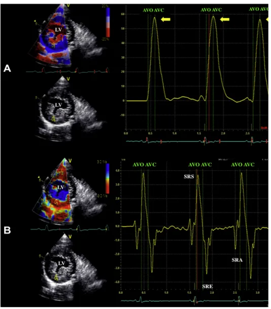
Figure 3 - Examples of normal regional radial strain (A) and strain rate (B) profiles recorded within the left ventricular free wall in a healthy dog (right parasternal transventricular short-axis view). (A) The radial
7
strain profile (expressed in %) is positive and maximal in end systole (arrows) reflecting regional systolic thickening of the left ventricular free wall. (B) The strain rate profile (expressed in s-1) is positive during systole (SRS), indicating regional thickening, then features 2 negative diastolic peaks during early filling and atrial contraction (SRE and SRA) corresponding to a biphasic thinning phase. The color displays of strain and strain rate are superimposed on the right parasternal transventricular short axis views (left upper panels). Strain length = 12 mm. Region of interest size = 6/3 mm. AVC, aortic valve closure; AVO, aortic valve opening; LV, left ventricle. Extracted from Chetboul 2010.
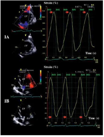
Figure 4 - Examples of radial (1A) and longitudinal (1B) strain profiles obtained from 2 dogs with dilated cardiomyopathy during 3 cardiac cycles. (1A) The radial strain profile is positive and maximal in end-systole; however, its maximal value (arrows) is very low as compared to the control group. (1B) The longitudinal strain profile obtained in a basal segment is negative and maximal in end-systole; however, its
8
maximal absolute value (arrows) is low compared to the control group. AVO: aortic valve opening; AVC: aortic valve closure; LA: left atrium; LV: left ventricle. Extracted from Chetboul et al. 2007.
3.3. Speckle tracking echocardiography
2D-STE is the most recently developed ultrasound technique to simultaneously assess regional myocardial function in multiple segments (Madron et al., 2015), providing a new approach to the assessment of LV function in humans and animals (Amundsen et al., 2006).
This imaging technique is based on the tracking, frame to frame, of speckles (natural acoustic markers). This tracking of speckle patterns is obtained by interference between the ultrasound beam and the myocardium on gray scale bi-dimensional (2D) images (Valerie Chetboul, 2010). These speckles appear as bright and small dots within the myocardium and represent natural acoustic tissue markers that can be tracked from frame to frame during the cardiac cycle (Valerie Chetboul, 2010).
In order to perform 2D-STE studies, special software is need, allowing to process spatial and temporal data on the 2D ultrasound images. The geometric shift of the speckles reflects local tissue movement and when a frame rate is known, the change in the position of the speckle determines its velocity (Dandel & Hetzer, 2009).
2D-STE St and SR analyzis has five steps until it’s complete: (1) delimitating the myocardial borders on the 2D image; (2) automatic detection of the region of interest (ROI) where the tracking is performed; (3) ROI is separated in six segments of same size, numbered from 1 to 6; (4) calculation of the chosen parameteres; (5) display of the six strain trackings as a function of time using a graphic (Madron et al., 2015). Systolic St and SR can be used as an index of global performance of the LV, by averaging all the segments, or as a regional deformation indicator, if we assess the 6 myocardial segments individually (Voigt et al., 2003).
Compared to TDI and its derived techniques, 2D-STE is independent of angle and of cardiac translation (once this method tracks the myocardium on the 2D image, it follows the direction of the wall and not the direction of the ultrasound beam), allowing any region of the heart to be assessed (Takano et al., 2011).
One more advantage is that it requires only one cardiac cycle. However it needs high-resolution image quality at high frame rates (Perk, Tunick, & Kronzon, 2007). In human medicine, the need for high image quality is a limitation for its use in patients with tachycardia and during stress echocardiography (Dandel & Hetzer, 2009).
9
2D-STE can be used to assess the complex pattern of regional myocardial motion in several segments and provide similar values to the TDI (velocity) and its derived techniques (St and SR), but can also allow to obtain more LV function parameters, such rotation and torsion angles, and segmental synchronism (Madron et al., 2015).
Regarding LV assessment, the reproducibility of 2D-STE strain measurements is considered to be better than TDI derived St, also, the intraobserver and interobserver variability between 2D-STE St and SR were found to be low (3,6% to 5,3% to 2D-strain and 7% to 11,8% to SR) (Perk et al., 2007). Ingul et. al also found lower values for interobserver variability regarding 2D-STE St measurements compared with TDI derived St and moreover 2D-STE also appeared significantly less time consuming as we described before (Ingul et al., 2005).
However, 2D-STE has known technical limitations, including its dependence on frame rate and image resolution, the decreasing of the reliability of the speckle tracking process and the potential out-of-plane movements of the speckles (Nesser & Winter, 2009).
In the following table, comparative advantages and disavantages using TDI and 2D-STE are shown.
Table 1 - Major advantages and disadvantages of 2D-STE imaging in comparison to TDI-derived St and SR imaging. Modified from: Dandel 2009.
Bi-dimensional speckle tracking (2D-STE) Tissue Doppler Imaging (TDI) Advantages
Deformation analysis in 2 dimensions High temporal resolution Tissue movement assessment relative to adjacent
segments Image quality less important
Angle independent
Better spatial resolution in comparison to the TDI technique
Less sensitive to signal noise
Better measurement reproducibility in comparison to the TDI technique
10 In comparison to the TDI technique, less time
consuming data acquisition and easy data processing. The automated tracking system allows
accurate measurements even for inexperienced observers
Disavantages Temporal resolution limited in comparison with TDI
technique 1-dimension measurements
Dependent on high-resolution image quality Tissue movement assessment in relation to the transducer
The lower optimal frame rate for speckle tracking (compared to TDI technique) technique limits the
reliability of measurements in patients with tachycardia
Measurement dependent on angle between ultrasound beam and direction of myocardial
movement Limited spatial resolution
Highly sensitive to signal noise; reduced to signal-to-noise ratio
Higher interobserver variability with 2D-STE Time consuming steps for data acquisition and processing. Important learning curve; necessity
of expert readers
Important: Althrough 2D-STE and TDI calculations correlate well, they do not yield the same values.
LV global and regional wall deformation can be better described by assessing normal and shear strains, and 2D-STE is a powerfull noninvasive method to analyze the three components of strain: longitudinal, circumferential and radial (Kusunose, Zhang, Mazgalev, Thomas, & Popovic, 2013).
In humans, several studies have demonstrated the efficacy of this technique in the diagnosis of various cardiac diseases and hemodynamic condicions such hyperthrophic cardiomyopathy (HCM) coronary disease and ventricular dyssynchrony (Amundsen et al., 2006).
Also in human medicine, the most commonly assessed strain parameter is global longitudinal strain, which is more sensitive than LV ejection fraction (EF) as a measure of systolic function and could be used to identify LV dysfunction in cardiomyopathies (Smiseth, Torp, Opdahl, Haugaa, & Urheim, 2016).
11 In veterinary medicine myocardial St and SR derived by 2D-STE have also been studied and established as a reliable method regarding several diseases and conditions: assessment of left ventricular regional function in affected and carrier dogs with duchenne muscular dystrophy (DMD) (Takano et al., 2011); radial and circumferential St analysis in cats with HCM (Takano, Isogai, Aoki, Wakao, & Fujii, 2015); assessment of left ventricular function in small breed dogs with hyperadrenocorticism (HAC) (Chen, Lien, & Huang, 2014); assessment of systolic myocardial deformations in dogs with chronic mitral valve insufficiency (CMVI) (Suzuki, Matsumoto, Teshima, & Koyama, 2013) and assessment of left ventricular function quantified by myocardial St imaging in small-breed dogs with chronic mitral regurgitation (MR) (Smith, Bonagura, Culwell, & Schober, 2012).
The recognition of earlier subclinical cardiac systolic and diastolic dysfunction could allow for an early medical approach and this could improve long-term cardiovascular outcomes (Takano et al., 2011).
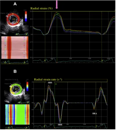
Figure 5 - Examples of normal left ventricular radial strain (A) and strain rate (B) profiles recorded in 6 myocardial segments using 2D-STE in a dog (right parasternal transventricular short-axis view). The software algorithm has automatically defined 6 equidistant myocardial segments within the interventricular septum and the LVFW. (A) The 6 corresponding LV radial strain versus time curves are shown on the right. All 6 LV segments undergo a homogeneous and coordinated systolic myocardial thickening during
12 systole (positive strain, maximal at end systole [ES], pink arrow); this may also be observed on the 2D and M-curves color-coded views (left) showing a positive strain during systole (red). (B) The 6 LV radial strain rate versus time curves, including a positive systolic wave (SRS) and 2 diastolic negative waves (SRE and SRA), are shown on the right; this may also be observed on the M-curves color-coded views (left) showing a positive strain rate during systole (red) and negative strain rate during diastole (green and blue). LV, left ventricle; LVFW, left ventricle free wall; 2D-STE, bi-dimensional speckle tracking. Extracted from Chetboul 2010.
4. Reference Values of Deformation
Despite the lack of validated reference values in veterinary medicine, studies in healthy animals have been performed. In attachment 1 a table with some of the values obtained in studies made in the past few years can be found. Some of them were obtained in healthy animals of different breeds, others were obtained from specific breeds, such as beagles and doberman pinschers.
However, even though these measurements provide new insights into ventricular motion and deformation, providing us with a more complete quantification of myocardial function in animals, further studies should be performed in a larger population of healthy individuals to evaluate the reliability and the possible role of this technique in animals (Mantovani et al., 2012).
5. Clinical Implications of Left Ventricular deformation assessment
St and SR measurements are increasingly popular in veterinary medicine, probably because of its promising results in human medicine.
These measurements appear to be a sensitive indicator of sub-clinical diseases in human medicine, such as diabetes, myocardial ischemia, aortic regurgitation, systemic sclerosis, arterial hypertension, isolated MR and non-ischemic cardiopathies. They also seem to be useful in the assessment of myocardial damage after infarction, in the evaluation of myocardial revascularization efficiency and prediction of heart failure (HF) (Marciniak et al., 2009; Yu, Sanderson, Marwick, & Oh, 2007), in sports medicine for the quantification of the LV systolic function in athletes, in sports with endurance, giving differentiation of physiologic hypertrophy from asymptomatic nonobstructive HCM (the major cause of sudden death in young athletes) (Poulsen et al., 2007) and also to differentiate hypertensive cardiac hypertrophy from cardiac hypertrophy (also known as “athlete’s heart) (Saghir, Areces, & Makan, 2007).
13 Also, in human medicine St and SR measurements are a preferred method for early detection of myocardial involvement in asymptomatic patients with amyloidosis, Duchenne’s progressive muscular dystrophy and Kawasaki syndrome (Arnold, Goebel, Ulmer, Gorenflo, & Poerner, 2007; D’Andrea et al., 2007).
Recently it was also found that 2D-STE is highly sensitive for the early detection of doxorubicin induced cardiac injury and radial St reduction in patients who undergo chemotherapy with histologic markers of doxorubicin cardiomyopathy (Migrino et al., 2008).
In veterinary medicine, some studies have been made, with healthy dogs, healthy cats, in dogs with DCM (Pedro, 2013), dogs with MR (Smith et al., 2012), cats with CHF (Sugimoto, Fujii, Sunahara, & Aoki, 2015), dogs with HAC (Chen et al., 2014), also in specific breeds, such Great Danes (Pedro, 2013), Irish Wolfhound (Westrup & McEvoy, 2013), or even wild canid like maned wolf (Mantovani et al., 2012). Studies in equines have also been made (Berli, Jud Schefer, Steininger, & Schwarzwald, 2015; Decloedt, Verheyen, Sys, De Clercq, & van Loon, 2011).
5.1. Cardiomyopathies
5.1.1. Dilated Cardiomyopathy
In human medicine it has been reported that patients with dilated cardiomyopathy (DCM) have significant changes in radial, circumferential and longitudinal St and SR, when compared with healthy individuals. In order to find if these changes are also a feature of canine DCM, some studies have been carried out in recent years (Pedro, 2013).
With the recognition of a genetic basis for DCM in some breeds (Meurs, 1998), the importance of prospective screening in lines with familiar prevalence of the disease is important (Valerie Chetboul et al., 2007), with the goal of obtain information in early stages.
In spite of DCM being diagnosed easily by 2D and M-mode echocardiography, with the identification of reduced contractility, dilatation of cardiac chambers and increased sphericity of the LV (Dukes-McEwan, Borgarelli, Tidholm, Vollmar, & Haggstrom, 2003), diagnosis of the early phase of the disease still remains a challenge for veterinary cardiologists because morphological and functional cardiac alterations may be equivocal or even absent at this asymptomatic phase (Dukes-McEwan et al., 2003). This explains the need for other more sensitive noninvasive indices, which can be used for early assessment of deterioration of myocardial disease or the beneficial effects of medication management (Valerie Chetboul et al., 2007).
14 Case reports and clinical studies evaluating TDI, showed that it’s a promising technique for the detection of early myocardial dysfunction in preclinical stages of dogs with DCM (Wess, Keller, Klausnitzer, Killich, & Hartmann, 2011).
Chetboul et al. 2007, did a study using TDI technique combined with St imaging, with the aim to provide sensitive indices for early detection and treatment of CMD. They used TDI and St imaging in dogs with overt DCM with a prerequisite before using these new criteria in prospective screenings of families predisposed or in clinical trials. In this study, radial and longitudinal right and left myocardial motion was assessed by TDI and St variables with the hypothesis of being altered in dogs with DCM. The results indicates that the DCM group reveals decreases in radial and longitudinal systolic velocity values of the LVFW, longitudinal and radial absolute values of peak systolic St of the LVFW, and longitudinal systolic right ventricular velocities (all of them P<.001 versus control) associated with post systolic contraction waves in 7 of the 14 dogs. Also early diastolic LVFW velocities were decreased for radial (P<.05) and longitudinal (P<.01) motions, being all the values negatively correlated with heart rate. Thus, this study reveals clinical importance showing that LV contractility assessed by TDI St imaging along both short and long axis is impaired in dogs with spontaneous DCM, as is diastolic LVFW function and systolic right ventriclular. However, the impact of these findings (sensitivity and prognostic values of these diastolic and systolic TDI St myocardial indices) should be assessed in future prospective studies to determinate the sensitivity of TDI and St variables for the early detection of canine DCM (Valerie Chetboul et al., 2007).
While in human medicine it has been reported that patients with DCM have significant changes when compared to healthy indiviaduals, until the study made by Pedro 2013, similar studies have not been performed in veterinary medicine. This study has been made with the goal to assess the function of the LV using 2D-STE in Great Danes to identify and report the differences in the left ventricular mechanics of normal dogs and dogs with DCM. The major objective of this study was to consider dogs that had two or more echocardiographic examinations and tried to detect changes in the left ventricular mechanics that could identify dogs that will develop DCM in the future. The results obtained was that when assessing the mechanics of the LV in healthy dogs, an increase magnitude and rate of deformation has been noticed from the base towards the apex of the LV and the same was noted in dogs with DCM. This contrasts with human medicine once even dogs with DCM demonstrated lower rate and magnitude of deformation, only a few variables were considered significant between them, supporting that more studies with different breeds and large groups are needed in veterinary medicine.
15 5.1.2. Hyperthrophic Cardiomyopathy
HCM is considered as the most common heart disease in cats (Ferasin et al., 2003). It is characterized by concentric left ventricle hyperthrophy (LVH) and both diastolic and systolic dysfunction. Diastolic dysfunction is the main feature of the disease, being the main cause of left atrium enlargement and subsequent congestive heart failure (CHF) (Sugimoto et al., 2015).
The definitive diagnosis of HCM is based on classical echocardiography, which is the method of choice to evaluate dysfunctions and to observe structural changes. (Brizard, Amberger, Hartnack, Doherr, & Lombard, 2009). Classical echocardiography is mainly the main tool in guiding clinicians in the management of the disease (A. Silva, Muzzi, Oberlender, Nogueira, & Muzzi, 2013).
In the early stages of the disease, cats are asymptomatic (Trehiou-Sechi et al., 2012), and the myocardial dysfunction evolves with the progression of the disease (Kittleson et al., 1999). HCM is still a challenging disease for veterinarians and remains as the major cardiac cause of mortality and morbidity in cats, with an associated risk of sudden death, arterial thromboembolism and heart failure (HF) (Valerie Chetboul, Blot, et al., 2006).
Although classical echocardiography is considered as the gold standard for he diagnosis of HCM, recent techniques such as TDI and 2D-STE have provided new parameters to assess myocardial function, including regional myocardial velocities and deformation, ventricular synchrony and torsion (Valerie Chetboul, 2010). One study has been published regarding these new parameters and their usefulness on the early diagnosis of HCM. Chetboul et al. 2006, using affected cats and carriers of dystrophin-deficient hyperthrophic muscular dystrophy as a model of HCM demonstrated that TDI could detect radial and longitudinal LVFW dysfunction in cats, even in those who didn’t presented significant left myocardial hyperthrophy.
Another study of Chetboul et al. 2006 demonstrated that classical echocardiography was unable to detect HCM in a young Maine Coon cat while TDI identified LVFW dysfunction. One year later, using classical echocardiography, the diagnosis of HCM was confirmed. Given that finding, it was suggested that TDI could have good sensitivity for detection of early stage HCM (A. Silva et al., 2013).
Silva et al. 2013, did a study using 2D-STE to evaluate LV longitudinal St and SR in non-sedated healthy cats, as well as longitudinal displacement and velocity. This study demonstrated that 2D-STE could be feasible for measuring LV longitudinal St and SR, velocity and displacement, establishing preliminary reference values for non-sedated healthy cats, which can provide feasibility of this technique in clinical context.
16 In 2015, Sugimoto et al. did a study, with the aim to assess global and segmental LV myocardial function using 2D-STE in cats with HCM whose TDI variables were within the reference range. Although impaired cardiac function using TDI in cats with HCM was previously shown, reference ranges using TDI in cats with HCM and in normal cats have been reported as widely variable. This study used 2D-STE since it was proven to be useful in the assessment of cardiac function in human patients with HCM but the clinical utility had not been validated in cats. Conventional echocardiography, TDI and segmental and global 2D-STE indices were evaluated and compared, and the results suggested that 2D-STE parameters are more sensitive compared with TDI parameters to detect early myocardial dysfunction in cats with HCM. They concluded that in addition to genetic screening, 2D-STE can be used in the preclinical diagnosis of HCM, providing the opportunity to prevent development of LVH using drug management in the future and to avoid unfavorable breeding programs.
Also using 2D-STE, Takano et al. 2014, proposed a study to investigate St analysis in cats with HCM. They measure circumferential and radial St and SR variables in healthy cats and global St and SR, and also segmental assessment of LVFW in cats with HCM were performed. Differently from the previous study, buprenorphine and acepromazine was used as sedation, which did not affected any global St nor SR variables. Finally, it was concluded that 2D-STE analysis using short axis images of LV appeared to be clinically feasible in cats, bringing the possibility to be useful for detecting myocardial dysfunctions in cats with heart disease.
These studies suggest that conventional echocardiography alone could be unable to identify the myocardial changes occurring in the early stages of the disease, and the association of recent modalities could improve diagnostic accuracy. In this regard, TDI and 2D-STE parameters have been found to be most promising in the recently available studies. However, more studies must be carried out in order to validate the role and reliability of this techniques in cats with cardiomyopathies (A. C. Silva et al., 2013).
5.1.3. Duchenne Muscular Dystrophy
DMD is related to a dystrophin mutation providing a dysfunctional protein (Valerie Chetboul, Escriou, et al., 2004). Dystrophin is a key linker protein between the sarcolemma of the myocyte and the contractile apparatus, the sarcomere (Cohn & Campbell, 2000). In pathologies like this, dystrophinopathies, in addition to the skeletal muscle disease it is common that patients suffer from cardiac changes over a variable period of time, before the occurrence of DCM, resulting in fatty infiltration with fatal outcome (Cullen & Mastaglia, 1980; Nigro, Politano, Nigro, Petretta, & Comi, 1994). Myocardial
17 dysfunction has been estimated to be responsible for 20% of the total mortality in human patients with DMD (Nigro, Comi, Politano, & Bain, 1990).
Conventional echocardiography is one of the noninvasive methods to assess cardiac function in patients and animals with DMD (Danilowicz, Rutkowski, Myung, & Schively, 1980). Some studies have demonstrated the usefulness of TDI to detect subclinical myocardial diastolic and systolic dysfunction in patients with normal parameters in classical echocardiography. In 2004, Chetboul et al determined the accuracy of TDI to detect dystrophin mutant Golden Retriever Muscular Dystrophy (GRMD) dogs early, before the occurence of CHF and myocardial dysfunction as determinated by conventional echocardiography. This study has proved that TDI is able to detect early changes in myocardial function that are not detectable with classical echocardiography. Also, other recent studies have confirmed that myocardial velocities, myocardial wall-ticknening velocities, myocardial velocity gradients and strain during systole and early diastole in LVFW were reduced in human patients (Mori et al., 2004) and in dogs with DMD (Valerie Chetboul, Carlos, et al., 2004).
Takano et al. 2011, did a study with the aim of demonstrating the applicability of 2D-STE to assess LV regional myocardial dysfunction in DMD model dogs without clinical signs of HF. They performed conventional echocardiography with systolic and diastolic function assessment by Doppler echocardiography, TDI and St indices with 2D-STE, and comparing all of them. The results obtained were significant differences in body weigh and trasmitral St and SR derived by TDI among the 3 groups (affected dogs, carrier dogs and controls), but no significant difference in global St indices. Althrough in segmental analysis, the peak radial SR in the early diastole in posterior segment at chordae the tendinease level appeared signifcant differences in the 3 groups. This study concluded, that the myocadial SR by 2D-STE could detect the impaired cardiac diastolic function in dogs with DMD without any LV dilation or clinical signs, thus, radial SR must be an important parameter to detect early myocardial impairment in dogs with this disease. However, more studies are needed to clarify the sensitivity of these new technique.
5.2. Valvular Disease
5.2.1. Mitral Valve Disease
Mitral valve disease (MVD) is considered as the most common cardiac disease in dogs. These patients are affected with volume overload caused by MR (Tidholm, Ljungvall, Hoglund, Westling, & Haggstrom, 2009). MR caused by MVD is the most common cause of HF and LV remodeling in dogs (Haggstrom, Duelund Pedersen, & Kvart, 2004).
18 Some dogs with MVD develop myocardial dysfunction as well as remodeling and enlargement of the heart (Richards, 2012). The presence of LV systolic dysfunction can influence the therapy and the prognosis of dogs with MVD and the assessment of LV function in MR by classical echocardiography is difficult, mainly by the changes in ventricular loading (Smith et al., 2012). Progressive increases in LV preload and reductions in LV afterload typically create an hyperdinamic LV with increases in EF and in the rate of early diastolic ventricular filling (Yared, Lam, & Hung, 2009). Consequently, conventional measures like EF and fractional shortening (FS) are insensitive for measuring early LV dysfunction (McGinley et al., 2007) becoming abnormal only in severe stages of the disease, suggesting that measurements of segmental deformation (St and SR) could provide increased sensitivity (Marwick, 2006).
McGingley et al. 2007, investigated the mechanics of contractile dysfunction in MVD, and found that even with a preserved EF, chronic severe MR results in a significant reduction in intrinsic contractile function and reserve and this is relevant for prognosis, helping the clinicians with the management of the disease.
Suzuki et al. 2013, did a study to clinically assess myocardial deformations in dogs with CMVI using 2D-STE. They placed the dogs into 1 of 3 classes based on the International Small Animal Cardiac Health Council classification and the dogs were examined for myocardial deformation (St and SR) in the circumferential, longitudinal and radial planes. They obtained results showing that Class II and III dogs had higher circumferential St than Class I (P=0.002 and P=0.001 respectively) and control dogs (P<0.001). Moreover, Class III dogs had higher radial St and SR than Class I dogs (P=0.006) and controls (P=0.001), and other deformations like longitudinal were not significantly different between classes of CMVI dogs and the controls. Suzuki and collegues, conluded that in the clinical progression of CMVI, myocardial deformations assessed by 2D-STE differed according with the myocardial contractile direction and with that, assessments of multidirectional myocardial deformations must be important for better assessment of clinical cardiac funtion in these dogs.
Another study, made by Zois et al. 2012, with the hypothesis that global St and SR are decresed in dogs with clinical signs of CHF due to MVD compared with healthy dogs and associated with classical echocardiography indices due MVD severity, obtained that assessed by 2D-STE, LV function appeared augmented in moderate-to-severe disease.
However, Smith et. al, 2012, congruous with the findings of Tidholm and associates who have used both TDI (Tidholm et al., 2009) and 2D-STE (Zois et al., 2012) for assessing myocardial St in dogs with spontaneous MR, demonstrated that there is no obvious advantage in applying mycoardial St imaging for the detection of LV systolic dysfunction in asymptomatic dogs with stage B2 MR, once myocardial contractility is preserved or St and SR analysis are perturbed by the hemodynamic factors that
19 cause hyperdynamic conventional indices seen in small-breed dogs with chronic MR, even with CHF. Thus, while radial St and SR can be measured in dogs with significant MR, these values are increased with ventricular remodeling, probably influenced by the same loading conditions that confound other echocardiography indices. So, a clear clinical application for this technique in dogs in stage B2 with MR is not evident considering the results of these studies.
To conclude, the clinical relevance of these results, including the relation to survival time and medication and an evaluation of the timing when the decrease in these deformation variables, should be investigated further using large groups of animals in differentes stages of the disease (Zois et al., 2012).
5.3. Cardiac Involvment in Systemic Diseases
5.3.1. Hyperadrenocorticism
HAC is a condition characterized by chronically elevated circulating glucocorticoid concentration and in human patients it is prooven that in response to stimulation of the regin-angiotensin system, glucocorticoid and mineralocorticoid receptors of myocytes are the responsible for the development of systemic hypertension, LVH (Chen et al., 2014), and myocardial fibrosis which are commonly observed (Brilla & Weber, 1992). Cardiac hyperthrophy resulting from hypertension and chronic exposure to excess circulating cortisol may also contribute to LV concentric remodeling in human patients and subsequently, to other functional abnormalities such as diastolic dysfunction (Muiesan et al., 2003). Sixty-eight to eighty-five percent of human HAC patients developed hypertension (Boscaro, Barzon, Fallo, & Sonino, 2001). In dogs, hypertension occurs in 47 to 86% of the cases (Ortega, Feldman, Nelson, Willits, & Cowgill, 1996), but more studies of cardiac function in dogs with HAC are needed to fully understand the consequences of such changes.
Hung-Yin Chen et al 2014, did a study with the aim of reporting the prevalence of LVH and assess LV systolic and diastolic function using 2D-STE in small breed dogs with HAC. They used 9 age-matched healthy dogs, 10 dogs with pituitary-dependent HAC and 9 dogs with adrenal-dependent HAC, and classical echocardiography, global circumferential and longitudinal St and SR were performed using 2D-STE. In this study, 2D-STE revealed changes in LV mechanics despite a preserved systolic function detected by conventional echocardiography. The indices of LV systolic function derived by conventional echocardiography were not significantly different between the HAC group and controls, but decreased systolic function was revealed by 2D-STE. These findings suggest that dogs with HAC may have subclinical systolic dysfunction that could remain hidden using conventional echocardiography. This is
20 also reported in human patients which subclinical LV diastolic dysfunction when using 2D-STE but with preserved FS and EF by conventional echocardiography (Muiesan et al., 2003).
In 2015, Oui et al., investigated the changes in longitudinal and radial LV function using TDI St imaging in beagles using 11 normal dogs, they administrated 2mg/kg of prednisone orally every 12 hours for 28 days which 7 out of 11 dogs to induce iatrogenic HAC. In this study, they measured the myocardial wall velocity of LV using color TDI and determinated the myocardial deformation by St and SR imaging. The results obtained were that conventional echocardiography revealed that diastolic LVFW and interventricular septum in the HAC group were tickened when compared with the control group and the St values in the HAC group were significantly lower than the values on the control group, particularly for the longitudinal wall. With the results obtained, they concluded that the lower values of myocardium from TDI and St imaging could be used to investigate subclinical LV systolic and diastolic dysfunction in dogs with iatrogenic HAC.
In the available publications, the limitations found were usually the low number of dogs, which suggests that more studies with larger group of animals are needed.
5.4 Congenital heart diseases
5.4.1. Patent Ductus Arteriosus
Patent ductus arteriosus (PDA) is one of the most common congenital heart defects in dogs, with an estimated prevalence of 21-32% of all the congenital heart diseases in dogs (Oliveira et al., 2011). PDA is a fetal vascular structure connecting the aorta to the main pulmonary artery and usually closes shortly after birth by a complex hemodynamic and neurohormonal process (Clyman, 2006).
The main hemodynamic effect of a left-to-right PDA is LV volume overload, causing an increase in preload, which in turn increases contractility following Starling’s law. The LV compensates this increase in preload by increasing stroke volume, and many times by developing eccentric hyperthrophy to normalize wall stress (Spalla, Locatelli, Zanaboni, Brambilla, & Bussadori, 2016a). Previous studies have analyzed the consequences of ductal patency and its closure on cardiac indices by M-mode and B-mode parameters (Saunders, Gordon, Boggess, & Miller, 2014).
Spalla et al. 2016, compared cardiac function and contractility by 2D-STE in dogs with PDA and in normal control dogs, suggesting that conventional parameters routinely used to assess systolic function, such as EF and FS, were not different between the 2 groups, while 2D-STE identified subtle
21 changes in cardiac systolic function and contractility between them. Based on this, 2D-STE may be a more appropriate tool to assess cardiac contractility in dogs with PDA. 2D-STE can also provide further insight into the effect of PDA closure on cardiac mechanics in dogs with PDA (Spalla, Locatelli, Zanaboni, Brambilla, & Bussadori, 2016b).
Concluding, dogs with PDA showed a marked increase in conventional echocardiographic parameters and an even more increase in advanced echocardiography parameters (longitudinal, circumferential and radial St and SR), whereas FS and EF were not different between the two techniques (Spalla et al., 2016a). However, the limitations of this recent study are related to a small sample size , which points to the need of additional investigation to clarify the clinical relevance.
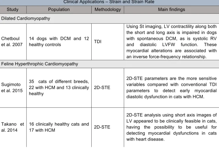
The scientific literature regarding the use of TDI and 2D-STE to assess strain parameters in the mentioned diseases is summed up on the next table.
Table 2 - Strain imaging clinical studies
Clinical Applications – Strain and Strain Rate
Study Population Methodology Main findings
Dilated Cardiomyopathy
Chetboul et al. 2007
14 dogs with DCM and 12
healthy controls TDI
Using St imaging, LV contractility along both the short and long axis is impaired in dogs with spontaneous DCM, as is systolic RV and diastolic LVFW function. These myocardial alterations are associated with an inverse force-frequency relationship. Feline Hyperthrophic Cardiomyopathy
Sugimoto et al. 2015
35 cats of different breeds, 22 with HCM and 13 clinically healthy
2D-STE
2D-STE parameters are the more sensitive variables compared with conventional TDI parameters to detect early myocardial diastolic dysfunction in cats with HCM.
Takano et al. 2014
16 clinically healthy cats and
17 with HCM 2D-STE
2D-STE analysis using short axis images of LV appeared to be clinically feasible in cats, having the possibility to be useful for detecting myocardial dysfunctions in cats with heart disease.
22 Duchenne Muscular Dystrophy
Takano et al. 2011
6 affected dogs, 8 carrier dogs with GRMD, 8 control dogs
2D-STE
The myocardial SR by 2D-STE served to detect the impaired cardiac diastolic function in DMD model dogs without any obvious dilation or clinical signs. The radial SR may be a useful parameter to detect early myocardial impairment in this disease.
Yamato et al. 2008
24 Golden Retriever dogs, divided in affected, carrier and controls
TDI
Differences observed in some variables of systolic and diastolic function especially between control and affected groups. It was possible to confirm that Golden Retriever Dogs, affected by CMD and without clinical signs of congestive heart failure, present changes of systolic and diastolic indices, detected by TDI, St and SR.
Mitral valve disease
Suzuki et al. 2013
87 dogs with chronic mitral
valve insufficiency 2D-STE
In the clinical progression of CMVI in dogs, myocardial deformations, as assessed by 2D-STE, differed according to myocardial contractile direction. Thus, assessments of multidirectional myocardial deformations may be important for better assessment of clinical cardiac function in dogs with CMVI. Smith et
al. 2012
40 healthy dogs: 20 controls, 20 with MR and LV remodeling (Stage B2)
2D-STE
LV diastolic diameter, diastole area, SF, average peak systolic and early diastolic radial St, global circumferential St, and average radial SR were significantly greater in the MR group.
Zois et al. 2012
93 dogs with different mitral
MVD severities 2D-STE
Assessed by 2D-STE, LV function appeared to be augmented in moderate-to-severe disease.
Hyperadrenocorticism
Chen et al. 2014
9 healthy dogs, 10 dogs with pituitary-dependent HAC, 9 dogs with adrenal-dependent HAC
2D-STE
2D-STE revealed significant decreases in systolic functions that were undetected using conventional echocardiography in the adrenal-dependent HAC and pituitary-dependent HAC.
Oui et al. 2015
11 3-year-old healthy male
beagles TDI
The St values from TDI strain imaging could be use to investigate subclinical LV systolic and diastolic dysfunction in dogs with iatrogenic HAC.
23 Spalla et
al. 2016
34 dogs with PDA, 10
healthy controls 2D-STE
2D-STE parameters identified subtle changes in cardiac systolic function and contractility. 2D-STE may be a more appropriate tool to assess cardiac contractility in dogs with PDA.
Spalla et
al. 2016 25 dogs with PDA 2D-STE
2D-STE can provide further insight into the effect of Patent Ductus Arteriosus closure on cardiac mechanics in dogs affected by PDA.
Others Arita et al. 2007
12 dogs without HF; 9 dogs with HF; 8 dogs with induced HF
TDI and 2D-STE
Radial St by 2D-STE is more accurate than TDI velocity to detect cardiac dyssynchrony in a canine model of dyssynchrony
Nakata et al. 2016
6 healthy female beagles instrummented with externally programmable pacemaker (EV4543 Pace Medical Inc., MA, USA)
2D-STE
2D-STE strain demonstrated to be a reliable tool for evaluation of LV myocardial deformation in tachycardia-induced cardiac dysfunction canine model showing an earlier significant wall motion abnormalities using radial strain and later using circumferential strain
Hamabe et al. 2013
5 female beagles with
implanted pacemakers 2D-STE
The results revealed the ability of 2D-STE to measure radial and circumferential strain in dogs with sustained high-electrical pacing, and allowed assessment of global and regional myocardial function and the degree of dyssynchrony
TDI: tissue Doppler imaging; 2D-STE: bi-dimensional speckle tracking; St: strain; SR: strain rate; HF: heart failure; LV: left ventricle; RV: right ventricle; LVFW: left ventricle free wall; CMVI: chronic mitral valve insufficiency; PDA: Patent Ductus Arteriosus DCM: dilated cardiomyopathy; HCM: hypertrophic cardiomyopathy; GRMD: golden retrievers muscular dystrophy; MVD: mitral valve disease; FS: fractional shortening
24 6. Conclusions and Future Perspectives
St and SR imaging are promising tools for the evaluation of myocardial function. These recent modalities are one of the most significant advances in cardiac ultrasound imaging.
In small animal studies, TDI, a noninvasive and precise quantification technique has been one of the most significant advances in cardiac ultrasound imaging, showing better sensitivity when compared to conventional echocardiography in both the prognosis and diagnosis of focal myocardial dysfunction (Madron et al., 2015).
These facts make TDI a powerful tool for cardiovascular research (Madron et al., 2015).
TDI-derived St measurements are less dependent of image quality, but the angle dependency is a major limitation when assessing certain regions of the myocardium (Dandel & Hetzer, 2009).
An even more recent method, 2D-STE, that uses grayscale imaging, is starting to be assessed through research and development studies, but the evidence for the utility of this modality is accumulating and it does offer major advantages over TDI, mainly its angle independency and its better signal to noise ratio (Artis et al., 2008).
Many authors have already shown that 2D-STE is a reproducible and repeatable method not only in Human Medicine but also in Veterinary Medicine, and it can be used for assessing the LV function (Westrup & McEvoy, 2013).
The high sensitivity of both TDI derived techniques and 2D-STE for the early detection of myocardial dysfunction is proven in some recent studies, arising interest in veterinarians.
New studies on large groups of animals with heart disease are needed to compare the diagnostic benefits of these new imaging techniques, as well as their adittional value in the prognosis and in therapeutic decision making (Madron et al., 2015). Currently these techniques are not yet considered part of a routine clincal echocardiographic study. We need more studies, particularly those focused on small animals and their most common cardiopathies, to be able to judge these techniques applicability and usefulness. The St imaging methodology is still undergoing improvement, and further clinical trials are needed to determine if clinical decisions based on these techniques result in a better outcome (Smiseth et al., 2016).
25 7. Bibliography
Amundsen, B. H., Helle-Valle, T., Edvardsen, T., Torp, H., Crosby, J., Lyseggen, E., … Slordahl, S. A. (2006). Noninvasive myocardial strain measurement by speckle tracking echocardiography: validation against sonomicrometry and tagged magnetic resonance imaging. Journal of the American College of Cardiology, 47(4), 789–793. http://doi.org/10.1016/j.jacc.2005.10.040
Andersen, N. H., & Poulsen, S. H. (2003). Evaluation of the longitudinal contraction of the left ventricle in normal subjects by Doppler tissue tracking and strain rate. Journal of the American Society of Echocardiography : Official Publication of the American Society of Echocardiography, 16(7), 716– 723. http://doi.org/10.1016/S0894-7317(03)00325-0
Arnold, R., Goebel, B., Ulmer, H. E., Gorenflo, M., & Poerner, T. C. (2007). An exercise tissue Doppler and strain rate imaging study of diastolic myocardial dysfunction after Kawasaki syndrome in childhood. Cardiology in the Young, 17(5), 478–486. http://doi.org/10.1017/S1047951107000959 Artis, N. J., Oxborough, D. L., Williams, G., Pepper, C. B., & Tan, L. B. (2008). Two-dimensional strain
imaging: a new echocardiographic advance with research and clinical applications. International Journal of Cardiology, 123(3), 240–248. http://doi.org/10.1016/j.ijcard.2007.02.046
Berli, A.-S. J., Jud Schefer, R., Steininger, K., & Schwarzwald, C. C. (2015). The use of strain, strain rate, and displacement by 2D speckle tracking for assessment of systolic left ventricular function in goats: applicability and influence of general anesthesia. Cardiovascular Ultrasound, 13(1), 1–17. http://doi.org/10.1186/s12947-015-0005-8
Boon, J. A. (2011). Veterinary echocardiography (Second Edi). Wiley-Blackwell.
Boscaro, M., Barzon, L., Fallo, F., & Sonino, N. (2001). Cushing’s syndrome. Lancet (London, England), 357(9258), 783–791. http://doi.org/10.1016/S0140-6736(00)04172-6
Brilla, C. G., & Weber, K. T. (1992). Mineralocorticoid excess, dietary sodium, and myocardial fibrosis. The Journal of Laboratory and Clinical Medicine, 120(6), 893–901.
Brizard, D., Amberger, C., Hartnack, S., Doherr, M., & Lombard, C. (2009). Phenotypes and echocardiographic characteristics of a European population of domestic shorthair cats with idiopathic hypertrophic cardiomyopathy. Schweizer Archiv Fur Tierheilkunde, 151(11), 529–538. http://doi.org/10.1024/0036-7281.151.11.529
Chen, H.-Y., Lien, Y.-H., & Huang, H.-P. (2014). Assessment of left ventricular function by two-dimensional speckle-tracking echocardiography in small breed dogs with hyperadrenocorticism. Acta Veterinaria Scandinavica, 56(1), 88. http://doi.org/10.1186/s13028-014-0088-5
Chetboul, V. (2002). Tissue Doppler Imaging: a promising technique for quantifying regional myocardial function. Journal of Veterinary Cardiology : The Official Journal of the European Society of Veterinary Cardiology, 4(2), 7–12. http://doi.org/10.1016/S1760-2734(06)70033-9
26 (2004). Assessment of repeatability, reproducibility, and effect of anesthesia on determination of radial and longitudinal left ventricular velocities via tissue Doppler imaging in dogs. American Journal of Veterinary Research, 65(7), 909–915.
Chetboul, V., Carlos, C., Blot, S., Thibaud, J. L., Escriou, C., Tissier, R., … Pouchelon, J.-L. (2004). Tissue Doppler assessment of diastolic and systolic alterations of radial and longitudinal left ventricular motions in Golden Retrievers during the preclinical phase of cardiomyopathy associated with muscular dystrophy. American Journal of Veterinary Research, 65(10), 1335–1341.
Chetboul, V., Escriou, C., Tessier, D., Richard, V., Pouchelon, J.-L., Thibault, H., … Derumeaux, G. (2004). Tissue Doppler imaging detects early asymptomatic myocardial abnormalities in a dog model of Duchenne’s cardiomyopathy. European Heart Journal, 25(21), 1934–1939. http://doi.org/10.1016/j.ehj.2004.09.007
Chetboul, V., Sampedrano, C. C., Testault, I., & Pouchelon, J.-L. (2004). Use of tissue Doppler imaging to confirm the diagnosis of dilated cardiomyopathy in a dog with equivocal echocardiographic findings. Journal of the American Veterinary Medical Association, 225(12), 1864,1877–1880.
Chetboul, V., Blot, S., Sampedrano, C. C., Thibaud, J.-L., Granger, N., Tissier, R., … Pouchelon, J.-L. (2006). Tissue Doppler imaging for detection of radial and longitudinal myocardial dysfunction in a family of cats affected by dystrophin-deficient hypertrophic muscular dystrophy. Journal of Veterinary Internal Medicine / American College of Veterinary Internal Medicine, 20(3), 640–647.
Chetboul, V., Sampedrano, C. C., Gouni, V., Nicolle, A. P., & Pouchelon, J.-L. (2006). Two-dimensional color tissue Doppler imaging detects myocardial dysfunction before occurrence of hypertrophy in a young Maine Coon cat. Veterinary Radiology & Ultrasound : The Official Journal of the American College of Veterinary Radiology and the International Veterinary Radiology Association, 47(3), 295– 300.
Chetboul, V., Gouni, V., Sampedrano, C. C., Tissier, R., Serres, F., & Pouchelon, J.-L. (2007). Assessment of regional systolic and diastolic myocardial function using tissue Doppler and strain imaging in dogs with dilated cardiomyopathy. Journal of Veterinary Internal Medicine / American College of Veterinary Internal Medicine, 21(4), 719–730.
Chetboul, V., Serres, F., Gouni, V., Tissier, R., & Pouchelon, J.-L. (2007). Radial strain and strain rate by two-dimensional speckle tracking echocardiography and the tissue velocity based technique in the dog. Journal of Veterinary Cardiology : The Official Journal of the European Society of Veterinary Cardiology, 9(2), 69–81. http://doi.org/10.1016/j.jvc.2006.11.002
Chetboul, V. (2010). Advanced techniques in echocardiography in small animals. The Veterinary Clinics of North America. Small Animal Practice, 40(4), 529–543. http://doi.org/10.1016/j.cvsm.2010.03.007 Chetboul, V., Bussadori, C., & Madron, É. (2015). Clinical echocardiography of the dog and cat. (E.
Madron, Ed.) (Vol. 1). Elsevier Health Science.