Mestrado em Engenharia Biomédica
Development and Characterization of
Nanocarrier Systems for the Delivery of
Antitubercular Drugs
Master Thesis developed in the course of Dissertation
R
ICARDOL
EANDROD
ELINDROR
IBEIROSupervisor
Acknowledgements
For different reasons, this work would not have been possible without the support of a number of high quality individuals, surrounded by which I found myself lucky to be most of the time.
I would like to start by thanking my thesis supervisor, Salette Reis, first of all for having accep-ted me in her group and having trusaccep-ted me with this work, but also for all the scientific, material and moral support, and mainly for making sure that everyone in the group treated me well. It must have been a hard task, I am sure.
Secondly, I would like to thank Marina Pinheiro, for all the guidance and patience in this work, (even through the rough times, the bad results, and the dangerously close deadlines), and for always being present when needed. This work would not be what it is without her.
I am in debt to a few ones that helped me with some experimental techniques: Fernanda Andrade, for the help with the MTT assays; José das Neves, for the help with the HPLC measure -ments; and Ana Cardoso, for the help with the DSC experiment.
I would also like to thank the ones who I shared the laboratory with, and who also shared with me their knowledge, experience and overall good mood. They told me not to write their names, but I'm not going to obey: to Catarina, Catarina, Catarina, Joana, Miriam and Nini, but also to dona Manuela, Patrícia, and Sofia. A special and warm thank you note goes out to Júlia, for all these years of healthy partnership, and for always laughing, laughing out loud.
To all my colleges in the MEB programme, specially to Raquel Almeida and André Carvalho, for all those lunatic lunches and (i)rational conversations.
A deep acknowledgement goes naturally to my parents, which are, and always will be, present in everything I do.
Finally, I would like to thank those two individuals, homo sapiens of the highest quality, that complete my life, and to whom this work is dedicated. To Sílvia, for always being there, for always supporting my choices, for being patient with my doubts, and, above all, for not having run away. And to Daniel, for having born at exactly the right moment.
Abstract
Tuberculosis (TB) is still an ongoing public health concern in African, Asian and South American countries, where it still has a strong prevalence, resulting in a heavy economic, social and human burden. In 2011, the World Health Organization (WHO) has reported an estimated 1.4 million deaths due to TB, a disease caused by the infection of Mycobacterium tuberculosis (MTb). There have been determined efforts to fight this disease, and the search for new antitubercular drugs plays a crucial role. In spite of these efforts, the most recent drug in the market dates back 50 years, and so new delivery strategies that improve the efficacy of existing treatments may become important in this fight.
The goal of this work was to develop a nanocarrier system for the delivery of antitubercular drugs. The chosen nanocarriers were lipid nanoparticles, more specifically nanostructured lipid con-jugates (NLCs). These particles were loaded with two anti-tubercular drugs: rifampicin (RIF) and rifabutin (RFB). Since the lung is the primary site of infection in TB, the proposed route of adminis-tration for this strategy is the pulmonary route. Once inhaled, the particles should be able to travel to the pulmonary alveoli and reach the alveolar macrophages (AMs). Produced particles thus must have an appropriate size, otherwise they will be trapped in the upper airways or leave the lung on exhaling. Also, it is known that AMs have specific receptors that bind to sugars. Surface modifica-tion by mannose coating was performed to take advantage of these receptors and improve cellular uptake by AMs.
The developed particles were characterized in terms of size, zeta and morphology. Results showed particles with size and morphology suitable to reach the pulmonary alveoli, and loading efficiency for both drugs was above 80%. The success of mannose coating was confirmed by FTIR analysis. Cytotoxicity of the formulation was evaluated by MTT assay with three different cell lines.
Although more studies are definitely needed, the results from the present work pose a strong argument for NLCs as a promising strategy for the pulmonary delivery of antitubercular drugs.
Resumo
A tuberculose (TB) apresenta-se ainda como um problema de saúde pública considerável em países Africanos, Asiáticos e Sul Americanos, onde ainda tem uma elevada prevalência, resultando num pesado fardo económico, social e humano. Em 2011, a Organização Mundial de Saúde estimou que a TB terá sido responsável, em todo o mundo, por 1.4 milhões de mortes. A TB é uma doença cau-sada pela infeção por Mycobacterium tuberculosis. Inúmeros esforços têm sido concentrados no combate a esta doença, e a pesquisa por novos fármacos tem, aqui, um papel preponderante. No entanto, e apesar destes esforços, o mais recente fármaco para o combate à TB tem já 50 anos. Novas estratégias de transporte e libertação de fármacos, que melhorem a eficácia dos tratamentos já existentes, poderão tornar-se importantes nesta luta.
O objectivo deste trabalho foi desenvolver um sistema de nanopartículas para o transporte e libertação de fármacos de combate à TB. As nanopartículas escolhidas foram nanopartículas lipídi-cas, mais especificamente partículas lipídicas nanoestruturadas (NLC). Nestas foram introduzidos dois fármacos: rifampicina (RIF) e rifabutina (RFB). Dado que os pulmões são o principal foco de infecção por TB, a via de administração proposta é a inalatória. Uma vez inaladas, as NLC deverão depositar-se nos alvéolos pulmonares, onde se encontram os macrófagos alveolares (AMs). As par-tículas produzidas deverão, portanto, ter um tamanho apropriado a este objectivo. É também conhe-cido que os AMs têm receptores de açúcares específicos. A superfície das partículas foi então modificada para expor moléculas de manose, com o ojbectivo de aumentar a
As nanopartículas desenvolvidas foram caracterizadas em termos de tamanho, potencial zeta e morfologia. Os resultados revelaram partículas com tamanho e morfologia adequados para atingir os alvéolos pulmonares. A taxa de incorporação para ambos os fármacos foi acima de 80%. A modi-ficação da superfície com manose foi confirmada por análise FTIR. A citotoxicidade foi avaliada por ensaios de MTT, com três linhas celulares.
Mais estudos são definitivamente necessários, mas os resultados do presente trabalho apresen-tam um forte argumento a favor da utilização de NLC como uma promissora estratégia para a admi-nistração pulmonar de fármacos no combate à TB.
Contents
Acknowledgements...i Abstract...iii Resumo...v List of figures...ix List of tables...xiAbbreviations and symbols...xiii
1 Introduction...1
1.1 Motivation...1
1.2 Characterization of TB...1
1.3 Traditional chemotherapy...3
1.4 Outline of the dissertation...4
2 Nanosystems for the pulmonary delivery of anti-tuberculosis drugs...5
2.1 Lung deposition...5
2.2 Pulmonary administration...6
2.3 Active targeting of alveolar macrophages...6
2.4 State of the art...7
2.4.1 Polymeric based NPs...8
2.4.2 Liposomes...12
2.4.3 Drug nanocrystals...17
2.4.4 NPs with effervescent activity...18
2.4.6 Lipid NPs...20
3 Materials and Methods...23
3.1 Development of Nanostructured Lipid Carriers...23
3.1.1 Initial formulation...23
3.1.2 Drug Loading...24
3.1.3 Choice of solid lipid for improved drug loading...24
3.1.4 Mannose coating...25 3.1.5 Lyophilization...26 3.2 Characterization...27 3.2.1 Particle Sizing...27 3.2.2 Zeta potential...29 3.2.3 Particle morphology...30 3.2.4 Loading efficiency...31
3.2.5 Schiff's base detection by FTIR spectroscopy...33
3.2.6 Citotoxicity...33
4 Results and discussion...35
4.1 Particle size and Zeta potential...35
4.2 Particle morphology...37
4.3 Loading efficiency...39
4.4 Schiff's base detection...41
4.5 Citotoxicity...41
5 Conclusions and future prospects...45
References...47
List of figures
Figure 1: Contagion and infection by MTb...2
Figure 2: Extrapulmonary TB...3
Figure 3: Problems associated with traditional TB chemotherapy...4
Figure 4: Influence of particle size in lung deposition and phagocitosys by AMs...5
Figure 5: Nanosystems currently in study for the treatment of TB...8
Figure 6: Schematic representation of the matrix of SLN and NLC...21
Figure 7: A schematic representation of the steps performed to develop simple NLCs...24
Figure 8: Schematic representation of method for mannosylation of SLNs.[87]...25
Figure 9: A schematic representation of the final process to achieve mannose coated, drug loaded NLC suspension...26
Figure 10: Illustration of variations in the electric potential from the surface of a nanoparticle.[102]...29
Figure 11: Frequency shift of scattered light due to movement of suspended particles when subjected to an electric field...30
Figure 12: Mean size and PDI (± SD) of NLC...36
Figure 13: Mean size and PDI (± SD) of NLC-M...36
Figure 14: - potential ± SD for NLC and NLC-Mζ ...36
Figure 15: SEM images for NLC and NLC-M...37
Figure 16: SEM images for NLC-RIF and NLC-RFB...38
Figure 17: Calibration spectrum and linear fit for RIF...39
Figure 19: FTIR spectra of NLC and NLC-M...41
Figure 20: MTT results for Raw cell line (Mean ± SD)...42
Figure 21: MTT results for CALU-3 cell line (Mean ± SD)...43
Figure 22: MTT results for A549 cell line (Mean ± SD)...43
List of tables
Table 1: Polymeric NPs for incorporation of anti-TB drugs...10
Table 2: Liposomes for the encapsulation of anti-TB drugs...15
Table 3: Other nanosystems for the delivery of anti-TB drugs...19
Table 4: Lipid NPs for the incorporation of anti-TB drugs...22
Table 5: Quantitative composition of prepared NLCs...23
Table 6: Mean hydrodynamic particle size and zeta potential for unloaded formulations...35
Table 7: Mean hydrodynamic particle size and zeta potential for loaded formulations...36
Table 8: Concentrations of RIF solutions used in dosing calibration...39
Table 9: Loading efficiency for non coated and coated formulations...40
Table 10: Concentrations of RFB solutions used in dosing calibration...40
Abbreviations and symbols
AM Alveolar macrophage CFC Chlorofluorocarbon CIP Ciprofloxacin DCP Dicetylphosphate
DMEM Dulbecco`s modified eagle medium DPI Dry powder inhaler
DPPC Dipalmitoylphosphatidylcholine DSC Differential scanning calorimetry EPC Egg phosphatidylcholine
FTIR Fourier transform infra-red HIV Human immunodeficiency virus IC50 Half maximum inhibitory concentration LEV Levfloxacin
LHLN 6-lauroxyhexyl lysinate
MBSA Maleylated bovine derum albumine MDI Metered-dose inhaler
MDR Multi drug resistant
MTb Mycobacterium tuberculosis
NLC Nanostructured lipid carrier
NP Nanoparticle
O-SAP O-steroyl amylopectin
OFX Ofloxacin
PBS Phosphate buffer saline PDI Polydispersity index PEG Polyethylene glycol
PLGA Poly(lactide-co-glycolide) acid pMDI Pressurized metered-dose inhaler PC Phosphatidylcholine
PS Pulmonary surfactant
RFB Rifabutin
RIF Rifampicin
SEM Scanning electron microscopy SLN Solid lipid nanoparticle
TB Tuberculosis
WHO World Health Organization XDR Extremely drug resistant
1 Introduction
1.1 Motivation
Tuberculosis (TB) is still far from being a health concern of the past. Although less frequent in European countries and North America, it has a strong prevalence in Africa, Asia and South Amer-ica. In 2011, the World Health Organization (WHO) reported an estimated 8.7 million new cases and 1.4 million deaths from TB, thus making it the second leading cause of death by infectious dis-eases in the world. To address this heavy public health burden, in 2006 the WHO lauched the Stop TB Strategy. The goals of this strategy are, for 2015, to reduce prevalence of and deaths due to TB by 50% compared with a baseline of 1990, and for 2050, to eliminate TB as a public health prob-lem [1].
The search for new anti-TB drugs is, of course, of key importance in this fight, but notwith-standing this search, new drug delivery strategies may also play an important role. Alternative deliv-ery systems, such as nanocarriers for anti-TB drugs, may reduce administration frequency and shorten periods of treatment, hence improving patient compliance and efficacy of treatment, and reduce drug related toxicity [2].
This constituted the major motivation factor behind this work. It's main goal was the develop-ment of a new delivery strategy for the treatdevelop-ment of TB, through the use of lipid nanoparticles as carriers for two rifamycins (i.e. rifampicin and rifabutin), commonly used as anti-TB drugs. The aim of this project was to produce a nanosystem for pulmonary administration, featuring both pass-ive and actpass-ive targeting strategies, in order to improve drug uptake by alveolar macrophages.
1.2 Characterization of TB
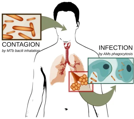
TB is a disease caused by the infection of Mycobacterium tuberculosis (MTb). It can affect practic-ally all organs of the human body, but the lung (pulmonary TB) is of particular high incidence. This is to be expected, since the infection starts with the inhalation of bacilli of MTb during breathing, leading the bacteria directly to the lung. Due to their size, the bacilli are able to reach the pulmonary alveoli, where they are phagocyted by the alveolar macrophages (AMs) [3] (Figure 1).
Development and Characterization of Nanocarrier Systems for the Delivery of Antitubercular Drugs
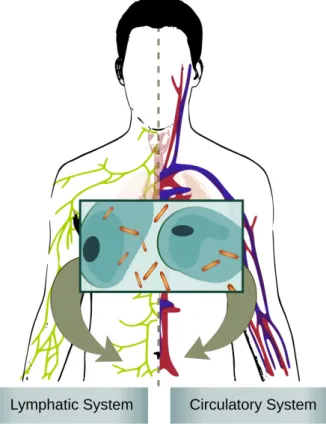
Inside the AMs, the bacilli reside in a membrane-bound vacuole, and for this reason some are able to avoid fusion with lysosomes and posterior digestion [4], ending up co-existing with the AMs [5]. They multiply and eventually escape the lung through the bloodstream and lymphatic system, spreading to other organs of the body, resulting in the extra-pulmonary TB [6](Figure 2). Moreover, MTb may exist within a granulomas consisting of macrophages and giant cells, T cells, B cells, and fibroblasts, and these granulomas can prevail not only in the lung, but in other organs as well. In lat-ent infections, the state of the bacteria within the granuloma is unknown. The estimates are that one third of the world's population is infected with the organism, although usually the infection is present in its dormant state [7].
Some symptoms may be associated with pulmonary TB and extra pulmonary TB, and they could be of help when diagnosing the disease. In pulmonary TB, symptoms include cough, produc-tion of sputum in later stages (due to inflammaproduc-tion and tissue necrosis), hemoptysis (only in rare cases), pleuritic pain, dyspnea (unusual, unless there is extensive disease), and may also cause severe respiratory failure. X-ray of the lung and examination of sputum is often used to confirm pulmonary TB. Extra pulmonary TB has a wider range of symptoms, depending on which organ is
2
Development and Characterization of Nanocarrier Systems for the Delivery of Antitubercular Drugs
affected, and in many cases infection produces sys-temic effects, rather than local ones. Moreover, these effects are many times associated with other ail-ments, such as human immunodeficiency virus (HIV) infection, diabetes mellitus, and neoplastic diseases, which considerably delays diagnosis and increases misdiagnoses, specially with patients co-infected with HIV [8].
1.3 Traditional chemotherapy
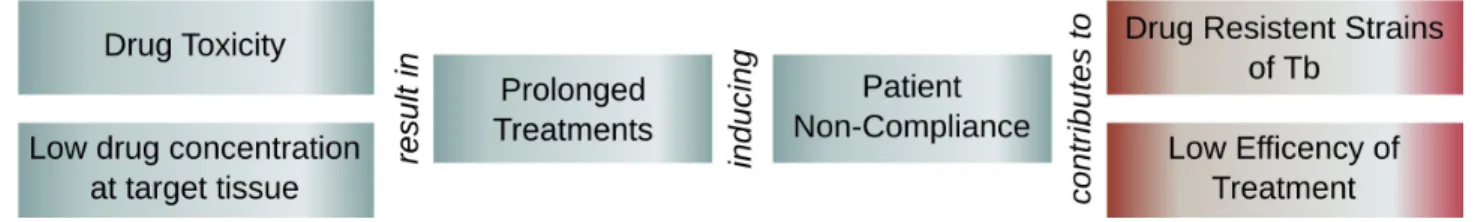
Treatment for TB almost always involves a cocktail of drugs administrated through long periods of time, which contributes to patient non-compliance, result-ing in multi drug resistant (MDR), extremely drug resistant (XDR) [1], and even totally drug resistant strains of TB, which are considerably harder to treat [9] (Figure 3). Also, progress on new drug therapies
has been developing slowly, and the most recent of anti-TB drugs currently in use dates back 50 years. Sarkar et al., in their review of the present TB chemotherapy available and of new and emer-ging drugs, stressed how essential further research in a new drug target is to fight MDR and XDR TB [2].
Currently available chemotherapy includes first-line drugs, such as isoniazid, pyrazinamide, rifampicin, and ethambutol, and second-line drugs, such as para-aminosalicylic acid, ciprofloxacin/ofloxacin, clofazimine, cycloserine, ethionamide, rifabutin, streptomycin, and thio-acetazone [10]. These second-line drugs are only used when treatment with first-line drugs fails. They are less effective, more toxic, and unavailable in many countries due to high costs [11]. The two drugs used in the present work were rifampicin (RIF) and rifabutin (RFB). They belong to the family of rifamycin antibiotics, which are among the most potent anti-tuberculosis agents known. They possess a unique ansa structure consisting of an aromatic nucleus linked on both sides by an aliphatic bridge [12]. RIF is a red crystalline powder. It exhibits a half life between 2.3 and 5 hours on initiation of therapy, but this value decreases to between 2 and 3 hours after repeated treatment. Rifabutin (RFB) is a violet crystalline powder. It has a longest half life, between 32 and 67 hours,
Development and Characterization of Nanocarrier Systems for the Delivery of Antitubercular Drugs
but it also shows increased toxicity, and adverse effects include rash, gastrointestinal disturbance, neutropenia, and occasional uveitis [10].
There are several new drug candidates currently in research and in clinical trials, and several exist-ing drugs are in a state of re-evaluation [9]. In December 2012, the FDA granted an accelerated approval of a new drug, bedaquiline, but only as part of a combination therapy to treat adults with MDR TB when other alternatives are not available [13]. However, bedaquiline has not yet gone through a phase III trial, and several accounts of heart failure have been reported, which may result in bedaquiline being removed from the market. This illustrates how important it is to find new strategies to fight this disease.
1.4 Outline of the dissertation
The present dissertation is divided in five chapters.
Chapter 1, the present chapter, constitutes a brief introduction to the theme and the main goals of the dissertation.
Chapter 2 presents some scientific considerations of TB and it's current treatment options, and it ends with a state of the art regarding nanosystems as carriers for anti-TB drugs. This state of the art, with appropriate modifications, is intended to be submitted for publication as a review article.
In Chapter 3, the materials and methods used in the present work are presented and explained. Results from the experimental work are shown and discussed in Chapter 4.
Finally, Chapter 5 constitutes an overall reflection on the goals and achievements of this work. It also tries to outline possible paths for future work.
4
2 Nanosystems for the pulmonary
delivery of anti-tuberculosis drugs
Since the lung is the most important point of access in the case of infection by MTb [14], [15], exploiting the inhalatory route for drug delivery becomes an exciting hypothesis to fight the dis-ease [16]–[18]. Indeed, the lung is the ideal target site for anti-TB drug delivery, and could provide a delivery portal requiring smaller doses for efficacy, exhibiting reduced toxicity and fewer side effects [3]. Also, the respiratory system behaves as an “aerosol filter”, a property that can be exploited to target particles having specific attributes to the lung [15], and since the lung mucosa has a large surface from which drugs may be systemically absorbed into the bloodstream, escaping the first-pass metabolism [14], enhancing overall bioavailability. This makes pulmonary delivery of drugs an interesting approach for the treatment of pulmonary infections. Adding to this, as was described in section 1.2, pathogenic TB bacilli establish infection mainly in alveolar macro-phages [16]. In this regard, it would be of interest not only to deliver the drugs to the lung, but also to achieve phagocitosys by AMs.
2.1 Lung deposition
To achieve lung deposition, particle size is the most important characteristic to take into account [19]. Figure 4 illustrates the influence of particle size in lung deposition. Particles with dia-meters greater than 5 μm deposit primarily in the mouth and upper airways, while particles with dia-meters ranging from 1-5 μm are the most efficient to reach the deep lung. With particles bellow 1µm, mechanisms such as diffusion and sedimentation become important in reaching the pulmonary alve-oli, and such could be exploited to optimize
Development and Characterization of Nanocarrier Systems for the Delivery of Antitubercular Drugs
an important characteristic in passive targeting of macrophages, since they affect the success of internalization within these cells. In this regard, particles with diameters of about 500 nm have been reported as ideal to undergo phagocytosis by AMs [18].
2.2 Pulmonary administration
Pulmonary administration of drugs must be done using a suitable device. Currently, there are three main delivery devices used for this purpose: nebulisers, pressurized metered-dose inhalers (MDIs), and dry powder inhalers (DPIs). They behave differently and are used with different kinds of particles [22]. MDIs and DPIs are popular choices for the treatment of pulmonary chronic diseases. DPIs are particularly popular, since they are propellant-free, portable, easy to operate and low-cost devices. Unfortunately, dry powders tend to result in particle aggregation, increasing the aerody-namic diameter and lowering the fraction that is respirable, compromising the technique, and ren-dering them unable to reach the deep lung regions where alveolar macrophages lie. Nebulisers may prove to be a better choice, since they can generally produce liquid droplets which are smaller, and thereby provide the opportunity for a larger proportion of the drug to reach the deep lung regions [17].
Although promising and vastly researched, these delivery strategies face obstacles difficult to overcome. With the particular case of anti-TB drugs, so far not one formulation has reach the mar-ket [14]. These difficulties have been reported throughout the scientific literature, and include: the use of safe and accepted excipients, developing scalable processes, developing droplets with proper particle size and morphology for lung deposition, and achieving satisfactory drug loading [14]. Also, usable strategies must be able to account for different lung structures, breathing patterns, and changes in the airway morphology by the pathogenic agent [15]. They must achieve access to poorly-aerated areas of the lung and extracellular bacteria in well-aerated lung tissue, overcoming induction of resistance due to depletion of intracellular drug concentrations, and surpassing limita-tions due to possible innate responses of the host [23]. The use of nanosystems may be of key interest in overcoming the above-mentioned obstacles.
2.3 Active targeting of alveolar macrophages
As stated before, by fine tunning the size of the carrier system, we can enhance phagocitosys by AMs, a desirable event in the case of pulmonary TB. To this passive targeting strategy, there are act-ive targeting strategies that can be used to improve treatment efficacy. In actact-ive targeting strategies,
Development and Characterization of Nanocarrier Systems for the Delivery of Antitubercular Drugs
the constitution and/or structure of the nanosystems is modified, so that certain ligands are present at their surface, changing the way the system interacts with surfaces and cells.
Macrophages exhibit a number of receptors that can be exploited by nanocarriers with appro-priate ligands. Sugars, such as mannose [24] and lactose [25], are among the most commonly used for this purpose, since these receptors are highly expressed in macrophages. Other ligands com-monly used for macrophage targeting include maleylated bovine serum albumin (MBSA), O - steroyl amylopectin (O-SAP), tetrapeptid tuftsin [26], and anionic lipids, such as dicetylphos-phate (DCP).
2.4 State of the art
Nanotechnology is an area of science regarding the design and study of structures, called nano-particles (NPs), in which at least one of the dimensions is measured at the nanoscale range (1 nm – 1000 nm). NPs display unique physical and chemical properties that significantly change with their size. In some cases, particles with dimensions greater than 1µm are considered nanoparticles, since they share some, or even most, of these physical and chemical characteristics.
NPs can be used for medical purposes, namely as nanocarriers for therapeutic and diagnostic agents by means of encapsulation, covalent attachment, or surface adsorption of these agents [27]. The use of NPs in strategies for pulmonary drug delivery is a promising area of research for several reasons. First, the size of these particles can be fine tuned to reach different areas of the lung, allow-ing for successful passive targetallow-ing strategies. Second, their surface can be modified and ligands attached to actively target bodies of interest, such as AMs [20]. Third, studies have demonstrated that pulmonary delivery of nanosuspensions favor higher lung tissue concentrations and markedly raise the lung to serum ratio of drugs, compared with other routes of administration [28]. This could improve bioavailability, reduce side effects, drug toxicity and dosing frequency, which ultimately leads to the increase of patient compliance and better efficacy of treatment [29].
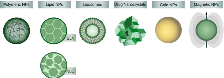
The most frequent approach in these strategies is the use of neutral nanoparticles as carriers for the drug. Common carriers to achieve pulmonary delivery are lipid NPs, polymeric NPs and lipos-somes. Other formulations currently in research include the production of drug nanocrystals, aero-sols with magnetic nanoparticles, nanoparticles with effervescent activity, and gold NPs for the study of internalization of NPs by AMs (Figure 5).
Development and Characterization of Nanocarrier Systems for the Delivery of Antitubercular Drugs
2.4.1 Polymeric based NPs
Natural and synthetic polymers are used to produce polymeric NPs as nanocarriers for drug deliv-ery [30]. Polymeric NPs are among the most widely researched systems for drug delivdeliv-ery in gen-eral, and many reports focus on pulmonary delivery in particular. Nanocarriers consisting of poly(lactide-co-glycolide) acid (PLGA), alginate, gelatine, and chitosan are widely found in the lit-erature. These delivery systems fulfill most requirements placed for pulmonary delivery, such as sufficient association of the therapeutic agent with the carrier particles, targeting of specific sites or cell populations in the lung, protection of the therapeutic agent against degradation, release of the therapeutic agent at a therapeutically optimal rate, ability to be transferred into an aerosol, low tox-icity, and stability against forces generated during aerosolization [31]. They are also interesting materials for the engineering of biodegradable nanocarriers [32]. Many reports on pulmonary deliv-ery using polymeric based nanocarriers have been proposed for a variety of therapeutic strategies, from gene delivery [31], [33]–[35] to more conventional drug delivery [36], [37].
Drug delivery formulations with anti-TB drugs have already been used with these nanocarriers. Jain et al. compared four different NP formulations for ciprofloxacin delivery, three of them being polimeric NPs [38]. The authors incorporated the drug within albumin, gelatin and chitosan NPs and studied their drug release profiles. Of the three polymers, chitosan and albumin NPs proved to be more capable of drug incorporation and sustained release.
Other studies usually focus on one type of nanosystem, although with multiple drugs. Alginate nanoparticles have been studied by Zahoor et al. for the incorporation of rifampicin, isonizid and
8
Development and Characterization of Nanocarrier Systems for the Delivery of Antitubercular Drugs
pyrazinamide [39]. The mentioned particles had aerodynamic diameters in the breatheble range, and presented high drug encapsulation efficiencies for each of the three drugs. Bioavailability of all for-mulations was studied, and these forfor-mulations showed better results than the administration of free drugs. Saraogi et al. used gelatin NPs for the delivery of isoniazid, and they associated them with active targeting by the inclusion of mannose in the formulations [40]. Their study included drug release, macrophage uptake, biodistribution and antitubercular activity studies. They obtained entrapment efficiencies of around 50%, and reported higher accumulation of isoniazid in the lungs when using mannosylated NPs, rendering them suitable for pulmonary delivery of anti-TB drugs.
Abdulla and coworkers used two different molecular weights of poly-(ethylene oxide)-block-distearoyl phosphatidyl-ethanolamine (mPEG2000–DSPE and mPEG5000–DSPE) polymers to pro-duce nanocarriers for pulmonary delivery of rifampicin [41]. They reported high drug loading and entrapment efficiencies, and noticed that these values were influenced by drug:polymer ratio, but not by mPEG–DSPE molecular weight. Particle size and aerodynamic characterization showed that prepared formulations are suitable for lung deposition through inhalation.
Chitosan has some important reported properties to act as an inert carrier, such as biocompatib-ility, low toxicity and biodegradabbiocompatib-ility, it is mucoadhesive and has the capacity of promoting macro-molecules permeation through well-organized epithelia [42]. Moreover, it has recently been shown that cross-linked chitosan NPs can be used with pressurized metered dose inhalers (pMDIs) [43]. This recent study also showed that this approach could be used for local therapy of lung diseases, such as TB. Pourshahab et al. used chitosan NPs as nanocarriers for isoniazid, and obtained a release profile with an initial drug release burst, followed by slow and sustained release in the fol-lowing 6 days [44].
PLGA NPs are extremely common in nanosystems, and have been used to encapsulate some anti-TB drugs. Sung et al. demonstrated that PLGA NPs loaded with rifampicin could be formu-lated, resulting in particles with aerosol properties suitable for lung delivery [45]. They have per-formed in vivo studies, and found evidence of delayed release of the drug. The presence of rifampicin in the lung was detected up to eight hours after the delivery. Jain et al. reported enhanced results when using PLGA NPs conjugated with lactose [25]. The conjugated particles resulted in greater average size and drug payload, slower drug release, and enhanced uptake in lung tissue, mainly due to active targeting of AMs with lactose. Pandey, Sharma and coworkers have used PLGA NPs for the incorporation of rifampicin, isoniazid and pyrazinamide, and administrated them through oral and pulmonary routes [46], [47]. They reported the presence of rifampicin in plasma
Development and Characterization of Nanocarrier Systems for the Delivery of Antitubercular Drugs
for 4-6 days, and of isoniazid and pyrazinamide for 8-9 days. Later, they reported that five doses of nebulized anti-TB PLGA NPs achieved the equivalent therapeutic benefits of 46 daily doses of orally administered free drug [46]. In 2004, the same authors, in further studies, coated similar NPs with wheat germ agglutinin, and reported an increased period during which all drugs were detect-able in plasma, namely 6-7 days for rifampicin and 13-14 days for isoniazid and pyrazinamide [47].
Incorporation of hydrophilic drugs in polymeric nanosystems proves to be challenging. Cheow and Hadinoto modified PLGA preparation methods to achieve higher encapsulation efficiencies of water soluble antibiotics, using levofloxacin as the model drug [48]. They have modified the single emulsification-solvent-evaporation method by including lecithin into the aqueous phase, and the double emulsification-solvent-evaporation method by increasing the water-miscibility level of the oil phase, and succeeded in enhancing encapsulation efficiency in both cases, with no loss regarding drug release profiles and antibacterial activity after spray drying. In other instance, they developed lipid-polymer hybrid NPs to incorporate levofloxacin, ciprofloxacin, and ofloxacin [49]. After ini-tial burst release, hybrid NPs showed a slower drug release than its non-hybrid counterparts. Table 1 summarizes the above mentioned studies on polymeric NPs.
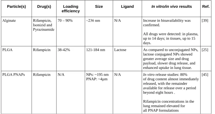
Table 1: Polymeric NPs for incorporation of anti-TB drugs
Particle(s) Drug(s) Loading
efficiency Size Ligand In vitro/in vivo results Ref.
Alginate Rifampicin,
Isonizid and Pyrazinamide
70 – 90% ~236 nm N/A Increase in bioavailability was
confirmed.
All drugs were detected: in plasma, up to 14 days; in tissues, up to 15 days.
[39]
PLGA Rifampicin 38-42% 121-184 nm Lactose As compared to unconjugated NPs,
lactose conjugated NPs showed greater average size and drug payload, slower drug release, and enhanced uptake in lung tissue.
[25]
PLGA PNAPs Rifampicin N/A NPs: ~195 nm
PNAP: ~4µm N/A In vitro release studies: 80% of drug content almost immediately released, with the remainder available for release over a period beyond eight hours .
Rifampicin concentrations in the lung remained elevated for all PNAP formulations
[45]
Development and Characterization of Nanocarrier Systems for the Delivery of Antitubercular Drugs
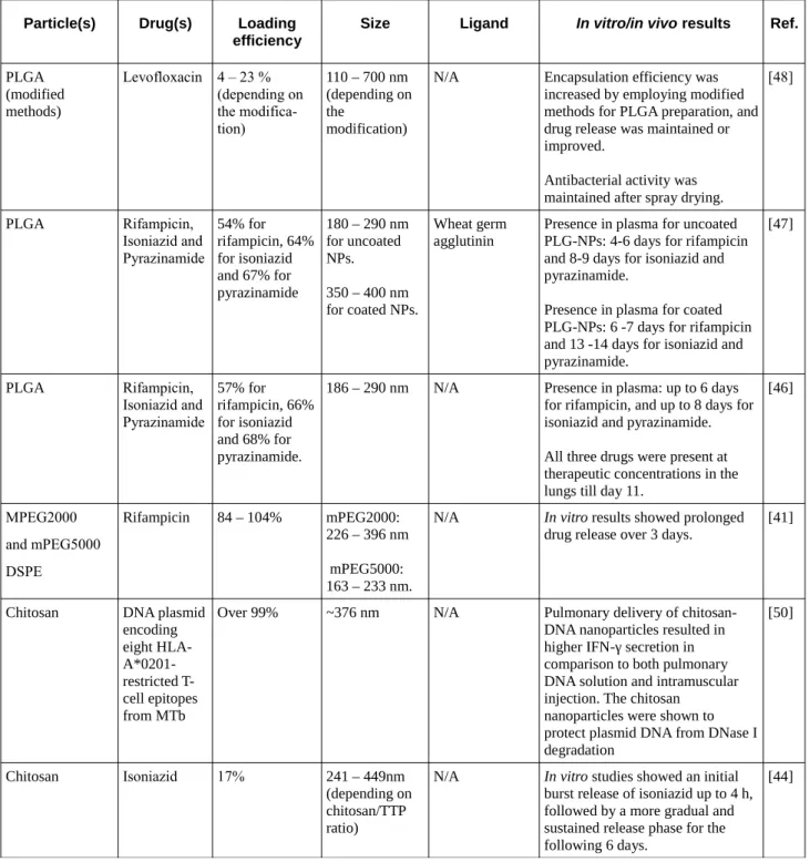
Table 1: Polymeric NPs for incorporation of anti-TB drugs
Particle(s) Drug(s) Loading efficiency
Size Ligand In vitro/in vivo results Ref.
PLGA (modified methods) Levofloxacin 4 – 23 % (depending on the modifica-tion) 110 – 700 nm (depending on the modification)
N/A Encapsulation efficiency was
increased by employing modified methods for PLGA preparation, and drug release was maintained or improved.
Antibacterial activity was maintained after spray drying.
[48] PLGA Rifampicin, Isoniazid and Pyrazinamide 54% for rifampicin, 64% for isoniazid and 67% for pyrazinamide 180 – 290 nm for uncoated NPs. 350 – 400 nm for coated NPs. Wheat germ agglutinin
Presence in plasma for uncoated PLG-NPs: 4-6 days for rifampicin and 8-9 days for isoniazid and pyrazinamide.
Presence in plasma for coated PLG-NPs: 6 -7 days for rifampicin and 13 -14 days for isoniazid and pyrazinamide. [47] PLGA Rifampicin, Isoniazid and Pyrazinamide 57% for rifampicin, 66% for isoniazid and 68% for pyrazinamide.
186 – 290 nm N/A Presence in plasma: up to 6 days
for rifampicin, and up to 8 days for isoniazid and pyrazinamide. All three drugs were present at therapeutic concentrations in the lungs till day 11.
[46] MPEG2000 and mPEG5000 DSPE Rifampicin 84 – 104% mPEG2000: 226 – 396 nm mPEG5000: 163 – 233 nm.
N/A In vitro results showed prolonged
drug release over 3 days.
[41]
Chitosan DNA plasmid
encoding eight HLA- A*0201-restricted T-cell epitopes from MTb
Over 99% ~376 nm N/A Pulmonary delivery of
chitosan-DNA nanoparticles resulted in higher IFN-γ secretion in comparison to both pulmonary DNA solution and intramuscular injection. The chitosan
nanoparticles were shown to protect plasmid DNA from DNase I degradation [50] Chitosan Isoniazid 17% 241 – 449nm (depending on chitosan/TTP ratio)
N/A In vitro studies showed an initial
burst release of isoniazid up to 4 h, followed by a more gradual and sustained release phase for the following 6 days.
Development and Characterization of Nanocarrier Systems for the Delivery of Antitubercular Drugs
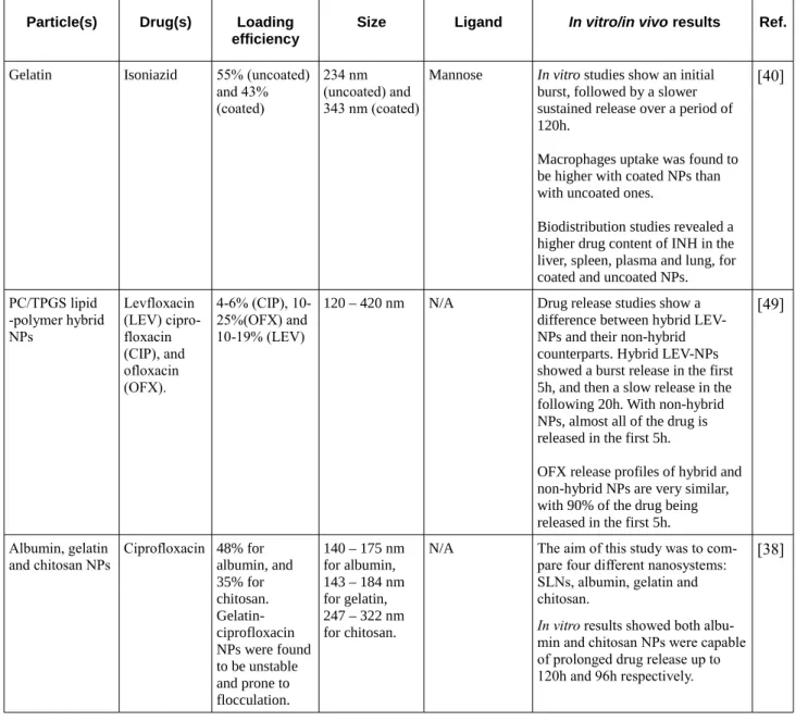
Table 1: Polymeric NPs for incorporation of anti-TB drugs
Particle(s) Drug(s) Loading efficiency
Size Ligand In vitro/in vivo results Ref.
Gelatin Isoniazid 55% (uncoated)
and 43% (coated)
234 nm (uncoated) and 343 nm (coated)
Mannose In vitro studies show an initial
burst, followed by a slower sustained release over a period of 120h.
Macrophages uptake was found to be higher with coated NPs than with uncoated ones.
Biodistribution studies revealed a higher drug content of INH in the liver, spleen, plasma and lung, for coated and uncoated NPs.
[40] PC/TPGS lipid -polymer hybrid NPs Levfloxacin (LEV) cipro-floxacin (CIP), and ofloxacin (OFX). 4-6% (CIP), 10-25%(OFX) and 10-19% (LEV)
120 – 420 nm N/A Drug release studies show a
difference between hybrid LEV-NPs and their non-hybrid counterparts. Hybrid LEV-NPs showed a burst release in the first 5h, and then a slow release in the following 20h. With non-hybrid NPs, almost all of the drug is released in the first 5h.
OFX release profiles of hybrid and non-hybrid NPs are very similar, with 90% of the drug being released in the first 5h.
[49] Albumin, gelatin and chitosan NPs Ciprofloxacin 48% for albumin, and 35% for chitosan. Gelatin-ciprofloxacin NPs were found to be unstable and prone to flocculation. 140 – 175 nm for albumin, 143 – 184 nm for gelatin, 247 – 322 nm for chitosan.
N/A The aim of this study was to
com-pare four different nanosystems: SLNs, albumin, gelatin and chitosan.
In vitro results showed both
albu-min and chitosan NPs were capable of prolonged drug release up to 120h and 96h respectively.
[38]
2.4.2 Liposomes
Liposomes are vesicular structures, constituted by phospholipid bilayers enclosing an aqueous medium. They were discovered in 1965 and have been attracting interest as nanocarriers for many years [26]. They possess a unique and versatile structure, with lipid and aqueous regions, which can be altered to make them better suited to carry hydrophilic, lipophilic, or both hydrophilic and lipo-philic particles. Their size can be fine tuned to achieve different regions of the lung by passive tar-geting, and their structure and composition can be changed to achieve active targeting to specific cells, namely AMs. Surface mannosylation is one of the most successful examples of this strategy,
Development and Characterization of Nanocarrier Systems for the Delivery of Antitubercular Drugs
as it has been shown to increase uptake of liposomes by AMs [51]–[54]. Liposomes seem particu-larly appropriate for pulmonary delivery, since they can be formulated from endogenous com-pounds, such as the components of pulmonary surfactant [55]. However, many aerosolization techniques can compromise liposome structure and integrity. In most aerosolized liposome formula-tions for targeted pulmonary delivery, liposomes are formed before packaging. This usually results in rupturing of vesicle structure during administration, thereby losing the ability for sustained release. However, it had already been demonstrated that PEGylated and plain Tf-conjugated lipo-somes are stable enough to undergo nebulisation in the course of an inhalational therapy [56]. Recently, Chattopadhyay et al. showed that changing their composition, by incorporation of charged lipids and cholesterol molecules into the bilayer, prevented particle aggregation and preserved bilayer integrity after air-jet nebulization [57]. Another approach is based on the fact that a drug– lipid mixture solubilized in chlorofluorocarbon (CFC) will form liposomes upon hydration in small airways. Gaur et al. explored the hypothesis of forming liposomes in situ, since the lung has a wet surface which could provide an aqueous phase for spontaneous formation [58]. They have reported that no vesicle rupture was observed with in situ formed liposomes, and prolonged drug release was achieved.
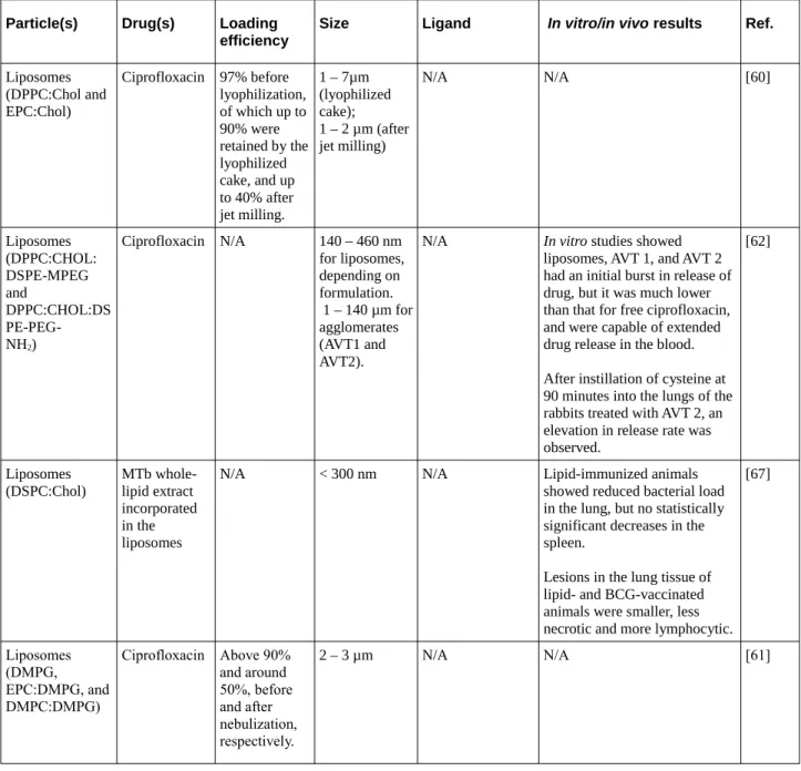
For all these reasons, it is therefore not uncommon to find a vast number of studies involving pulmonary delivery of drugs with liposomal formulations, many of them focusing on anti-TB drugs. Ciprofloxacin was one of the first anti-TB drugs to be used with liposomes. Wong, Finlay and coworkers explored liposomal ciprofloxacin in 1998. They studied liposome disruption during aerosolization, using 25 nebulizers [59]. Later, they published results on spontaneous forma-tion of liposomes on dispersion of phospholipid-based powder formulaforma-tions [60], [61]. With these liposomes, they achieved entrapment efficiencies of 44% for ciprofloxacin, but the value increases up to 96% with the incorporation of negatively charged lipids. Bhavane et al. developed liposome agglomerates that could be triggered by the instillation of a biologically acceptable agent [62]. They used cysteine as such agent, and proposed that this strategy could facilitate post-administration modulation of the drug release rate. It could allow for treatment regimens where the administration of one single dose would be sufficient for an extended period of time, since drug release could be periodically accelerated. They also found that progressive release of the drug does not cause signi-ficant inflammation, unlike the administration of free ciprofloxacin.
Other anti-TB drugs have already been studied with liposomes. Justo and Moraes studied the possibility of passive liposomal encapsulation of isoniazid, pyrazinamide, rifampicin, ethionamide,
Development and Characterization of Nanocarrier Systems for the Delivery of Antitubercular Drugs
and streptomycin [55]. However, under the tested conditions, rifampicin and ethionamide were not successfully encapsulated. Low encapsulation efficiencies were obtained for isoniazid and pyrazin-amide, being the encapsulation of streptomycin only higher at a drug to lipid molar ratio of 0.04. Gaur et al. published a feasibility study where they used rifampicin as the model drug [58]. In this study, in situ formed liposomes showed better sustained release profile than the preformed lipos-somes, but both liposomal aerosols showed improved delivery of rifampicin over plain drug aero-sols, with encapsulation efficencies around 30%. Liposomes for the delivery of isoniazid have been developed and evaluated in vitro [63] by Chimote and Banerjee. They observed a sustained release of isoniazid encapsulated in liposomes, tooking place over 24 h after a burst release in the first 5h. They have also conducted biocompatibility and stability studies, and found the formulations to be haemocompatible and cytocompatible, and stable for the duration of at least one month.
The possibility of surface coating to achieve active targeting with liposomes has also been a subject of interest. Vyas et al. used rifampicin when studying liposomes coated with macro-phage-specific ligands, and reported a preferential accumulation of lingad-coated formulations in the lung macrophages, namely MBSA and O-SAP coated liposomes [64]. In vivo tissue distribution studies are on par with these results, by showing higher lung drug concentration for ligand-coated liposomes. O-SAP surface modification was also the focus of Deol and Khuller, who developed coated liposomes for the encapsulation of both rifampicin and isoniazid [65]. They compared the results with uncoated ones, and reported that encapsulating drugs within liposomes reduced toxicity, and that O-SAP coating succeeded in enhancing lung accumulation. Tuftsin functionalization of liposomes encapsulating rifampicin was studied by Agarwal and coworkers [66]. They reported interesting results: considering one single administration, tuftsin functionalization did not give bet-ter results than uncoated formulations, but with regular administration over two weeks, tuftsin lipo-somes were more efficient in controlling tuberculosis.
The use of aerosolized liposomes as vaccines to fight TB is a different strategy already con-sidered. Dascher et al. incorporated lipids from MTb into liposomes, and administrated them to guinea pigs [67]. They succeeded in reducing bacterial burden in the lung, but regarding the spleen results were not statistically significant. Moreover, lipid-vaccinated lungs showed significantly less pathology, with granulomatous lesions being smaller and more lymphocytic.
Gene therapy has also been the subject of many studies with liposomes for the past twenty years, but despite these efforts, little progress towards developing an effective pharmaceutical product has been done, and the vast majority of clinical trials still uses viral delivery of DNA, a
Development and Characterization of Nanocarrier Systems for the Delivery of Antitubercular Drugs
much more effective approach, despite the associated toxicity issues [68]. There is ongoing research to address these problems. One recent study used single-tailed cationic lipid 6-lauroxyhexyl lysinate (LHLN) to prepare cationic liposomes, and in vivo results showed that, compared with commer-cially available Lipofectamine2000/DNA complexes, LHLN-liposomes exhibited lower cytotox-icity, and higher pulmonary gene transfection efficiency [69]. Table 2 summarizes the currently found studies with liposomes as carriers for anti-TB drugs.
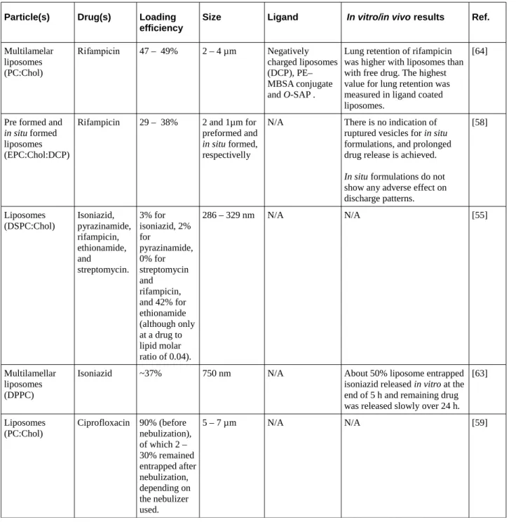
Table 2: Liposomes for the encapsulation of anti-TB drugs
Particle(s) Drug(s) Loading efficiency
Size Ligand In vitro/in vivo results Ref.
Multilamelar liposomes (PC:Chol) Rifampicin 47 – 49% 2 – 4 µm Negatively charged liposomes (DCP), PE– MBSA conjugate and O-SAP .
Lung retention of rifampicin was higher with liposomes than with free drug. The highest value for lung retention was measured in ligand coated liposomes.
[64]
Pre formed and
in situ formed
liposomes (EPC:Chol:DCP)
Rifampicin 29 – 38% 2 and 1µm for
preformed and
in situ formed,
respectivelly
N/A There is no indication of
ruptured vesicles for in situ formulations, and prolonged drug release is achieved.
In situ formulations do not
show any adverse effect on discharge patterns.
[58]
Liposomes
(DSPC:Chol) Isoniazid, pyrazinamide, rifampicin, ethionamide, and streptomycin. 3% for isoniazid, 2% for pyrazinamide, 0% for streptomycin and rifampicin, and 42% for ethionamide (although only at a drug to lipid molar ratio of 0.04). 286 – 329 nm N/A N/A [55] Multilamellar liposomes (DPPC)
Isoniazid ~37% 750 nm N/A About 50% liposome entrapped
isoniazid released in vitro at the end of 5 h and remaining drug was released slowly over 24 h.
[63] Liposomes (PC:Chol) Ciprofloxacin 90% (before nebulization), of which 2 – 30% remained entrapped after nebulization, depending on the nebulizer used. 5 – 7 µm N/A N/A [59]
Development and Characterization of Nanocarrier Systems for the Delivery of Antitubercular Drugs
Table 2: Liposomes for the encapsulation of anti-TB drugs
Particle(s) Drug(s) Loading efficiency
Size Ligand In vitro/in vivo results Ref.
Liposomes (DPPC:Chol and EPC:Chol) Ciprofloxacin 97% before lyophilization, of which up to 90% were retained by the lyophilized cake, and up to 40% after jet milling. 1 – 7µm (lyophilized cake); 1 – 2 µm (after jet milling) N/A N/A [60] Liposomes (DPPC:CHOL: DSPE-MPEG and DPPC:CHOL:DS PE-PEG- NH2) Ciprofloxacin N/A 140 – 460 nm for liposomes, depending on formulation. 1 – 140 µm for agglomerates (AVT1 and AVT2).
N/A In vitro studies showed
liposomes, AVT 1, and AVT 2 had an initial burst in release of drug, but it was much lower than that for free ciprofloxacin, and were capable of extended drug release in the blood. After instillation of cysteine at 90 minutes into the lungs of the rabbits treated with AVT 2, an elevation in release rate was observed.
[62]
Liposomes
(DSPC:Chol) MTb whole-lipid extract incorporated in the liposomes
N/A < 300 nm N/A Lipid-immunized animals
showed reduced bacterial load in the lung, but no statistically significant decreases in the spleen.
Lesions in the lung tissue of lipid- and BCG-vaccinated animals were smaller, less necrotic and more lymphocytic.
[67] Liposomes (DMPG, EPC:DMPG, and DMPC:DMPG) Ciprofloxacin Above 90% and around 50%, before and after nebulization, respectively. 2 – 3 µm N/A N/A [61] 16
Development and Characterization of Nanocarrier Systems for the Delivery of Antitubercular Drugs
Table 2: Liposomes for the encapsulation of anti-TB drugs
Particle(s) Drug(s) Loading efficiency
Size Ligand In vitro/in vivo results Ref.
Liposomes
(EPC:Chol) Isoniazid, rifampicin 8 – 10% for isoniazid. 44 – 49% for rifampicin. ≥200 nm for O-SAP coated liposomes. <200nm for DSPE-PEG liposomes.
O-SAP Encapuslated drugs were found to be less toxic than free drug. Drug uptake in macrophages was found to be similar between encapsulated and free drugs.
Slow and controlled drug release was achieved in encap-sulated drugs.
O-SAP coating enhanced lung
accumulation. Also, pre-admin-istration of PC and Chol lipo-somes before the injection of lung specific stealth liposomes, further enhanced their uptake in lungs.
[65]
Liposomes (EPC) Rifampicin. 28 – 32% 25 – 65nm Tuftsin With 10 mg/kg dose of
lipo-somal RIF, a significant reduc-tion in the lung bacillus load and an increase in MST were observed, compared with those in free RIF treated animals. Regarding tuftsin functionaliza-tion, one single treatment with coated liposomes was only mar-ginally better than that observed with uncoated ones, but coated liposomes given twice weekly for 2 weeks was considerably more effective than uncoated ones in controlling TB.
[66]
2.4.3 Drug nanocrystals
The pure use of therapeutic agents in the form of nanocrystals has been proposed as a system for drug delivery. They are used as dispersions of pure drug nanoparticles kept stable through the pres-ence of a minimum amount of a surfactant – nanosuspensions. Drug nanocrystals dissolve rapidly in the lung lining fluid leading to a high concentration, which is helpful for localized treatment of res-piratory diseases such as pulmonary TB. Results show that these could be used in drug delivery for-mulations to improve pharmacokinetic, pharmacodynamic and targeting properties of poorly soluble drugs. Gao et al. reported two different kinds of pulmonary formulations containing drug nanocrystals [28]: aqueous nanosuspension packaged and administered by a nebulizer; drug nano-crystals collected and transported into the lung by the small aerosol droplets generated by the nebulizer.
Development and Characterization of Nanocarrier Systems for the Delivery of Antitubercular Drugs
Spore like drug particles for deep lung deposition have also been proposed as an innovative system [70]. Hollow and spore like nanoagglomerates were obtained by mixing the drug solution with an antisolvent in a high gravity environment. The fabrication of drug particles similar to spores may improve the pulmonary drug delivery efficiency in DPIs, and is a more efficient, cost-effective and easy to scale up method over milling, homogenization, spray freezing into liquid, and supercrit-ical antisolvent precipitation to prepare nanosuspensions. According to the authors, uniform particle size and controlled morphology can be achieved with this technique.
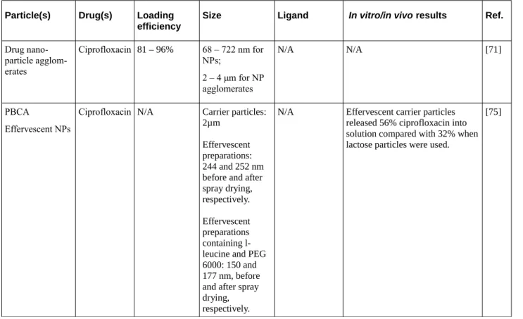
Currently, only one report was found regarding the production of nanocrystals or nanoagglom-erates of an anti-TB drug. El-Gendy et al. prepared ciprofloxacin nanosuspensions that were then flocculated to form nanoparticle agglomerates [71]. Nanoparticle size ranged from 68 – 722 nm, depending on the formulation, and agglomerates exhibited a particle size range of 2 – 4 μm. They performed dissolution studies, and compared the results with the stock drug. Results showed that the dissolution rate was improved, demonstrating that these techniques may help to overcome some of the solubility issues presented by new anti-TB drugs, specially by molecules that, although did not pass from the clinical trials due to solubility issues, shown higher potential as anti-TB drugs.
2.4.4 NPs with effervescent activity
Nanoparticles with effervescent activity have recently been suggested for pulmonary delivery. Oral drug delivery associated with effervescent pharmaceutical formulations is used for a long time, in stomach distress medications, vitamin supplements and analgesics. Effervescent activity of the car-rier particles occurs when the carcar-rier particles are exposed to humidity, adding an active release mechanism to the pulmonary route of administration. Additionally, effervescent particles can be synthesized with adequate size for deep lung deposition, and the technology appears to be safe for pulmonary delivery [72].
Although effervescent NPs have been mostly studied as a promising pulmonary delivery strategy for anti-cancer drugs [72]–[74], one report has been found regarding their use for the deliv-ery of ciprofloxacin [75]. Ely and coworkers have developed and studied different powder composi-tions with effervescent activity, and found two formulacomposi-tions suitable for pulmonary delivery. These formulations had the addition of l-leucine and PEG 6000, which improved the aerodynamic charac-teristics of the powder particles. Effervescent activity of the prepared formulations resulted in the release of nanoparticles with less agglomeration compared to the carrier particles made just of lactose.
Development and Characterization of Nanocarrier Systems for the Delivery of Antitubercular Drugs
Table 3: Other nanosystems for the delivery of anti-TB drugs
Particle(s) Drug(s) Loading efficiency
Size Ligand In vitro/in vivo results Ref.
Drug nano-particle agglom-erates Ciprofloxacin 81 – 96% 68 – 722 nm for NPs; 2 – 4 μm for NP agglomerates N/A N/A [71] PBCA Effervescent NPs
Ciprofloxacin N/A Carrier particles:
2µm Effervescent preparations: 244 and 252 nm before and after spray drying, respectively. Effervescent preparations containing l-leucine and PEG 6000: 150 and 177 nm, before and after spray drying, respectively.
N/A Effervescent carrier particles
released 56% ciprofloxacin into solution compared with 32% when lactose particles were used.
[75]
2.4.5 Gold and magnetic NPs
Gold NPs have recently been used to study internalization and intracellular translocation of inhaled nanoparticles in rat AMs [76]. Particles used had mean hydrodynamic radius of 16 nm. Results showed AMs had efficiently internalized NPs by endocytosis. Gold NPs have been conjugated with streptomycin [77], and it has been demonstrated that ciprofloxacin binds to gold NPs [78], but the cited studies do not focus on pulmonary delivery, and no other reports have been found regarding the use of these particles for pulmonary delivery, regardless of the model drug.
The use of magnetic aerosols using superparamagnetic iron oxide NPs has also been suggested as a way to improve drug delivery to the lung [79], and ciprofloxacin has been used as a model drug in the development of superparamagnetic nanocomposites with magnetically mediated release of the loaded anti-TB drug [80]. However, no study has been found combining these two strategies to achieve magnetically mediated pulmonary delivery of anti-TB drugs.
Development and Characterization of Nanocarrier Systems for the Delivery of Antitubercular Drugs
2.4.6 Lipid NPs
Lipid NPs are the last nanosystems presentd in this state of the art and are the focus of this thesis. Generally speaking, and by contrast with liposomes and polymeric nanoparticles, lipid NPs show higher drug loading capacity, higher stability, and require the use of lower amounts of organic solvents during production [81]. As with liposomes and most polymeric NPs, these nanocarriers are biocompatible and can be produced with appropriate size and morphology for lung targeting and deposition [82], and have been studied as a viable pulmonary drug delivery strategy [83]. It is also possible to modify the surface of lipid NPs to achieve active targeting of AMs. Mannose is a com-mon surface modification with lipid nanocarriers [84].
Solid lipid nanocarriers (SLNs) and nanostructured lipid carriers (NLCs) are the two most common lipid NPs used. The published results by Jain and coworkers, who compared four different nanocarriers for the incorporation of ciprofloxacin, showed that SLNs are capable of prolonged drug release [38]. This work is one of the three reports that were found regarding pulmonary deliv-ery of SLNs loaded with drugs for the treatment of TB, namely rifabutin, isoniazid, rifampicin and pyrazinamide. Nimje et al. prepared rifabutin loaded SLNs, and compared uncoated formulations with formulations coated with mannose [24]. Results showed cellular uptake in AMs was almost six times enhanced due to mannose coating. Coated formulations also showed to be less immunogenic and more suitable for sustained delivery. Pandey and Kuller have prepared SLNs for pulmonary delivery through nebulization [85]. They incorporated isoniazid, rifampicin and pyrazinamid, of which rifampicin showed the highest incorporation due to the lipid-based nature of the formulation and lipophilic characteristics of the drug. The nebulized SLNs were successfully deposited in the lungs, and were detected in other organs up to 7 days after administration. Administrated free drug was cleared from the system within 24 – 48 h. Jain and Banerjee included SLNs in their list of nanosystems to deliver ciprofloxacin, and concluded that these NPs were suitable for drug loading, and capable of sustained drug release [38].
Development and Characterization of Nanocarrier Systems for the Delivery of Antitubercular Drugs
The matrix of SLNs consists of solid lipids only, with perfect crystallinity. This results in lower drug loading, since there are very few empty spaces in which the drug can be found. It also results in expulsion of drug content during long storage due to changes in lipid packaging. NLCs are differ-ent structures. The matrix consists of both solid and liquid lipids, consequdiffer-ently showing lower crys-tallinity and higher incidence of nanostructures, which won't result in denser lipid packaging over time. Thus, higher drug loading and stability during long storage is achieved, when compared with SLNs [86]. Figure 6 illustrates the differences in the matrix of SLNs and NLCs, and the overall influence of such differences in drug loading and expulsion over time. However, it should be noted that no study was found regarding the use of NLCs as carriers for any anti-TB drug. Table 4 sum-marizes currently found studies regarding the use of lipid nanoparticles for the treatment of TB.
Figure 6: Schematic representation of the matrix of SLN and NLC.
A:SLN exhibits a high order matrix, while NLC exhibits a low order matrix. B: Drug is loaded into SLNs and NLCs. C: Over time, SLNs tend to a denser lipid packaging and higher drug expulsion than NLCs.
Development and Characterization of Nanocarrier Systems for the Delivery of Antitubercular Drugs
Table 4: Lipid NPs for the incorporation of anti-TB drugs
Particle(s) Drug(s) Loading efficiency
Size Ligand In vitro/in vivo results Ref.
SLN
(tristearin) Rifabutin 82% (uncoated)
and 87% (coated) 251 nm (uncoated) and 389 nm (coated)
Mannose In vitro studies showed a
sustained drug release for 120h, during which uncoated SLNs showed higher drug release. Macrophage uptake was higher for coated SLNs.
In vivo results showed higher
drug presence for coated SLNs in the lungs. [24] SLN (Stearic acid) Isoniazid, rifampicin and pyrazinamid 51% for rifampicin, 45% for isoniazid and 41% for pyrazinamide.
1 – 2 µm N/A In vitro results varied for
simulated gastric or intestinal fluid. The drug released was <20% in the first 6 h and 11– 15% during 6–72 h for isoniazid/pyrazinamid; 9% in the first 6 h and 11% during 6– 72 h for rifampicin, although rifampicin release was in the range of 8–12% during the entire study period for intestinal fluid.
All the three drugs could be detected in the lungs, liver and spleen of the animals up to day 7 following the nebulization.
[85]
SLN (stearic acid)
Ciprofloxacin 39% 74 – 99 nm N/A The aim of this study was to
compare four different nanosys-tems: SLNs, albumin, gelatin and chitosan.
In vitro results showed SLNs
were capable of a prolonged drug release up to 80 h.
[38]
3 Materials and Methods
3.1 Development of Nanostructured Lipid Carriers
3.1.1 Initial formulation
NLCs were initially prepared with with Cetyl Palmitate (C32H64O2;M=480.83 g mole-1; Gattefossé)
as the solid lipid, and Mygliol 812 (from Acofarma) as the liquid lipid. Polysorbate 60 (C64H126O26; M=1 310 g mol-1; Sigma Aldrich) was used as surfactant, to stabilize the emulsion.
Quantities used for each are presented in table 5:
There are several methods to produce NLCs in the laboratory, such as high pressure homogenization, microemulsion technique, emulsification-solvent evaporation, emulsification-solvent diffusion method, solvent injection (or solvent displacement) method, phase inversion, multiple emulsion technique, ultra-sonication and membrane contractor technique [86].
In the present work, an ultra-sonication method was used. In detail, the two lipids and the surfact-ant were heated in a water bath up to 70º C, temperature at which both lipids are in the liquid state. When the solid lipid was fully melted, 4.4 mL of heated ultrapure water (T = 70º C) was added to the mixture. Mixture then went through ultraturrax (Ystral X10/20 E3) at 3500 rpm for 30 s, and sonication (Sonics Vibra-cell, with CV18 probe) at 70% power for 5 min, which resulted in a nanoemulsion. This nanoemulsion was finally left to cool at room temperature and stored. Figure 7 features a schematic representation of the process.
Mass / mg
Cetyl palmitate 350
Mygliol 812 150
Polysorbate 60 100
Development and Characterization of Nanocarrier Systems for the Delivery of Antitubercular Drugs
3.1.2 Drug Loading
Two anti-TB drugs were used in the present work, one first-line drug (RIF) and one second-line drug (RFB). A third first-line drug (isoniazid) was considered, but its high hidrofilicity rendered it unsuitable for lipid NPs. RIF (C43H58N4O12; M=822.94 g mol ¹; Sigma Aldrich) ⁻ and RFB (C46H62N4O11;M=847.00468 g mol ¹; ⁻ Sigma Aldrich) are lipofilic molecules, and in principle suit-able to be loaded with high efficiency in lipid NPs.
Drug content in loaded NLCs (NLC-RIF and NLC-RFB) was added (~7 mg) to the lipid mix-ture prior to the melting stage. When the melted lipid mixmix-ture and the drug form a homogeneous liquid mixture, ultra-pure water is added, followed by ultraturrax and sonication, as described before.
3.1.3 Choice of solid lipid for improved drug loading
Drug loading and release are essential parameters to evaluate a drug delivery system. Solubility of the drugs in the lipid mixture plays a major role in the amount of drug a nanosystem can be loaded with. When it comes to lipid NPs, it is important to choose lipids where the used drugs exhibit high solubility. For this purpose, qualitative solubility studies where performed to choose a solid lipid that would yield better results than the initially used Cetyl Palmitate.
In brief, the same amount of rifampicin was added to the same amount of five solid lipids: Cetyl Palmitate, Dynassan 116, Compritol 888 ATO, Golucire 43/01, Precirol ATO 5 (all lipids from Gattefossé). Lipids Compritol 888 ATO and Precirol ATO 5 both gave satisfactory results, showing the highest solubility for the used drugs. However, Compritol 888 ATO has a much higher melting
24
Development and Characterization of Nanocarrier Systems for the Delivery of Antitubercular Drugs
point (above 70 ºC) and it was deferred in favor of Precirol ATO 5. The final lipid mixture used in the formulation was Precirol ATO 5 as the solid lipid, and Mygliol 812 as the liquid lipid.
3.1.4 Mannose coating
As described in detail in the state of the art, mannose (C6H12O6; M=180.16 g mol-1) has already been
used to enhance uptake by AMs with gelatin NPs [40], liposomes [51] and SLNs [24], and it was chosen as the ligand in the presented work to achieve active targeting.
Coating of NLCs with mannose (NLC-M) was initially adapted from the methods described by Jain et al. for SLNs used for site specific delivery of anti-cancer drugs [87]. First, NLCs were modified in order to exhibit amine groups at the surface. To achieve this, stearyl amine (Octadecylamine; C18H39N; M=269.51 g mol-1; Sigma Aldrich) was added to the
lipid mixture described at 3.1.3, and the resulting mixture was dissolved in acetone:ethanol (1:1 v/v) at 70ºC. An aqueous phase was prepared, containing the surfactant and the drug. The lipid phase was slowly mixed with the aqueous phase, at 70ºC, using a micropipette, while slowly agitating. The mix-ture was then ultraturraxed and sonicated as described before. For mannosylation of NLCs, a mannose (Sigma Aldrich) solution (50 mM) was prepared in acetate buffer (pH = 4), and subsequently added to the NLC suspension and left under constant and gentle stirring for a period of 48h. The rationale behind this procedure follows: the acidic environment would result in the ring opening of the mannose molecules; the alde-hyde group would then react with free amine present at the surface of the stearyl amine functionalized NLCs, leading to the formation of a Schiff's base (–N=CH–) (Figure 8), which
Development and Characterization of Nanocarrier Systems for the Delivery of Antitubercular Drugs
This first method resulted in a very thick formulation, and modifications were performed to achieve a workable liquid suspension, as well as to try to avoid using an organic solvent in the process (acetone). The final method consisted in the development of the NLCs as described in 3.1.1, with the lipid mixture stated in 3.1.3, to which the drug content was added. Afterwards, the solution of mannose in acetate buffer was added to the NLC suspension, together with stearyl amine. The mix-ture was then left under constant and gentle stirring for a period of 48h. Finally, the resulting sus-pension was washed through dialysis in ultra-pure water for 30 min, using a regenerated cellulose tubular membrane (CelluSep T3; Nominal MWCO: 12 000 – 14 000; Wall thickness: 20 μm; Orange Scientific), to remove any excess mannose and stearyl amine. Figure 9 shows a schematic representation of the final process to achieve mannose coated, drug loaded NLCs.
3.1.5 Lyophilization
Lyophilization (or freeze-drying) is the process of removing water from a frozen sample, using sub-limation and desorption under vacuum. Many pharmaceutical products, mainly heat sensitive
26
Figure 9: A schematic representation of the final process to achieve mannose coated, drug loaded NLC suspension.
Development and Characterization of Nanocarrier Systems for the Delivery of Antitubercular Drugs
pounds, are dried using this technique, since it improves the long-term physico-chemical stability and prevents degradation reactions such as hydrolysis. Lyophilization is also used with NPs to pre-vent particle aggregation [88].
Liofilization was the final step of the present formulation, not only to improve physico-chemical stability, prevent degradation and particle aggregation, but also to allow a successful detection the Schiff's Base (thus confirming mannose coating) using Infrared Spectroscopy, and to provide a better material to perform citotoxicity studies, since re-suspension could be done using cell medium. The quality of the lyophilized product is highly dependent on the parameters of the process. The ideal lyophilization parameters for NLCs were based on the literature. Varshosaz et al. performed an exhaustive study on lyophilization parameters of NLCs, experimenting with various temperatures, pressures and cryoprotectants [88]. Considering the differences in materials available, a lyophilization process for NLCs was adapted from the procedure considered ideal by Varshosaz and coworkers.
A VirTis freeze dryer (Advantage Plus EL-85; SP Scientific) was used in this process. Samples were prepared with Aerosil 2% (m/m) as cryoprotectant. Initial freezing was done at – 60 ºC for 720 min. Condensatin was made at – 80 ºC under 150 mTorr of pressure. Drying was done at 20º C for 1 200 min, under 150 mTorr of pressure. Secondary drying was performed at 25 ºC, for 1 200 min, under 100 mTorr of pressure.
3.2 Characterization
3.2.1 Particle Sizing
To determine the size of the developed NLCs, dynamic light scattering (DLS) – also called photon correlation spectroscopy – was used. The advantages of using this technique over other techniques to determine the average size of a large population of spherical particles include the short time required to perform the measurements, the statistical validity of the results and the relatively low cost of the necessary devices. There are, however, drawbacks, such as the influence in the results of dust particles and nanoparticle aggregation [89].
The determination of particle size by DLS works by studying the scattering of light by particles in suspension, assuming they are spherical in geometry and animated with Brownian motion. In a typical DLS experiment, a beam of polarized light, usually a LASER, passes through a scattering medium. A detector measures the scattered light at a fixed angle, called scattering