VANESSA MENDES HENRIQUES
Characterization of TRIB2-mediated resistance to
pharmacological inhibition of MEK
Faro, 2017
VANESSA MENDES HENRIQUES
Characterization of TRIB2-mediated resistance to
pharmacological inhibition of MEK
Faro, 2017
Oncobiology Master Thesis
Supervisors: Dr. Wolfgang Link Dr. Bibiana Ferreira
i
pharmacological inhibition of MEK”
Declaração de autoria do trabalho
Declaro ser a autora deste trabalho, que é original e inédito. Autores e trabalhos
consultados estão devidamente citados no texto e constam da listagem de
referências incluída.
Copyright Vanessa Mendes Henriques
_____________________________
A Universidade do Algarve tem o direito, perpétuo e sem limites geográficos, de
arquivar e publicitar este trabalho através de exemplares impressos reproduzidos
em papel ou de forma digital, ou por qualquer outro meio conhecido ou que venha
a ser inventado, de o divulgar através de repositórios científicos e de admitir a sua
cópia e distribuição com objetivos educacionais ou de investigação, não
comerciais, desde que seja dado crédito ao autor e editor.
ii
First, I would like to thank the greatest opportunity given by professor doctor Wolfgang Link to accepting me into his team, contributing to my scientific and personal progress. Thank You for all Your knowledge and help across the year.
A special thanks to Bibiana Ferreira who stayed by me all year and trained me. Thank You for all you taught me, thank you for all your patience and time and all the support. Thank you for all the great times that You provided me. It was an amazing experience to learn and work with You, which made me grow as a scientist and also as a person. Thank You for everything.
To the other members of my team Susana Machado and Pedro Charlito, for their help and support all the time and for the good moments in the lab.
To João Santos who I promised a special thanks. Thank You for all the help and support in the lab, thank You for all the conversations in the TCU and for being my friend.
To all the Professors of Oncobiology Master at Algarve’s University for the knowledge transmitted and for all the support.
To my brother and also best friend Tiago Henriques, who has been one of the most important persons of my life. Thank You for all the kindness and support across the years. Thank You for being always there for me.
To my father António Henriques and mother Carmo Mendes who made everything possible, to provide me this amazing opportunity and for all the support. To my godmother Carla Silva who has been a second mother to me. Thanks for every happy moment by Your side, for all the laughs and mainly for being always there when I need.
To my friends of a life-time: Alexandra Borges, Tiago Dias, Lara Neves e Carolina Roque for all the good moments, the support during this year and to make me who I am. I hope to have You at my side for the eternity.
To my friends specially to Elisabete Silva, who has revealed one of my best friends in the past few years, to Marisa Flook, Cátia Salvador. Thank You for all the conversations and confidences, for all the nights out, for all the laughs and all the support.
To my friends that I had the pleasure to meet during my master and who also revealed to be incredible friends: Cristina Fernandes, Gabriela Carrasqueiro and Rita Bastos. Thank you Cristina for being there in the good and in the bad moments, you’re an example of strength and
iii for all the good moments You provided and for being there when I most needed. Thank You Rita, one of the weirdest and amazing persons that I had the pleasure to meet, thanks for all the conversations, support and good moments. I will miss You.
iv Chemoresistance and metastasis are the main reasons for treatment failure in melanoma patients. MAPK pathway is often hyperactivated in melanoma due to BRAF mutations. BRAF and MEK inhibitors revolutionized the standard-care of patients with advanced melanoma. Yet, patients develop resistance to these drugs very fast.
Previous studies showed that Tribbles homolog 2 (TRIB2) is overexpressed in melanoma and confers resistance to chemotherapeutic and targeted drugs such as darcarbazin, PI3K and mTOR inhibitors. Furthermore, TRIB2 protein contains a MEK1 binding site. Taking this into account, we hypothesize that TRIB2 might confer resistance to MEK inhibition.
In order to test our hypothesis, we generated isogenic melanoma cell lines with TRIB2 knockdown, using shRNA, and cells with TRIB2 depletion using CRISPR technique.
Since the members of the Tribbles protein family might be functionally redundant and compensate for TRIB2 depletion, we decided to determine mRNA and protein levels of TRIB1, TRIB2 and TRIB3 using q-PCR and Western-Blot techniques, respectively, on a panel of melanoma and non-melanoma cell lines. We treated these isogenic cell lines with the MEK inhibitor Refametinib for 72h. The resistance was evaluated through cell death analysis, using cell counting based on trypan-blue and annexin V/ Propidium iodide staining.
The isogenic cell lines were successfully established and determined that compensation of TRIB2 through TRIB1 or TRIB3 only plays a minor role. Importantly Refametinib treatment of melanoma cell lines with different levels of TRIB2 showed that cell death correlated with TRIB2 expression level suggesting that TRIB2 confers resistance to MEK inhibitors.
Understanding the resistance mechanisms to the therapeutic agents can improve the outcomes of current therapies and contribute to the development of new therapeutic approaches.
v Melanoma é uma das formas mais agressivas do cancro da pele, sendo responsável por 80% das mortes para este tipo de cancro. Trata-se de um cancro é potencialmente metastático altamente resistente à terapia, levando a uma baixa taxa de sobrevivência. Existem duas vias de sinalização que estão comummente mutadas ou hiperactivas neste cancro, que contribuem para a proliferação celular e para a resistência a algumas terapias que atuam segundo as vias de sinalização PI3K e MAPK.
A via-de-sinalização MAPK está frequentemente hiperactiva devido a mutação numa das serinas/treoninas kinases que compõem a via, BRAF. Vemurafenib foi o primeiro fármaco “alvo” aprovado pela FDA no melanoma, e sem dúvida revolucionou a terapia no melanoma. Trata-se de um inibidor do RAF, específico para a mutação V600E. Contudo, o melanoma é um cancro altamente heterogéneo e os pacientes eventualmente adquirem resistência a esta terapia. Por isso, têm se apostado no desenvolvimento de inibidores de MEK, que se localiza jusante de BRAF na via de sinalização. No entanto, os mecanismos de resistência continuam a ser das maiores preocupações, e das principais causas de morte nestes pacientes. Recentemente o nosso grupo identificou um novo mecanismo de resistência aos inibidores de PI3K/ mTOR, BEZ235, a inibidores de PI3K, BAY236, BAY439, inibidores do mTOR, Rapamycin e até mesmo a fármacos citotóxicos utilizados na quimioterapia (DTIC, gemcitabine and 5-fluorouracil) mediado por TRIB2. TRIB2 é uma pseudokinase que pertence à família de proteínas Tribbles, constituída por três elementos: TRIB1, TRIB2 e TRIB3, altamente conservados e homólogos. Na sua estrutura, TRIB2 possui um domínio pseudokinase, um domínio COP1 e um domínio de ligação às proteínas MAPK. Este estudo em que foi identificado um mecanismo de resistência mediado por TRIB2, demonstrou que TRIB2 se liga ao AKT via domínio COP1 ativando o AKT através da fosforilação da serina 473. Uma vez fosforilado e ativo, o AKT fosforila MDM2, que regula a atividade de p53, sendo considerado um oncogene. Quando MDM2 está fosforilado, fosforila o p53 enviando-o para degradação, bloqueando assim os mecanismos apoptóticos mediados por p53. Os autores demonstraram ainda que o AKT, uma vez ativado, fosforila também FOXO3a, um gene supressor de tumores, enviando o para degradação. Estudos anteriores demonstraram que a proteína TRIB2 é sobreexpressa em linhas celulares de melanoma e também em pacientes com melanoma. Considerando estas três principais observações: (a) TRIB2 na sua estrutura tem um domínio de ligação MAPK, (b) TRIB2 está sobreexpressa em Melanoma e (c) TRIB2 confere resistência
vi TRIB2 pertence à família de proteínas tribbles que são altamente conservados entre espécies e apresentam alta homologia, podendo ter funções redundantes. Deste modo, antes de testarmos a nossa hipótese, decidimos averiguar os níveis de mRNA, através de q-PCR, e de proteína, através de um western blot, dos diferentes tribbles em linhas celulares de melanoma (G361, SK-Mel-28 e A375), osteossarcoma e HEK293T. Os resultados mostram que os níveis de mRNA de TRIB1 e TRIB2 são maiores em linhas celulares de melanoma comparativamente às linhas HEK293T e osteossarcoma, enquanto os de TRIB3 são mais elevados na linha celular HEK293T em relação às linhas celulares de melanoma e a de osteossarcoma. Os resultados de expressão de proteína mostram que todas os membros da família Tribbles são mais expressos nas linhas celulares de melanoma comparativamente às linhas celulares de Osteossarcoma e HEK293T.
Para testar a nossa hipótese criámos dois sistemas diferentes em linhas celulares de melanoma: uma linha celular com níveis de expressão de TRIB2 mais reduzidos (knockdown) através de shRNA; outro onde eliminamos a expressão de TRIB2 utilizando a técnica CRISPR-Cas9. Para obtenção de knockdowns para TRIB2 transfetámos um plasmídeo que codifica com shRNA que codifica para TRIB2que é depois processado a small interference (si)RNA, e liga-se ao mRNA específico promovendo a sua degradação. O knockdown foi conseguido na linha celular G361. Nas restantes (SK-Mel-28 e A375) o controlo da técnica, shGFP interferiu também com a expressão de TRIB2. A técnica de CRISPR Cas9 baseia-se o sistema imune de E. coli: este sistema é constituído por single-guide RNA (sgRNA) e pela Cas9, uma nuclease que reconhece a sequência especifica e causa quebras duplas no DNA, que são depois corrigidas pelo sistema de reparação de material genético NHEJ levando a pequenas inserções ou deleções, culminando na perda de função do gene alvo. As células foram transfetadas com um plasmídeo que codifica para sgRNA e também para a Cas9. Foram testados vários clones para a obtenção de TRIB2 knockouts (KO), apenas uma parte está representada neste trabalho. Optámos por utilizar um KO de SK-Mel-28 (#8) e um de G361 (#14). Estas duas técnicas já tinham sido previamente validadas no nosso laboratório.
O processo de obtenção de linhas celulares é bastante moroso, por isso decidimos otimizar algumas condições para depois testarmos a nossa hipótese de que TRIB2 confere resistência à inibição de MEK. Testámos duas concentrações para o inibidor de MEK (Refametinib) 100nM e 1µM onde é possível observar que ambas as concentrações inibem a via de sinalização e
vii longos de exposição ao fármaco, e verificámos que após 72 horas a via ainda está inibida. Deste modo, optámos por este período de incubação pois facilita a análise da morte celular. Testámos também plaquear diferentes números de células, para ter a certeza que estas não morriam por falta de espaço, mas sim devido ao inibidor, e observámos que o número de células plaqueadas não exerce influência na morte celular. Decidimos também averiguar qual o melhor tempo de incubação do controlo positivo para morte celular (etoposide) onde verificámos que 48horas de incubação causa mais morte celular.
Após a obtenção das linhas celulares, as células foram submetidas ao tratamento com um inibidor de MEK, Refametinib, durante 72 horas. A morte celular foi avaliada através de contagem de células com trypan blue (células mortas surgem com citoplasma azul), através da técnica Annexin V / Propidium Iodide (PI), um método para identificar as células em apoptose que se baseia na integridade da membrana celular (as células em apoptose apresentam mudanças na morfologia da membrana celular que permite a estes componentes se ligarem aos alvos e emitir fluorescência) e apenas com PI. As técnicas Annexin V/PI e apenas marcação com PI foram analizadas no aparelho FACs Calibur utilizando o programa CellQuestPro. Todos os dados foram tratados/ analisados utilizando GraphPad Prism6. Os nossos resultados mostram que a morte celular se correlaciona com os níveis de expressão de TRIB2: nos vários sistemas utilizados, as células com reduzida ou sem expressão de TRIB2 morreram mais que as que tinham TRIB2, sugerindo que esta proteína pode, de algum modo, conferir resistência à inibição do MEK.
Em suma, este trabalho mostra evidencias que sugerem que TRIB2 confere resistência à inibição do MEK tornando TRIB2 um alvo importante na terapia do melanoma. Estudos anteriores sugerem TRIB2 como um biomarcador no Melanoma, uma vez que este prediz a resposta clinica a uma dada terapia. Neste estudo demonstramos evidências que TRIB2 confere também resistência à inibição do MEK e que poderá ser útil no futuro, para diferenciar os doentes que poderão beneficiar da terapia. Um estudo aprofundado dos mecanismos de resistência aos fármacos contribui para o desenvolvimento e melhoria das terapias, aumento a esperança de vida dos doentes oncológicos.
viii
Acknowledgements ... ii
Abstract ... iv
Resumo ... v
Figures Index ... xi
Tables Index ... xii
Abbreviations ... xv 1. Introduction ... 1 1.1 Cancer ... 1 1.2 Melanoma ... 3 1.2.1 Melanoma Classification ... 4 1.2.2 Melanoma Genetics ... 6 1.2.3 Melanoma Treatment ... 7 Surgical Resection ... 8 Chemotherapy ... 10 Immunotherapy ... 11 Targeted Therapies ... 13 1.2.4 Resistance mechanisms ... 16
1.2.5 The Role of TRIB2 in resistance to anti-melanoma drugs ... 20
2. Methods ... 26
2.1 Cell culture ... 26
2.2 Cell lines characterization ... 27
2.3 q-PCR ... 28
RNA extraction ... 28
CDNA synthesis ... 28
ix
Protein Extraction ... 29
Protein Quantification ... 30
SDS-PAGE ... 31
Protein Transference and detection ... 31
2.5 Cell lines generation ... 32
2.5.1 shTRIB2 ... 32
2.5.2 TRIB2 KO – CRISPR ... 33
2.5.3 TRIB2-FLAG KI – CRISPR ... 34
2.6 Experimental Conditions Optimization ... 34
2.6.1 Drug concentration and time-points ... 34
2.7 MEK inhibition Experiments... 36
3. Results ... 37
3.1. Characterization of Cell Lines ... 37
3.1.1. TRIB1 and TRIB2 mRNA levels are higher in melanoma cell lines. ... 37
3.1.2. Tribbles protein levels are higher in melanoma cell lines. ... 39
3.2 Optimizing Experimental Conditions ... 40
3.2.1. 100nM of Refametinib is sufficient to inhibit MAPK pathway. ... 40
3.2.2. Refametinib treatment for 72hours induces cell death. ... 40
3.3. Generation of cell lines with different TRIB2 status ... 43
3.3.1. A375 ... 44
3.3.1.1. A375: TRIB2 Knockdown ... 44
3.3.1.2. Refametinib treatment caused increased cell death in A375 TRIB2 knockdown. ... 44
3.3.2. G361 ... 45
3.3.2.1 G361: TRIB2 knockdown ... 45
x
3.3.2.4. Refametinib caused increased cell death in G361 TRIB2 knockout. ... 49
3.3.3. SK-Mel-28 cell line ... 50
3.3.3.1. SK-Mel-28: TRIB2 Knockout ... 50
3.3.3.2. Refametinib treatment in SK-Mel-28 with different levels of TRIB2 shows that cell death correlates with TRIB2. ... 50
3.3.3.3.SK-Mel-28: TRIB2 Knock-In ... 53
4. Discussion ... 54
5. Conclusion and Future Perspectives ... 59
6. Bibliographic References ... 61 ANNEX A- E.Z.N.A Total RNA Kit (Omega) Protocol ... I ANNEX B- NZY First Strand CDNA Synthesis Kit ... IV ANNEX C- Antibodies list ... VI ANNEX D- Plasmids used in shRNA technique ... VII ANNEX E- Annexin V/ PI Apoptosis Detection Kit: SC-4252 AK ... XIII ANNEX F- Propidium Iodide Protocol ... XV ANNEX G: MAPK Pathways ... XVI
xi
Figure 1.1. The hallmarks of Cancer………2
Figure 1.2. Anatomy of the normal skin………...4
Figure 1.3. The four stages of Melanoma……….5
Figure 1.4. The MAPK signaling pathway………..7
Figure 1.5. Treatment applied in metastatic melanoma………8
Figure 1.6. Immune checkpoint blockade in Melanoma………12
Figure 1.7. Mechanisms of acquired resistance to cancer therapy………..17
Figure 1.8. Structure of the Tribble protein family……….22
Figure 1.9. Proposed model of TRIB2 mediated drug-resistance………...25
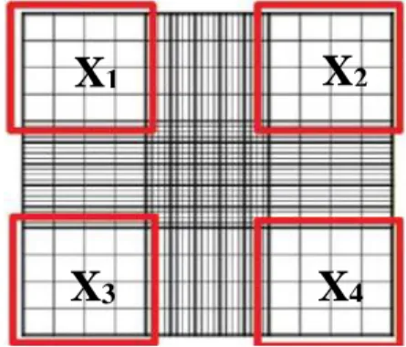
Figure 2.1. Scheme of a Neubauer Chamber………..27
Figure 2.2. Different cell systems to test if TRIB2 confers resistance to MEK inhibition……32
Figure 3.1. Tribbles mRNA levels of a panel of melanoma and non-melanoma cell lines…….38
Figure 3.2. Tribble protein levels in melanoma and non-melanoma cell lines………..39
Figure 3.3. Effect of Refametinib treatment on MAPK pathway in SK-Mel-28 cell line…….40
Figure 3.4. SK-Mel-28 cell line treated with Refametinib for 72hours………..41
Figure 3.5. Effect of Refametinib treatment on apoptotic cell death in SK-Mel-28 cell line…..42
Figure 3.6. Effect of Refametinib treatment on MAPK pathway in SK-Mel-28 cell line……...43
Figure 3.7. TRIB2 Knockdown in A375 cell line………..44
Figure 3.8. Cell death analysis of Refametinib treatment in A375 TRIB2 Knockdown cell line……….45
Figure 3.9. TRIB2 Knockdown in G361 cell line………..46
Figure 3.10. Cell death analysis of Refametinib treatment in TRIB2 knockdown……….47
xii Figure 3.13. Influence of Refametinib treatment in cell death using G361 TRIB2 knockout
cells………...49
Figure 3.14. TRIB2 Knockout (KO) in SK-Mel-28 cell line………..…50
Figure 3.15. Cell death analysis of Refametinib treatment in TRIB2 knockout……….51
Figure 3.16. Cell death analysis after 72hours of Refametinib treatment in SK-Mel-28 TRIB2 KO cell line………52
Figure 3.17. TRIB2-FLAG Knock-In (KI) in SK-Mel-28 cell line………53
Tables Index
Table 1.1- Clinical Classification of Melanoma………..5Table 1.2. Drugs used in melanoma……….9
Table 2.1. Genetic characterization of the melanoma cell lines used……….27
Table 2.2. Primers used in q-PCR………..29
Table 2.3. Details of MEK inhibitor Refametinib (BAY766) experiments in different cell lines………...36
xv
Abbreviations
A
ABC proteins -ATP – Binding Cassette (ABC) proteins
AJCC - American Joint Committee on Cancer (AJCC)
AML- Acute myelogenous leukemia ANOVA- Analysis of Variance APS- Ammonium Persulfate ATCC – American Type Culture Collection
ATP – Adenosine Triphosphate
B
BRAF - B-type Raf kinase BSA – Bovine serum albumin
C
Cas9 – Clustered Regularly Interspaced
Short Palindromic Repeats associated
protein
CO2 - Carbon Dioxide
cDNA - Complementary DNA C/EBPs - CCAAT/ enhancer binding proteins
CRISPR - Clustered Regularly
Interspaced Short Palindromic Repeats
D
DMEM - Dulbecco’s Modified Eagle’s Medium
DMSO - Dimethyl-sulfoxide
DNA - Deoxyribonucleic acid DOC- Sodium deoxycholate dsbreaks - Double strand breaks DTIC- Dacarbazine
E
ECL - Enchanced chemiluminescente solution
E.coli – Escherichia coli
EGFR - Epidermal Growth Factor Receptor
EMT- Epithelial-mesenchymal-transition ERK - Extracellular signal-regulated kinases
F
FACS - Fluorescent Activated Cell Scanning
FDA - Food and Drug Administration FBS - Fetal Bovine Serum
FOXO - Forkhead transcription factor
G
GRB2 - Growth factor receptor-bound protein 2
GST - Glutathione – S- Transferase GTP - Guanosine-5'-triphosphate
H
HCl - Hydrochloric acid
xvi HEK - Human Embryonic Kidney – HEK
HGF - Hepatocyte growth factor HR - Homologous recombination HRP - Horseradish peroxidase
I
IARC - International Agency for Research on Cancer
IC50 - half maximal inhibitory concentration
IL-2 – Interleukine -2
J
JNK - c-Jun N-terminal kinases
K
KD- Knockdown kDa – Kilo daltons KI- Knock-in KO- Knockout
M
MAPK – Mitogen-activated protein kinases
MAPKK - Mitogen-activated protein kinase kinase
MDM2 - Double minute 2 homologue MMR - DNA-mismatch repair
MMP2 - Metalloprotease 2
MEK – Mitogen-activated protein kinase kinase
MRP - Multidrug resistance protein mTOR - Mechanistic target of rapamycin
N
NER - Nucleotide Excision Repair NF-ƙb - Nuclear factor kappa B NHEJ - Non-Homologous End Joing NP40 - Nonidet P 40
O
OVO4 - Sodium Orthovanadate
P
PAM - Protospacer adjacent motif (PAM) PARP - oly(ADP-ribose) polymerase PBS - Phosphate buffered saline PCR- Polymerase Chain Reaction pH – Potential of hydrogen PI - Propidium Iodite
PIC- Protein Inhibitors Cocktail PI3K - Phosphatidylinositol 3 kinase Pgp - P- glycoprotein
PS - Phosphatidylserine
PTEN - Phosphatase and tensin homolog
Q
q- PCR - quantitative Polymerase Chain Reaction
R
RNA - Ribonucleic acid rpm - Revolutions per minute RTKs - Receptor Tyrosine Kinases
xvii
S
SD - Standard Deviation SDS - Sodium dodecyl sulfate sgRNA - Single guide RNA
shGFP – Short hairpin against Green Fluorescent Protein
shRNA - Short hairpin RNA siRNA - Small interference RNA
shTRIB2 - Short hairpin against Tribbles homolog 2
SNL - Sentinel lymph node SOS - Son of Sevenless(SOS)
T
TBS – T - tris-buffered saline with tween TEMED - Tetramethylethylenediamine TMZ - Temozolomide
TNF- Tumor necrosis factor
TRIB1 - Tribbles homolog 1 TRIB2 - Tribbles homolog 2 TRIB3 - Tribbles homolog 3
TSC2 - Tuberous Sclerosis Complex 2 TSG – Tumor suppressor gene
U
UV- Ultraviolet radiation
V
VEGF - Vascular endothelial growth factor
VEGFR - Vascular endothelial growth factor
VLS- Vascular leak syndrome
W
WB – Western Blot
1
1. Introduction
1.1 Cancer
Cancer is among the leading causes of death worldwide. In 2013 there were 14.9 million of new cancer cases and 8.2 million deaths worldwide. Cancer incidence has been increasing in most countries since 1990. By 2030 it is expected 21.7 million new cases and 13 million cancer deaths (1, 2). Genetic differences and environmental factors, including infectious agents, lifestyle and culture, such as smoking, dietary patterns, sun exposure, physical inactivity and reproductive behaviors have been known to be the major risk factors for cancer (3-5).
Cancer is usually viewed as an evolutionary process that results from the accumulation of mutations (usually somatic mutations) or epigenetic events (which do not alter DNA sequence, conferring a selective growth advantage and ultimately uncontrolled proliferation (6). There are two major types of mutations: the hereditary that arise on a germ cell (7, 8) and somatic mutations that occur in any non-germ cell. The latter include base pair substitutions, small insertions or deletions, chromosomal rearrangements and gain or losses of gene copy number. Tumorigenesis is a multi-step process that can arise from the alteration in three main types of genes: oncogenes with dominant gain of function: genes that stimulate cell division, inhibit cell differentiation and halt cell death; tumor suppressor genes loss-of-function: genes that inhibit cell proliferation and regulate apoptosis; and DNA repair genes (9, 10). Traditionally, the accumulation of genetic mutations has been considered the major cause of cancer progression. However, this paradigm has changed and is currently accepted that epigenetic changes also play an important role in cancer development (11).
After centuries of research it is now established that cancer is a very complex group of diseases. In 2001 Hanahan and Weinberg described for the first time “rules that govern the transformation of normal cells into malignant cancers”, known as the “hallmarks of cancer”. These hallmarks can be defined as a small number of molecular, biochemical and cellular characteristics shared by most of all human cancer (12, 13). The first six hallmarks of cancer described were (figure1.1): self-sufficiency in growth signals, insensitivity to anti-growth signals, limitless replicative potential, sustained angiogenesis, evading apoptosis and tissue invasion and metastasis (12). In 2011, Hanahan and Weinberg purposed two new hallmarks essential for malignant transformation: genomic instability which confers tumor heterogeneity and
2 inflammation which is believed to foment multiple hallmarks functions. There are two more capabilities emerging: a reprograming metabolism and avoid immune destruction (13).
Figure 1.1. The hallmarks of Cancer. Almost every cancer has acquired some capabilities during its development. The first 6 hallmarks of cancer suggested in 2000 were: sustaining proliferative signaling, reducing their dependence on growth factors from normal tissue microenvironment, evading growth suppressors, resisting cell death, allowing cells to proliferate out of control, angiogenesis in order to obtain oxygen and nutrients, immortality and invasion & metastasis mainly due to morphological cell changes and activation of metalloproteases. In 2011, the same authors suggested 2 new hallmarks of cancer involved in cancer pathogeny: genomic instability that allows cancer cells with driver mutations to proliferate and gives rise to tumor heterogeneity and tumor promoting inflammation that can support and enhance the other capabilities. New capabilities are emerging: one of them involves the ability of cancer cells to reprogram its metabolism in order to sustain neoplasic proliferation and the other involves the ability to avoid immune system mediated destruction. Adapted from
3
1.2 Melanoma
Melanoma is the most dangerous form of skin cancer and represents less than 5% of all skin cancers, yet is responsible for 80% of skin cancer deaths (14). Metastatic melanoma has a poor clinical outcome, about 5% after six months. The World Health Organization (WHO) estimates that each year are diagnosed 132,000 of new cases of melanoma. The International Agency for Research on Cancer (IARC) estimates that in Europe there is 100,000 new cases and 22,000 deaths each year (15-17).
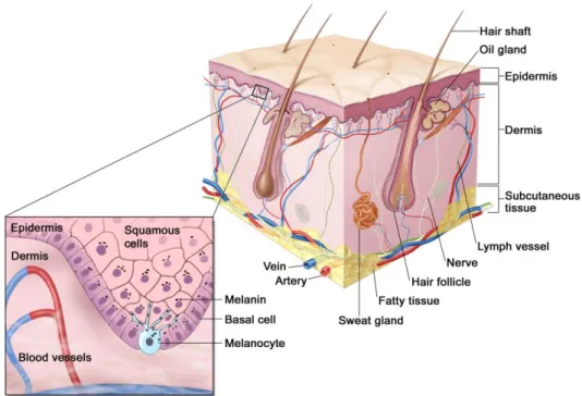
Melanoma is a cancer that arises from melanocytes, which are specialized pigmented cells (figure 1.2), derived from the neural crest and are found predominantly in the skin and hair follicles. A major risk associated with melanoma is the ultraviolet radiation (UV), along with the family history, fair skin and immunosupression. Melanoma has a high somatic mutation rate, among the highest of any cancer type, largely attributed to UV radiation (18). In response to UV radiation, keratinocytes, which are cells that secrete the major structural components of the epidermal barrier, synthesize factors that regulate melanocyte survival, differentiation, proliferation and motility. In this way, keratinocytes stimulate melanocytes to produce melanin resulting in the tanning response. When exposed to UV radiation, melanocytes are activated and secrete melanin and protect the neighboring cells from further damage (15, 18-21). Increased survival features of melanocytes depend not only on themselves, but also on paracrine stimulation from fibroblasts and keratinocytes. Melanocytes can escape their regulation by keratinocytes through disrupted intracellular signaling due to mutations in growth regulatory genes, production of autocrine growth signals and loss of adhesion receptors. Therefore, melanocytes can proliferate and spread, leading to the formation of a naevus (a pre-malignant lesion) (15, 21).
Melanoma is highly metastatic, and highly resistant to treatment (22-24). As mentioned before, melanocytes derived from neural crest cells. These cells undergo epithelial-mesenchymal-transition (EMT) in order to migrate and exit from the neural tube. In a similar way, melanoma cells are able to undergo EMT in the initial events of metastasis to dissociate from surrounding keranocytes (22). In fact, metastasis are the main cause of the death in melanoma patients (25).
4
1.2.1 Melanoma Classification
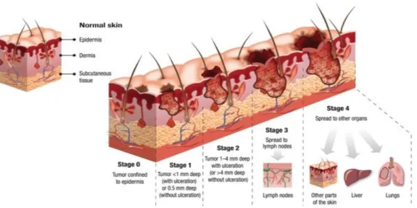
Melanoma can be categorized into five different stages according to their tumor thickness, number of metastatic nods and distant metastasis (figure 1.3). The first stage, stage 0 is the less aggressive one, when cell proliferations is limited to the epidermis and has not reached the underlying dermis. In these stage the treatment applied is surgical resection. Melanomas in stage I and II differ on tumor thickness and ulceration and are treated by surgical resection followed by drug or radiation treatment. When melanoma spreads to the lymph nodes is classified as stage III. In this case surgical removal of the lymph nodes is required. Stage IV refers to a cancer that has spread into distant organs, and it is treated with chemotherapy. According to American Joint Committee on Cancer (AJCC), Melanoma can be clinically categorized in 5 different subtypes including: superficial spreading melanoma, amelanotic melanoma, nodular melanoma, acral lentiginous melanoma, and uveal melanoma (table 1.1) (15, 18, 21, 26-29).
Figure 1.2. Anatomy of the normal skin. Melanocytes are specialized pigmented cells that produce melanin and reside in the basal layer of human skin. Adapted from National Cancer
5
Subtype Description Frequency Common Site Superficial
Spreading Melanoma
Form of melanoma in which cancer cells tend to stay within the tissue of origin: epidermis.
70 % Trunk of men
Legs of Women
Amelanotic Melanoma
Type of skin cancer in which the cells do not produce melanin, they have lack of
pigment.
2-8 % Glabrous skin
(skin that is normally devoid of
hair)
Nodular melanoma Melanoma cells proliferate downwards through the skin
(vertical growth). 10-25% Trunk of men Legs of Women Acral Lentiginous Melanoma Form of melanoma characterized by its site of origin: palm, sole, or
beneath the nail.
5% Palms
Soles Nails
Uveal Melanoma Melanoma of the eye. 3-5% Iris, ciliary body or choroid
Table 1.1- Clinical Classification of Melanoma. Adapted from Chudnovsky Y. et al, JCI
2005.
Figure 1.3. The four stages of Melanoma. Melanoma is staged depending on tumor thickness, number of metastatic nodes and distant metastasis. Stage 0-II is confined to the epidermis, stage III includes lesions spread to the lymph nodes and on stage IV the lesions spread to other organs. Adapted from Colegio Oficial de Enfermeros de Badajoz, 2017.
6
1.2.2 Melanoma Genetics
The MAPK signaling cascade plays a key role in melanoma, making it an important therapeutic target. In normal cells, the MAPK pathway (figure 1.4) is activated by mitogens or hormones and extracellular growth factors. This signaling pathway controls fundamental cellular processes such as growth, proliferation, differentiation, migration and apoptosis (30, 31). The MAPK pathway includes a small G protein (RAS) and three serine/threonine protein kinases: B- type RAF kinase (RAF), Mitogen-activated protein kinase kinase (MEK) and Extracellular signal-regulated kinase(ERK). The binding of mitogens, hormones, cytokines or neurotransmitters to tyrosine kinase receptors causes its dimerization, which triggers the activation of RAS. Mechanistically, the phosphorylated SH2 (Src Homology 2) of GRB2 (Growth factor receptor-bound protein 2), an adaptor protein, brings Son of Sevenless(SOS) into close proximity to GDP-Ras and converts it into Guanosine-5'-triphosphate (GTP) -Ras (activated form) by catalyzing the GDP to GTP (32). This guanine nucleotide exchange leads to the activation of RAS signaling. Once activated, RAS attracts and binds RAF, which usually is found in cytosol, via effector loop. Therefore, RAF becomes attached to the membrane via RAS. In this way, RAF becomes activated and is able to activate a second kinase, MEK, by phosphorylating its serine / threonine domains (33). MEK is considered a “dual specificity kinase”, which means that it is able to phosphorylate serine/ threonine residues as well as tyrosine residues. By phosphorylation, MEK activates ERK1 and ERK2 that, once activated, each of these ERKs phosphorylates downstream substrates regulating several cellular processes (33). The activation of MAPK signaling potentiates PI3K signaling. These pathways can interact at different levels creating a complex network. The resulting signaling cascade culminates with translocation of ERK to the nucleus where it activates transcription factors, resulting in gene expression (15, 17, 32, 34, 35). Some transcription factors activated by ERK are cdc25 (phosphatase), MSK1/2 (stress activated kinases) and CREB. Once activated, these transcription factors regulate cell proliferation and survival (36, 37). Most cancer lesions that lead to constitutive activation of ERK signaling occur during the early steps of tumorigenesis. The constitutive activation of ERK signaling can result from the overexpression of receptor tyrosine kinases (RTKs), activating mutations in receptor tyrosine kinases, sustained autocrine production of activating ligands, RAS mutations and BRAF mutations (38).
MAPK and PI3K pathways are key regulators of cell proliferation in melanoma. The most common mutation found in melanoma is in BRAF (~50%) (39, 40), followed by
7 figure 1.4 (39, 42). AKT3 is activated in ~60% of melanomas, due to its overexpression or alterations in upstream regulators such as PTEN (43). BRAF and NRAS mutations can result in hyperactivated ERK, which is present in up to 90% of human melanomas (26).
1.2.3 Melanoma Treatment
Until 2010 the standard care for metastatic melanoma included surgical resection, chemotherapy and high interleukine 2 (IL-2) doses (figure 1.5) (17). When detected early, melanoma can be treated by surgical resection, which has over 95% success rate at stages I/II (44). If detected in advanced stages, melanoma is difficult to treat since currently there is no effective treatment. Melanoma lesions can be asymptomatic for long periods, or be detected at stage IV without a clearly identified primary lesion. The main drugs used in melanoma patients are chemotherapy, immunotherapy and targeted therapies (table 1.2). Despite all the efforts, melanoma is still one of the most aggressive cancers, with extremely poor prognosis (21, 44, 45).
Figure 1.4. The MAPK signaling pathway. Growth factors bind to the tyrosine kinase receptor, which brings SOS into close proximity. GDP-RAS is converted into GTP-RAS and phosphorylates RAF. RAF phosphorylates MEK, and MEK phosphorylates ERK. ERK translocates into the nucleus and stimulates transcription of target genes. Mutations in NRAS are found in ~20% of melanoma patients. MAPK pathway is frequently activated by mutations on BRAF (~50%). The PI3K pathway can be activated due to PTEN mutations (30-50%) or
8 Surgical Resection
Surgical resection is still the first treatment choice for patients with early stages melanoma having huge success rate in stage I/II. In cases of metastatic melanoma, surgical resection has a minimal impact in treatment (44, 46). The treatment choice for melanoma patients in stage I-III is surgery. An important prognostic indicator, which provides information about disease progression independently of the treatment, is the analysis of sentinel lymph node (SNL), the first node draining the primary melanoma in the lymphatic system (47). The first rout of metastasis in melanoma is the lymphatic system, making the study of SNL an important toll because it allows the detection of locoregional dissemination (46, 47). If melanoma is spread to the SLN it is performed a complete lymphadenectomy as a gold standard treatment in order to remove metastatic cells present in the lymphatic drainage (48).
Figure 1.5. Treatment applied in metastatic melanoma. Dacarbazine was the first chemotherapeutic drug used, followed by high doses of Interleukine 2 (IL-2). After 2010 new therapeutic strategies became available such as immunotherapies (exp. Ipilimumab, Nivolumab) and targeted therapies (examples: Vemurafenib and Trametinib). Adapted from
9
Drug Group Drug Class Examples Effects
C H EMO TH ER A PY Alkylating Agentes Nitrosaureas Fotemustine/
Carmustine SsDNA breaks
Nitrogen Mustards Cyclophosphamide DNA crosslinking
Triazenes
Dacarbazine/ Temolozomide
Inhibition of nucleic acid and protein
synthesis
Antibiotics Anthracyclines Doxorubicin
(adriamycin)
SsDNA breaks DNA crosslinking Inhibition of DNA and RNA replication
Plant-derived
products Vinca Alkaloids Vincristine
Altered cell division, motility
Taxanes Taxol Altered cell division, motility
Hormonal
Analogs Antiestrogen Tamoxifen
Altered estrogen signaling
Platinium
Drugs Cisplatin
SsDNA and dsDNA breaks IMM U N O TH ER A PY Cytokines IL-2 Growth and activation of T-cells
and natural killers, promoting tumor regression Checkpoint inhibitors Ipilimumab (antibody against CTLA-4) Promotes T-cell activation and proliferation ; amplifies T cell immunity Nivolumab / Pembrolizumab ( antibodies against PD-1) Promotes T cell activation, IL-2 production and mediates immune toxicity TA R G ET ED TH ER A PIES BRAF inhibitors Vemurafenib (against V600E mutation) Anti- proliferative effects MEK inihibitors Trametinib
Induces cell cycle arrest and reduces
tumor growth Table 1.2. Drugs used in melanoma.
10 Chemotherapy
Cytotoxic chemotherapy has been used for the treatment of metastatic melanoma for the last decades. Chemotherapy is based on the inhibition of the division of rapidly growing cells, which is a characteristic of cancerous cells, but it is also a characteristic of normal cells with fast proliferation rates, such as the bone marrow, skin cells, gastrointestinal tract cells and hair follicles cells. The fact that chemotherapeutic agents non-specifically target cells that are dividing rapidly is the major reason for their toxicity (49-52). The first chemotherapeutic agent used to treat advanced melanoma was dacarbazine (DTIC), an alkylating agent (figure 1.5). The alkylating agents are the most widely used anti-cancer drugs and have the ability to covalently bind an alkyl group to the DNA bases (commonly to the N7 guanine) forming an adduct, thereby preventing multiplication of rapidly growing cells. DTIC has an overall response rate ranging between 10-20% and only allow a complete remission on 5% of patients (21, 53, 54). Temozolomide (TMZ) is another alkylating agent widely used in melanoma that has some advantages over many alkylating agents because of its unique chemical structure and pharmacokinetic properties. In particular, its small weight allows the compound to cross the blood brain barrier. This drug has shown efficacy in the treatment of malignant brain tumors and metastatic melanoma in the brain (55, 56). Other cytotoxic chemotherapeutic drugs have been tested such as nitrosaureas (Carmustine), vinka alcaloides (Vincristine), taxanos (Taxol) and platinium compounds (Cisplatin) (table 1.2) but they had no better results than DTIC (21, 53, 54). Another nitrosourea used is Fotemustine, which was proven to be efficient, mainly in brain metastasis giving its high lipophilicity (57). Other conventional chemotherapeutic drugs have also been used to treat melanoma, such as plant-derived products, antibiotics and hormonal analogs (table 2). Alkylating agents, along with most other cytotoxic agents, are not “magic bullets” envisioned by Paul Erhlich: drugs that go straight to their intended cell-structural
targets. The resistance to conventional chemotherapeutic agents in melanoma leads to an
extremely poor prognosis (21, 53, 54, 58-63).
The scientific progress during the last decades, allowed for a deeper study of molecular mechanisms driving melanoma progression, leading to an improvement in melanoma treatment. Since the last decade two new therapeutic approaches improved the standard care for melanoma patients: Immunotherapies and targeted therapies (17, 64).
11
Immunotherapy
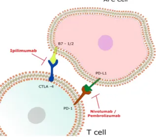
Melanoma is a highly immunogenic type of cancer, and melanocytes have the ability to induce adaptive immune responses. The primary effector cells of the adaptive immune response against cancer are the T lymphocytes that include helper T cells and cytotoxic T lymphocytes (65, 66). The ability of melanoma cells to induce adaptive immune responses was associated with the fact that melanoma has a high mutation load that leads to the presentation of immune stimulatory neoantigens. Neoantigens are antigenic proteins that have new epitopes that have not been previously exposed / recognized by the immune system, leading to an immune response (67-69). The statement that melanoma is highly immunogenic is supported on several observations: (a) spontaneous remissions occur; (b) in about 5% of the melanomas the primary tumor is not found; (c) it was found that primary tumor and metastasis have infiltrated lymphocytes; (d) studies demonstrated that tumor infiltrating T lymphocytes can recognize some melanoma antigens; (e) melanomas respond to immunotherapy. In cases of immunosuppression the risk of developing melanoma is higher (70). Immunotherapy is defined as the use of the immune system to treat cancer (71, 72). Immunotherapy, including cytokine and vaccine treatments are an alternative to conventional chemotherapeutic drugs (73). One of the first “immunotherapeutic tools” used was IL-2, started in the 90’s. Initial studies revealed that 2 is able to induce tumor regression in melanoma and other malignancies (65). Yet, IL-2 has shown some degree of toxicity mainly associated to vascular leak syndrome (VLS) (65). VLS is a phenomenon characterized by an increased vascular permeability along with protein and fluid extravasation, resulting in interstitial edema and organ failure (74). Recently, three new immunotherapeutic drugs have been approved by the FDA to treat melanoma: Ipilimumab, an antagonist monoclonal antibody to CTLA-4 (approved in 2011), (figure 1.6) Pembrolizumab and Nivolumab. Pembrolizumab and Nivolumab are both antagonist monoclonal antibodies to PD-1 and were approved in 2014 (65, 73, 75). Immune checkpoints are negative regulators of the immune system, important to maintain self-tolerance and avoid an auto-immune response. Melanoma cells can take advantage of this mechanism and block an immune response against them. The anti-CTLA-4 antibody binds to CTLA-4 receptor blocking CTLA-4 signaling. This blockade contributes to T cell activation and proliferation, amplifying T cell mediated immunity against melanoma cells (76). Similar to CTLA-4, PD-1 is also a checkpoint inhibitor, playing a key role in immune tolerance. In cancer cells, PD-1 interaction with its ligand promotes T cell apoptosis limiting T cell proliferation and inhibiting IL-2 production. The PD-1 pathway blockade induces T cell activation and proliferation, enhancing anti-tumoral activity (70, 77). However, these therapies have an extremely high cost, and do not benefit the majority of the
12 patients. In fact, the immunotherapy is only beneficial to 15- 50% of melanoma patients. Some patients have intrinsic resistance in tumor cells that have genetic or non-genetic changes that contributes to natural cell survival. One example is that tumors can express proteins with few molecular exchanges, making the immune system unable to recognize these antigens as foreign. It is also possible that, with tumor development, cancer cells lose a proportion of its non-silent mutations, producing lower ratio of antigenic epitopes leading to a phenomenon called the immunoadaption of tumors. Moreover, cancer cells have developed mechanisms to escape the immune system resulting in a less efficient therapy (14, 64, 70-72, 74). Another promising strategy is the use of this dual inhibition combined with immunotherapy including IL-2, interferon, anti-CTLA4, anti-PD1 (78, 79). The future of immunotherapies includes the understanding of resistance mechanisms and the development / improvement of biomarkers in order to provide information about the patient response to the treatment. Hopefully, the ability to distinguish patients that may benefit from these treatments may improve the clinical outcome of melanoma patients (37). In the past few years much attention has been focused on the development of targeted therapies (17, 64).
Figure 1.6. Immune checkpoint blockade in Melanoma. Ipilimumab (against CTLA-4) blocks the immunosuppression induced by the interaction between the B7 family and CTLA-4 proteins. Nivolumab or Pembrolizumab (against PD-1) blocks the interaction of PD-L1 ligand to its receptor. The inhibition of these immune checkpoints allows the immune system to target cancer cells.
13
Targeted Therapies
Over the past years a new generation of cancer treatment arose, such as targeted therapies. Targeted therapies interfere with disease-specific proteins involved in tumorigenesis (49, 50, 80). Target-based therapies are considered to be the future of cancer treatment and much attention has been focused on developing inhibitors for MAPK signaling pathway. MAPK pathway is often hyperactivated in melanoma due to BRAF and NRAF mutations (two thirds of melanomas) (15, 18, 38).
Deregulation of the MAPK pathway, described in section 1.2.2, is frequent in melanoma leading to increased cell proliferation, invasion, metastasis and angiogenesis, making this pathway an important target in melanoma treatment. Despite recent therapeutic advances in the treatment of advanced melanoma, targeting RAS has not been so successful. NRAS mutations are commonly found in codon 12, 13 and 61 and have been associated and aggressive clinical which is easy to understand since active RAS can activate both MAPK and PI3K pathways leading to tumor progression and cancer cell survival. Although much effort has been made to target NRAS, to date no effective anti- RAS therapies have been successfully developed. Previous strategies were focused mainly in posttranslational modifications of NRAS using farnesyltransferase inhibitors. Nowadays the efforts are focused on targeting NRAS with small molecules or siRNA and mainly on downstream effectors of NRAS (81-83). BRAF, one of the downstream effector of NRAS, is one of the three human RAF genes (together with A-RAF and C-RAF) and it is one of the most common mutated genes in melanoma ~50%. The most common mutation leads to a substitution of a glutamic acid for a valine at position 600 (V600E). The mutant V600E BRAF protein results in increased kinase activity (10 fold more activity) which induces hyperactivity of MAPK pathway, stimulating proliferation, survival and neo-angiogenesis by stimulating autocrine vascular endothelial growth factor (VEGF), contributing to the development of nevi. Some studies have shown that V600E BRAF regulates expression of IL-8 a pro-inflammatory chemokine to promote tumor growth and angiogenesis. This mutant form also induces metastasis by triggering invasive cellular behavior and by promoting IL-8 mediated anchoring of melanoma cells to the vascular endothelium, which helps cell extravasation and the development of lung metastasis. As mentioned before, the most common gene mutated in melanoma is BRAF. Patients with BRAF mutations were associated with a poor prognosis. (15, 32, 38, 84). Sorafenib is a nonselective inhibitor of tyrosine kinases (like BRAF) and RTKs such as vascular endothelial growth factor receptor (VEGFR) and was the first BRAF inhibitor investigated in clinical trials in melanoma (38). Clinical trials using
14 sorafenib as a monotherapy failed to demonstrate anti-tumor activity (85). Studies using sorafenib along with other therapeutic agents such as DTIC, carboplatin and paclitaxel in patients with metastatic melanoma were also clinically ineffective. (85-88). The limited activity of sorafenib in tumors with BRAF mutations contributed to the development of new inhibitors with greater selectivity such as Vemurafenib. This inhibitor was the first molecularly targeted therapy approved by the FDA in 2011 for the treatment of advanced melanoma (89). This drug has shown potent anti-proliferative effects in several preclinical models, including the ones harboring the V600E mutation. The mechanism of action involves selective inhibition of the mutated BRAF V600E kinase, which leads to reduced MAPK signaling activity. A phase III clinical trial comparing Vemurafenib and DTIC as first line therapy showed that Vemurafenib improved overall and progression-free survival compared to DTIC group. However, were detected some adverse effects associated with Vemurafenib such as arthralgia, rash, fatigue, alopecia, photosensitivity, nausea and diarrhea. In fact, there are some cutaneous adverse effects described in 92-95% of melanoma patients treated with BRAF inhibitors. There are also some benign and malignant lesions associated with Vemurafenib treatment, being the most commons squamous cell carcinoma and keratocanthoma (73, 84, 89, 90). The mechanism behind the neoplasia development points to MAPK re-activation in skin with mutated RAS. BRAF inhibitors activate C-RAF in wildtype cells, that can induce ERK signaling, leading to squamous cell carcinoma development. Some of the patients treated with Vemurafenib also developed basal cell carcinoma (84, 91). The major problem / concern using Vemurafenib (and also other inhibitors) is that patients eventually develop resistance to therapy, leading to a poor prognosis. Actually, there are already some resistance mechanisms associated with BRAF inhibitors such as re-activation of MAPK signaling, changes in ERK1/2 regulated cell cycle events, activation of alternative signaling pathways and chromatin-regulating events (92). Re-activation of MAPK signaling can emerge due to mutations on RAS, which promotes C-RAF dimerization and activation and due to ERK mutations. In fact, a study has demonstrated that elevated expression of C-RAF was associated with a mutant BRAF melanoma cell resistance to AZ628, a RAF inhibitor (92-94). Herkert B. et al., also showed that ~40% of melanoma patients with BRAF mutations have concomitant loss of PTEN, contributing to the hyperactivation of PI3K pathway and consequently to cancer cell survival (95-97) .
Vemurafenib revolutionized the standard care of melanoma patients. Yet, a big part of melanoma patients dies from resistance once drugs stop having a clinical effect. An intrinsic mechanism of resistance to Vemurafenib is the expression of Hepatocyte growth factor (HGF),
15 which leads to increased cell proliferation (98). Acquired resistance mechanisms were also described such as upstream mutations on NRAS, downstream mutations of MEK and BRAF splice variants. Considering these complications, an alternative strategy is the development of inhibitors for downstream effectors of BRAF, such as MEK (99-103).
Nowadays, selective MEK inhibitors represent a promising new therapeutic option in BRAF and NRAS mutated melanomas. Some studies demonstrate that preclinical models with BRAF mutations are sensitive to MEK inhibitors. Patients harboring NRAS mutations were found to be partially sensitive to MEK inhibitors (104, 105). In BRAF mutated melanoma murine xenografs, MEK inhibitors contributed to tumor regression through increased apoptosis and reduced angiogenesis and proliferation (104, 106) The first MEK inhibitor, PD098059, was described in 1995 (104, 107, 108). Until now, about thirteen MEK inhibitors have been tested in the clinic. The first MEK inhibitor approved by FDA in 2013 was Trametinib (GSK1120212), a selective inhibitor of MEK1 and MEK2 (100, 104, 108). MEK inhibitors can be classified in two major classes: Adenosine Triphosphate (ATP) competitive or non-ATP competitive inhibitors (108). The ATP competitive inhibitors bind to the ATP binding site of MEK, preventing MEK to be phosphorylated. E6201 is an ATP-competitive MEK inhibitor that proved to be effective against Vemurafenib resistance melanoma harboring a MEK1 mutation in a preclinical model (109). However, the sensitivity to E6201 was correlated to wildtype PTEN suggesting that parallel signaling of PI3K pathway may play a role in resistance to this inhibitor (110). Most of MEK inhibitors are non-ATP competitive, which means that they bind to an allosteric binding site close to the ATP binding site preventing MEK activation. MEK 1 and 2 are very similar and consists in a N-terminal sequence, a kinase domain and a C-terminal sequence. In the N-C-terminal sequence MEK1/2 contains an inhibitory/allosteric segment, which is only present in MEK1/2 and not in the other MAPKK. This allosteric segment present in MEK1/2 is relatively unique making the ATP non- competitive MEK inhibitors highly specific (108, 111). Trametinib is an orally available, small molecule, non-ATP competitive MEK inhibitor that induces cell cycle arrest, reducing tumor grow. It was proven to be clinically effective in the presence of BRAF and NRAS mutations. Therefore, it was accepted by the FDA as a single agent for the treatment of patients with V600E BRAF and in combination with dabrafenib (104, 112). Refamatinib is a non-ATP competitive MEK inhibitor very similar to Trametinib, which is still in clinical trials (108). Another MEK inhibitor approved by FDA in 2015 for the treatment of advanced melanoma is Cobimetinib in combination with Vermurafenib. Cobimetinib is also an ATP non-competitive MEK 1/2
16 inhibitor (113, 114). Although all the efforts in developing an effective treatment, resistance to therapy is still the most difficult issue to be overcome. Patients develop resistance to almost all drugs, including to MEK inhibitors, such as the mutation MEK1 P124L (the substitution of a leucine by a proline), resulting in a gain-of-function mutation. Mutations on ERK were also associated with MEK inhibitors resistance leading to MAPK hyperactivation (94, 115-117). Another resistance mechanism is the activation of PI3K pathway. It was already shown that PI3K and MAPK pathways interact in order to regulate several cellular processes like cell proliferation and apoptosis. The MAPK pathway cross-activates PI3K signaling through regulation of PI3K, Tuberous Sclerosis Complex 2 (TSC2) and mTORC1. GTP-RAS can bind and activate directly PI3K kinase. When RAS or PTEN are mutated, even in the presence of a MEK inhibitor, the PI3K pathway remains active contributing to tumor growth. Taking this into account, the inhibition of both PI3K and MAPK pathways might be used to more efficiently treat melanoma patients. Nowadays, there are several fair options for melanoma treatment. Yet, there are still significant obstacles to be overcome, like resistance mechanisms, that should be treated as a priority in melanoma care (32, 118, 119).
1.2.4 Resistance mechanisms
Understanding the mechanisms underlying the resistance associated with different therapeutic agents can improve the outcome of current therapies and contribute to the development of new therapeutic approaches. As mentioned before, one of the biggest concerns in melanoma is the development of resistance to treatment. Resistance can be intrinsic, meaning that it exists before the treatment or acquired when the resistance occurs after the treatment, which means that the tumor was initially sensitive to the treatment. There are some acquired resistance mechanisms already described, such as mutation on drug target / pathway, drug inactivation, drug efflux pumps, DNA damage repair, activation of alternative pathways, tumor heterogeneity and one of the most commons, defects in cell death control (figure 1.7) (120-122).
17
Most of the anti-cancer drugs must undergo metabolic activation in order to have a clinical effect. The toxicity to the normal tissues is a limiting factor to the amount of drug that can be administered. The amount of drug that reaches the tumor mass is also limited by the drug pharmacokinetics (absorption, distribution, metabolism and elimination) (121). Cancer cells can develop resistance to the treatments due to a decreased drug activation or drug inactivation. This phenomenon can occur, for example, due to Glutathione – S- Transferase (GST) superfamily, a group of detoxifying enzymes that protect cellular macromolecules from attack by reactive electrophiles. GST play an important role in the regulation of MAPK pathway via protein- protein interactions (122-124). Some studies show an increased expression of GST in cancer allowing the detoxification of the anticancer drugs, which culminates in less efficient cytotoxic damage of cells (122, 123, 125). Glutathione transferase levels were found to be Figure 1.7. Mechanisms of acquired resistance to cancer therapy. The main resistance mechanisms to cancer therapy involve changes in drug metabolism, like drug inactivation, mutation of drug target or target pathway, and drug efflux pumps that decreases the amount of drug that has an effect on cancer cells. The crosstalk between oncogenic pathways is also an important resistance mechanism. Some cancer cells are also able to increase DNA repair allowing mutated cells to survive. Tumor heterogeneity plays also a crucial role in therapy resistance: not all cells are sensitive to treatment, and resistant cells can proliferate and contribute to tumor growth. One of the main resistance mechanisms is dysregulated cell death control which leads to cancer cell survival, and consequently, to cell proliferation and tumor grow.
18 higher in melanoma cells compared to normal melanocytes, which allows cancer cells to protect themselves against oxidative stress (126, 127). This increased expression of GST has also been associated with resistance to apoptosis (122, 123, 125).
A drug’s efficacy is influenced by the drug target or mutations in the drug target pathway. Many anticancer drugs target topoisomerase II (example Etoposide) which is a nuclear enzyme essential for DNA replication, chromosome condensation and chromosome segregation. This enzyme forms a complex with DNA that is normally transient. When a topoisomerase II inhibitor is present, the complex stabilizes leading to DNA damaged and later results in cell death. Some cancer cells acquire mutations in topoisomerase II gene, conferring resistance to this type of anticancer drugs (122, 128, 129). A study shows that melanoma cells exposed to etoposide (which induces DNA damage) have tenfold reduced topoisomerase II activity corresponding to an increased drug resistance (126, 130, 131). Another example is the mutation of cellular receptors such as Epidermal Growth Factor Receptor (EGFR) or in one of its downstream targets (122, 128, 129).
The efficacy of a drug depends also in the real amount of drug able to reach the tumor. One of the most resistance mechanisms studied is the drug efflux that results in a reduced drug accumulation. Several cell membrane transport proteins, such as the ATP – Binding Cassette (ABC) proteins have been associated with drug resistance by promoting drug efflux (121, 122). The ABC superfamily proteins function as ATP-dependent efflux transports, mediating drug efflux resulting in lower drug accumulation (126, 132). P- glycoprotein (Pgp) and multidrug resistance protein (MRP) belongs to ABC transports superfamily and are thought to contribute to treatment failure (133). Melanoma cells express MRP, yet a study shows that its expression did not increase after chemotherapy (134).
The response to anticancer drugs culminates direct or indirectly in DNA damage, leading to cell death. An increased repair of drug-induced DNA damage is an important mechanism of chemo-resistance. The DNA damage response can occur through the nucleotide excision repair (NER), or homologous recombination (HR). This mechanism can reverse the effect induced by anticancer drugs, such as cisplatin, that causes DNA crosslinks leading to apoptosis. There are some studies showing that some drug resistant melanoma cell lines present an increased NER of DNA damage (126, 135). Furthermore, DNA-mismatch repair (MMR) deficiency results in drug-resistance by changing the ability of cancer cells to repair DNA damage (136).
The crosstalk between signaling pathways in cancer is also a mechanism that can contribute to drug resistance (137). Connections between signaling pathways give the cell the ability to deal with perturbations of homeostasis. In this way, cancer cells are able to activate a similar
19 mechanism through the activation of an alternative pathway which will compensate the drug effect on one pathway. It has been shown that the crosstalk between MAPK and PI3K/AKT signaling pathways contributes to resistance in melanoma (137, 138). RAS is a small G protein located upstream of this two pathways. Upon MAPK inhibition, cancer cells display a strong PI3K activation leading to cell survival and melanoma progression (64, 138).
The deregulation of the apoptotic pathway is probably one of the most important mechanisms of resistance in melanoma cells. Apoptosis, often called programmed cell death, involves two different pathways: an intrinsic and an extrinsic pathway. The extrinsic pathway is triggered by binding of Fas ligand to death receptors that belong to the Tumor Necrosis Factor (TNF) superfamily. These are extracellular membrane receptors, which activates caspase 8 an important component of the apoptotic pathway. Caspases are enzymes that cleave after aspartic acid and become activated upon cleavage by other caspases (proteolytic cascade). The intrinsic pathway can be triggered by different stimuli, including death receptor signaling and intracellular signals like the absence of growth factor, hormones or cytokines (negative stimuli) and radiation, toxins, hypoxia and free radicals (positive stimuli). Once activated, the intrinsic pathway leads to the release of mitochondrial cytochrome-c, which in combination with Apaf-1 results in caspase 9 activation. The intrinsic pathway is mainly controlled by Bcl2 proteins, which include proteins with pro- and anti- apoptotic activity. The two pathways converge with the activation of caspase 3 and 7 that cleave proteins responsible for nuclear membrane and cytoskeletal structure, replication systems and DNA repair (126, 139). Dysregulated cell death control can be associated with three main molecular changes: enhanced survival signals, activation of anti-apoptotic factors and inactivation of pro-apoptotic effectors(21, 126, 140). Tumor heterogeneity also plays a crucial role in anticancer drug resistance. Tumor heterogeneity can be defined as the differences between tumors of the same type in different patients and between cancer cells within the same tumor mass (141, 142). Resistance can develop from a clone with a specific characteristic that allows it to survive to a certain drug, that proliferates originating a resistant cell subpopulation. Recent studies have demonstrated that a fraction of cells that compose part of the heterogeneous tumor mass have stem cell properties and are usually drug resistance. The cancer treatment affects the sensitive cells, but not the resistant cells that survive and can expand contributing to the disease relapse (122, 141).
20
1.2.5 The Role of TRIB2 in resistance to anti-melanoma drugs
Recently, the Link lab has discovered a novel resistance mechanism to anti-cancer drugs currently in clinical trials, namely to BEZ235, a PI3K/mTOR inhibitor, BAY236 and BAY1082439, both PI3K inhibitors and Rapamycin, a mTOR inhibitor and also to conventional cytotoxic drugs DTIC, gemcitabine and 5-fluorouracil, mediated by Tribbles homolog 2 (TRIB2) (143). TRIB2 is a protein that belongs to the Tribbles family of proteins, enhanced cell resistance to these drugs (143) . Furthermore, TRIB2 protein levels were correlated with AKT activation. TRIB2 was able to inhibit Forkhead BoxO (FOXO), a tumor suppressor gene, contributing to the malignant phenotype of melanoma cells. These findings suggest that TRIB2 is conferring resistance by reducing cell death induced by PI3K/mTOR inhibitors (143, 144). In fact, previous studies from this group proved that TRIB2 is overexpressed in melanoma cell lines and in melanoma patients. TRIB2 expression correlates with disease stage and clinical progression, suggestingTRIB2 as a potential biomarker for diagnosis and prognosis of melanoma (14, 144).
The Link lab found that TRIB2 is a repressor of FOXO through a screening for FOXO repressors (144). FOXO suppresses cell survival and proliferation through regulation of the expression of apoptotic proteins and cell cycle regulators. Some studies have shown that hyperactivation of MAPK pathway leads to FOXO inactivation (37, 145). Since FOXO functions as a tumor suppressor gene inactivated in many human cancers inhibiting its repressor proteins might represent an attractive therapeutic strategy to reactivate them. ERK and p38 are known to phosphorylate FOXO1 at various sites, suggesting that MAPK signaling cascade may play a key role in FOXO regulation (37, 119, 146). FOXO regulation is receiving increasing attention in cancer research since FOXO family members were found to be associated with cancer initiation, progression and resistance (36). FOXO proteins represent a subfamily of transcription factors that belong to the forkhead family (145, 147). In mammals there are 4
FOXO genes: FOXO1, FOXO3, FOXO4, and FOXO6 involved in crucial cellular processes like
regulation of stress resistance, metabolism, cell cycle arrest and apoptosis (146-148). FOXOs activity is regulated at three different levels: subcellular localization, stability and transcriptional activity (144). This regulation is mediated by different processes such as phosphorylation, acetylation and ubiquitination FOXO proteins regulate biological processes involved in cell proliferation, cell cycle progression, cell differentiation, tissue homeostasis, angiogenesis and apoptosis through apoptotic genes such as cyclin-dependent kinase inhibitor
21 p27, BIM, Fas ligand and Bcl-6 (36, 37, 146, 147, 149). As result, it is not surprising that deregulation of FOXO proteins may be involved in some pathological processes such as cancer. It was shown that the activation of PI3K or MAPK pathways leads to the repression of FOXO-mediated growth arrest and apoptosis (37). FOXO transcription factors are tumor suppressors that are inactivated in some human cancers (36, 150). It was shown that FOXO overexpression inhibits tumor growth in vitro and tumor size in vitro in breast cancer, correlating FOXO cytoplasmic localization of FOXO with a poor prognosis. Contrarily, when FOXO has a nuclear localization cell cycle stops, angiogenesis is reduced and apoptosis is induced, contributing to tumor regression. The tumor suppressor role of FOXO was also described in leukemia, prostate cancer, and glioblastoma (150-154). Most importantly, recent studies, have revealed that the cytostatic and cytotoxic effects of many chemotherapeutic agents, including paclitaxel, doxorubicin, lapatinib, gefitinib, imatinib, cisplatin and tamoxifen are mediated by FOXO activity (36). Therefore, FOXO can be determinant to the sensitivity to chemotherapeutic drugs. It has been established that AKT phosphorylates FOXO proteins, promoting cell survival, since FOXO regulates pro-apoptotic proteins including TRAIL and BIM. FOXO phosphorylation by AKT induces its translocation to cell cytoplasm and posterior degradation, in particular FOXO3a, which is also regulated by MAPK pathway. FOXO can be inactivated, also by the crosstalk between PI3K and MAPK pathways (36, 155). Thus, FOXO is considered a very important target to melanoma treatment. An interesting approach would be the reactivation of FOXO to take advantage of its tumor suppressor properties. Importantly Zanella F. et al., discovered, TRIB2 as a novel FOXO-repressor, that might be useful as a target to reactivate FOXO factors(144).
Tribbles
TRIB2 belongs to the tribble family of genes, first described in 2000 as a Drosophila protein
that coordinates morphogenesis by inhibiting mitosis. Tribble family members were identified in a genetic screen that aimed at identifying mutations that control cell division and cell migration during embryonic Drosophila development. The name originates from the fictional small animal that vexed the crew of the Enterprise in the “Trouble with Tribbles” episode from Star Trek television series (156, 157). Tribbles encodes an evolutionarily conserved protein family that influences cell proliferation, motility and metabolism (158). Tribbles homologs are characterized by the presence of a N-terminal portion, a central serine/threonine kinase like domain and a C-terminal that contains a COP-1 binding site for E3 ubiquitin ligases and a