Faro, 2015
TRIB2 confers resistance to MAPK
and mTOR1 inhibitors
Neuton Pedro Gorjão da Silva
Oncobiology Master’s Thesis
Supervisors: Dr. Wolfgang Link
Faro, 2015
TRIB2 confers resistance to MAPK
and mTOR1 inhibitors
Neuton Pedro Gorjão da Silva
Oncobiology Master’s Thesis
Supervisors: Dr. Wolfgang Link
TRIB2 confers resistance to MAPK and
mTOR1 inhibitors
Declaração de autoria do trabalho
Declaro ser o autor deste trabalho, que é original e inédito. Autores e trabalhos consultados estão devidamente citados no texto e constam da listagem de referências incluída.
Copyright- Neuton Pedro Gorjão da Silva. Universidade do Algarve. Departamento de Ciências Biomédicas e Medicina.
A Universidade do Algarve tem o direito, perpétuo e sem limites geográficos, de arquivar e publicitar este trabalho através de exemplares impressos reproduzidos em papel ou de forma digital, ou por qualquer outro meio conhecido ou que venha a ser inventado, de o divulgar através de repositórios científicos e de admitir a sua cópia e distribuição com objetivos educacionais ou de investigação, não comerciais, desde que seja dado crédito ao autor e editor.
ii
Acknowledgements
This last year, at the Cancer Signalling Laboratory, was an amazing experience. It allowed me to grow up as a person and as a researcher, so I would like to express my gratitude to everyone who helped me to accomplish this important step in my life.
My first words go to my Supervisors dr. Wolfgang Link and dr. Richard Hill. Thank you for having accepted me so kindly in his research group and giving me the opportunity to do my master´s thesis in such a remarkable group. Thank you for your support, guidance and help since the first day, and more important, thank you for your friendship.
Thank you to the other Lab members, Inês, Marta, Pedro and Joel for your help, kindness and friendship.
Special thanks to my friend Tânia Saramago that helped me to edit the thesis. Thank you to all my closest friends, Nicole, António Brazio, Andreia Mendes, Mariana, Celso Pinto for your friendship and support.
Thank you to all members of Bragança´s, Patricia Madureira´s and Ana Teresa Maia´s Labs, to Dino, Gonçalo Pinheiro, Daniel, Frederica, Joana Sousa, Om Rathor and Gil Carraco for your advices, support and friendship.
I also would like to express my gratitude to my beloved family. You have been always present even 300 km away.
At least I would like to dedicate this thesis to my girlfriend Cátia Lopes. I cannot imagine finish this journey, without her presence and love.
“It has been a beautiful fight. Still is.”
Resumo
A incidência mundial de cancro é elevadíssima, estima-se que um em cada cinco de nós irá morrer como consequência desta doença. U m e s t u d o f e i t o à e s c a l a gl o b a l d e m o n s t r a q u e e x i s t e m 14.1 milhões de casos por ano, resultando em 8,2 milhões de mortes. Em Portugal, o cancro é a segunda principal causa de morte, a seguir às doenças do aparelho circulatório, em particular o AVC (acidente vascular cerebral). Apesar de se encontrar no grupo restrito de tumores malignos mais facilmente detetáveis, o melanoma em fases mais avançadas da doença, é a forma mais mortal de cancro da pele que conduz a um tempo médio de sobrevivência bastante reduzido.
Do ponto de vista clínico, apesar de existirem pacientes em fases iniciais que poderão ser tratados com sucesso através de cirurgia, em fases mais avançadas a aquisição de resistência à quimioterapia continua a ser um problema bastante comum. É imperativo encontrar alternativas de tratamento e melhores métodos de diagnóstico para o combate a esta doença.
A medicina personalizada assume-se cada vez mais como uma excelente alternativa aos tratamentos convencionais. Uma vez que o cancro é uma doença bastante heterogénea, a medicina personalizada exige um conhecimento bastante profundo e especifico de cada paciente para que o tratamento possa ser aplicado de uma forma mais individual, ou seja, será necessário ao nível do diagnóstico, uma capacidade de hierarquizar pacientes com base no êxito perante a resposta ao tratamento com um fármaco que seja específico, por oposição às terapias convencionais. Prevê-se então uma mudança de paradigma no tratamento de cancro: dos agentes citotóxicos mais convencionais, até aos agentes mais específicos, concebidos com base nas especificidades de cada tipo de célula cancerígena. Com base neste pressuposto, é necessária a identificação e validação de novos alvos terapêuticos.
O desenvolvimento de novas técnicas de biologia molecular tem alterado a maneira como nós entendemos a origem do cancro. Estudos “large-scale” ou “image-based”, permitem interpretar os diferentes fenótipos resultantes de alterações químicas, genéticas ou epigenéticas pertencentes a cada tipo de tumor. Desta forma é possível
iv compreender as inúmeras vias moleculares orquestradas por diferentes genes, que dão origem controlam e regulam diferentes tumores.
Numa célula normal, uma via de sinalização é desencadeada quando uma molécula (sinal) se liga a um recetor extracelular. É ativado então uma cascada bioquímica de eventos, evolutivamente conservados. O fim de uma via de sinalização passa pelo núcleo, cujo objetivo fundamental passa pela transcrição de genes que controlam diversos tipos de processos celulares fundamentais. Numa célula tumoral, algumas destas vias moleculares encontram-se alteradas. A partir de screens genéticos, é possível identificar as mutações responsáveis pela desregulação de genes e consequentemente das proteínas constituintes destas vias. As principais vias de sinalização alteradas em melanoma são a PI3K/AKT/m-TOR (principais membros alterados: PTEN (Phosphatase and tensin homolog) - 15–50% deletado, mutado ou silenciado; PI3K (Phosphoinositide 3-kinase) - 6% mutado; recetores membranares - 10%–20% constitutivamente ativados e AKT (Protein kinase B) - 60% amplificado ou ativo), as via MAPK (Mitogen-activated protein kinase), também conhecida como via MEK/ERK (principais membros alterados: RAS (20%) e B-RAF (60%)).
Outro membro com enorme relevo da via PI3K é o gene tribbles2. Para além de funcionar como um oncogene, tem sido descrito também como biomarcador para melanoma, característica que permite distinguir e avaliar se um paciente tem ou não desenvolvido este tipo de tumor. Quando expresso, TRIB2 assume também importante destaque na resistência ao tratamento com alguns fármacos e drogas anti tumorais, nomeadamente drogas que atuam ao nível da via PI3K. Tendo em conta todos estes fatores, questionamos se esta proteína também desempenha um papel importante no mecanismo de resistência criado após a administração de agentes inibidores de outro elemento desta via, m-TOR. Outra via de sinalização frequentemente alterada em melanoma é a via MEK/ERK. Para além disso trata-se de uma via alternativa à via PI3K, pelo que testamos a hipótese de que TRIB2 também regula a resistência a inibidores MEK. Examinámos o impacto da sobre expressão de TRIB2 ao nível celular, proteico, RNA e de DNA, na presença de inibidores proteicos MEK ou mTOR.
Tendo como ponto de partida resultados anteriores do nosso laboratório, em que foi possível demonstrar que TRIB2 confere resistência a diferentes tipos de agentes
quimioterapêuticos convencionais, assim como a inibidores de PI3K, começamos por averiguar se TRIB2 também confere resistência em células tratadas com inibidores m-TOR. Foram criadas linhas isogénicas celulares, com o intuito de formar células com diferentes concentrações proteicas: elevados ou baixos níveis de TRIB2.
Após o tratamento com rapamicina, um potente inibidor do complexo mTORC1, verificou-se que apenas as células que contêm elevados níveis de TRIB2, resistem ao tratamento com este tipo de inibição.
De seguida, submetemos de novo linhas isogénicas ao tratamento com um inibidor tanto do complexo mTORC1 como do mTORC2, TORIN1. Os resultados são bastante claros e revelam que TRIB2 não confere qualquer tipo de resistência, quando mTORC2 é inibido.
Quando as células que sobre expressam TRIB2 são tratadas com BAY 766, um poderoso inibidor MEK, são criados mecanismos de evasão e estas células resistem. O mecanismo fundamental pelo qual as células ultrapassam a resistência, é determinada pela interação entre MEK e TRIB2 que formam um complexo proteína- proteína. Estes resultados são enfatizados quando os sinais imunofluorescentes de TRIB2 e MEK de células resistentes se sobrepõem. Observamos também que a ativação da via de sinalização MEK/ERK regulada por TRIB2, reprime a ação de FOXO3a um importante fator de transcrição. Por outro lado, os nossos resultados apontam para que TRIB2 leva a um aumento de afinidade de NF-κB se ligar ao promotor, sugerindo que a sua concentração aumenta no interior do núcleo destas células resistentes a inibidores MEK.
De seguida, testou-se a ativação da via PI3K, verificando se a proteína AKT se encontrava fosforilada. Os resultados são semelhantes para todas as linhas celulares, ocorrendo fosforilação de AKT. Averiguamos de que forma esta proteína seria ativada, e verificou-se que a ativação de AKT ocorre pela fosforilação do resíduo serina na posição 473, sempre que as células sobre expressam TRIB2.
Como é sabido, mTORC2 desempenha um papel importante na ativação de AKT através da fosforilação da serina 473. Um dos elementos fundamentais deste complexo é a proteína RICTOR. Procedemos ao seu knock down, e a resistência criada na presença de diferentes drogas (BEZ235, BAY236 e BAY439), é perdida. O mesmo resultado é obtido quando o Knock down é feito para FOXO3a.
vi O melanoma, em fases mais evoluídas da doença (estádio IV), tem-se revelado como uma das formas mais mortais de cancro. É imperativo que se consiga encontrar novas formas de combate a este tipo de doença com contornos tão trágicos. A importância biológica deste projeto prende-se exatamente com esta necessidade: tentar perceber qual o papel da proteína TRIB2, do ponto de vista molecular, descrita anteriormente como um biomarcador para melanoma no mecanismo de resistência a múltiplas drogas.
Após o término do projeto, foi possível verificar que TRIB2 confere resistência, em diferentes linhas celulares após a administração de drogas que inibem a estrutura proteica mTOR. TRIB2 funciona como uma molécula ativadora de AKT, uma proteína que desempenha um papel fundamental na via de sinalização PI3K/AKT/mTOR, uma das vias mais alteradas em melanoma. Por sua vez um dos ativadores moleculares de AKT é o complexo mTOR. Verificámos que quando TRIB2 é sobre expresso, a ativação de AKT acontece apenas através do complexo 1 de mTOR e não pelo complexo 2. Indo um pouco mais fundo nesta análise concluímos que AKT é ativado por mTOR1 que tem a capacidade de fosforilar o resíduo serino na posição 473, um dos requisitos para que ocorra a ativação completa de AKT.
Uma outra via de sinalização, que se encontra ativa em melanoma é a via MAPK, mais em concreto a via MEK/ERK. À imagem do que acontece aquando a administração de diferentes inibidores, TRIB2 exerce um papel chave no mecanismo de resistência a inibidores MEK. Em células resistentes, TRIB2 forma um complexo proteína-proteína com MEK, que conduz a ativação de ERK e por consequência de toda a via. Esta ativação, culmina com a presença de vários fatores de transcrição no núcleo, como é exemplo NF-κB, conduzindo à transcrição de diferentes genes fundamentais nos processos de evasão, crescimento celular e proliferação.
Palavras-chave:
Melanoma; inibidores MEK; inibidores mTOR; fármaco-resistência; TRIB2Malignant melanoma is the deadliest form of skin cancer that leads to a median survival time of only 6 to 9 months. Besides the patients in early stages that could be successfully treated by surgery alone, actually, there is no efficient diagnostic procedure. It is imperative to find alternatives and better methods of diagnosis for metastatic melanoma. Acquisition of resistance to chemotherapy agents, namely cytotoxic drugs such as dacarbazine, remains a major problem in melanoma therapy.
Since TRIB2 (tribbles2) protein has recently been implicated as a biomarker for melanoma, and as a mediator in the process of resistance to conventional chemotherapeutics or PI3K (Phosphoinositide 3-kinase) inhibitors, we hypothesised that resistance to the inhibition of different components of this and other signalling pathways such as mTOR (mammalian target of rapamycin) and MEK (MAPK/ERK Kinase) is mediated by TRIB2.
In this study, we examined the impact of ectopic expression of TRIB2 in the presence of mTOR or MEK inhibitors.
We demonstrated that in the presence of rapamycin, a potent inhibitor of mTORC1 (mammalian target of rapamycin complex 1 or mechanistic target of rapamycin complex 1), TRIB2 confers resistance to this kind of treatment. However, when we exposed cells to a mTOR2 inhibitor, TORIN1, resistance is not observed. We demonstrated that TRIB2 acts as an adaptor, recruits the mTOR complex 1 that phosphorylates AKT (also known as Protein Kinase B, PKB) on residue Serine 473.
Furthermore, we investigated the effect of TRIB2 expression, when cells where exposed to pharmaceutical inhibition of MEK. We found that TRIB2 expression protects cells against the effect of the MEK inhibitor BAY 766. We have seen that TRIB2 and MEK co-localize, interact and form a protein-protein complex. We also observed that the inhibition of MEK/ERK and PI3K pathways regulated by TRIB2, represses FOXO3 (Forkhead box O3a) a binding to p27 promoter, a well-known and studied transcription factor. On the other hand, high levels of TRIB2 leads to an increase of NF-κB (factor nuclear K B) affinity for promotor. NF-κB enhances the transcription of different genes crucial for melanoma cells proliferation and survival.
mechanism in melanoma cells. This molecular process determines and modulates important cell decisions which include cell growth, proliferation and survival.
Contents
CHAPTER 1. INTRODUCTION 1 1. Cancer ... 1 2. Melanoma ... 2 2.1 A clinical perspective ... 2 2.2. Diagnosis... 22.3 Clinical Grading System ... 3
2.4. Conventional Therapies ... 4
3. Molecular pathways in melanoma ... 4
3.1. RAS/RAF/MAPK molecular pathway ... 6
3.2. PI3K/AKT/FoxO signalling axis ... 7
3.3. Mechanistic target of rapamycin (mTOR) ... 9
3.4. Transcription Factors ... 10
3.4.1. Nuclear factor KB (NF-κB) ... 10
3.4.2. Forkhead transcription factors of the O class (FOXOs) ... 11
4. Biomarkes ... 15
4.1. TRIB2 ... 13
5. Melanoma Treatment ... 15
5.1 mTOR inhibitors ... 16
5.2 MEK inhibitors ... 17
6. Resistance toMelanoma Treatment ... 15
7. Crosstalking ... 15
8 Hyphotesis ... 23
CHAPTER 2. METHODS 25 1. Cell culture and Tissue samples ... 25
1.2 Transfection ... 26
1.2.1. RNAi technique ... 28
2. Drug Time Courses ... 30
3. RNA/Protein extraction and FACS (Fluorescence- activated cell sorting) samples .. 30
3.1. RNA ... 30
3.2. Protein ... 31
3.2.1. Protein Quantification/Bradford Assay ... 31
3.3. FACS (Fluorescence-activated cell sorting) ... 32
4. Western Blotting ... 33
5. Co-Imunnoprecipitation (Co-IP) ... 37
6. Chromatin Imunnoprecipitation (ChIP) ... 38
7. cDNA synthesis ... 39
8. Real Time Polymerase Chain Reaction (qRT-PCR) ... 41
8.1. DNA electrophoresis ... 42
9. Dual staining Microscopy ... 42
CHAPTER 3. RESULTS 45 1. TRIB2 confers resistance to mTOR1 complex inhibition ... 45
2. TRIB2 does not confer resistance to mTORC2 complex inhibition. ... 46
3. Confirmation of PI3K, mTOR1 and mTOR1/2 inhibitor efficiency ... 48
4. RICTOR and FOXO3a knock down increases resistance to dual... 50
5. BAY766 effectively inhibits ERK phosphorylation ... 53
6. Microscopy Analysis ... 54
7. TRIB2 and MEK interact and form a protein complex... 56
8. Confirmation of co-localization using confocal microscopy ... 57
9. Cells that over express TRIB2 repress the transcription factor FOXO3a ... 59
10. TRIB2 over expressing cells have higher levels of NF-K B in their nucleus ... 61
1. Discussion ....………....64 2. Future directions ... …..67
Figures List
FIGURE 1.1-CLASSIFICATION OF MELANOMA DEVELOPMENT ... 3
FIGURE 1.2-MAPK SIGNALLING PATHWAY ... 6
FIGURE 1.3-THE PI3K/AKT/MTOR SIGNALING PATHWAY ... 8
FIGURE 1.4-MTOR COMPLEXES ... 10
FIGURE 1.5–DIFFERENT MOTIFS WITHIN THE TRIBBLES PROTEIN STRUCTURE ... 14
FIGURE 1.6–MECHANISMS OF INTRINSIC RESISTANCE, DEVELOPED BY MELANOMAS THAT CARRY BRAFV600E MUTATION DUE TO EXPOSURE TO B-RAF INHIBITORS ... 19
FIGURE 1.7-MECHANISMS OF RESISTANCE TO RAF/MEK INHIBITORS ... 21
FIGURE 1.8-MODEL THAT SUMMARIZES THE DIFFERENT GENETIC NETWORKS THAT RESULT FROM THE CROSS TALKING IN MELANOMA ... 22
FIGURE 1.9-HYPOTHESIS MODEL IN WHICH TRIB2 CONFERS RESISTANCE TO MTOR/MEK... 23
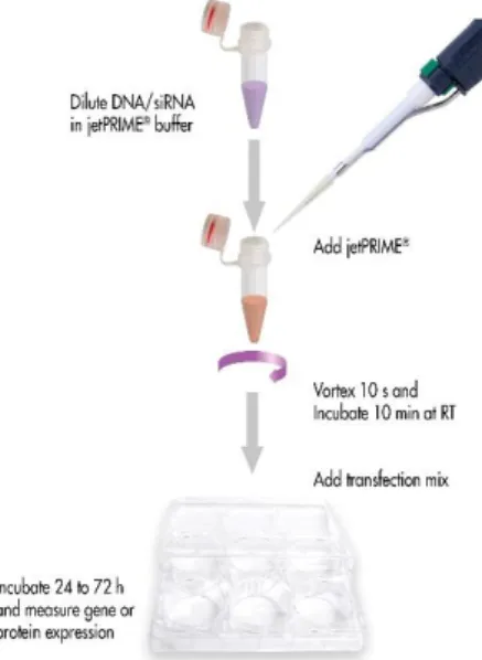
FIGURE 2.1-CHEMICAL TRANFECTION METHOD ... 26
FIGURE 2.2-DNA TRANSFECTION PROTOCOL. ... 28
FIGURE 2.3-RNAI MECHANISM. ... 29
FIGURE 2.4-BEER´S LAW ... 31
FIGURE 2.5-WESTERN BLOT ELECTROPHORESIS ... 33
FIGURE 2.6-WESTERN BLOT SEMI-DRY TRANSFER APPARATUS. ... 34
FIGURE 2.7-WESTERN BLOT BLOCKING AND INCUBATION. ... 34
FIGURE 2.8-CHROMATIN. IMMUNOPRECIPITATION WORKFLOW ... 38
FIGURE 2.9- CDNA SYNTHESIS WORKFLOW.65 ... 40
FIGURE 3.1- TRIB2 STATUS CONFERS RESISTANCE TO RAPAMYCIN. ... 45
FIGURE 3.2-TRIB2 DO NOT PROMOTE RESISTANCE WHEN CELLS ARE TREATED WITH MTORC2 INHIBITORS. ... 47
FIGURE 3.3-THE TRIB2 KNOCK DOWN IS EFFICIENT. ... 47
FIGURE 3.4-TRIB2 RESISTANCE OCCURS DUE TO PHOSPHORYLATION OF SER473 OF AKT. ... 49
FIGURE 3.5-THE RICTOR KNOCK DOWN IS EFFICIENT. ... 50
FIGURE 3.6-TRIB2 RESISTANCE IS LOST, WHEN RICTOR OR FOXO3A ARE KNOCKED DOWN ... 51
FIGURE-3.7-TRIB2 IS A GOOD CANDIDATE FOR MEK INHIBITION
RESISTANCE. ... 52
FIGURE 3.8-INCREASED PTYR204-ERK IN HIGH TRIB2 EXPRESSING CELLS. . 54 FIGURE 3.9-MAPK PATHWAY IS ACTIVATED WHEN TRIB2 IS
OVEREXPRESSED ... 55
FIGURE 3.10-TRIB2 AND MEK FORM A PROTEIN COMPLEX IN ISOGENIC
MATCHED CELL LINES. ... 57
FIGURE 3.11-MEK AND TRIB2 CO-LOCALIZE. ... 58 FIGURE 3.12-FOXO3A IS REPRESSED BY OVER EXPRESSION OF TRIB2. ... 59 FIGURE 3.13-FOXO3A HAS MORE AFFINITY TO PROMOTER WITH HIGH
LEVELS OF TRIB2 ... 60
FIGURE 3.14-NF-KAPPA B HAS MORE AFFINITY TO BIND THE PROMOTER,
WHEN TRIB2 IS PRESENT ... 61
FIGURE 3.15-NFKAPPA B HAS MORE AFFINITY TO PROMOTER WITH HIGH
LEVELS OF TRIB2 293T ... 62
FIGURE 4.1-MECHANISM IN WHICH TRIB2 REGULATES THE RESISTANCE
Tables List
TABLE 1.1-MALIGNANT MELANOMA STAGING SYSTEM APPROVED BY AJCC
... 3
TABLE 1.2-MOST COMMON INHIBITORS BEING TESTED (CLINICAL TRIALS) FOR THE TREATMENT OF MELANOMA ... 18
TABLE 2.1- CHARACTERISTICS OF THE ISOGENIC CELL LINES CREATED FOR EXPERIMENTS ... 26
TABLE 2.2-CHEMOTHERAPEUTIC DRUGS USED IN TIME COUSES. ... 30
TABLE 2.3-SOLUTIONS FOR STACKING GELS FOR TRIS-GLYCIMIDE SDS-POLYCRIMIDE GEL ELECTROPHORESIS ... 35
TABLE 2.4-SOLUTIONS FOR RESOLVING GELS FOR TRIS-GLYCIMIDE SDS- POLYCRIMIDE SDS- POLYCRIMIDE GEL ELECTROPHORESIS ... 62
TABLE 2.5-PRIMARY ANTIBODIES ... 62
TABLE 2.6-SECONDARY ANTIBODIES ... 37
TABLE 2.7-REAGENTS NECESSARY TO c-DNA SYNTHESIS ... 40
Abbreviations List
AJCC- American Joint Committee on Cancer AKT- Protein Kinase B
BRAF-B-type Rafkinase
ChIP- Chromatin Imunnoprecipitation Co-IP Co-Imunnoprecipitation
DNA -Deoxyribonucleic acid DTIC- Dacarbazine
ERK-Extracellular signal-regulated kinases FACS -Fluorescent Activated Cell Scanning FasL- Fas ligand
FDA-Food and Drug Administration FOXO- Forkhead transcription factor IGF-1- Insulin-like growth factor 1 IgG- ImmunoglobulinG
Il-2- Interleukin-2 IP- Immunoprecipitation KD- Kinase Domain
MAPK- Mitogen-activated protein kinases
MAPKK- Mitogen-activated protein kinase kinase MDM2- Mouse double minute 2 homolog
MEK-MAPK/ERK Kinase
mTOR-Mechanistic target of rapamycin NF-KB- Nuclear factor KB
PBS-Phosphate buffered saline
PDK1-Pyruvate dehydrogenase lipoamide kinase isozyme 1 PI3K-Phosphatidylinositol 3 kinase
PIP3-Phosphatidylinositol (3,4,5)-trisphosphate pRAS-proline-rich AKT substrate
PTEN-Phosphatase with tensin homology
RICTOR-Rapamycin-Insensitive Companion of mTOR RTK-Receptor tyrosine kinases
TRIB2- Tribbles pseudokinase 2 USA-United States o fAmerica
CHAPTER 1. INTRODUCTION
1. Cancer
Cancer is a leading cause of death and it is estimated that about one in five of us will die as a consequence of cancer. The worldwide incidence of cancer is extremely high, a 2012 study revealed that there are 14.1 million cases per year, resulting in 8,2 million deaths. It is also estimated that there are 32,6 million people living with cancer (within 5 years of diagnosis). 1
The evolution of molecular biology techniques has changed the way we see the origin of cancer. Nowadays, the focus of carcinogenesis studies has shifted from the identification of gene mutations to understanding the pathways that these genes control. This paradigm shift has suggested that tumorigenesis, at the molecular level, results after several genetic alterations that have occurred within the cell. It is a multistep process, in which every gene affected encode proteins with different functions.2,3
In this way, the most mutations are observed in genes related with cell growth and proliferation (including oncogenes such as RAS, cMYC and SRC), tumour suppressor genes that inhibit cell proliferation (p53, RB and FOXO´s), control of apoptosis (p53, FOXO´s and BCL-2), cell-contact inhibition (E-CADHERIN), promotion of survival (telomerase and PI3K/AKT) and stability genes that are responsible to repair mistakes in genome made during normal DNA replication or induced by exposure to environmental factors (mismatch repair, nucleotide-excision repair and base-excision repair genes).2
A cell needs to overcome a number of mechanisms in order to become cancerous. This theory proposes that cells, to achieve transformation, must activate some proliferative signal transduction, evade the growth suppression mechanisms, resistance to apoptosis, enable replicative immortality, induce angiogenesis, activate invasion and promote metastasis.4,7
Recently, two other hallmarks were added: reprogramming of energy metabolism and evading immune destruction.8 Acquisition of drug resistance and avoidance of oncogene (it will be discussed in next sections) and inducing of senescence are also another important features in tumour development.
2. Melanoma
2.1
A clinical perspective
In the last 50 years melanoma has risen faster than any other cancer type and unlike other kind of cancers, melanoma incidence is not strongly dependent on age, and is one of the most common causes of cancer deaths between the ages of 20–35. It remains one of the cancers most resistant to treatment.9 Melanoma provides one of the best sources to study the relationship between environmental factors and cancer cells. It arises within any anatomic place that contains normal pigment cells (neural crest-derived melanocytes), located on the basement membrane of epithelial surfaces. The main function of melanocytes is the synthesis, storage, and transfer of melanin pigments to surrounding epithelial cells9.
2.2. Diagnosis
Histological patterns have been well described. Microscopic features that correlate with clinical subgroups (including superficial spreading melanoma, nodular melanoma, acral lentiginous melanoma (it is the most common subtype of melanoma in people with darker skins.), and lentigo malignant melanoma (also known as Hutchinson melanotic freckle., lentigo maligna has a lower rate of transformation to invasive melanoma than the other forms)) have been thoroughly codified. As an example, cutaneous lesions detected as little as 1 mm in Breslow thickness could have microscopic evidence of lymph node metastasis which, when present, confers a significantly increased risk for metastasis.910
The traditional Clark model is a multi-step system to predict the progression of melanoma. Several models that try to explain the genetic basis of melanoma development and progression are based on this Clark model. It also predicts that the acquisition of a BRAF mutation can be a founder event in melanocytic neoplasia.
It emphasizes the stepwise transformation of melanocytes to melanoma, from the formation of nevi to the subsequent development of dysplasia, hyperplasia, invasion, and metastasis 9 (Fig.1.1).
Melanoma progression can also be classified according to different stages of the disease in a clinical grading system (stages 0, I, II, III, and IV).11
2.3
Clinical Grading System
Melanoma disease progression is typically evaluated using the American Joint commission on Cancer (AJCC) system (Table 1.1). It is a four stage system that incorporates tumour thickness, presence of ulceration and how widespread the melanoma is in a patient (if it has spread to nearby lymph nodes or any other organs).
The fact is that melanoma diagnosis is very much based on the professional’s experience and on complex laboratory tests, being a very subjective factor. Therefore, more accurate ways to diagnose and stage this disease are being searched for.
.
Table 1.1-Malignant melanoma staging system approved by AJCC11 Figure 1.1-Classification of melanoma development55
Melanoma can be classified according to the Clark´s model based on the level of invasiveness: I-Epidermis (normal); II to IV- dermis (Nevus, RGP, VGP,); V- subcutaneous tissue (metastatic melanoma). The Breslow approach considers the millimetres of invasion depth, ranging from 0 to 2mm. (representation of a vertical section of skin)
2.4. Conventional Therapies
The standard therapy options for melanoma are surgery (surgical removal of the tumour and surgical excision margins based on Breslow’s tumour thickness) for non- metastatic tumours detected early, surgery, radiation, chemotherapy and supportive care. The median survival time for melanoma patients with stageIV melanoma ranges from 0.7 to 5 months depending on age and performance status (how patients react to treatment).12
The chemotherapeutic drugs used in the treatment of several tumours can be classified according to their function: DNA-modifying agents (nitrogen mustard, first used in a patient with lymphoma), anti-metabolites (5-fluorouracil (5-FU)), spindle poisons (paclitaxel), topoisomerase inhibitors (doxorubicin) and cytotoxicantibiotics (bleomycin). Some chemotherapeutic agents have activity in patients with metastatic melanoma, including dacarbazine (DTIC) which is an alkylating agent as well as the nitrosoureas, platinum analogs, vinca alkaloids (vinblastine), and the taxanes (paclitaxel). Numerous trials of single agents or combinations of chemotherapy have been performed, but DTIC remains the standard regimen and the most used chemotherapeutic agent for melanoma. The response rate observed with DTIC chemotherapy alone ranges from 15% to 25%, indicating that improved therapies are desperately required.12
Although the clinical criteria for atypical nevi and traditional therapies have been useful in diagnose and improvement of treatment, they are not precise or highly reproducible and contain some pitfalls: for instance, only a small percentage of primary melanomas arise from nevi (10-20%)9and chemotherapy for advanced melanoma (stage IV) remains largely palliative with low survival rates after diagnosis.12Therefore, new therapy strategies are required.
3. Molecular Pathways in Melanoma
It is crucial to develop new strategies, so over the past 30 years, many groups have helped to decipher the complex genetic networks involved in melanoma proliferation, progression and survival and our understanding has significantly improved. Many of these oncogenic loci and pathways are being well studied and have become crucial targets
for pharmacological drug development. This continuous understanding of the disease can revolutionize the treatment of cancer.13,14
In a normal cell, the general signalling can start when a signalling molecule binds to a cell receptor (extracellular signals). The signal molecule can be a growth factor, mitogens, hormones, neurotransmitters or cytokines. It can take place either through direct cell-cell contacts and cell-matrix interactions (integrins and cadherins) or through the action of secreted signalling molecules. Secreted signalling can be classified into endocrine, paracrine and autocrine signalling. In endocrine mechanism, hormones are carried through the circulatory system to act on distant target cells. In paracrine signalling, a molecule released from one cell acts locally to affect nearby target cells and in autocrine signalling, a cell produces a molecule to which it also responds. Then the signal is usually transmitted into a cascade of events evolutionarily conserved, that passes a message into the nucleus. Here, different transcription factors, such as FOXO3a, NF-KB, c-MYC, AP-1, p53 and E2F1 for example, have the capability to enhance transcription. Different genes are transcribed controlling fundamental cellular processes such as proliferation cell growth, apoptosis, cellular energy production, cell transport or homeostasis10-15. In tumour cells, these pathways are modified and the scientific community has been performing high throughput genomic screens to identify many of the driver mutations responsible for malignancy. The dysregulation of these genes affecting upstream proteins like cell receptors or downstream proteins plays an important role in the development of melanoma.10,15Different studies suggest the involvement of pathways like Notch and
WNT/βb-catenin 16,17. Our attention is directed towards the most mutated molecular
signalling pathway implicated in melanoma development. MAPK pathway is the most important pathway in melanoma, and has been reported to be activated in over 80% of all cutaneous melanomas, making it the focus of many scientific studies in the melanoma field. The most common mutations are found in RAS (25%); B-RAF (70%). The second most important signalling axis is PI3K/AKT, with several of its members being mutated or modified are PTEN - 15–50% deleted, mutated or silenced; PI3K – 6% mutated;10%– 20% receptor-independent activated and AKT - 60% amplificated or activated.
3.1
RAS/RAF/MAPK molecular pathway
Mitogen-activated protein kinase (MAPK) cascades are key signalling pathways involved in the regulation of cell proliferation, survival and differentiation being activated by a wide variety of receptors involved in growth and differentiation including receptor tyrosine kinases (RTKs), integrins, and ion channels. 20
The ERK pathway is the best studied of the mammalian MAPK pathways, and is deregulated in approximately, one-third of all human cancers.
The signalling cascade is triggered depending on the molecular stimuli (growth factors and mitogens), the scaffold of the pathway usually includes a set of adaptors (Shc, GRB2, Crk, etc.) linking the receptor to a guanine nucleotide exchange factor (SOS, C3G,
Figure 1.2-MAPK signalling pathway.54
Although there are more pathways as shown in this figure, the three most significant arms of the mitogen-activated protein kinase (MAPK) pathway are ERK (extracellular signal-regulated kinase), JNK (c-Jun N-terminal kinase) and p38. They can be activated through different stimulus such as growth factors, integrins and interleukines . The three-tiered kinase dynamic cascade leads to activated MAPKs entering the nucleus to trigger immediate early gene and transcription factor activation for cellular responses such as cytokine production, apoptosis, cell proliferation and migration.
etc.). Then, the signal is transduced to small GTP-binding proteins (RAS). RAS proteins (H-, K- and N-Ras) function as a GDP/ GTP-regulated switch. GDP/GTP cycling is regulated by guanine nucleotide exchange factors (RAS GEFs; e.g., Sos) that promote formation of active RAS-GTP, whereas GTPase-activating proteins (GAPs; e.g., NF1 neurofibromin) stimulate GTP hydrolysis and formation of inactive RAS-GDP. In normal cells, Ras is bound to GDP and inactive. Extracellular stimuli cause transient formation of the active Ras. Activated Ras-GTP binds to downstream effector targets, of which the Raf kinases are the best characterized. The core unit of the cascade of these three kinases: is composed by MAPKKK (Raf-1, B-RAF and A-RAF)) that activates MEK1/2 by phosphorylating serines 218 and 222 in the activation loop, MAPKK (MEK1/2), and MAPK (ERK). An activated ERK dimer can both regulate targets in the cytosol and phosphorylate a variety of transcription factors in the nucleus regulating gene expression. (Fig. 1.2)20–23
The second most important molecular network involved in melanomagenesis is the PI3K/AKT/FoxO signalling axis. There is growing evidence that activation of this pathway plays a significant role in melanoma, frequently in the setting of concurrent activation of RAS-RAF-MEK-ERK signalling. This evidence includes the identification of genetic and epigenetic events that activate this pathway in melanoma cell lines.
3.2. PI3K/AKT/FoxO signalling axis
Phosphoinositide 3-kinase (PI3K) is a major component of the signalling pathway and can be activated by receptor tyrosine kinases (RTKs) like HER2 and EGFR or G protein-coupled receptors. The GTPase RAS can also recruit and activate PI3K through direct binding.18,19
PI3K is a heterodimer with a catalytic subunit p110 and a regulatory subunit p85 that regulates many normal cellular processes including cell proliferation, survival, growth, and motility. PI3K catalyses the production of the lipid second messenger phosphatidylinositol-3,4,5-triphosphate (PIP3) at the cell membrane phosphorylating phosphatidylinositol-4,5-bisphosphate (PIP2) at the 3′ position on its inositol ring. Pip3 is now able to recruit other downstream molecules such as AKT and PDK1 via their
pleckstrin-homology (PH) domains.18,19 In an opposite way there is a phosphatase PTEN that converts PIP3 back to PIP2 acting as a tumour suppressor (Fig.1.4).
AKT can be phosphorylated at two critically conserved residues (threonine 308 and serine 473) and only reaches its full activity if both are phosphorylated. The kinase phosphoinositide- dependent kinase 1 (PDK1) targets the threonine 308 residue whereas the serine 473 residue is phosphorylated by the Mechanistic Target of Rapamycin Complex 2 (mTORC2). When AKT is fully activated, regulates a wide range of different targets with different cell functions (Fig.1.3).
Figure 1.3-The PI3K/Akt/mTOR signaling pathway.18
This pathway is up-regulated in melanoma via either direct upstream stimulation (growth factor receptors and their ligands) or indirect activation via cross-talk with RAS. The crucial step in this pathway is the AKT full activation that can regulate members of the apoptotic pathway: inhibit Bad, and FOXO transcription factors and activates NF-kB and CREB that regulate anti-apoptotic genes. It inhibits GSK3β (Glycogen synthase kinase-3) responsible for the degradation of β-catenin resulting in cell cycle progression.
The activation of mTOR leads to regulation of cell growth by controlling mRNA translation, ribosome biogenesis, autophagy, and metabolism.
AKT also targets survival and cell cycle regulation. AKT phosphorylates Mdm2 that controls the levels of p53 in the cell.
3.3. Mechanistic target of rapamycin (mTOR)
The mechanistic target of rapamycin (mTOR) protein is a 289-kDa serine/threonine kinase that belongs to the phosphoinositide 3-kinase (PI3K)-related kinase family and is conserved throughout evolution. It has emerged as a critical growth-control node, receiving stimulatory signals from Ras and PI3K downstream from growth factors, as well as nutrient inputs in the form of amino-acid, glucose and oxygen availability Aberrant mTOR signaling is involved in many disease states including cancer, cardiovascular disease, and diabetes. It is present in two distinct multi-protein complexes, mTOR complex 1 (mTORC1) and mTOR complex 2 (mTORC2)67 (figure 1.4)
mTORC1, is composed of regulatory-associated protein of mTOR (Raptor), mammalian lethal with Sec13 protein 8 (mLST8, also known as GbL); proline rich AKT substrate 40 kDa (PRAS40); and DEP-domain-containing mTOR-interacting protein (Deptor) and is inhibited by rapamycin. After entering the cell, rapamycin binds to FK506-binding protein of 12 kDa (FKBP12) and interacts with the FKBP12- rapamycin binding domain (FRB) of mTOR, thus inhibiting mTORC1 functions. mTORC1 is known for its role in regulating cell growth and proliferation through modulation of
protein synthesis. The AKT promotes cell growth (i.e., accumulation of cell mass)
predominantly when activation of complex 1 of the mammalian target of rapamycin (mTORC1) occurs Recent research has identified novel mTORC1 cell signalling mechanisms that modulate mitochondrial biogenesis, hypoxia signalling and cell cycle progression.
The second complex, mTOR complex 2 (mTORC2), is composed of mTOR, RICTOR, GβL, Sin1, PRR5/Protor-1, and DEPTOR. It ca be inhibited with BEZ235 and it is not sensitive to rapamycin. Functions for Mtorc2 is still unclear. However, the first function ascribed to mTORC2, based on the previously known function of TORC2 in yeast, was the regulation of the actin cytoskeleton. Knockdown of mTORC2-specific components in cultured cells results in alteration of the actin cytoskeleton68. AKT was identified as a mTORC2 substrate, it was found that phosphorylates Ser473 in the hydrophobic motif of AKT66. Although earlier knockdown studies of RICTOR also showed reduced phosphorylation of Thr308 in the activation loop, further studies in knockout mice suggested that phosphorylation of Thr308, by phosphoinositide-dependent kinase 1 (PDK1), does not depend on prior Ser473 phosphorylation66,67
3.4. Transcription Factors
3.4.1. Nuclear factor Kappa B (NF-κB)
Increased expression of pro-inflammatory and proangiogenic factors is associated with aggressive tumour growth and decreased survival of patients with cancer. In
Figure 1.4-mTOR complexes69.
mTOR nucleates two different complexes: mTOR complex 1 (MTORC1) on the left and mTOR complex 2 (mTORC2) on the right. AKT is activated by mTORC2, through phosphorylation of Ser 473.
particularly, recent genetic and cancer genome studies, support the involvement of nuclear factor Kappa B (NF-κB) transcription factors, and the signalling pathways that control its activity, in human cancer32,33.
Nuclear factor Kappa B (NF-κB) was first discovered in 1986 as a nuclear factor that binds to the enhancer element of the immunoglobulin kappa light-chain of activated B cells34.This transcription factor has a key role in many physiological processes such as innate and adaptive immune responses, cell proliferation, cell death, and inflammation. It has become clear that aberrant regulation of NF-κB leads to tumour progression, as well as to resistance to chemotherapy and radiotherapy35.
The genes regulated by NF-κB (NF-κB-dependent transcription) are not only tightly controlled by positive and negative regulatory mechanisms but also closely coordinated with other signalling pathways. The list of NF-κB-dependent target genes can be further extended to regulators of apoptosis (antiapoptotic Bcl family members and inhibitor of apoptosis proteins/IAPs), proliferation (cyclins and growth factors) genes such IκBα, p105, or A20 that generate auto-regulatory feedback loops in the NF-κB response. Other NF-κB target genes are central components of the immune response, e.g., immune receptor subunits or MHC molecules. Inflammatory processes are controlled through NF-κB-dependent transcription of cytokines, chemokines, cell adhesion molecules, factors of the complement cascade, and acute phase proteins This intricate crosstalk is crucial to shaping the diverse biological functions of NF -κB into cell type– and context-specific responses36.
3.4.2.
Forkhead transcription factors of the O class (FOXOs)
One of the most important AKT and ERK targets are the mammalian forkhead transcription factors of the O class (FoxOs) that include FoxO1, FoxO3, FoxO4 and FoxO6. 28–30 FOXO family members have overlapping but different patterns of expression of genes that influence cell proliferation, survival, metabolism and response to stress indicating that they may have redundant as well as distinct functions. These proteins contain multiple levels of regulation including phosphorylation, acetylation/deacetylation, ubiquitination and protein–protein interactions.28,30 They were first reported in fusion genes in human soft-tissue tumours and leukemias. It was observed that three of the four known FOXO genes were found at chromosomal translocation breakpoints in this type
of tumours. Nowadays it is known that FOXO factors are deregulated in several tumour types including breast cancer, prostate cancer, glioblastoma, rhabdomyosarcoma, melanoma and leukemia. Interestingly, these translocations occur at the identical position, immediately N terminus to the recognition helix (H3) of the FOXO family members and these fusions are no longer controlled by AKT and are constitutively retained in the nucleus.
Another role for FOXO3a inactivation in cellular transformation can also be inferred from the fact that FOXO3a negatively regulates cell survival and cell cycle progression in mammalian cells and that FOXO family members are regulated by the PTEN tumour suppressor. Furthermore, nuclear exclusion of FOXO3a correlates with expression of IKKb or phosphorylated AKT in many primary tumours. It is a good candidate to biomarker, linking poor survival of the patients with breast tumours.
In response to stress stimuli or to nutrient deprivation, FOXO3a has been found to interact with the tumour suppressor p53 in vitro. Given that FOXO3a shares similar target genes including p21, GADD45, WIP1 and PA26 with p53, it suggests that these two proteins may coordinate mechanisms of tumour suppression.28,30,31 FOXO3a is known as a tumour suppressor and activation of FOXO3a factors by mutation of the phosphorylation sites (thereby restricting the localization of the FOXO proteins to the nucleus) leads to cell-cycle arrest or cell death. Therefore, FOXO3a represent an interesting potential target to develop novel therapeutic approaches for cancer28
4. Biomarkers
Biomarkers are tumour related factors that correlate with tumour biological behaviour and patient prognosis. In a very general sense, a biomarker describes any measurable diagnostic indicator that is used to assess the risk or presence of disease. For example, current methods of detection, prognostication, and monitoring of melanoma focus on clinical, morphologic, and histopathologic characteristics of measurable tumour, based on the conventional American Joint Committee on Cancer (AJCC) staging system are Clark and Breslow tumour thickness, presence of ulceration and extent of nodal involvement for primary cutaneous melanoma, and site of metastases for distant metastatic disease.55 Although this information provides some insight into disease behaviour and outcome, melanoma is still an unpredictable disease. It should be a molecule that can be
measured in blood or other body fluid that are accessible, minimally invasive to the patient and is low cost. Nowadays, the only melanoma biomarker being incorporated in the prognostic classification system established by the AJCC was lactate dehydrogenase (LDH).56,57
Given the hypoxic environment of melanoma cells with resultant inability to produce adenosine triphosphate from glucose through oxidative phosphorylation, LDH catalysis the conversion of pyruvate to lactate when oxygen supply is low or absent. It is not a secreted enzyme; thus, an elevated serum level is thought to be secondary to spillage of LDH when melanoma cells outgrow their blood supply. Besides LDH, other circulating tumour markers in melanoma are being tested (melanoma inhibitory activity, lipid bound sialic acid, neuron specific enolase, TA90 immune complex, S-100B protein, 5-S-cysteinyldopa, tyrosinase, cytokines, metalloproteinases).58
Modern personalised medicine intends to use individual molecular markers and patterns of markers to subdivide traditional tumour stages into subsets that behave differently from each other, based on current molecular information which indicates that melanoma should be viewed as a heterogeneous group of disorders with molecularly distinct defects in important cellular processes that include cell cycle regulation, cell signalling, cell adhesion, cell differentiation and cell death. Advances in genomics, proteomics, molecular pathology, technologies such as mass spectrometry, protein and DNA arrays, combined with our understanding of the human genome, have generated many candidate biomarkers of potential clinical value. 37These prognostic and therapeutic markers might help to define molecular targets involved in different pathways, in order to achieve improvements in the clinic.
4.1
TRIB2
TRIB2 is a member of Tribbles (Trib) gene Family. The tribbles gene was identified first in Drosophila mutational screens for genes that control cell proliferation and migration. The members of this family share a TRIB domain, which is homologous to protein serine-threonine kinases, but lacks the active site lysine lacking a catalytic activity. These proteins function as adaptors in signalling pathways, instead of direct phosphorylation and interact with various transcription factors including ATF4, p65, CtIP.
MAPKK and COP1 are also known to interact with TRIB2. They are also involved in a series of non-neoplastic disorders including metabolic, neurological diseases and cancer.
TRIB2 is a FOXO repressor that contributes to the maintenance of oncogenic properties by cells in melanoma such as growth and survival.38–40 The TRIB2 domain(s)
responsible for oncogenic activity are currently unknown but the integrity of the KD is essential for cell proliferation and survival. Mutations in this domain significantly interferes with these activities. 39 TRIB2 is associated with high expression in melanoma and this high expression will induce the repression of FOXO family members, that are known to play a central role in diverse physiological processes including cellular energy storage, growth, and survival and protect the organism from stress and aging28. Recently, TRIB2 was found to be a melanoma biomarker that reflects the stage and progression of melanoma and as well the predicting the clinical outcome following patient treatment 52
TRIB proteins share four motifs: a pseudo kinase domain/TRIB domain (divergent kinase region with undetermined catalytic activity), a catalytic loop, a COP1 site used to direct key target proteins to the proteosome , and a MEK1 site that binds and modulates MAPKK kinase activity 40 (Fig.1.5)
Figure 1.5–Different motifs within the TRIBBLES protein structure.40
The tribbles family of genes encodes pseudokinase proteins that share a divergent kinase region (Trib domain), a catalytic loop, a COP1 site used to direct key target proteins to the proteosome for degradation, and (3) a MEK1 site that binds and modulates MAPKK kinase activity. These proteins are highly conserved in evolution as we can see through the similarity between species (Human, Mouse and Drosophila).
The role of TRIB2 in the development of several diseases has been described in the literature: TRIB2 negatively regulates WNT signalling in liver, its over expression induces acute myelogenous leukemia in mice through inactivation of C/EBPα, TRIB2 is over expressed and often amplified in lung cancer and the down regulation of the tumour suppressor gene FOXO3a is mediated by TRIB2 in human melanoma cells38–40
5. Melanoma Treatment
Cancer medicine is increasingly moving toward a new era of personalized diagnostics and therapeutics. The identification and validation of tumour relevant molecular targets in the different pathways discussed previously, is the first step in the discovery of these therapeutic compounds. Targeted therapeutic agents, includes small molecules or monoclonal antibodies that interact with a molecular target.
From 2011 until now, the U.S. FDA has approved seven novel agents, such as BRAF-inhibitors (vemurafenib 2011, dabrafenib 2013), MEK-inhibitors (trametinib 2013), anti-PD1 antibodies (nivolumab 2014, pembrolizumab 2014), anti-CTLA-4 antibody (ipilimumab 2011), or peginterferon-alfa-2b (2011) In the recent years, the use of traditional chemotherapy for metastatic melanoma has given no evidence for survival benefit. Recent advances have led to the first clinical trials in history where treatment has been associated with a survival benefit16. A phase III trial compared vemurafinib, BRAF inhibitor specific to melanomas harbouring the BRAF V600E, to dacarbazine, a traditional chemotherapeutic drug (alkylating agent), in patients with previously untreated melanoma. The vemurafenib arm demonstrated superior overall survival (86% versus 64% at 6 months) and progression-free survival (median 5.3 months versus 1.6 months when compared to dacarbazine alone).51
Another two different phase III trials, shows that the median progression-free survival was 5·1 months for dabrafenib and 2·7 months for dacarbazine.52
Trametinib (GSK1120212, GlaxoSmithKline Pharmaceuticals) is an orally available, small-molecule, selective inhibitor of MEK1 and MEK2. In 2013, after
treatment cutaneous squamous cell carcinomas were not observed and the median progression-free survival was 5·7 months for these patients.53
In table 1.2 is represented the bulk of agents used to target different pathways in melanoma. The majority of these compounds like PI3K/AKT or MEK/RAF inhibitors, are still in experimental development or clinical trials, but they seem to be very promising in melanoma treatment. However, some of this drugs have failed in clinical trials or the tumour has developed mechanisms of resistance by deregulation of other pathways by other oncogenes, suggesting that other points might be more promising to target, and the mechanisms of resistance must be discovered.
5.1
mTOR inhibitors
The mechanistic target of rapamycin (mTOR) is a critical kinase in the regulation of gene translation and has been suggested as a potential target to treat melanoma. So, over the past few years a number of components of the PI3K/mTOR pathway have been the subject of intense drug discovery both in pharmaceutical companies and in academic research. Rapamycin is one of the most efficient inhibitors being tested. It is an allosteric inhibitor of mTOR, and was approved as an immuno-suppressant in 1999. Other drugs such as rapamycin analogues or rapalogs (Temsirolimus- (CCI-779)), competitive inhibitors (PP242) and other mTOR inhibitors like Everolimus (RAD001), are being developed and tested.
Rapamycin and rapalogs have the interesting characteristic that are very specific for mTORC1 complex. Since it is known that PI3K regulates mTOR activity, most of the non-rapalog mTOR inhibitors described to date in the scientific literature were developed to inhibit other enzymes, especially class I PI3Ks. As an example we have BEZ235, used in our experiments and GSK2126458. These two compounds are known to dual inhibit PI3K and mTOR.
A new generation of ATP-competitive inhibitors that directly target the mTOR catalytic site, with potent mTOR inhibition are now being tested in early clinical trials.
5.2
MEK inhibitors
MEK1 and MEK2 are homologous dual specificity kinases that share ERK as their only known catalytic substrate, making MEK an appealing target for cancer drug
development. Besides the ATP-binding region that is also present in other proteins, MEK contains a hydrophobic allosteric pocket adjacent to the ATP-binding site that is specific for this kinase allowing the design highly selective inhibitors of MEK. Another important fact is that unaffected PI3K-AKT status may contribute to increased sensitivity to MEK inhibitors in melanomas whose MAPK pathway is activated through oncogenic mutations in BRAF gene.43 One of the promising MEK inhibitors is AZD6244. In vitro studies reported that the majority of cell with mutations in Raf gene are sensitive to this drug. In the other way, none of the resistant cell lines possessed a B-Raf mutation, suggesting that cell lines containing mutant B-RAF are dependent on MEK activity and therefore sensitive to MEK inhibition. Another study suggests the use of MEK inhibitors in combination with PI3k inhibitors, in the treatment of cancers with coexisting PIK3C and KRAS mutations44
Pathway Mol. Target drug Mode of action Ref. PI3K/Akt c-kit Imatinib
PI3K/Akt PI3K ZSTK474an ATP-competitive inhibitor that inhibits all four PI3K isoforms 40
PI3K/Akt PI3K/mToR BEZ235 Kinase inhibitor(PI3K and mTor) 40
PI3K/Akt GSK2126458 A small-molecule pyridylsulfonamide inhibitor of PI3k 40
PI3K/Akt BYL719 PI3K inhibitor 40
PI3K/Akt PI3K/mToR CCI-779 An ester analog of rapamycin. Inhibits (Temsirolimus) mTOR 41
PI3K/Akt PI3K/mToR RAD001 Inhibitior of mTOR 2
(Everolimus)
PI3K/Akt akt Isoselenocyanates Small molecule that inhibits akt (ISC-4)
MAPK/ERK Raf-1 Bay439006 Competes with ATP MAPK/ERK V600Eb-Raf Sorafenib inhibits cell proliferation
MAPK/ERK V600Eb-Raf
(BAY43-9006) PLX4032
and vascular development Inhibits directly V600Eb-Raf
(Vemurafenib)
MAPK/ERK GSK2118436 inhibitor of B-raf 40
MAPK/ERK V600Eb-Raf (dabrafenib)
MAPK/ERK Mek AS703026 small-molecule inhibitor of MEK1 and MEK2
MAPK/ERK Mek AZD6244 ATP-independent inhibitor of MEK1 and MEK2
MAPK/ERK Mek E6201 inhibitor of MEK1 and MEK2 MAPK/ERK Mek GSK1120212 inhibitor of MEK1 and MEK2 MAPK/ERK Mek GDC0973 MEK1 inhibitor
MAPK/ERK Mek MEK162 inhibitor of MEK1 and MEK2
(binimetinib) MAPK/ERK Mek D0325901 inhibitor of MEK1 and MEK2 40
Table 1.2- Most common inhibitors being tested (clinical trials) for the treatment of melanoma.
39 42 43 44 44 40 40 40 40 40 40
6. Resistance to melanoma treatment
Tumour cells typically adapt to be dependent on their specific driving oncogene and the pathways it regulates; this is termed oncogene/pathway addiction. This process is present in the majority of tumours that develop resistance to inhibitors. As the most mutant melanoma cells are
V600EB-RAF, these tumours are common addicted to ERK1/2 signalling.
Figure 1.6–Mechanisms of intrinsic resistance, developed by melanomas that carry BRAFV600E mutation due to exposure to B-Raf inhibitors47:
1- Activation of molecular pathway by Ras activation or mutation; 2, 3- activation of receptor tyrosine kinases (PDGFRβ and IGF-1R) leads to RAF dimerization and consequent activation of downstream targets. Activation of RTKs also results in activation of other survival pathways, in an MEK-independent way; 4- COT is a kinase that can activate MEK and resist RAF inhibition.
The first approach consisted in creating small molecules to target this pathway (BRAF and MEK inhibitors). Although BRAF and MEK inhibitors have proven clinical benefits in melanoma, most patients develop resistance when submitted to monotherapy. This is an important issue for any targeted therapy and it is crucial to find out the mechanisms of resistance to develop an effective therapeutic strategy for their reversal. In melanoma, response to B.RAF inhibitors are not durable and resistance to
treatment develops in 6-8 months from the initiation of therapy. Resistance is very common, leading to insensitivity of the RAF kinase to the inhibitor.
Two principal mechanisms are suggested: intrinsic and acquired resistance. Some tumours after exposure to AZD6244, show intrinsic resistance resulting in the activation of the PKB pathway. It is achieved through 4 different mechanisms: activation of RAS due to RAS mutations; activation of receptors RTKs (PDGFRβ and IGF-1R) leads to dimerization of RAF compromising the suppression of ERK signalling and promote different/independent survival pathways. As an example, IGF-R Mediates PI3K Signalling in BRAF-Inhibitor Resistant Cells 45; the last mechanism is the activation of another MEK kinase (COT) (Fig. 1.6). Combination with MEK inhibitors appears to be more effective than single agent treatment.
The acquired resistance is basically composed of two different concepts. The first involves the emergence of mutations in the targeted kinase that abrogate drug binding, such as the “gatekeeper” mutations, it usually makes the kinase constitutive activated. Gatekeeper mutations in the ATP-binding domain often result in increased kinase activity. The second one involves a “kinase switch” in which separate oncoproteins are up-regulated (amplification of a gene, for example) to substitute the driving oncoprotein, re-activating the pathway43,45
Although promising findings suggest that simultaneous MEK and IGF-1R/PI3K inhibition leads to cytotoxicity in melanomas resistant to BRAF inhibitors, some tumours create mechanisms of evasion.22,45–48This is very surprising, since MEK1/2 mutations
are extremely rare in cancer and do not consistently increase MEK1/2 activity. So, a question emerges: if acquired resistance to MEK inhibitors arises, how does it happen?
It is a result of intrinsic and acquired resistance: loss of PTEN tumour suppressor protein, and increased levels of AKT signalling; gene amplification or overexpression of cyclin D1; promoting activation of RAS by silencing NF1; protein kinase D3 is activated leading to PI3K/AKT pathway activation. This way, tumour cell proliferation becomes independent or less dependent on activation of the BRAF-MEK-ERK pathway.49
7. Crosstalking
To restore the intracellular balance, it is considered a more appropriate approach, targeting an entire signalling pathway, as well as the networks in which several proteins from distinct cascades interact which other in a phenomenon called cross talking, rather
7. Crosstalking
To restore the intracellular balance, it is considered a more appropriate approach, targeting an entire signalling pathway, as well as the networks in which several proteins from distinct cascades interact which other in a phenomenon called cross talking, rather than a single protein.
In order to understand what mechanisms are behind the resistance in treated tumours and in the parallel activation of different pathways, several studies were performed and reported the existence of cross talking between different proteins, making part of large genetic networks.11(Fig.1.8)
Figure 1.7-Mechanisms of resistance to RAF/MEK inhibitors.49
In the presence of BRAF inhibition different pathways are triggered loss of PTEN, a tumour suppressor protein, increases basal levels of AKT that promotes survival. Gene amplification or overexpression of cyclin D1, stimulates the RB pathway. Silencing of the NF1 gene promotes RAS activation and regulates the senescence process and control of cell proliferation. Increased activity of protein kinase D3 (PRKD3) induces activation of the PI3K-AKT signalling;
Figure 1.8-Model that summarizes the different genetic networks that result from the cross talking in melanoma13.
The PI3K/AKT/mTOR and the MAPK pathways are the two most important and mutated genetic networks in melanoma. Cross talking between these two pathways usually occurs.
In most cancers two or more pathways must be activated simultaneously to maintain a positive feedback loop that promotes resistance. For instance, a study revealed that BRAFV600E (V600E mutation results in an amino acid substitution at position 600 in BRAF, from a valine (V) to a glutamic acid (E)), combined with PTEN tumour suppressor gene silencing resulted in development of melanoma with 100% penetrance, with metastases observed in lymph nodes and lungs41. Another study reveals that activated ERK increases the level of c-Jun expression by affecting its transcription and stability in human melanoma.24. Melanoma cells are also capable of bypassing the negative crosstalk from p38 to ERK that exists in other cancer cells and promote malignancy.42
8. Hypothesis
Recent data within our group revealed that TRIB2 is an oncogene and novel biomarker for melanoma. Furthermore, our group has demonstrated that TRIB2 confers resistance to a range of PI3K inhibitors as well as to another anti-cancer therapeutics.
Based on our results regarding TRIB2-resistance to PI3K inhibitors, we hypothesised if TRIB2 conferred resistance to inhibitors that target mTOR, and considering that MEK signals the alternate arm of this network and a putatively identified MEK 1 binding domain within TRIB2 has been identified, we hypothesized, that TRIB2 could confer resistance to MEK inhibitor exposure as well as bind MEK.
Our previous results demonstrate that TRIB2 confers resistance to PI3K, so we also hypothesized if TRIB2 confers resistance to mTOR, another member of PI3K pathway (Fig.1.9).
Figure 1.9-Hypothesis model in which TRIB2 confers resistance to mTOR/MEK inhibitors.
CHAPTER 2. M
ETHODS1. Cell culture and Tissue samples
1.1
Cell Culture
Cell culture is one of the major tools used in cellular and molecular biology, providing excellent model systems for studying the normal physiology, behaviour and biochemistry of cells. In this project we used cell culture to reproduce in vitro the effects of several drugs in melanoma cells. The great advantage of this approach is the consistency and reproducibility of results that can be obtained from using a batch of clonal cells.
The artificial environment in which the cells are cultured invariably consists of a suitable vessel containing a substrate or medium that supplies the essential nutrients (amino acids, carbohydrates, vitamins, minerals), growth factors, hormones, and gases (O2, CO2), and regulates the physicochemical environment (pH, osmotic pressure, temperature). Most cells are anchorage-dependent and must be cultured while attached to a solid or semi-solid substrate (adherent or monolayer culture)
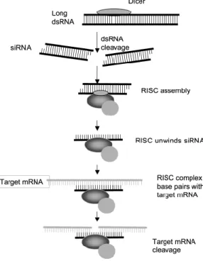
Isogenic cell lines (Table 2.1) were created in order to express different levels of TRIB2: 293T and U2OS were transfected to overexpress Trib2 because the basal levels of Trib2 in these cells are low. In the other hand, SK-Mel28 and G361 were transfected to knockdown the Trib2 expression (RNAi technique) because these melanoma cell lines have a high expression of TRIB2.
After cell seeding (cell culture hood), the cells grew in DMEM (Sigma) with 10% heat inactivated FCS (Sigma) supplemented with Pen/Strep (Gibco) in T-75 flasks at the humid CO2 incubator.
Table 2.1-Characteristics of the isogenic cell lines created for experiments.
1.2
Transfection
Transfection is a powerful analytical tool, that introduces foreign nucleic acids (DNA or RNA) into cells to produce genetically modified cells. This technique allows to study the genes and gene products in cells (gene function, regulation and proteins). The transfection
Cell line Amount of protein expressed (TRIB2)
293T Empty(GFP) (Human Renal cancer) low
293T TRIB2 (Human Renal cancer) high
U2OS Empty(GFP) (Human Osteosarcoma) low
U2OS TRIB2 (Human Osteosarcoma) high
G361 scramble(sc.) sh RNA (Human melanoma) high
G361 TRIB2 sh RNA (Human melanoma) low
SK-ML28 scramble(sc.) sh RNA (Human melanoma) high
SK-ML28 TRIB2 sh RNA (Human melanoma) low
Figure 2.1-Chemical tranfection method.59
In this mechanism, DNA/siRNA is wrapped in the transfection reagent and delivered into the cell via endocytosis.