Acta Cir. Bras. 2018;33(2):144-155 DOI: http://dx.doi.org/10.1590/s0102-865020180020000006
Mariana Barbosa Dias CampeloI, Joelita de Alencar Fonseca SantosII, Antonio Luiz Martins Maia FilhoIII, Daniel Cabral Leão FerreiraIV, Luciana Barros Sant’AnnaV, Rauirys Alencar de OliveiraVI, Leonardo Fonseca MaiaVII, Emilia Ângela Loschiavo ArisawaVIII
Effects of the application of the amniotic membrane in
the healing process of skin wounds in rats
1Abstract
Purpose: To evaluate the efficacy of the application of the human amniotic membrane (HAM)
on the inflammatory process, fibroblast proliferation, formation of collagenand reduction of skin wound areas in rats.
Methods: Thirty six rats were submitted to a surgical injury induction and divided into two
groups (n = 18): group C (control) and T (treated with the HAM). The macroscopic evolution in the wound area and the histological characteristics of the skin samples were evaluated.
Results: The regression of the wound area was greater in group T. The histological analysis
revealed a significant reduction (p < 0.05) in the inflammatory infiltrate in group T at all experimental periods compared with that in the control group. Furthermore, the group T presented a significant increase in the proliferation of fibroblasts at 14 and 21 days compared with group C (p < 0.05). Regarding the deposition of mature collagen fibers, there was an increase in the replacement of type III collagen by type I collagen in group T (p < 0.05).
Conclusion: Treatment with the HAM reduced the healing time as well as the inflammatory
responses, increased the proliferation of fibroblasts, and induced a higher concentration of mature collagen fibers.
Key words: Wound Healing. Amnion. Collagen. Inflammation. Rats.
IFellow PhD degree, Postgraduate Program in Biomedical Engineering, Universidade do Vale do Paraíba (UNIVAP),
Sao Jose dos Campos-SP, Brazil. Conception, design, intellectual and scientific content of the study; acquisition and interpretation of data, technical procedures, manuscript preparation.
IIFellow PhD degree, Postgraduate Program in Biomedical Engineering, UNIVAP, Sao Jose dos Campos-SP. Assistant
Professor, Nursing Department, Universidade Federal do Piauí (UFPI), Teresina-PI, Brazil. Technical procedures, manuscript preparation.
IIIPhD, Associate Professor, Biotechnology and Biodiversity Laboratory, Universidade Estadual do Piauí (UESPI),
Teresina-PI, Brazil. Technical procedures.
IVVeterinary, Biotechnology and Biodiversity Laboratory, UESPI, Teresina-PI, Brazil. Technical procedures. VPhD, Immunology Laboratory, UNIVAP, Sao Jose dos Campos-SP, Brazil. Technical procedures.
VIPhD, Associate Professor, Department of Physiotherapy, UESPI, Teresina-PI, Brazil. Technical procedures. VIIAssistant Professor, Department of Medicine, UFPI, Teresina-PI, Brazil. Technical procedures.
VIIIPhD, Biostimulation and Tissue Repair Laboratory, UNIVAP, Sao Jose dos Campos-SP, Brazil. Conception, design,
antigenicity, antimicrobial action, and ability to decrease exudates and adhesions, accelerate epithelization, reduce local pain, and act as a substrate for tissue growth5-8
.
The amniotic membrane, a tissue of an embryonic origin, is composed of three layers: a single epithelial layer, a thick basement membrane, and an avascular mesenchyme. Because it contains no nerves or lymphatic vessels, it is nourished and oxygenated by the chorionic fluid and amniotic fluid3.
The more internal epithelial layer consists of simple cuboidal epithelium that lines the amniotic membrane and is in direct contact with the amniotic fluid4. This layer is
supported by the basal membrane, composed
mainly of type IV collagen, elastin, fibronectin,
laminin, and proteoglycans.The mesenchymal
layer has three regions: an acellular layer that constitutes the main fibrous skeleton of the HAM, composed of types I and III collagen and fibronectin; a layer of mesenchymal cells similar to dispersed fibroblasts; and a randomly disposed sponge layer of collagen fibers separating the amniotic membrane from
the chorion7-9 .
Pre-clinical studies have employed the HAM successfully and showed that its use can accelerate the regeneration of various tissues5,10,11. However, only a limited number
of studies have investigated its application as
biological dressings.
It is important to increase our
knowledge on the influence of the HAM on skin wounds in animal models, given that the efficacy and action of the HAM are not fully understood to promote the validation and improvement of therapeutic protocols. Thus, we hypothesized that the use of the HAM fragments as biological dressings can accelerate the tissue repair process. The objective of the present study was to evaluate the effectiveness of this biomaterial in the inflammatory process and fibrinogenesis, mainly of types I and III
■
Introd
uction
Chronic nonhealing skin wounds are one of the major health problems in several countries. It is estimated that 6 million individuals are affected by chronic wounds in the United States, with a cost estimated at
more than 25 million dollars annually1
.
Growing advances have occurred in
the treatment of skin lesions, considering the high availability of different coverages,
whose development demands advanced
technology and high costs. However, they are not completely effective in tissue regeneration
and pain reduction2
.
Given the socioeconomic and clinical
significance of skin wounds and their impact on public health, constant research is required
to improve treatment protocols. Innovative
solutions, particularly the application of extraembryonic stem cells derived from the
placenta, chorion, and amniotic membrane,
have been the subject of numerous studies in regenerative medicine and skin engineering, especially in recent years. The cells from these
biomaterials are considered multipotent and
have a high proliferation rate, good plasticity,
and no immune response3 .
According to these studies, the human
amniotic membrane (HAM), the inner layer of the fetal membranes, stands out for its ability to act in the healing process and reduce the tissue inflammatory response4. It should
be emphasized that the use of the HAM has a long history in the treatment of wounds because since the first half of the last century, researchers have been studying its use in the reconstitution of tissue lesions, especially of the skin. In addition, it has been used in
ophthalmology and in burns5-7
.
As a part of the placenta, the HAM is discarded after birth; however, its application
collagen, in different stages of the wound healing process in surgically induced skin
lesions in rats.
■
Methods
This study was approved by the Research Ethics Committee Universidade do Vale do Paraíba (1.647.871) and the Committee of Ethics in Animal Experimentation of the
Universidade Estadual do Piauí with n°
147.66-16.
Thirty-six 40-day-old male Wistar rats (Rattus norvegicus albinus) weighing 230 ±
20 g were used and housed individually in polypropylene cages containing ration and
water ad libitum in a 12/12-h light-dark cycle.
The animals were divided into two groups according to the experimental protocols: Control (C)- rats submitted to surgical injury induction and simulation of application of the HAM and Treated (T)- animals also submitted to surgical injury induction followed by the application of a fragment of the HAM. Thereafter, these groups were subdivided on the basis of the experimental periods of 7, 14, and 21 days, totaling six animals in each group.
Harvesting and processing of the placenta
Two human placentas were harvested and processed on the basis of the methodology described by Sant’Anna et al.11. Term human
placentas were donated by patients with normal pregnancy (gestational age greater than or equal to 37 weeks and negative results for HIV-1 and 2, hepatitis B and C, and syphilis) who underwent elective cesarean sections and who signed the Informed Consent Form.
The placenta was placed in sterile plastic
bags refrigerated at 10-15°C, accommodated in
thermal boxes with ice packs, and transported
to the laboratory. The biomaterial was prepared in sterile conditions in a laminar flow
hood, including the manual separation of the
HAM from the chorionic membrane, followed by washing with phosphate buffered saline (Sigma, St. Louis, MO, USA) containing 100-U/mL penicillin, 100-μg/mL streptomycin and
amphotericin (Lonza, Basel, Switzerland)11.
The HAM was then sectioned into pieces of
an appropriate size (4×4 cm), which was larger than the surgical wounds.
The fragments were stored separately, as reported by Hennerbichler et al.12, in
50-mL vials filled with a solution containing glycerol and serum-free DMEM without
phenol (volume 1:1) as a cryoprotectant agent
and frozen at -80°C. The HAM was thawed
immediately before use, until it reached an
ambient temperature (240C), according to the
procedure described by Baradaran-Rafii et al.13.
It is important to note that the HAM
fragments were applied on the wound bed
of the animals previously cleaned with 0.9% saline solution and with the stromal surface in contact with the bloody area, based on
previous studies by Niknejad and Yasdanpanah9
who reported better results when this protocol
was used.
Experimental surgical procedures
The animals were anesthetized with xylazine hydrochloride (2%, 0.01 mL/kg) and ketamine hydrochloride (10%, 0.005 mL/kg) intramuscularly after weighing. Next, a 6-cm2 area on the dorsal region of the animal was shaved using an electric clipper, and local
antisepsis was provided using 0.5% topical chlorhexidine alcohol. The surgical lesions,
measuring 3 cm in diameter and 1 cm in depth, were created in a standardized manner
with the help of a circular metal instrument with a metal blade (nº 4) removing the entire thickness of the skin.
of the HAM, exceeding its edges; the ends were secured in four points using methyl methacrylate.
After completing the surgical
procedures, the rats in groups C and T were
kept in individual cages and received the
following treatments: antibiotic- amoxicillin (0.001 mL/kg, intramuscularly, single dose); anti-inflammatory agent- flunixin meglumine (0.001 mL/kg, subcutaneously, 3 days); and analgesic- metamizole (0.001 mg/kg, orally, 3 days).
Macroscopic analysis
The wound area was macroscopically evaluated 0, 7, 14, and 21 days after surgery using the images captured with a digital camera
(Nikon Coolpix P100, with 10.3-megapixel resolution), without a flash and using natural light, fixed on a tripod at a distance of 20 cm from the wound. Macroscopic morphometry was performed using the software ImageJ to calculate the area of the wound at the different
experimental periods; the results were
expressed as the percentage of regression of the skin wound (area of the initial wound - area of the final wound / area of the initial wound × 100)14
.
Histological analysis
At 7, 14, and 21 days after surgery, the
animals in each group were euthanized by an overdose of anesthesia (Sodium Pentobarbital 100 mg/kg, intraperitoneally) and disposed of
in accordance with the guidelines established
by the National Council for control of Animal Experimentation (CONSEA). The skin area
where the wound was created and the
surrounding area were removed and fixed in neutral buffered formalin (10%, Synth, Diadem-SP, Brazil) and processed for routine histological processing. Thereafter, 5-μm sections were stained with hematoxylin & eosin (H&E) and
sirius red for the qualitative histological and morphometric analyses.
The H&E-stained images were scanned
using the Leica DM 2500 microscope coupled
to the Leica DFC 425 camera and Leica
Application Suite Program LAS v3.7. Images
were obtained from the cross sections of six sequential fields of each wound (40X objective) in a light microscope (final amplification ×400). To quantify the number of inflammatory cells and fibroblasts, the images were analyzed using the software ImageJ, which enabled the elaboration of a grid and the individual marking of cell nuclei with the aid of the manual counting tool. The average score of 10 random microscopic fields per section was used to create a single score for each specimen in each experimental group.
The slides stained with picrosirius red were quantitatively evaluated via digital image analysis to calculate the area occupied by the deposition of types I and III collagen in the skin wound. The images were captured using the digital video camera (Leica DF425, Germany)
coupled to the polarized light microscope
(Leica DM2500) and digitalized in 1024×768 pixels and resolution of 24 bits/pixel with a general amplification of x100. The images were processed using CellProfiler (Broad Institute of Harvard and MIT), which identified, isolated, and measured the areas occupied by type I and type III collagen in relation to the total area of
the image. All histomorphometric analyses
were performed blindly.
Statistical analysis
The statistical analysis was performed using the software Statistic 7.0. Statsoft©. The
■
Results
Macroscopic analysis
The skin wounds of the animals were evaluated macroscopically 0, 7, 14, and 21 days after the surgical procedures in terms of the formation of a scab and reduction in the
wound area.
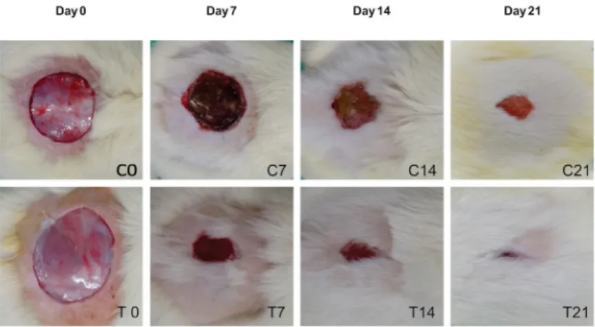
Among the animals in group C, no
evidence of infection was observed 7 days after
the surgical procedures, although the wounds
were covered by extensive scabs (Figure 1-C7). At 14 days, the presence of granulation tissues was observed (Figure 1-C14), while at 21 days,
the wound area showed a significant reduction,
presenting a small and not yet re-epithelialized
area (Figure 1-C21).
Conversely, infection was not also observed among the animals in group T at 7 days owing to the application of the HAM,
which remained adhered to the wound area,
without forming a thick scab (Figure 1-T7). At 14 days, the animals had an advanced healing process, with a reduction in the surgical wound area and the presence of granulation tissues (Figure 1-T14). At 21 days, the wounds were practically closed and re-epithelialized, with no signs of the presence of the HAM fragment (Figure 1-T21).
Figure 1 -Macroscopic images of the surgical wound area of the groups C and T animals 0, 7, 14, and 21 days
after surgery.
Microscopic analysis
7 days
The qualitative analysis of the
H&E-stained slides on the 7th day revealed the
presence of superficial scabs in the group C animals and an intense inflammatory response
below the scab (Figure 2-C7) with a predominance
of neutrophil polymorphonuclear cells and moderate vascular congestion in the hypodermis. The histological sections of group T then revealed that the wounds treated with the HAM presented a discrete acute inflammatory infiltrate, with the beginning of
the wound healing process characterized by an intense angiogenesis, increased deposition of extracellular matrix resulting from young and large fibroblasts (Figure 2-T7), and proliferation of type III collagen.
14 days
In the fragments of group C, a mild
inflammatory infiltrate was observed, with a
observed, with mild inflammatory response as well as the presence of polymorphonuclear leukocytes, absence of edema and vascular
congestion, and presence of well-vascularized
granulation tissues and organized collagen deposition (Figure 2-T14).
21 days
At 21 days, the specimens of group
C showed a deposition of collagen fibers,
with the formation of granulomas, which
may have interfered with the wound healing
process (Figure 2-C21). In the group treated
with the HAM fragment, there was a closure of the wound area by second intention and an organization of the collagen fibers (Figure 2-T21). In this group, proliferation of keratinized squamous epithelium, presence of fibroblasts, and discrete vascularization, were
observed.
Figure 2 - Qualitative histological aspects of the wound healing process. Note the inflammatory response
and proliferation of fibroblasts and collagen fibers in the region of the wound in groups C (control) and T (treated with the human amniotic membrane) at 7, 14, and 21 days (Hematoxylin & Eosin, x400). Yellow arrows indicate the inflammatory cells; red arrows, the blood vessels; blue arrows, the fibroblasts; and green arrows, the collagen fibers.
Analysis of the collagen fibers
During the wound healing process, type
III collagen (immature) is replaced by type I collagen. The qualitative analysis of the sirius red-stained slides showed the progression of
this process in both groups throughout the
experiment.
The analysis of the images captured
using the polarized light microscope in the
control group showed a predominance of type
III collagen (immature) as shown in Figures 3A (non-polarized) and 3B (polarized) at 14
days and in Figures 3C (non-polarized) and 3D (polarized) at 21 days.
Conversely, in the samples of the
HAM-treated group (T) at 14 days, there was a concentration of the subtypes of partially organized types I and III collagen, with an evident prevalence of type III collagen, both in the microscopic images of the non-polarized
light (Figure 3E) and polarized light (Figure 3F).
At 21 days, the histological sections showed that the wound area was fully re-epithelialized and that types I and III collagen were aligned and fully organized, with a predominance of type I collagen as shown in Figures 3G
(non-polarized) and 3H ((non-polarized), and an advanced
Figure 3 - Qualitative aspects of the collagen fibers in groups C (control) and T (treated with the human
amniotic membrane) at 14 and 21 days, stained with sirius red (x100)
Macroscopic analysis of the percentage of regression of the areas of cutaneous lesions
The macroscopic analysis of the percentage of regression of the wounds showed a significant increase in group T
when compared with that in group C. On the
7th day and 21st day, a statistically significant
difference was observed (p < 0.05) with respect to the areas of regression of the wound in group T compared with that in the
control group. However, at 14 days, there
was no significant difference between groups C and T (Figure 4).
Figure 4 - Percentage of regression of the surgical wounds in groups C (control) and T (treated with the human
Quantitative analysis of the inflammatory cells and fibroblasts
There was a reduction in the inflammatory infiltrate in group T compared with that in group C. The intergroup analysis showed a statistically significant difference (p <
0.05) at 7, 14, and 21 days. Group T showed a significant increase in the number of fibroblasts (p < 0.05) when compared with group C only
on the 14th and 21st days of the wound healing
process. However, no significant difference was observed in the number of fibroblasts between both groups at 7 days (Figure 5).
Figure 5 - Histological analysis of groups C (control) and T (treated with the human amniotic membrane) at 7,
14, and 21 days. (A) Number of inflammatory cells (20.000 µm2) and (B) number of fibroblasts (20.000 µm2);
Mann-Whitney U test. The values with a statistical significance are indicated by * (p < 0.05).
Quantitative analysis of the collagen concentration
Regarding the percentage of types I
and III collagen present in the wound healing
process at 7 days, the average percentage of
types I and III collagen was practically equal in both groups, with a predominance of the latter. However, at 14 days, there was a statistically significant increase in the replacement of type III collagen by type I collagen in group T compared with that in group C (p ≤ 0.05). At 21 days, a similar distribution of types I and III collagen subtypes was observed in both groups, with no statistical significance (Figure 6).
Figure 6 - Percentage of types I and III collagen in
the surgical wounds of groups C (control) and T (treated with HAM) at 7, 14, and 21 days;
In the present study, the typification of the collagen fibers via the analysis of the
polarized light allowed us to observe a balance
between the distribution of types I (yellow, orange, or red) and III collagen (green) from the margins to the center of the wound.
■
Discussion
The amniotic membrane acts as a
transplanted basal membrane that serves as
a substrate for epithelization3. Recent studies
have revealed that the HAM is a rich source of MSCs, collagen matrix, and growth factors. In addition, it serves as a support for tissue repair and regeneration15.
The objective of this study was to evaluate the efficacy of the application of the HAM regarding the inflammatory response, fibrinogenesis, and remodeling and organization of collagen during the wound healing process; thus, the periods of 7, 14, and
21 days were selected, allowing us to evaluate
this process14 .
The results showed that the application of the HAM accelerated the wound healing process, with closure of the injury. The anti-inflammatory action of the HAM reduced the duration of the initial healing phase, favoring the early initiation of the formation of collagen fibers, as in the remodeling phase, characterized by the replacement of type III collagen (immature) by type I collagen
(mature).
After tissue injury, the inflammatory response plays an important role in the processes of normal and pathological healing because it provides protection against invading
pathogens and removes damaged tissues16.
However, a prolonged inflammatory response determines a delay in the progression in the different phases of the wound healing
process14-17
. The results obtained in this study
showed that the HAM played a protective
role in the wound area, providing a favorable microenvironment to repair the skin, compared with the results of the control group. The histological analysis demonstrated
that the inflammatory response in group T was
less intense at all stages of the process than that in group C, favoring a more organized progression. The considerable decrease in the number of inflammatory cells at 14 days and 21 days in group T demonstrated that the HAM did not increase the inflammatory response
in the wounds, which corroborates their low
immunogenicity and its possible allogeneic
use18-19 .
Manuelpillai et al.14
, reported that the
anti-inflammatory properties are associated with the presence of several factors in the stroma of the HAM, such as interleukin (IL)-10, IL-1 receptor antagonist (IL-1), hyaluronic
acid, and prostaglandin E2 (10). Tseng et
al.20 reported that the anti-inflammatory
activity of the HAM requires a close contact
with its stromal matrix. The high content
of proteoglycans and glycoproteins of the intermediate layer of the stromal matrix contains a non-fibrillar mesh, mainly of type III
collagen6. In addition, the microenvironment
maintained with the use of fragments allows the receptor tissue of this biomaterial to use all growth factors and other products present to potentiate the development of tissue repair.
The results obtained showed that
during the proliferative phase (7 to 14 days), the number of fibroblasts increased in both groups
studied. However, the histomorphometric
It is evident that the repair process had
an early initiation in the wounds protected compared with that in the fully exposed
wounds because the membrane acts as a
biological bandage protecting the injured tissues; this favors local hydration and avoids contamination by microorganisms and
contaminated materials5.
Meller et al.6, Tahan and Tahan8, and
Sant’Anna et al.23
, demonstrated that the
HAM exerts beneficial actions in the repair and regeneration of tissues owing to its anti-inflammatory, analgesic, anti-bacterial, re-epithelization, and healing effects on wounds.
Histological staining by picrosirius red is the method of choice to evaluate the presence of collagen fibers, staining them red. These fibers, when visualized under an optical
microscope with polarizing lens, assume
different colors. They become orange if they are type I collagen or greenish if they are type III collagen. This birefringence is highly specific for collagen24.
Histologically, the dermis of rodents is
composed of dense connective tissue formed by types I and III collagen and a small amount of elastic fibers. The proliferative phase of healing occurs 3 to 5 days after tissue injury and is characterized by an increase in the number of fibroblasts, which produce and deposit a large amount of type III collagen. Later, the maturation of these fibers occurs, with their remodeling and replacement by type I collagen, which is thicker and more
resistant25. The present study demonstrates
that the collagen fibers of the group T animals reached maturity and early macromolecular orientation compared with that of the control
animals, which had a slower wound healing process.
Duarte et al.5 reported that in the
wound healing process of rats treated with the HAM, a predominance of mature collagen fibers was observed. Vascular formation was
more significant in the animals that received the HAM, indicating that even its conservation at lower temperatures does not inactivate pro-angiogenic substances previously identified in
the cryopreserved membrane26.
In addition, the harvesting of the amniotic membrane does not offer any risk
to the newborn or the parturient, and the
membrane can be easily obtained and stored in laboratories for later use. The use of these
biomaterials does not raise ethical or religious
conflicts, besides being a cost-effective alternative, when compared to the isolation and culture of stem cells, which require more complex processes4-18.
Therefore, the results confirm the biocompatibility of the HAM and that its application promoted a greater formation of collagen fibers and total re-epithelialization of the wound area. We emphasize that the use of the HAM offers the advantages of high availability, ease of application, and good storability12-18. In this context, it is possible to
affirm that by presenting properties that favor the re-structuring of the damaged portions of the tissues, the amniotic membrane could be used as a biological coverage of acute and
chronic wounds18-26.
■
Conclusions
■
References
1. Tamama K, Kerpedjieva SS. Acceleration of wound healing by multiple growth factors and cytokines secreted from multipotential stromal cells/mesenchymal stem cells. Adv Wound Care. 2012;1(4):177–82. doi: 10.1089/wound.2011.0296.
2. Lorenzo Hernández MP, Hernández Cano RM, Soria Suárez MI. Heridas crónicas
atendidas en un servicio de urgencias.
Enfermería Glob. 2014;13(3):23–31.
3. Toda A, Okabe M, Yoshida T, Nikaido T. The potential of amniotic membrane/amnion-derived cells for regeneration of various tissues. J Pharmacol Sci. 2007;105(3):215–8. doi: 10.1254/jphs.CR0070034.
4. Niknejad H, Peirovi H, Jorjani M, Ahmadiani A, Ghanavi J, Seifalian AM. Properties of the amniotic membrane for potential use in tissue engineering. Eur Cell Mater. 2008;15(April):88–9. doi: 10.22203/eCM.
v015a07.
5. Duarte IGL, Duval-Araujo I. Amniotic
membrane as a biological dressing in
infected wound healing in rabbits. Acta Cir Bras. 2014;29(5):334–9. doi: 10.1590/ S0102-86502014000500008.
6. Meller D, Pauklin M, Thomasen H, Westekemper H, Steuhl K-P. Amniotic membrane transplantation in the human eye. Dtsch Arztebl Int. 2011;108(14):243–8. doi: 10.3238/arztebl.2011.0243.
7. Mamede A, Carvalho M, Abrantes A, Laranjo M, Maia C, Botelho M. Amniotic membrane: from structure and functions to clinical applications. Cell Tissue Res. 2012;349(2):447–58. doi: 10.1007/s00441-012-1424-6.
8. Tahan AC, Tahan V. Placental amniotic epithelial cells and their therapeutic potential in liver diseases. Front Med (Lausanne). 2014;1:48. doi: 10.3389/ fmed.2014.00048.
9. Niknejad H, Yazdanpanah G. Opposing effect of amniotic membrane on angiogenesis originating from amniotic epithelial cells. J Med Hypotheses Ideas. 2014;8(1):39–41. doi: 10.1016/j.jmhi.2013.08.002.
10. Manuelpillai U, Moodley Y, Borlongan C V., Parolini O. Amniotic membrane and amniotic cells: potential therapeutic tools to combat tissue inflammation and fibrosis?
Placenta. 2011;32(Suppl. 4):S320-5. doi:
10.1016/j.placenta.2011.04.010
11. Sant’Anna LB, Cargnoni A, Ressel L, Vanosi G, Parolini O. Amniotic membrane application reduces liver fibrosis in a bile duct ligation rat model. Cell Transplant. 2011;20(3):441– 53. doi: 10.3727/096368910X522252. 12. Hennerbichler S, Reichl B, Pleiner D,
Gabriel C, Eibl J, Redl H. The influence of various storage conditions on cell viability in amniotic membrane. Cell Tissue Bank. 2007;8(1):1–8. doi:
10.1007/s10561-006-9002-3.
13. Baradaran-Rafii A, Aghayan H-R, Arjmand B, Javadi M-A. Amniotic membrane transplantation. J Ophthalmic Vis Res. 2007;2(1):58–75.
14. Estevão LRM, Mendonça FDS, Baratella-Evêncio L, Simões RS, Barros MEG De, Arantes RME,Rachid MA, Evêncio-Neto J . Effects of aroeira (Schinus terebinthifoliu Raddi) oil on cutaneous wound healing in rats. Acta Cir Bras. 2013;28(3):202–9. doi: 10.1590/S0102-86502013000300008.
15. Gibbons GW. Grafix®, a cryopreserved
placental membrane, for the treatment of chronic/stalled wounds. Adv Wound Care. 2015;4(9):534–44. doi: 10.1089/ wound.2015.0647.
16. Qian LW, Fourcaudot AB, Yamane K, You T, Chan RK, Leung KP. Exacerbated and prolonged inflammation impairs wound healing and increases scarring. Wound Repair Regen. 2016;24(1):26–34. doi:
10.1111/wrr.12381.
17. Demidova-Rice TN, Hamblin MR, Herman IM. Acute and impaired wound healing: pathophysiology and current methods for drug delivery, part 1: normal and chronic wounds: biology, causes, and approaches to care. Adv Skin Wound Care. 2012;25(7):304– 14. doi: 10.1111/wrr.12381.
18. Nicodemo M de C, Neves LR das, Aguiar JC, Brito F de S, Ferreira I, Sant’Anna LB, Raniero LJ, Martins RAL,Barja PR, Arisawa EALS. Amniotic membrane as an option for treatment of acute Achilles tendon injury in rats. Acta Cir Bras. 2017 Feb;32(2):125–39. doi: 10.1590/s0102-865020170205.
19. Duan-Arnold Y, Uveges TE, Gyurdieva A, Johnson A, Danilkovitch A. Angiogenic potential of cryopreserved amniotic
of all tissue components in their native state. Adv Wound Care. 2015;4(9):513–22. doi: 10.1089/wound.2015.0638.
20. Tseng SCG, Espana EM, Kawakita T, Di Pascuale MA, Li W, He H, Liu TS, Cho TH, Gao YY, Yeh LK, Liu CY. How does amniotic membrane work? Ocular Surface.
2004;2:177–87.doi:
10.1016/S1542-0124(12)70059-9.
21. Frykberg RG, Banks J. Challenges in the treatment of chronic wounds. Adv Wound Care. 2015;4(9):560–82. doi: 10.1089/ wound.2015.0635.
22. Gutiérrez-Moreno S, Alsina-Gibert M, Sampietro-Colom L, Pedregosa-Fauste S, Ayala-Blanco P. Estudio coste-beneficio del trasplante de membrana amniótica para úlceras venosas de extremidades inferiores refractarias a tratamiento convencional. Actas Dermosifiliogr. 2011;102(4):284–8. doi: 10.1016/j.ad.2011.01.003.
23. Sant’Anna LB, Brito FS, Barja PR, Nicodemo MC. Long-term effects of human amniotic membrane in a rat model of biliary fibrosis.
Brazilian J Med Biol Res. 2017;50(7):e5692. doi: 10.1590/1414-431X20175692.
24. Junqueira LCU, Bignolas G, Brentani RR. Picrosirius staining plus polarization microscopy, a specific method for collagen detection in tissue sections. Histochem J. 1979;11(4):447–55. doi: 10.1007/
BF01002772.
25. Baptistella E, Malafaia O, Czeczko NG, Ribas-Filho JM, Nassif PAN, Nascimento MM do, Pachnicki, JPA. Comparative study in swines’ vocal cords healing after excision of fragment with CO2 laser with
mitomycin and 5-fluorouracil postoperative topical application. Acta Cir Bras. 2009
Jan-Feb;24(1):13-8. PMID: 19169536.
26. Barbuto RC, de Araujo ID, Bonomi DO, Tafuri LSA, Neto AC, Malinowski R, Bardin VSS, Leite MD, Duarte IGL. Use of the amniotic membrane to cover the peritoneal cavity in the reconstruction of the abdominal wall with polypropylene mesh in rats . Rev Col Bras Cir. 2015;42(1):49–54. doi: 10.1590/0100-69912015001010.
Correspondence:
Mariana Barbosa Dias Campelo Departamento de Enfermagem, UFPI
Campus Universitário Ministro Petrônio Portela Avenida Nossa Senhora de Fátima, Bloco SG 12 64.049-550 Teresina - PI Brasil
Tel.: (55 86)3215-5881
marianadias@hotmail.com
Received: Oct 10, 2017 Review: Dec 12, 2017 Accepted: Jan 11, 2018
Conflict of interest: none Financial source: none
1Research performed at Laboratory of
Experimental Surgery, and Laboratory of Core Research in Biotechnology and Biodiversity (NPBIO), Faculty of Medical
Sciences, Universidade Estadual do Piauí
(UEPI), Teresina-PI, Brazil. Part of PhD degree
thesis, Postgraduate Program in Biomedical Engineering, Universidade do Vale do Paraíba
(UNIVAP). Tutor: Profa. Emilia Ângela Loschiavo