Printed version ISSN 0001-3765 / Online version ISSN 1678-2690 www.scielo.br/aabc
Morphological and hematological studies of
Trypanosoma
spp.
infecting ornamental armored catfish from Guamá River-PA, Brazil
RODRIGO Y. FUJIMOTO1, MIKAELLE S. NEVES2, RUDÃ F.B. SANTOS2, NATALINO C. SOUZA2, MÁRCIA V.S. DO COUTO2, JOSIANE N.S. LOPES2, DANIEL G. DINIZ3 and JORGE C. EIRAS4,5
1Embrapa Tabuleiros Costeiros, Avenida Beira Mar, 3250, Caixa Postal 44, 49025-040 Aracaju, SE, Brasil 2
Universidade Federal do Pará, Instituto de Estudos Costeiros, Rua Leandro Ribeiro, s/n, Aldeia, 68370-000 Bragança, PA, Brasil
3
Universidade Federal do Pará, Instituto de Ciências Biológicas,
Laboratório de Investigações em Neurodegeneração e Infecção, Hospital Universitário João de Barros Barreto, Rua dos Mundurucus, 4487, Guamá, 66073-005 Belém, PA, Brasil
4
Departamento de Biologia, Faculdade de Ciências, Universidade do Porto, Rua do Campo Alegre, s/n, 4169-007 Porto, Portugal
5
CIIMAR, Centro Interdisciplinar de Investigação Marinha e Ambiental/CIMAR, Universidade do Porto, Rua dos Bragas, 289, 4050-123 Porto, Portugal
Manuscript received on October 30, 2012; accepted for publication on February 27, 2013
ABSTRACT
A total of 281 specimens of freshwater armored ornamental fish species (Leporacanthicus galaxias,
Lasiancistrus saetiger, Cochliodon sp., Hypostomus sp., Pseudacanthicus spinosus, Ancistrus sp. and
Rineloricaria cf. lanceolata) were captured at the hydrological basin of Guamá River, Pará, Brazil. The infection by Trypanosoma spp. was inspected. The morphological and morphometric characterization of the parasites and the hematological parameters were determined. Leporacanthicus galaxias and Pseudacanthicus spinosus
presented 100% infection prevalence, and the other species showed a variable prevalence of infection. The parasites showed clearly different morphotypes and dimensions, and probably belong to different species. The hematological response to the infection varied with the host. Cochliodon sp. showed no differences between infected and not infected fish. In other species several modifications on some hematological parameters were found, but apparently without causing disease. It is emphasized the possibility of introduction of the parasites in new environments due to the artificial movements of these ornamental fish.
Key words: freshwater fish, hematological parameters, infection, Trypanosoma spp.
Correspondence to: Daniel Guerreiro Diniz E-mail: [email protected]
INTRODUCTION
The northeast of Pará State, in Brazil, especially
the hydrographic basin of Guamá River, is an
important area for the fishery of ornamental
freshwater fish. This is due to the facility of
reaching the fishing grounds and to the proximity
There are some observations on the parasites
of ornamental fish in Brazil, mostly about
ectoparasites (Garcia et al. 2009, Piazza et al. 2006,
Prang 2007, Tavares-Dias et al. 2010), and some
studies deal with the infection by
Trypanosoma
spp. These parasites may not harmful to the fish
hosts (Untergasser 1989), or cause anemia, damage
of the hematopoietic tissues and, finally, the death
of the fish (Noga 1996). Massive infections in some
hosts (over 10
5specimens /mm
3), like
Cyprinus
carpio
and
Carassius auratus
, may cause anemia,
anorexia and ascites (Paperna 1996).
According to Eiras et al. (2010, 2012), there
are at least 62 nominal species of trypanosomes
infecting freshwater fish in Brazil, a number of them
parasitizing armored fish like
Hypostomus
spp. and
Pterodoras
spp. (D’Agosto et al. 1985, Lopes et al.
1991, Bara et al. 1985, Fróes et al. 1979). It must
be stressed out that a number of these parasites
were identified only with basis on morphology and
morphometry, and in most of the times assuming
specificity in parasitization.
In this paper we report the infection of 7
different species of ornamental armored freshwater
fish from the Guamá River by
Trypanosoma
spp.
The morphology and morphometry of each form is
described, and the prevalence, abundance and mean
intensity of infection is reported. Furthermore,
the hematological characteristics of the hosts are
referred to.
MATERIALS AND METHODS
A total of 281 armored ornamental specimens
were captured at Guamá River, including
41
Leporacanthicus galaxias
(Isbrüker and
Nijssen 1989) (common name: acari pinima),
48
Lasiancistrus saetiger
(Armbruster 2005)
(acari canoa), 35
Cochliodon
sp. (acari pleco),
10
Pseudacanthicus spinosus
(Castelnau 1855)
(acari assacu), 57
Rineloricaia cf. lanceolata
(acari
loricaria), 42
Hypostomus
sp. (acari picoto), and 48
Ancistrus
sp. (acari ancistrus).
Immediately after capture, a blood sample was
taken from the caudal vein with syringes coated
with 10% EdTA. After blood sampling the fish were
measured (total and standard length) and inspected
for ectoparasites, and integument or gill lesions.
The following blood parameters were determined:
glucose (mg/dL) using the automatic meter Prestige
IQ 50, hematocrit (Ht, at 13,000 rpm during 3
minutes), total plasma protein (g/dL) using a Quimis
refractometer, total hemoglobin (HB, g/dL) using
a Celmi 500 and Celmi 550 meter, and number of
erythrocytes per mm
3counted in Neubauer chamber.
Blood smears were air dried and stained with
May Grunwald Giemsa modified by Rosenfeld
(1947). The smears were used for leukocyte
differential counting. The determination of mean
corpuscular volume (MCv = ht / Er x 10, fentoliter),
mean corpuscular hemoglobin (MCh = hb / Er x
10, picograms), and mean corpuscular hemoglobin
concentration (MChC= hb / ht x 100, g/dL) were
recorded according to Vallada (1999).
Blood smears and hematocrit were also used
for determining the presence of trypanosomes. In
the positive samples the mean intensity of infection
was determined indirectly relating the number of
parasites with the amount of 1,000 erythrocytes
(modified from Ranzani-Paiva 1995 method of
differential counting of white blood cells). The
parasites were photographed using a digital camera,
and the photographs were used latter (employing
the software Motic Images Advanced 3.0) to
determine the cell characteristics: total length (TL),
maximum width of the body (W), nucleus length
(NL) and width (NW), distance between the middle
of nucleus and anterior (DNA) and posterior (DNP)
extremities, length of the free flagellum (FF) and
number of folds of the undulating membrane (UM).
With the data obtained, the nuclear index (IN)
according to D’Agosto and Serra-Freire (1993) and
Gu et al. (2006) was calculated.
variance (BioEstt 4.0 programme). For F significant
values, it was employed the Tukey test (5% of
probability) to compare the means values. For the
analysis of the variance of folds of the undulating
membrane, it was employed the Kruskal-Wallis
method for non-parametric data. It was also used
the normality test with basis in the deviations
values to verify the existence of outliers which
were eliminated.
RESULTS
The species
Pseudacanthius
spinosus and
Leporacanthicus galaxias
presented 100% of
prevalence of infection, the other hosts showed
a prevalence value varying between 22.6%
(
Cochliodon
sp.) and 58.3% (
Lasanciastrus
sp.) (Table I). Therefore, most of the fishes were
infected, and a high proportion of them were also
infected by unidentified leeches. however, some
of the specimens infected with leeches were not
parasitized by trypanosomes.
Hypostomus
sp. presented the highest intensity
of infection (1.3 parasites by 1,000 erythrocytes),
and
Rineloricaria cf. lanceolata
showed the lowest
one (0.5). The other species presented intermediate
values (Table I).
Pseudacanthicus spinosus
had
the highest abundance level, and
Rineloricaria cf.
lanceolata
the smallest one, while the other species
presented intermediate values (Table I).
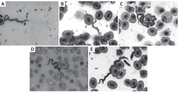
The morphology (Fig. 1) of the parasites varied.
In general, they had a rounded anterior extremity
and a tapered posterior one. In some cases both the
extremities were slightly tapered. The nuclei were
most of the times oval-shaped, sometimes almost
circular, in some cells occupying all the cell width.
The kinetoplast was mostly rounded, in most of
cases having a sub-terminal location. Usually the
small part of the cell located before the kinetoplast
was difficult to observe clearly due to poor staining
of this part of the body.
The undulating membrane was well defined,
developing all over the body length, or about half
of the length. In some cases it was observed only
near the extremity, presenting only two folds in
the specimens with smaller values of body width.
The undulating membrane was especially evident
in
Cochliodon
sp., presenting in this host more
folds. The cytoplasm varied from basophilic to
eosinophilic. The free flagellum was sometimes hard
to distinguish because it was not so intensely stained,
and its length varied between short and long.
There was a great morphometrical variation in
the several characteristics as it can be seen in Table
II, and several features presented a great variation
depending from the host species.
The hematological study showed the infection
caused varied effects on the hosts. Interestingly,
in
Cochliodon
sp. no hematological alterations
were found between infected and not infected
specimens. In
Ancistrus
sp. and
Hypostomus
sp. the repercussions were minimal – the first
specimens showed only a pronounced increase of
the concentration of mean corpuscular hemoglobin,
and the second revealed increase in the percentage
of lymphocytes. In
Rineloricaria cf. lanceolata
the infection caused decrease of mean corpuscular
hemoglobin concentration and glucose, and
modifications in the white blood cells (decrease
of lymphocytes and neutrophils and increase in
monocytes). Finally,
Lasiancistrus saetiger
showed
increase in the hematocrit and erythrocytes, and
decrease of total plasma proteins and of mean
corpuscular hemoglobin.
DISCUSSION
probability of leech infection. The armored
fish have a benthic behavior that facilitates the
infection by leeches, and high values of infection
by trypanosomes are not uncommon. D’Agosto and
Serra-Freire (1990) reported 100% of prevalence
for
Trypanosoma chagasi
and
T. guaiabensis
infecting the armored
Hypostomus punctatus
from lake Açú at Rio de Janeiro. Other reports on
infections in several species of armored fish showed
a high variability on the prevalence and intensity of
infection values (Fróes et al. 1978, 1979, Lopes et
al. 1989, Ribeiro et al. 1989, Eiras et al. 1989, 1990).
Considering these facts, and the fact that apparently
the probability of leech infection in the fishes from
our sample was the same for all the host species, it
is possible to conclud that the resistance of the fish
to the infection varies with the fish species.
There are in Brazil at least 62 species of
trypanosomes described from freshwater fish, and at
least 28 from those were described from armored fish
(Eiras et al. 2010). Most of the descriptions were done
assuming a strict specificity of infection, and a form
observed in a new host was considered a new species
(Thatcher 2006). Today it is recognized that strict
specificity may be an exception but not a role, and it is
urgent to review the Brazilian fish trypanosomes as it
was done with trypanosomes from Africa performed
by Baker (1960), resulting in a substantial reduction
of the number of blood flagellate species. Besides,
one confusing factor is the variability in length during
infection and the existence of pleomorphic species
(Gibson et al. 2005).
Our data do not allow the identification of
the parasite species and, for the reasons described
above, a comparison with the Brazilian species of
trypanosomes would be useless. The identification
based solely on morphological features is usually
not possible, and the absence of specific infections,
Cochliodon sp. Ancistrus sp. Lasiancistrus saetiger.
Not infected Infected Not infected Infected Not infected Infected W 22.0±5.73ns 21.8±12.03ns 39.2±13.98ns 26.6±5.46ns 28.4±15.68ns 32.7±15.23ns SL 9.2±1.90ns 9.8±1.77ns 11.2±1.43ns 11.4±0.30ns 10.1±1.76ns 9.8±1.99ns TL 12.2±1.02ns 12.9±1.99ns 13.9±2.07ns 14.1±0.81ns 11.4±2.30ns 11.4±3.50ns
P _ 26.66 _ 20 _ 58.3
MI _ 1 _ 1 _ 1
AB _ 0.26 _ 0.2 _ 0.58
GLUC 92.4±10.80ns 42.7±11.74ns 72.4±28.72ns 95±13.85ns 55.2±18.37ns 44.1±8.75ns HT 20.6±10.76ns 21.8±15.56ns 20.4±10.46ns 14.3±8.96ns 17.8±7.49 b 28.0±11.69 a
TPP 7.8±2.45ns 8.4±3.16ns 4.6±1.69ns 3.0±1.17ns 8.9±2.30 a 6.5±2.53b
HB 6.8±4.20ns 5.2.54ns 8.9±5.54ns 9.3±5.95ns 9.23±4.52ns 9.7±4.97ns
ER 0.3±0.46ns 0.2±0.18ns 0.6±16.99ns 0.6±0.26ns 0.3±0.32 b 1.3±0.06 a
MCV 1097.9±286.90ns 568.9±250.54ns 373.7±156.62ns 289.2±264.15ns 845.8±786.60ns 230.9±99.29ns MCH 270.8±175.63ns 211.9±108.40ns 170.5±107.62ns 162.3±87.09ns 1106.4±1917.10ns 71.6±37.18ns CMCH 38.6±29.07ns 35.7±2.94ns 41.3±17.67 b 155.4±216.18 a 63.4±39.44 a 29.4±10.97 b
LYM 56.1±20.65ns 47.2±20.12ns 75.6±19.56ns 81.3±2.51ns 94.4±13.23 64.4±15.33 NEU 39.5±21.16ns 48.2±21.29ns 19.9±16.89ns 11.3±1.52ns 4.0±9.6 28.0±13.8
MON 4.2±1.48ns 4.5±2.51ns 3.2±3.24ns 7.3±3.21ns 1.2±0.8 7.5±4.4
TABLE I
Total weight (W), standard (SL) and total length (TL) of fish, prevalence of infection (P), mean intensity of infection (MI), abundance of infection (AB), and hematological parameters in infected and uninfected fish. Figures highlighted in
Leporacanthicus galaxias
Pseudacanthicus
spinosus Hypostomus sp. Rineloricaria cf. lanceolata
Infected Infected Not infected Infected Not infected Infected
W 28.4±14.6 25.2±27.9 21.4±8.76ns 23.9±6.05ns 19.4±6.88ns 13.0±5.38ns
SL 10.1±2.0 9.5±2.7 8.7±1.33ns 9.1±0.96ns 15.3±2.10ns 15.5±2.53ns
TL 12.9±2.6 12.6±2.7 11.0±1.53ns 11.±1.51ns 18.0±2.80ns 17±4.69ns
P 100 100 _ 20 _ 46.6
MI 1.1 1.2 _ 1.3 _ 0.5
AB 1.1 1.2 _ 0.2 _ 0.2
GLIC 60.2±27.8 27.6±21.6 62.3±15.29ns 48.6±8.5ns 102.4±30.04a 59.5±22.12b
HT 31.6±13.2 10.1±5.9 16.9±7.01ns 21±7.54ns 16.0±6.76ns 17.5±3.93ns
TPP 8.6±3.4 6.3±2.6 8.2±2.18ns 10.3±0.91ns 9.0±2.00ns 9.0±1.85ns
HB 10.9±3.8 5.7±3.2 9.9±5.09ns 9.7±0.92ns 9.5±4.73ns 4.9±2.86ns
ER 0.4±0.3 0.2±0.2 0.6±0.30ns 0.4±0.20ns 0.6±0.34ns 0.2±0.18ns
MCV 843.7±661.4 1887.0±180.6 693.7±740.41ns 490.9±167.64ns 557.4±848.57ns 1070.4±1073.02ns MCH 353.6±348.3 1541.2±145.1 342.1±354.24ns 246.2±108.77ns 199.5±1066.66ns 216.5±110.35ns CMCH 37.4±17.7 62.7±17.2 58.8±24.71ns 50.4±18.28ns 58.3±47.05a 26.4±11.84b
LYM 27.3±7.8 62.8±8.7 29.4±5.85a 14.66±9.23b 80.2±7.71a 64.7±9.46b
NEU 68.9±8.5 28.8±6.9 67.5±5.61ns 76±15.09ns 14.6±7.16b 25±5.29a
MON 3.7±1.9 8.4±2.6 3±1.75ns 9.3±7.57ns 4.3±3.25b 10.2±4.27a
TABLE I (continuation)
Abbreviations: GLUC, glucose (mg/dl), HT, hematocrit, TPP, total plasma proteins g/dl, HB, hemoglobin g/dl, ER, number of erythrocytes (number of cells x 106/mm3); MCV, mean corpuscular volume (fentoliter); MCH, median corpuscular hemoglobin (pg); CMCH, concentration of the median corpuscular hemoglobin in g/dl; LYM, total number of lymphocytes; NEUT, neutrophils (%); MON, monocytes (%).
at least in most of the cases, do not allow a positive
identification without the aid of molecular tools, and
characterization of the development of the parasite
within the vector, which were not considered in the
present research. However, it is highly probable that
we face different species due to the so pronounced
differences in morphology and morphometry of the
parasites, as depicted in Figure 1 and Table II. It is
the authors’ intent to pursue this study in the future
in order to elucidate this question.
According to the hematological data obtained,
it seems that some host species (
Lasiancistrus
saetiger
and
Rineloricaria cf. lanceolata
) were
more affected than others (
Ancistrus
sp. and
Hypostomus
sp.), while
Cochliodon
sp. apparently
had the hematological parameters not altered by the
infection. Therefore, it can be concluded that some
hosts adapted better than others to the infection.
Some results of other authors for different
freshwater hosts species show results sometimes
similar to ours: anemia in
Carassius auratus
infected with
Trypanosoma danilewskyi
(Dyková
and Lom 1979), in
Barilius blendelisis
parasitized
by
Trypanosoma
sp. (Rauthan et al. 1995), and in
Cyprinus carpio
infected with
T. borreli
(Clauss et
al. 2008). Other authors reported minimal changes
in blood parameters for different species of parasites
and hosts, and Aguilar et al. (2005) consider that
T. granulosum
has only minor effects in the host
Anguilla anguilla
. According to Lom (1979), “it
seems, on the evidence obtained from observations
of natural infections, that species of this genus live
in a more or less balanced state with their host”.
In summary, we conclude that the armored
ornamental freshwater fish species studied are
highly infected by trypanosomes (representing most
probably different species) and react differently
to the infection as showed by the hematological
observations.
Furthermore, it is important to emphasize
the risk of dissemination of these parasites, due
to the artificial movements of the hosts, in spite
of the need of a vector to transmit the parasites
to uninfected fish. This problem is especially
important because the infection is not detectable
by visual inspection of the fish.
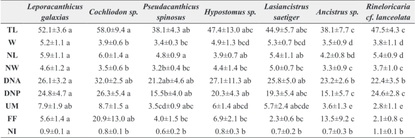
Abbreviations: TL, total length; W, width; NL, nucleus length; NW, nucleus width; DNA, distance from the nucleus till the anterior extremity; DNP, distance from the nucleus till the posterior extremity; UM, number of folds of the undulating membrane; FF, length of the free flagellum; NI, nuclear index. values followed by different letters in the same line indicate significant differences (5% probability in Tukey test).
TABLE II
Morphometric characteristics (average plus standard deviation, figures
in micrometers) of Trypanosoma spp. infecting armored fish.
Leporacanthicus
galaxias Cochliodon sp.
Pseudacanthicus
spinosus Hypostomus sp.
Lasiancistrus
saetiger Ancistrus sp.
Rineloricaria cf. lanceolata TL 52.1±3.6 a 58.0±9.4 a 38.1±4.3 ab 47.4±13.0 abc 44.9±5.7 abc 38.1±7.7 c 47.5±4.3 c
W 5.2±1.1 a 3.9±0.6 b 3.4±0.3 bc 4.9±1.3 bcd 5.3±0.7 bcd 3.5±0.9 d 3.8±1.1 d
NL 5.9±1.1 a 6.0±1.4 a 4.8±0.9 a 3.9±0.7 ab 5.4±1.1 ab 4.2±0.8 bd 5.4±0.9 d
NW 4.6±1.2 a 3.5±0.6 b 3.2b±0.4 bc 4.4±1.4 bc 5.0±0.7 bc 3.3±0.9 c 3.7±1.0 c
DNA 26.1±3.2 a 32.0±2.5 ab 21.2ab±4.6 ab 27.1±11.3 ab 25.8±5.0 ab 23.2±2.6 b 22.4±3.5 b
DNP 24.8±4.7 a 26.3±5.4 a 15.5b±4.0 ab 20.3±4.3 ab 19.3±5.4 abc 15.1±5.7 c 24.6±2.8 c
UM 7.9±1.9 ab 8.7±1.5 a 3.5cd±0.9 abc 6±1.4 abcd 5.7±2.4 abcde 3.6±1.3 e 2.8±1.1 e
FF 5.6±1.4 a 20.9±13.0 ab 4.0±1.5 bc 6.9±2.1 bc 2.3±0.6 bc 13.5±9.2 c 2.1±0.8 c
ACKNOWLEDGMENTS
Paticipation of J.C. Eiras on this research was
partially supported by the European Regional
development Fund (ERdF) through the COMPETE
- Operational Competitiveness Programme and
national funds through FCT – Fundação para a
Ciência e a Tecnologia, under the project “PEst-C/
MAR/LA0015/2011
RESUMO
Um total de 281 espécimes de peixes ornamentais de água doce – das espécies Leporacanthicus galaxias,
Lasiancistrus saetiger, Cochliodon sp., Hypostomus sp.,
Pseudacanthicus spinosus, Ancistrus sp. e Rineloricaria
cf. lanceolata – foram capturados na bacia hidrográfica
do rio Guamá, Pará, Brasil. A infecção por Trypanosoma
spp. foi inspecionada. A caracterização morfológica e morfométrica dos parasitas e os parâmetros hematológicos foram determinados. Todas as espécies foram infectadas e todos os espécimes de Leporacanthicus galaxias e
Pseudacanthicus spinosus estavam parasitados. As outras espécies mostraram uma prevalência variável da infecção. Os parasitas mostraram claramente morfotipos e dimensões diferentes, e provavelmente, pertencem a espécies diferentes. A resposta hematológica à infecção variou de acordo com o hospedeiro. Em Cochliodon
sp. não houve diferença entre peixes infectados e não infectados. Em outras espécies diversas modificações em alguns parâmetros hematológicos foram encontrados, mas aparentemente sem causar doença. Ressalta-se a possibilidade de introdução de parasitas em novos ambientes devido aos movimentos artificiais destes peixes ornamentais.
Palavras-chave: peixes de água doce, parâmetros hematológicos, infecção, Trypanosoma spp.
REFERENCES
AGUILAR A, áLVAREz MF, LEIRO JM AND SANMARTÍN ML. 2005. Parasite populations of the european eel (Anguilla anguilla L.) in the rivers Ulla and Tea (Galicia, Northwest Spain). Aquaculture 249: 85-94.
BAKER JR. 1960. Trypanosomes and dactylosomes from the blood of freshwater fish in East Africa. Parasitology 50: 515-526.
BARA MA AND SERRA-FREIRE NM. 1985. Aspectos epidemio-lógicos de infecção por tripanossomas em Hypostomus punctatus no Lago-Açú da UFRRJ. Rev Bras Med Vet 7: 46-49.
CLAUSS TM, DOvE ADM AND ARNOLd JE. 2008. Hematologic Disorders of Fish. Vet Clin Exot Anim 11: 445-462. D’AGOSTO M, BARA MD AND SERRA-FREIRE NM. 1985.
Aspectos epidemiológicos da infecção por tripanossomas em (Hypostomus punctatus) Valenciennes, 1840 (Osteichthyes, Loricariidfae) no lago Açú da uFRRJ, Brasil. Rev Bras Med Vet 7: 46-49.
D’AGOSTO M AND SERRA-FREIRE NM. 1990. Taxonomia de tripanosomas parasitas de peixes cascudo-pedra (Hypostomus punctatus) do lago Açu, Rio de Janeiro, Brasil. Parasitologia al dia 4: 14-18.
D’AGOSTO M AND SERRA-FREIRE NM. 1993. Estádios evolutivos de Tripanossomas de Hypostomus punctatus
valenciennes, 1840 (Osteichthyes, Loricariidae) em infecção natural de Batracobdella gemmata Blanchard (Hirudinea, Glossiphoniidae). Rev Bras zool 10(3): 417-426.
DYKOvÁ I AND LOM J. 1979. Histopathological changes in
Trypanosoma danilewskyi Laveran and Mesnil, 1904 and Trypanoplasma borreli Laveran and Mesnil, 1902 infections of goldfish, Carassius auratus (L.). J Fish Dis 2: 381-390.
EIRAS JC, REGO AA AND PAVANELLI GC. 1989.Trypanosoma guairaensis sp. n. (Protozoa, Kinetoplastida) parasita de Megaloancistrus aculeatus (Perugia, 1891) (Pisces, Loricariidae). Mem I Oswaldo Cruz 84: 389-392.
EIRAS JC, REGO AA AND PAVANELLI GC. 1990.Trypanosoma nupelianus sp. n. (Protozoa, Kinetoplastida) parasitizing
Rhinelepis aspera (Osteichtyes. Loricariidae) from Paraná
River, Brazil. Mem I Oswaldo Cruz 85: 183-184. EIRAS JC, TAKEMOTO RM, PAVANELLI GC AND AdRIANO EA.
2010. Diversidade dos Parasitas de Peixes de água Doce do Brasil. Clichtec, Maringá, 333 p.
EIRAS JC, TAKEMOTO RM, PAVANELLI GC AND LUQUE JL. 2012. Checklist of protozoan parasites of fishes from Brazil. zootaxa 3221: 1-25.
FRóES OM, FORTES E, LIMA DC AND LEITE VRV. 1978. Três espécies novas de tripanossomas de peixes de água doce do Brasil (Protozoa, Kinetoplastida). Braz J Biol 38(2): 461-468.
FRóES OM, FORTES E, LIMA DC AND LEITE VRV. 1979. Tripanossomas (Protozoa, Kinetoplastida) de peixes de água doce do Brasil. II. Novos tripanossomas de cascudos (Pisces, Loricariidae). Braz J Biol 39(2): 425-429. GARCIA F, FuJIMOTO RY, MARTINS ML AND MORAES FR.
2009. Protozoan parasites of Xiphophorus spp. (Poeciliidae) and their relation with water characteristics. Arq Bra Med Vet zootec 61(1): 156-162.
GIBSON WC, LOM J, PECKOvÁ H, FERRIS VR AND HAMILTON
GU z, WANG J, zHANG J AND GONG X. 2006. Redescription of
Trypanosoma ophiocephali Chen 1964 (Kinetoplastida: Trypanosomatina: Trypanosomatidae) and first record from the blood of dark sleeper (Odontobutis obscura
Temminck and Schlegel) in China. Parasitol Res 100: 149-154.
LOM J. 1979. Biology of the Trypanosomes and Trypanoplasms of fish. In: Biology of the Kinetoplastida. Lumsden W and Evans D (Eds), Academic Press, New York 2: 270-336. LOPES RA, LOPES OvP, RIBEIRO RM, ALBUQUERQUE S,
SATAKE T AND GARAvELLO JC. 1991. Trypanosoma of brazilian fishes. xI. Trypanosoma valerii sp. n. from
Pterodoras granulosus Valenciennes 1833 (Pisces, Doradidae). Naturalia 16: 19-24.
LOPES RA, SATAKE T, BRENTEGANI LG, NUTI-SOBRINhO A, BRITSKI HA AND RIBEIRO RD. 1989. Trypanosomes of Brazilian fishes. III. Trypanosoma dominguesi sp. n. from armored catfish Hypostomus alatus Castelnau 1855 (Pisces, Loricariidae). Ann Parasit Hum Comp 64(2): 83-88. NOGA EJ. 1996. Fish Disease. Diagnosis and Treatment.
St. Louis, Missouri: Mosby-Year Book, Inc., 367 p. PAPERNA I. 1996. Parasites, infections and diseases of fishes
in Africa: An update. CIFA Technical Paper. N.31. Rome, FAO, 220 p.
PIAzzA RS, MARTINS ML, GUIRALDELLI L AND YAMASHITA M. 2006. Parasitic diseases of freshwater ornamental fishes commercialized in Florianópolis, Santa Catarina, Brazil. Bol Inst Pesca 32(1): 51-57.
PRANG G. 2007. An industry analysis of the freshwater ornamental fishery with particular reference to the supply of Brazilian freshwater ornamentalsto to the UK market. Uakari 3(1): 7-51.
RANzANI-PAIVA MJT. 1995. Células do sangue periférico e contagem diferencial de leucócitos de taínha Mugil platanus Günther, 1880 (Osteichthyes, Mugilidae) da
região estuarino-lagunar de Cananéia – SP (Lat. 25° 00’S – Long. 47°55’W). Bol Inst Pesca 22(1): 23-40.
RAUTHAN JVS, GROvER SP AND JAIWAL P. 1995. Studies on some haematological changes in a hill stream fish Barilius bendelisis (Hamilton) infected with trypanosomes. Flora and Fauna (Jhansi) 1: 165-166.
RIBEIRO RD, SATAKE T, NUTI-SOBRINhO A, BRENTEGANI LG, BRITSKI HA AND LOPES RA. 1989. Trypanosomes of Brazilian fishes. Iv. Trypanosoma lopesi sp. n. from armored catfish Rhinelepis aspera Agassiz 1829 (Teleostei, Loricariidae). zool Anz 222(3/4): 244-248. ROSENFELd G. 1947. Corante pancrômico para hematologia e
citologia clínica: nova combinação dos componentes do May-Grunwald e do Giemsa num só corante de emprego rápido. Mem Inst Butantan 20: 329-334.
TAVARES-DIAS M, GONzAGA LEMOS JR AND MARTINS ML. 2010. Parasitic fauna of eight species of ornamental freshwater fish species from the middle Negro River in the Brazilian Amazon Region. Rev Bras Parasitol Vet 19(2): 103-107.
THATCHER VE. 2006. Amazon fish parasites. 2a ed., Pensoft Publishers, Sofia, Moscow, p. 205-251.
TORRES MFA. 2007. Pesca Ornamental na Bacia do Rio Guamá: Sustentabilidade e Perspectivas ao manejo. [Tese]. Núcleo de Altos Estudos Amazônicos, NAEA, universidade Federal do Pará, Belém, Pará, 264 p.
UNTERGASSER D. 1989. Handbook of Fish Disease. Plaza, Neptune City: TFH publications Inc., 160 p.