Sergio Santoro
Master in Materials Science
Molecular probes: an innovative technology
for monitoring membrane processes
Dissertation for obtaining the Doctoral degree in
Membrane Engineering
Jury:
President: Prof. José Paulo Barbosa Mota, FCT, Universidade Nova de Lisboa Examiner(s): Prof. Frederico Castelo Alves Ferreira, IST, Universidade de Lisboa Prof. Thomas Schafer, University of the Basque Country
Members: Prof. João Carlos Lima, FCT, Universidade Nova de Lisboa Prof. Reyes Mallada, Universidad de Zaragoza
Dr. Alberto Figoli, Institute for Membrane Technology (ITM-CNR) Prof. Isabel Maria Rôla Coelhoso, FCT, Universidade Nova de Lisboa Prof. João Paulo Serejo Goulão Crespo FCT, Universidade Nova de Lisboa
May 2016
Supervisors:
Isabel Maria Rôla Coelhoso, Assistant Professor, Faculdade de
Ciências e Tecnologia, Universidade Nova de Lisboa
João Paulo Serejo Goulão Crespo, Professor, Faculdade de
Ciências e Tecnologia, Universidade Nova de Lisboa
Co-supervisors:
Reyes Mallada, Associate Professor, Department of Chemical
and Environmental Engineering and Aragon Nanoscience
Institute, Universidad de Zaragoza
Sergio Santoro
Master in Materials Science
Molecular probes: an innovative
technology for monitoring
membrane processes
Dissertation presented to Faculdade de Ciências e Tecnologia, Universidade Nova de Lisboa for obtaining the Doctoral degree in Membrane Engineering
Molecular probes: an innovative technology for monitoring membrane processes
Copyright @ Sergio Santoro, FCT/UNL and UNL
“To all the dreamers,
to those who speak to the wind. To the ones in love, to the day-dreamers, to all who will still give their life away to make a dream come true. To the rejects, the not wanted, to all whom are left out. To the crazy ones, them being real or just assumed to be. To the good hearted men, to those who still want to believe that real feelings exist. To those who can still be touched. A tribute to the great soakers, to big ideas, to dreams. To those who will never give up, to who is mocked or judged. To the everyday poets. To the possible winners and so to all the losers who are still urged to rise again and to keep fighting. To the forgotten heroes and to those who wander around. To those, that after having fought and lost for their ideals, still feel they are invincible. To those who are not scared to express their ideas. To those who went around the world and to who one day will do the same. To those who do not want to distinguish between reality and fiction. To all the errant knights. And somehow, it is fair and well said…..to all the performers.”
Don Quixote
i
Acknowledgements
“Inside us there is something that has no name, that something is what we are.” Though only my name appears on the cover of this dissertation, a great many people from Portugal, Spain and Italy have contributed to its production. I owe my gratitude to all those people who have made this dissertation possible.
My deepest gratitude is to Prof. João Paulo Crespo. I have been amazingly fortunate to have an advisor who gave me the freedom to explore on my own, and at the same time the guidance to recover when my steps faltered.
I would like to express my sincere gratitude to Prof. Isabel Coelhoso for the continuous support of my Ph.D study: her guidance helped me in all the time of research and writing of this thesis.
I will always be grateful to Prof. João Carlos Lima, who introduced me to the wonders of the Photochemistry. I am indebted to him for his continuous encouragement and guidance.
This dissertation would not have been possible without Dr. Artur Moro. He has been always there to teach me, listen, give advice, read my reports, comment on my views and help me understand and enrich my ideas.
I am grateful to Dr. Carla Portugal for holding me to a high research standard and enforcing strict validations for each research result by insightful comments and constructive criticisms.
Thanks also go to my colleagues and friends of the Universidade Nova de Lisboa, who expressed an interest in the project and provided useful feedback.
I will always be grateful to Prof. Reyes Mallada because she provided me the opportunity to join her team at the Institute of Nanoscience of Aragon. I benefited from her advice, particularly so when exploring new ideas. Her positive outlook and confidence in my research inspired me.
I owe my gratitude to Dr. Victor Sebastian for the continuous support of research during my stay in Spain, for his patience, motivation, and immense knowledge.
Dr. Manuel Arruebo’s insightful comments were thought-provoking and they helped me focus my ideas.
The members of the Institute of Nanoscience of Aragon have contributed immensely to my personal and professional time at Zaragoza. The group has been a source of friendships as well as good advice and collaboration.
I would also like to express my thanks to Dr. Alberto Figoli who has been always played a key role in my studies and research. It was though his persistence, understanding and kindness that I completed my graduate training and my PhD. The joy and enthusiasm he has for her research was contagious and motivational for me.
ii A very special thanks goes out to Giovanni Desiderio and Dr. Giuseppe Lombardo. They truly made a difference in the last part of my studies.
I am grateful to all my friends from the Institute on Membrane Technology for being my surrogate family.
My sincere thanks also goes to Prof. Enrico Drioli, Dr. Curcio Efrem and all the members of the The Erasmus Mundus Doctorate in Membrane Engineering (EUDIME) for the assistance throughout my PhD program and the exchange of ideas in the annual meetings.
“Quando ti viene nostalgia non è mancanza. E' presenza di persone, luoghi, emozioni che tornano a trovarti.” Grazie ai luoghi e agli amici di sempre, che mi sono mancati e che hanno fatto sentire sempre la loro presenza. Soprattutto quando ne avevo bisogno.
Grazie al mio fianco, Marika, gioia che ha contagiato la mia esistenza. Con la promessa che il più bello dei mari è quello che non navigammo e i più belli dei nostri giorni non li abbiamo ancora vissuti. Grazie ad Adriana, riparo nella tempesta e arcobaleno nel sereno, e a Francesco, il fratello che non ho mai avuto.
Grazie a Letizia, che mi ha insegnato a restare bambino. E’ difficile chiederti scusa per un mondo che è quel che è, io nel mio piccolo tento qualcosa per farlo assomigliare a te.
Grazie a mia mamma e mio papà, invincibili eroi del quotidiano. A voi devo quello che sono ed il senso che ho acquisito della vita.
iii
Abstract (300 words)
The ultimate objective of this study is to use molecular probes as an innovative and alternative technology contributing to the advance of membrane science by monitoring membrane processes in-situ, on-line and at sub-micron scale.
An optical sensor for oxygen sensing was developed by the immobilization of tris (1, 10-phenanthroline) ruthenium (II) (Ru(phen)3) in a dense polymeric membrane made of polystyrene (PS) or Poly(3-hydroxybutyrate-co-3-hydroxyvalerate) (PHBV). The emission of the probe was quenched by both the temperature and by the oxygen. Moreover, the oxygen sensitivity was affected by the oxygen permeability of the membrane. The evaluation of the oxygen concentration is prone to errors since the emission of a single probe depends on several parameters (i.e. optical path, source intensity). The correction of these artefacts was obtained by the immobilization of a second luminescent molecule non-sensitive to the oxygen, Coumarin. The potential of the luminescent ratiometric sensor for the non-invasive monitoring of oxygen in food packaging using polymeric films with different oxygen permeability was evaluated. Emphasis was given to the efficiency of the optical sensor for the on-line, in-situ and non invasive monitoring of the oxygen by comparing the experimental data with a model which takes into account the oxygen permeability of the packaging materials evaluated independently.
A nano-thermometer based on silica nano-particles doped with Ru(phen)3 was developed. A systematic study shows how it is possible to control the properties of the nano-particles as well as their temperature sensitivity. The nano-thermometer was immobilized on a membrane surface by dip-coating providing information about the temperature on the membrane surface.
v
Resumo
O objectivo final deste trabalho é a utilização de sondas moleculares como uma tecnologia inovadora e alternativa contribuindo para o avanço da ciência em processos de membranas, monitorizando esses processos in-situ, on-line e numa escala submicrométrica.
Foi desenvolvido um sensor óptico para a detecção de oxigénio imobilizando tris (1, 10-fenantrolina) de ruténio (II) (Ru(phen)3) numa membrana polimérica densa de poliestireno (PS) ou de poli (3-hidroxibutirato-co -3-hidroxivalerato) (PHBV). A sensibilidade da sonda para o oxigénio é afectada pela permeabilidade da membrana a este gás. Verificou-se que a avaliação da concentração de oxigénio é propensa a erros uma vez que a emissão de uma única sonda depende de vários parâmetros, tais como, o percurso óptico, a intensidade de fonte, entre outos. A correcção destes erros foi obtida pela imobilização de uma segunda molécula luminescente não-sensível ao oxigénio, a cumarina. O potencial deste sensor raciométrico luminescente para a monitorização não invasiva de oxigénio em embalagens de alimentos usando filmes poliméricos com diferentes permeabilidades ao oxigénio foi avaliada. Os resultados obtidos através da monitorização não invasiva do oxigénio com o sensor foram comparados com os obtidos usando um modelo que tem em conta a permeabilidade ao oxigénio dos materiais de embalagem avaliada independentemente.
Desenvolveu-se ainda, um nano-termómetro baseado em nano-partículas de sílica dopadas com Ru(phen)3. Um estudo sistemático mostra como é possível controlar as propriedades das nano-partículas, bem como a sua sensibilidade à temperatura. O nano-termómetro foi imobilizado sobre a superfície de uma membrana por dip-coating sendo possível obter deste modo, a temperatura na superfície da membrana.
vii
Resumen
El objetivo final de este estudio es el uso de una tecnología innovadora basada en el uso de sondas moleculares que actúan como sensores. La tecnología desarrollada contribuye al avance de la ingeniería de membranas, específicamente al control in situ de los procesos fisico-químicos que tienen lugar en su estructura porosa. El desarrollo de esta tecnología ha hecho posible obtener un buen control en la cuantificación de la temperatura y concentración de oxígeno presente en las membranas, alcanzándose una resolución sub-micrométrica.
Se ha desarrollado un sensor óptico (sonda) para detectar y cuantificar la presencia de oxígeno mediante la inmovilización de tris (1, 10-fenantrolina) rutenio (II) (Ru(phen)3) en una membrana polimérica densa de poliestireno (PS) o poli (3-hidroxibutirato-co -3-hidroxivalerato) (PHBV). La emisión espectral de la sonda era sensible tanto a la temperatura de la membrana como a la presencia de oxígeno. Por otra parte, la sensibilidad de la sonda al oxígeno se vio afectada por la permeabilidad de las moléculas de oxígeno a través de la membrana. La cuantificación de la concentración de oxígeno no es trivial y suele estar sujeta a errores de emisividad generados por artefactos espectrales, ya que la emisión de una única sonda depende de varios parámetros, como por ejemplo el camino óptico o la intensidad de la fuente de excitación. La corrección de estos artefactos se obtuvo mediante la inmovilización de una segunda molécula luminiscente no sensible al oxígeno, cumarina.
En este proyecto se ha evaluado el potencial del sensor luminiscente radiométrico para la monitorización no invasiva de oxígeno en el envase de alimentos, utilizando para ello películas poliméricas con diferente permeabilidad al oxígeno. Se ha estudiado en mayor profundidad la eficiencia del sensor óptico para el análisis on-line, utilizando para ello la medida in situ y no invasiva de la luminiscencia de las sondas. La monitorización de la presencia de oxígeno se realizó mediante la comparación de los datos experimentales de la emisión de las sondas con los datos de permeación de oxígeno a través del embalaje.
Además, se ha elaborado una sonda térmica, nanotermómetro, basada en el dopado de nanopartículas de silice con Ru(phen)3. Un estudio sistemático muestra cómo es posible controlar las propiedades luminiscentes de las nanopartículas, así como su sensibilidad a la medida de la temperatura. El nano-termómetro se inmovilizó sobre la superficie de la membrana, proporcionando información muy precisa sobre la temperatura en la superficie de la misma.
ix
Riassunto
L'obiettivo finale di questo studio è l’impiego di sonde molecolari come tecnologia innovativa ed alternativa che contribuisca al progresso della scienza delle membrane favorendo il monitoraggio del processo a membrana in-situ, on-line e su scala inferiore al micron.
E’ stato sviluppato un sensore ottico per il monitoraggio dell’ossigeno tramite l’ immobilizzazione di tris (1,10-fenantrolina) rutenio (II) (Ru(phen)3) in una membrana polimerica densa fatta di polistirolo (PS) e poli(3-idrossibutirrato-co-3-idrossivalerato) (PHBV). L'emissione della molecola è influenzata sia dalla temperatura che dall'ossigeno. Inoltre, la sensibilità all’ossigeno è condizionata dalla permeabilità all'ossigeno della membrana. Tuttavia, la determinazione della concentrazione di ossigeno è soggetta ad errori, poiché l’emissione di una singola molecola dipende da diversi parametri (per esempio cammino ottico, intensità sorgente). La correzione di questi artefatti è stata ottenuta tramite l'immobilizzazione di una seconda molecola luminescente non sensibile all'ossigeno, ovvero la Cumarina. Abbiamo valutato le potenzialità del sensore basato sul rapporto delle intensità delle emissioni delle due molecole luminescenti nel monitorare la concentrazione di ossigeno nell'imballaggio alimentare fatto da film polimerici con diversa permeabilità all'ossigeno. E’ stata data enfasi all'efficienza del sensore ottico per il monitoraggio non invasivo, in-situ e on-line dell'ossigeno, confrontando i dati sperimentali con un modello che tiene conto delle permeabilità all'ossigeno dei materiali di imballaggio valutate indipendentemente.
È stato sviluppato un termometro costituito da nano-particelle di silice drogate con Ru(phen)3. Uno studio sistematico mostra come sia possibile controllare le proprietà delle nano-particelle, nonché la loro sensibilità alla temperatura. Il nano-termometro è stato immobilizzato su una superficie di una membrana mediante dip-coating fornendo informazioni riguardo la temperatura sulla superficie della membrana.
xi
Nomenclature
A: Packaging area, m2 a.u.: Arbitrary unit b: Optical path, cm
BOPP: Biaxially oriented polypropylene (byp): (2,2’-bipyridine)
c: Concentration probe, mol CA: Contact angle,°
: Oxygen concentration in the enclosed package
CO2out: Oxygen concentration in the atmosphere (21%) Coumarin: 7-Methoxy-4-methylcoumarin
C(T): Temperature compensation dp: Pore size, μm
(dpp): (4,7-diphenyl-1,10-phenanthroline)
dR/dT: Temperature sensitivity of the ratiometric response, °C-1 ENM: Electrospun Nanofibrous Membrane
EDS: X-ray microanalysis f1: Fractional intensity I: Intensity of emission, a
FPP: Tetrakis pentrafluoropheny porphine I: Intensity of emission in the presence of oxygen I0: Intensity of emission in absence of oxygen, a.u. I21: Intensity of emission at atmospheric pressure, a.u. Iexc: Intensity of exciting light, a.u.
I(N2): Fluorescence intensities in the absence of oxygen I(O2): Fluorescence intensities in the 100% oxygen k: Constant of excitation/detection geometry kq: Rate of bimolecular quenching
KSV: Stern-Volmer constant, %-1
KSV(Tref): Stern-Volmer constant at 25°C, %-1 l: Thickness of packaging, m
NA: Avogadro’s number, mol-1 N2: Nitrogen
O2: Oxygen
OEP: Octaethylporphyrin
xii p(O2): Oxygen partial pressure, bar
P(O2): Oxygen permeability, m2 s-1 PAN: Poly(acrylonitrile)
PBA: 1-pyrene butyric acid, OEP: octaethylporphyrin PHVB: Poly(β-hydroxybutyrate-β-hydroxyvalerate) PDMS: Poly(dimethylsiloxane)
Pe: Oxygen permeability, barrer
(phen): Phenanthroline (ppy):[2-phenylpyridinato-C2,N] PS: Polystyrene
PSS: Poly(sodium 4-styrene sulfonate)
PtTFPP: Ttetrakis(pentafluorophenyl)porphyrin PDMS: Polydimethylsiloxane,
PVA: Polyvinyl alcohol R: Ratiometric response
Rm: Non-temperature dependent ratiometric response R0: Ratiometric response in absence of oxygen R0: Ratiometric response in an atmosphere of oxygen
Rm: Non-temperature dependent ratiometric response in absence of oxygen R0(T): Ratiometric response at a certain oxygen level and temperature R0(Tref): Ratiometric response in absence of oxygen at 25°C
R21: Ratiometric response at atmospheric pressure, a.u. Ru(phen)3: Tris(1,10-phenanthroline)ruthenium(II) s: Electrical conductivity, μS cm-1
T: Temperature, °C
TGA: Thermogravimetric analysis TTA: Thenoyltrifluoroacetonate,
V: Volume enclosed by the packaging, m3 yO2: Volumetric oxygen concentration, %
Greek symbols
β: Geometric parameter of the permeation cell, m-1 δ: Collision radius, m
ε: Molar extinction coefficient, m2 mol-1 λ: Wavelenght, nm
xiii
Contents
Acknowledgements ... i
Abstract (300 words) ... iii
Resumo ... v
Resumen ... vii
Riassunto ... ix
Nomenclature ... xi
List of Figures ... xvii
List of Tables ... xxi
Chapter 1 ... 1
Introduction: state of art and thesis motivations ... 1
1.1 Membrane technology: advances and limitations ... 3
1.2 Monitoring of membrane processes ... 4
1.3 Importance of temperature and oxygen in membrane processes ... 5
1.4 Molecular probes: an innovative technology for monitoring membrane process... 6
1.5 Oxygen molecular probe ... 7
1.6 Temperature molecular probes ... 8
1.7 Objectives and thesis outline ... 10
1.8 References ... 13
Chapter 2 ... 17
Development of Oxygen and Temperature Sensitive Membranes Using Molecular Probes as Ratiometric Sensor ... 17
2.1 Introduction ... 19
2.2 Materials and Methods ... 21
2.2.1 Materials ... 21
2.2.2 Methods ... 22
2.2.2.1 Fluorescence measurements ... 22
2.2.2.2 Oxygen and temperature sensitive membrane preparation ... 23
2.2.2.3 Characterization of the membranes with the ratiometric sensor ... 24
2.3 Results and Discussion ... 24
2.3.1 Oxygen sensitive membrane ... 24
2.3.2 Development of an oxygen sensor based on ruthenium ... 25
2.3.3 Development of a ratiometric sensor based on ruthenium ... 27
2.3.4 Oxygen sensitivity of the two probe system fluorescent membrane ... 30
2.3.5 Temperature sensitivity of the two probe system fluorescent membrane ... 32
xiv
2.3.7 Temperature compensation of the oxygen sensor ... 35
2.4 Conclusions ... 37
2.5 References ... 38
Chapter 3 ... 41
Monitoring oxygen permeation through packaging films using ratiometric luminescent sensors ... 41
3.1 Introduction ... 43
3.2 Experimental section ... 45
3.2.1. Materials and Methods ... 45
3.2.2. Preparation of the optical sensor ... 46
3.2.3 Response of the sensor ... 46
3.2.4 Oxygen permeability trough dense films ... 47
3.3. Results and discussion ... 50
3.3.1 Optical sensor sensitivity ... 50
3.3.2 Food packaging mimicking experiments ... 52
3.4 Conclusions ... 55
3.5 References ... 56
Chapter 4 ... 59
Development of Fluorescent Nano-thermometers for temperature monitoring on membrane surfaces... 59
4.1 Introduction ... 61
4.2. Experimental section ... 62
4.2.1. Materials ... 62
4.2.2. Synthesis of SiO2NPs ... 63
4.2.3 Immobilization of SiO2NPs on PVDF membranes ... 64
4.2.4 Characterization ... 65
4.3 Results and Discussion ... 66
4.3.1 Optimization of fluorescent silica nanoparticles as nanothermomethers ... 66
4.3.2 Fluorescent Ru(phen)3-Shell SiO2NPs immobilized in the surface of membranes as nanothermometers ... 73
4.4 Conclusions ... 76
4.5 References ... 77
Chapter 5 ... 81
On-line and non-invasive mapping of temperature on electrospun nanofibrous PVDF membrane in direct contact membrane distillation by means of molecular probes ... 81
5.1 Introduction ... 83
5.2 Materials and methods ... 85
xv
5.2.2 Membrane preparation ... 85
5.2.3 Electrospinning ... 85
5.2.4 Membrane characterization ... 86
5.3 Results and Discussion ... 90
5.3.1 Membrane composition and morphology ... 90
5.3.2 Membrane Characterization ... 92
5.3.3 DCMD results ... 93
5.3.4 Evaluation of temperature on the membrane surface ... 94
5.4 Conclusions ... 100
5.5 References ... 101
Chapter 6 ... 105
Conclusions ... 105
6.1 Oxygen monitoring ... 107
6.2 Temperature monitoring ... 108
6.3 Future works ... 110
Capítulo 6 ... 111
Conclusiones ... 111
6.1 Medición de oxígeno ... 113
6.2 Medición de la temperatura ... 114
6.3 Perspectivas futuras ... 116
Capitolo 6 ... 119
Conclusioni ... 119
6.1 Monitoraggio dell’ossigeno ... 121
6.2 Monitoraggio della temperatura ... 122
6.3 Prospettive future ... 124
xvii
List of Figures
Figure Page Figure 1.1: Membrane separation ... 3
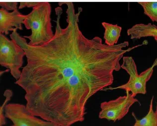
Figure 1.2: Membrane process ... 5 Figure 1.3: Bovine pulmonary artery endothelial cells marked with different luminophores. Source:
www.flickr.com ... 6
Figure 1.4: Luminescence of an oxygen molecular probe in argon (Ar), aerated and oxygen
atmosphere. Source: http://www.scivaxls.com. ... 8
Figure 1.5: Representative scheme of working of an optical sensor for temperature detection [46]. .... 9 Figure 2.1: Membrane separation: Chemical structures of polymers and molecular probes employed
in the development of oxygen and temperature sensitive membranes. ... 21
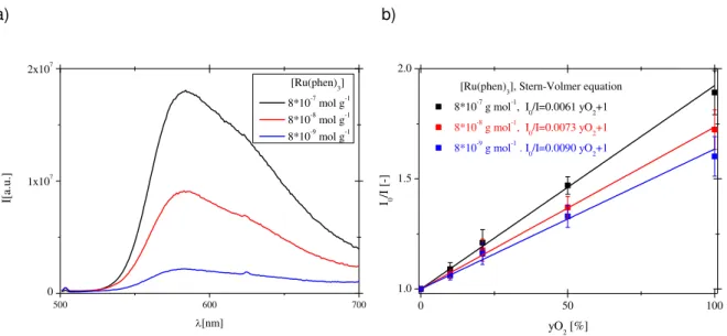
Figure 2.2: Scheme of the spectrofluorimeter and cuvette used for preliminary experiments. ... 22 Figure 2.3: Scheme of experiments for the evaluation of the oxygen and temperature sensitivity. .... 23 Figure 2.4: Effect of the oxygen concentration on the emission spectra of Ru(phen)3 (b) immobilized in PS films (T=21°C, probe concentration 8x10⁻⁷mol g-1). Experiments were performed using the set-up reported in Figure 2.2. ... 25
Figure 2.5: Effect of the concentration of Ru(phen)3 on the absolute intensity of the emission (a) and on the Stern-Volmer plot (b) (T=21°C). Experiments were performed using the set-up reported in Figure 2.2. ... 26
Figure 2.6: Stern-Volmer plots obtained for membranes prepared with PS and PHVB (T=21°C, probe
concentration 8x10⁻⁷mol g-1). Experiments were performed using the set-up reported in Figure 2.3. .. 27
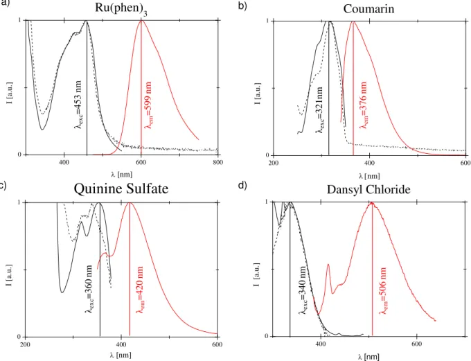
Figure 2.7: Normalized spectra of Absorbance (black dashed), excitation (black solid) and emission
(red solid) of solution of Chloroform containing 10 µM at 21°C of Ru(phen)3(a), Coumarin (b), Quinine Sulfate (c) and Dansyl Chloride (d). ... 28
Figure 2.8: Emissions of membranes containing the oxygen sensitive probe (8x10⁻⁷mol g-1,
λmax=589nm) and different reference compounds: (a) Coumarin (8x10⁻⁷mol g-1, λmax=363nm), (b) Quinine Sulfate (8x10⁻⁷mol g-1, λmax=403nm), (c) Dansyl Chloride (8x10⁻⁶mol g-1, λmax=506nm). Experiments were performed at T=21°C using the set-up reported in Figure 2.2. ... 29
Figure 2.9: Oxygen sensitivity at T=21°C of (a) the intensities of emission of Ru(phen)3 and Coumarin in PS and (b) the ratiometric signal. Experiments were performed using the set-up reported in Figure 2.3. ... 31
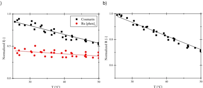
Figure 2.10: Effect of the temperature on (a) the intensities of the emission of Ru(phen)3 and Coumarin in PS and (b) the ratio of their intensities , recorded at yO2=21%. ... 33
xviii
Figure 2.12: Effect of the temperature on Stern-Volmer constant (a) and of oxygen on the temperature sensitivity (b). ... 34
Figure 2.13: (a) Effect of the temperature on the Volmer ratiometric response and (b)
Stern-Volmer trend of the compensated ratiometric response at different temperatures... 36
Figure 3.1: Mechanism of dynamic quenching... 44 Figure 3.2: Set up used for experiments ... 47
Figure 3.3: Permeation of oxygen in food package ... 48
Figure 3.4: Set-up for measuring oxygen permeability ... 49
Figure 3.5: Intensity of the emission of Ru(phen)3 in aerated and inert atmosphere (a) and kinetics of the maximum of emission during degassing. ... 50
Figure 3.6: Emission of the optical sensor collected beyond PDMS and BOPP films. ... 51 Figure 3.7: Calibration of the optical sensor in food packaging mimicking experiments ... 52 Figure 3.8: Oxygen concentration in the food packaging simulation for PDMS employing the
ratiometric optical sensor. Results obtained using the optical sensor were predicted by the model and confirmed by chromatographic analyses (GC). ... 53
Figure 3.9: Oxygen concentration in the food packaging simulation for BOPP a) Results obtaining using the optical sensor compared with by the model (constant permeability) and confirmed by chromatographic analyses (GC). b) Model (solid line), Dual mode model (dashed line) and results in the food packaging simulation for 24 hours using BOPP. ... 54
Figure 4.1: Scheme of preparation of Core and Shell doped silica nanoparticles. ... 63 Figure 4.2: SEM pictures of a)Bare, b) Ru(phen)3-Core and c) Ru(phen)3-Shell SiO2100% NPs. The size of the scale bar corresponds to 1 micron in all the images. ... 67
Figure 4.3: TEM picture of Bare SiO2 NP (a) and Ru(phen)3-Shell SiO2 5%NP. The size of the scale bar corresponds to 50nm in all the images. ... 68
Figure 4.4: Normalized a) excitation and b) emission spectra of Bare, Ru(phen)3-Core and Ru(phen)3 -Shell 100% SiO2NPs. ... 69
Figure 4.5: Normalized Intensity of emission with respect to SiO2 5% NPs doped with 1.35 wt% of Ru(phen)3 at 590 nm of Ru(phen)3-Shell SiO2 5% NPs prepared with different concentrations of Ru(phen)3. ... 70
Figure 4.6: Temperature sensitivity of the maximum emission (590 nm) of Ru(phen)3-Core and Ru(phen)3-Shell SiO2 100%NPs. ... 70
Figure 4.7: Particle size (black) and shell thickness (red) as a function of the mass ratio of silica precursor employed to grow the shell and the mass of Bare SiO2 NPs coated. The values correspond to DLS measurements. ... 71
xix
Figure 4.9: a) Effect of the temperature on the intensity of the emission at 590 nm of Ru(phen)3-Shell SiO2 NPs doped with different amounts of Ru(phen)3, b).Temperature sensitivity of Shell SiO2 NPs as a function of Ru(phen)3 immobilized. ... 73
Figure 4.10: SEM picture of PVDF membrane load with hydrophobic 100% Ru- Ru(phen)3-Shell SiO2 NPs (a) compared with bare PVDF membrane (b). ... 74
Figure 4.11: SEM picture of PVDF membrane load with hydrophobic 100% Ru- Ru(phen)3-Shell SiO2 NPs: a) cross-section, b)zoom of the cross-section in proximity of the surface, c) top view of the surface. ... 74
Figure 4.12: (a) Normalized emission of Ru(phen)3-Shell SiO2 NPs immobilized on PVDF membranes and (b)their temperature sensitivity. ... 75
Figure 5.1: Thermal polarization ... 84 Figure 5.2: a) Scheme of the set-up used for monitoring the temperature in DCMD process, b)Picture
of the set-up. ... 88
Figure 5.3: Scheme of the developed membrane module for optical observations. Pictures evidencing
the window (a), the cover (b) and the optical-fiber (c). ... 89
Figure 5.4: SEM picture of PVDF ENMs (Magnification: 10,000 X): a)PVDF 6wt%, b)PVDF 10wt%, c) PVDF 10wt%+0.85wt% Ru(phen)3, d) PVDF 10wt%+0.85wt% Ru(phen)3+0.43wt% LiCl. ... 90
Figure 5.5: SEM picture of PVDF ENMs (Magnification: 100,000 X): a)PVDF 6wt%, b)PVDF 10wt%,
c) PVDF 10wt%+0.85wt% Ru(phen)3, d) PVDF 10wt%+0.85wt% Ru(phen)3+0.43wt% LiCl. ... 91
Figure 5.6: Flux of 10wt%PVDF/ Ru(phen)3/LiCl prepared via electrospinning for 4 hours at different temperatures (a) and mean value at the steady state (b). ... 93
Figure 5.7: Emission spectra of Ru(phen)3 immobilized in PVDF ENM normalized with respect the value of the maximum of the emission (572 nm) collected at 40°C (a) and temperature sensitivity of the maximum of the emission (572 nm) (b). ... 95
Figure 5.8: Profile of temperature on the membrane surface along membrane module at different
temperature of the feed: a) 60°C, b) 50°C, c) 40°C ... 96
Figure 5.9: Topographic Maps of the temperature on membrane PVDF ENM using discrete optical
fiber measurements together with griddedinterpolant function, Matlab, at different temperature of the feed: a) 60°C, b) 50°C, c) 40°C. ... 97
xxi
List of Tables
Table Page Table 1.1: List of optical sensor prepared employing common molecular probes for oxygen sensing .. 7
Table 1.2: List of optical sensor prepared employing common molecular probes for temperature
sensing ... 9
Table 2.1: Comparison of the emissive properties of the ratiometric sensors: wavelength of emission
(λ); intensities of emission in aerated condition (I21); ratio of emission intensities of reference over Ruthenium complex (R21) at atmospheric pressure; sensitivity of the intensity of the reference (I0/I21); sensitiviy of the ratiometric response (R0/R21) to oxygen removal. ... 30
Table 2.2: Permeability, diffusion coefficient and solubility of different gases in the membranes of PS and PS doped with Ru(phen)3 and Coumarin. [Probe]=8x10⁻⁷ mol g-1. ... 32
Table 3.1: Oxygen permeability and thickness of the optical sensor (PS) and the films used for food
packaging simulations. ... 52
Table 4.1: Preparation of solutions used for the growth of the shell on bare SiO2NPs. ... 64
Table 4.2: Size and polydispersity index of Bare, Ru(phen)3-Core and Ru(phen)3-Shell SiO2NPs. .... 66
Table 5.1: Electrical conductivity (s) and viscosity (ν) of PVDF polymeric solutions. ... 92 Table 5.2: Characterization results of 10wt%PVDF/ Ru(phen)3/ LiCl ENM ... 92
1
Chapter 1
3 The following chapter includes the state-of-the-art related with the technology of interest in the present thesis (molecular probes for oxygen and temperature monitoring in membrane processes), the motivations for the explored approach and the main objectives of this thesis.
1.1 Membrane technology: advances and limitations
Membranes have been considered an interesting technology providing a crucial contribute in solving critical issues of the 21th century, such as pollution, energy and water demands. Membrane processes have been gained considerable interest for environmental friendly, non energetic intensive and economical separation of species in a wide range of application (i.e. water purification and desalination, recovery or release of target molecules of interest in medical and pharmaceutical field, production of biomass, bio-fuel and biogas).
The key activity of the membrane is the preferential transport of chemical species useful for separation processes or controlled release molecules [1].
Figure 1.1: Membrane separation.
In the evaluation of the membrane performance, three parameters must be considered [1]: i) Selectivity: favouring the selective transport of the target molecules; ii) Permeability: ensuring high mass transport of permeant species;
iii) Stability: guarantying long term and stable performance of the transport.
Efforts have been devoted for the improvement of membrane processes on the basis of production of high-performance membrane materials and the optimization of operating conditions of the processes. The high-performance of the membrane is usually guaranteed by the employment of novel materials and designed membrane morphologies adequate for the separation of interest, whereas the optimization of the membrane processes is guaranteed by dedicated membrane modules and optimized operating conditions aimed to improve the interactions between the permeant species and the membrane modules which govern the mass transport trough the membrane [2].
Permeate
Retentate
4 In fact, the breakthrough in membrane science was achieved as a consequence of development of extremely selective and ultra-thin membranes and of high surface-area membrane modules which made membrane processes competitive and economical viable [3-4]. Furthermore, progresses in material science and nanotechnology favoured the development of new material with tailored properties and high performance [5-7].
Besides these advances, there are major issues limiting the membrane processes performance in terms of selectivity, permeability and stability, in particular fouling, concentration and thermal polarization [8-10]. In fact, in the recent years, the development of membrane modules and the design of membrane process are becoming increasingly important to minimize those major persistent factors and to maximize the membrane performance [11].
For this reason, the evaluation of membrane properties and membrane performance at sub-micron scale, in-situ and on-line are crucial for better understanding the chemical and physical interactions between the permeant species and the membrane surface in order to improve membrane technology overcoming its limitations.
1.2 Monitoring of membrane processes
Membrane technology applications focus considerable interest on the evaluation of the performance of membranes and efforts are directed towards the improvement of the techniques enable to detect on-line the membrane processes performance. It is well know that there are dramatically changes during the processes because of variations in membrane properties, such as deterioration of the material, swelling or fouling, and of operating conditions, such as temperature polarization or feed composition.
The efficiency of the membrane is usually expressed in terms of permeability obtained by quantifying the permeate. On the other hand, the quantitative analyses of the permeate concentration and the evaluation of its different chemical composition with respect to the feed and the retentate allows the evaluation of the selective mass transport. Then, the long-term observation of those properties consents to estimate the stability of the membrane and of the membrane process.
The qualitative and the quantitative analyses of the streams in/out-coming to/from the membrane module provide a global information about the performance of the process that is a consequence of permeant/membrane interactions at molecular scale and the chemical and physical parameters (i.e. temperature, concentration of the permeants in the boundary layer) which affect the process. Furthermore, it is well known that those interactions and parameters dramatically change along the module and time as a consequence of the membrane nature and of the operating conditions.
In fact, for a complete picture the “wish list” for monitoring membrane processes is: in real time, non-invasive, in situ, at a molecular scale and using pattern recognition approaches [12].
5
Figure 1.2: Membrane process.
1.3 Importance of temperature and oxygen in membrane processes
Oxygen is fundamental in a wide range of applications involving membrane technologies such as gas separation for oxygen enriched air, fuel cell for energy production, aerobic membrane reactor treating wastewater, membrane contactors employed in liquid oxygenation (i.e. water, blood) [13-16].
Furthermore, oxygen is essential for life and its monitoring provides information about biological activity crucial for the monitoring of the aerobic bio-fouling on membrane surface, the evaluation of food quality stored in packaging and efficiency of membrane bioreactor [17-19].
Temperature is an omnipresent factor influencing nearly every chemical process, including membrane processes. In fact, it is well know that temperature dramatically affects the performance of the membrane, usually increasing the productivity of the processes by increasing the membrane permeability [1]. Moreover the temperature improves the yield of reactions in membrane reactors [20]. On the other hand, temperature significantly affects microbial growth and the physicochemical properties of organic molecules dissolved in the treated solutions [21].
Nowadays, electrochemical sensors such as thermocouple for the monitoring of the temperature and Clark electrode for the detection of the oxygen levels are employed as accurate and inexpensive technologies with a rapid response time employed in monitoring membrane processes [22-23]. However, the miniaturization of the sensors is the most critical issue of these technologies that are unable to monitor the parameters of interest with a high spatial resolution at micron and submicron scale, typically demanded in innovative research fields such as nano-medicine, biotechnology and micro-fluidics as well as in membrane technology [24-25].
Feed
Sweep
Retentate
6
1.4 Molecular probes: an innovative technology for monitoring membrane process
Luminophores are chemical compounds that can re-emit light upon light excitation usually employed as markers in bio-medical applications. Their employment as molecular probes could be considered a promising and suitable alternative technology in monitoring membrane processes in-situ, on-line, non-invasive and at sub-micron scale [26].
Figure 1.3: Bovine pulmonary artery endothelial cells marked with different luminophores.
Source: www.flickr.com.
In fact, the emission of the luminophores (fluorescence or phosphorescence) strongly depends on their chemical compositions and molecular structures, but it is also affected by environmental conditions (i.e. pH and temperature) and vicinity (i.e. solvents and presence of quenchers) [27]. Optical sensors are successfully applied in different fields, such as: marine, environmental, medical, chemical and molecular biotechnology analysis, industrial production monitoring, bioprocess control and automotive industry [28] and they are based on the immobilization of luminophores in a matrix. The properties of the emission of optical sensor (intensity, lifetime or quantum yield) enable to monitor changes in its vicinity or in the environmental, providing physical or chemical information [29].
The role of the matrix load with the luminophore is to support and protect the probe by the encapsulation guaranteeing at the same time high degree of interactions with the target in case of chemical sensing.
The desired properties of the molecular probes are: high emission, long photochemical stability and elevated and exclusive sensitivity towards the chemical or physical parameter of interest. In this contest, a wide variety of molecular probes and optical sensors for oxygen and temperature sensing were developed since are two key factors affecting chemical processes [30-31].
7
1.5 Oxygen molecular probe
Optical sensors are based on quenching of phosphorescence of luminophores immobilized generally in a polymeric matrix. In fact, the luminescence of oxygen molecular probes is quenched by the analyte: higher oxygen concentration leads lower intensity of emission.
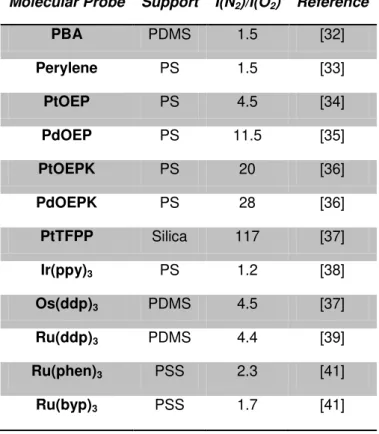
Table 1.1: List of optical sensor prepared employing common molecular probes for oxygen
sensing.
Molecular Probe Support I(N2)/I(O2) Reference
PBA PDMS 1.5 [32]
Perylene PS 1.5 [33] PtOEP PS 4.5 [34] PdOEP PS 11.5 [35]
PtOEPK PS 20 [36]
PdOEPK PS 28 [36] PtTFPP Silica 117 [37] Ir(ppy)3 PS 1.2 [38]
Os(ddp)3 PDMS 4.5 [37]
Ru(ddp)3 PDMS 4.4 [39]
Ru(phen)3 PSS 2.3 [41]
Ru(byp)3 PSS 1.7 [41]
I(N2): the fluorescence intensities in the absence of oxygen, I(O2): the fluorescence intensities in the 100% oxygen, PBA: 1-pyrene butyric acid, OEP: octaethylporphyrin, OEPK: octaethylporphyrinketone, FPP: tetrakis pentrafluoropheny porphine, (ppy): [2-phenylpyridinato-C2,N], (dpp): (4,7-diphenyl-1,10-phenanthroline), (phen): phenanthroline, (byp): (2,2’-bipyridine), PDMS: Polydimethylsiloxane, PS: Polystyrene, PSS: poly(sodium 4-styrene sulfonate).
8 In recent years more advanced probes, typically organo-metallic compounds, are employed for the development of optical sensors because of their long excited state lifetimes, which increase the sensitivity to lower concentrations of oxygen. In general, organo-metallic compounds sensitive to oxygen are classified into two categories: transition metal polypyridyl complex (in general complexes of Ru2+,Re+, Os3+ and Ir3+) and metallo-porphines, (in particular Pt2+ and Pd2+porphyrins).
Numerous sensors are based on the employment of Pt2+ and Pd2+porphyrins because of the high phosphorescence quantum yields at room temperature and, in particular, high sensitivity to oxygen [34,43]. However, porphyrins are very reactive towards singlet oxygen, which leads to photobleaching and limits the long term stability of these sensors.
Nowadays, efforts are devoted to chemical modification of the probes to ensure long term stability or to the development of sensors based on the lifetime monitoring [44]. Unfortunately, the monitoring of the decay time is more sophisticated with respect to the monitoring of the emission intensity, limiting the application of these sensors in real situations.
Figure 1.4: Luminescence of an oxygen molecular probe in argon (Ar), aerated and oxygen
atmosphere. Source: http://www.scivaxls.com.
On the other hand, transition Ru polypyridyl complexes, such as tris(bipyridine)ruthenium(II) (Ru(bpy)3) and tris(phenantroline)ruthenium(II) (Ru(phen)3) show photostability combined with significant luminescence sensitive to oxygen, but lower with respect to metallorphines due to shorter excited state lifetime [45-46].
1.6 Temperature molecular probes
9
Figure 1.5: Representative scheme of working of an optical sensor for temperature detection
[46].
Popular organic dyes were studied as valid candidates for in-situ luminescent nano-thermometry in biological media. The interest is related to the fact that luminescent molecular probes enable high spatial resolution with fast response, often in a time scale comparable to that of cellular processes, combined with a non-invasive monitoring.
Table 1.2: List of optical sensor prepared employing common molecular probes for
temperature sensing
Molecular Probe Support Temperature range (K) Reference
Rhodamine B Silica 283-368 [48]
Fluorescein Starch 273-333 [49]
Ru(phen)3 PAN 273-393 [50]
Ru(byp)3 PVA 50–290 [51]
PtOEP PS 300-450 [52]
Eu(TTA) PS 278-333 [53]
(phen): phenanthroline, (byp): (2,2’-bipyridine), OEP: octaethylporphyrin, TTA: thenoyltrifluoroacetonate, PAN: Poly(acrylonitrile), PVA: Polyvinyl alcohol , PS: Polystyrene.
Among the organic dyes, Rhodamine B and Fluorescein have been extensively studied because of their good brightness, emission in the visible and linear temperature dependence of the intensity of the emission in ambient conditions (273-333 K) [47-48]. Furthermore, Fluorescein has been employed as pH sensitive molecular probes. However, limitations in the employment of organic dyes are related to their weak thermal stability.
10 fact that the emission of phosphorescent luminescent metal–ligand complexes is generally affected by the oxygen that often are immobilized in matrix with low oxygen permeability to minimize interferences related to oxygen concentration drifts.
In fact, in the last decade, metallo-porphines of platinum (i.e. Platinum octaethylporphyrin) and Ru polypyridyl complexes have been selected in several studies as fluorescent nano-thermometers immobilized in different matrixes.
1.7 Objectives and thesis outline
Membrane is an emerging technology allowing non-intensive separation processes without the employment of chemicals. The development of high performance membranes based on innovative and devoted materials and the design of the membrane processes allowed the achievement of the success of membrane technologies in different fields, from gas separation to water treatment, in particular desalination.
Besides the easy scale-up of membrane processes which favour the employment of membrane technologies at industrial scale, the performance of the process is related to the chemical-physical interactions at molecular level between the permeant species and the membrane. For this reason, it is evident the capital importance of studying of membrane processes at molecular level in order to better understand the selective permeation of chemicals, providing an important feedback for the preparation of innovative materials and to overcome the limitations of membrane science. In fact, major factors limiting membrane technology (i.e. concentration polarization, thermal polarization, fouling) are related to phenomena taking part on membrane surface. The study of these critical issues is really challenging because of the lack in term of experimental data related to the problems of studying them with high spatial resolution without disturbing the membrane processes.
Luminophores, molecules emitting light upon light exposure, are successfully employed as molecular probes for the in-situ, at submicron scale, non-invasive and on-line monitoring of several parameters (i.e. temperature, oxygen, and pH) typically demanded in innovative fields of nano-science. The aim of this work is to propose molecular probe as an innovative technology for monitoring membrane processes.
In this study, tris (1,10-phenanthroline) ruthenium (II) chloride hydrate, (Ru(phen)3, was selected as molecular probe on the basis of its high brightness, photostability and pronounced oxygen and temperature sensitivity.
11 operating conditions, in particular fluctuations of the optical path, drifts in the intensity of the excitation light and temperature variations, limiting the effectiveness of the oxygen sensing. Therefore, a second probe, 7-Methoxy-4-methylcoumarin (Coumarin), non-sensitive to the oxygen concentration was immobilized in the polymeric membrane, allowing the corrections of artefacts induced by changes in the geometry and intensity of the light source. On the other hand, the implementation of simple model compensated the thermal effect, obtaining a signal that depends exclusively on the oxygen concentration. This work performed at the Universidade Nova de Lisboa was submitted to Journal of Membrane Science.
The developed oxygen sensitive membranes were employed as optical sensor for the on-line and non invasive monitoring of oxygen in food packaging. The sensor was placed in a degassed atmosphere sealed by polymeric films reproducing a typical configuration in modified atmosphere packaging (MAP): the polymeric packaging in an interface between the enclosed degassed atmosphere (without oxygen) and the aerated atmosphere (oxygen concentration of 21v%). Packaging films made of polymer with different oxygen permeability such as of biaxially oriented polypropylene (BOPP) and polydimethyl siloxane (PDMS) were tested. The oxygen content in the enclosed atmosphere was evaluated by measuring the fluorescence emission of the optical sensor using a bifurcated optical fiber connected to the excitation source and the detector. The experiments showed the efficiency of the optical sensor in the on-line and non-invasive monitoring of the oxygen permeated trough the packaging materials. As expected, the increase of the oxygen concentration is more pronounced in the MAP sealed with PDMS than BOPP due to its oxygen permeability one order of magnitude higher. Moreover, the results were confirmed by the solution-diffusion model which predicted the rate of permeation of oxygen trough the packaging material considering its oxygen permeability evaluated with an independent method. This work was carried out at the Universidade Nova de Lisboa and submitted to Journal of Food Engineering.
12 developed at the Universidad de Zaragoza and submitted to Journal of Colloids and Interface Science.
Once again, Ru(phen)3 was selected as molecular probe and immobilized into/onto hydrophobic porous membrane made of Poly(vinylidene fluoride). The membranes prepared using electrospinning present excellent performance in direct contact membrane distillation process because of their open pore morphology made of a 3D network of nano-fibers. The membranes presented luminescent activity due to the immobilized molecular probe which enables the mapping of temperature on membrane surface and the evaluation of the thermal polarization. A paper is in preparation to be submitted to Journal of Membrane Science.
13
1.8 References
[1] Baker, R W. Overview of Membrane Science and Technology, in: Membrane Technology and Applications. Wiley, second edition, 2004.
[2] Figoli A, Santoro S, Galiano, F, and Basile, A. Pervaporation membranes: preparation and characterization, in: Vapour Permeation and Membrane Distillation Principles and Applications. Elsevier Woodhead Publishing, 2015.
[3] Uemura, T, and Henmi, M. Thin-Film Composite Membranes for Reverse Osmosis, in : Advanced Membrane Technology and Applications. Wiley, 2008.
[4] Koutsou, C P, Karabelas, AJ, and Kostoglou, M. Membrane desalination under constant water recovery – The effect of module design parameters on system performance. Separation and Purification Technology, 147: 90-113, 2015.
[5] Lau, M L, Ismail, A F, Misdan, N, and Kassim, M A. A recent progress in thin film composite membrane: A review. Desalination, 287: 190-199, 2012.
[6] Bastan, D, Esmaeili, N, and Asadollahi, M. Polymeric mixed matrix membranes containing zeolites as a filler for gas separation applications: A review. Journal of Industrial and Engineering Chemistry, 19(2): 375-393, 2013.
[7] Briscoe, W H. Polymers and Nanoscience, In: Colloidal Foundations of Nanoscience. Elsevier, 2014.
[8] Mohammad, A W, Teow, T Y, Ang, W F, Chung, Y T, Oatley-Radcliffe, D L, and Hilal, N. Nanofiltration membranes review: Recent advances and future prospects. Desalination, 356: 226-254, 2015.
[9] Ali A, Macedonio F, Drioli, E, Aljlil S, and Alharbi, A O. Experimental and theoretical evaluation of temperature polarization phenomenon in direct contact membrane distillation. Chemical Engineering Research and Design, 91(10): 1966-1977, 2013.
[10] Jogdand, A, and Chaudhuri, A. Modeling of concentration polarization and permeate flux variation in a roto-dynamic reverse osmosis filtration system. Desalination, 375: 54-70, 2015.
[11] She, Q, Wang, R, Fane, A G, and Tang, C Y. Membrane fouling in osmotically driven membrane processes: A review. Journal of Membrane Science, 499: 201-233, 2015.
[12] Abetz, V, Brinkmann, T, Dijkstra, M, Ebert, K, Fritsch, D, Ohlrogge, K, Paul, D, Peinemann, K-V, Nunes, SP, Scharnagl, N, and Schossig, M. Developments in Membrane Research: from Material via Process Design to Industrial Application. Advanced Engineering Materials, 5: 328-358, 2006. [13] Nyström, M, and Mänttäri, M. Introduction: Opportunities and Challenges of Real Time Monitoring
on Membrane Processes, in: Monitoring and Visualizing Membrane-Based Processes. Wiley, 2009.
14 [15] Banham, D, Ye, S, Pei, K, Ozaki, J-I, Kishimoto, T , and Imashiro, I. A review of the stability and durability of non-precious metal catalysts for the oxygen reduction reaction in proton exchange membrane fuel cells. Journal of Power Sources, 285: 334-348, 2015.
[16] Razavi, S M , and Miri, T. A real petroleum refinery wastewater treatment using hollow fiber membrane bioreactor (HF-MBR). Journal of Water Process Engineering, 8: 136-141, 2015. [17] Tabesh, H, Amoabediny, Rasouli, A, Ramedani, A, Poorkhalil, A, Kashefi, A, and Mottaghy, K.
Simulation of blood oxygenation in capillary membrane oxygenators using modified sulfite solution. Biophysical Chemistry 195: 8-15, 2014.
[18] Wisniewski, N, and Reichert, M. Methods for reducing biosensor membrane biofouling. Colloids and Surfaces B: Biointerfaces,18(3-4):197-219, 2000.
[19] Mills, A. Oxygen indicators and intelligent inks for packaging food. Chem. Soc. Rev. 34, 1003– 1011, 2005.
[20] Murat S, Insel H C, Cokgor, E U, and Orhon, D. Effect of low dissolved oxygen on simultaneous nitrification and denitrification in a membrane bioreactor treating black water. Bioresource Technology, 102 : 4333-4340, 2011.
[21] Iulianelli A, Ribeirinha P, Mendes A, and Basile, A. Methanol steam reforming for hydrogen generation via conventional and membrane reactors: A review. Renewable and Sustainable Energy Reviews, 29: 355-368, 2014.
[22] Arévalo, J, Ruiz, L M, Pérez, J, and Gómez, M A. Effect of temperature on membrane bioreactor performance working with high hydraulic and sludge retention time. Biochemical Engineering Journal, 88: 42-49, 2014.
[23] Childs, P R N. Advances in temperature measurement. Advances in Heat Transfer 36: 111-181, 2003.
[24] Ramamoorthy, R, Dutta, P K, and Akbar, S A. Oxygen sensors: Materials, methods, designs and applications. Journal of Materials Science 38(21): 4271 – 4282, 2003.
[25] Gosse, C, Bergaud, C, and Löw, O. Molecular Probes for Thermometry in Microfluidic Devices, in: Thermal Nanosystems and Nanomaterials, Topics in Advanced Physics 118. Springer-Verlag, 2009.
[26] Grist M, Chrostowski L, and Cheung, KC. Optical oxygen sensors for applications in microfluidic cell. Culture, 10(10): 9286-9316, 2010.
[27] Chu C S, Lo, Y-L, and Sung, T-W. Review on Recent Developments of Fluorescent Oxygen and Carbon Dioxide Optical Fiber Sensors. Photonic Sensors 1(3): 234-250, 2011.
[28] Stich, M I J, Fischer, L H, and Wolfbeis, O S. Multiple fluorescent chemical sensing and imaging, Chem. Soc. Rev., 39: 3102-3114, 2010.
[29] Quaranta, M, Borisov, SM, and Klimant, I. Indicators for optical oxygen sensors. Bioanal Rev, 4:115-157, 2012
15 [31] Brites, C D S, Lima, P P, Silva, N J O, Angel, M, Amaral, V S, Palacio, F, and Carlos, L D.
Thermometry at the nanoscale. Nanoscale, 4: 4799-4829, 2012.
[32] Sharma, A, and Wolfbeis, O S. Fiber optic Oxygen Sensor Based on Fluorescence Quenching and Energy Transfer. Applied Spectroscopy, 42(6): 1009-1011, 1988.
[33] Fujiwara, Y, Ishikawa, Y, and Amao, Y. Nippon Kagaku Kaishi, 2002.
[34] Lee, S-K, and Okura, I. Photoluminescent determination of oxygen using metalloporphyrin-polymer sensing systems. Spectrochimica Acta Part A, 54(12): 91-100, 1998.
[35] Amao, Y, Miyashita, T, and Okura. Optical oxygen sensing based on the luminescence change of metalloporphyrins immobilized in styrene-pentafuorostyrene copolymer film. Analyst,125(5): 871-875, 2000.
[36] Hartmann, P, and Trettnak, W. Effects of polymer matrices on calibration functions of luminescent oxygen sensors based on porphyrin ketone complexes. Anal Chem., 68: 2615-2620, 1996. [37] Chu, C-S. and Lo, Y-L. Optical fiber dissolved oxygen sensor based on Pt(II) complex and
core-shell silica nanoparticles incorporated with sol–gel matrix. Sensors and Actuators B, 151(1): 83-89, 2010.
[38] Amao, Y, Ishikawa, Y, and Okura, I. Optical Oxygen Sensing Material: Terbium(III) Complex Adsorbed Thin Film. Bull Chem Soc Jpn, 74: 2445-2449, 2001.
[39] Xu, W, Kneas, K A, Demas, J N, and DeGraff, B A. Oxygen sensors based on luminescence quenching of metal complexes: Osmium complexes suitable for laser diode excitation. Anal. Chem, 68(15): 2605-2609, 1996.
[40] Carraway, E R, Demas, J N, and DeGraff, B A. Luminescence quenching mechanism for microheterogeneous systems. Anal. Chem., 63(4): 332-336, 1991.
[41] Amao, Y, and Okura, I. Optical oxygen sensing materials: chemisorption film of ruthenium(II) polypyridyl complexes attached to anionic polymer. Sensors and Actuators B, 88(2): 162-167, 2003.
[42] Bergman, I. Rapid-response Atmospheric Oxygen Monitor based on Fluorescence Quenching. Nature, 218: 396, 1968.
[43] Basu, B J, Thirumurugan, A, Dinesh, A R, Anandan, C, and Rajam, K. Optical oxygen sensor coating based on the fluorescence quenching of a new pyrene derivative. Sensors and Actuators B, 104(1): 15-22, 2005.
[44] Papkovsky, P B, Olah, J, Troyanovsky, I V, Sadovsky, N A, Rumyantseva, V D, Mironov, F A, Yaropolov, A I, and Savitsky, A P. Phosphorescent polymer films for optical oxygen sensors. Biosens. Bioelectron. 7: 199-206, 1992..
[45] Puklin, E, Carlson, B, Gouin, S, Costin, C, Green, E, Ponomarev, S, Tanji, H, Gouterman, M, 2000, Ideality of pressure-sensitive paint. I. Platinum tetra(pentafluorophenyl)porphine in fluoroacrylic polymer. J. ApplPolym. Sci., 77(3): 2795-2804
16 [47] Wang, Z, McWilliams, A R, Evans, C E B, Lu, X, Chung, S, Winnik, M A, and Manners, I. Covalent attachment of RuIIphenanthroline complexes to polythionylphosphazenes: the development and evaluation of single-component polymeric oxygen sensors. Adv. Funct. Mater., 12(6-7): 415-419, 2002.
[48] Wang, X-D, Wolfbeis, O S, and Meier, R J. Luminescent probes and sensors for temperature. Chem. Soc. Rev.,42(19): 7834-7869, 2012.
[49] Ross, D, Gaitan, M, and Locascio, L E. Temperature measurement in microfluidic systems using a temperature-dependent fluorescent dye. Anal. Chem., 73(17): 4117-4123, 2001.
[50] Guan X, and Su, Z. Synthesis and characterization of fluorescent starch using fluorescein as fluorophore: potential polymeric temperature/pH indicators. Polym. Adv. Technol., 19(5): 385-392, 2008.
[51] Baleizao, C, Nagl, S, Schäferling, M, Berberan-Santos, M N, and Wolfbeis, O S. Dual Fluorescence Sensor for Trace Oxygen and Temperature with Unmatched Range and Sensitivity. Anal. Chem., 80(16): 6449-6457, 2008.
[52] Mills, A, Tommons, C, Bailey, R T, Tedford, M C, Crilly, P J. Luminescence temperature sensing using poly(vinyl alcohol)-encapsulated Ru(bpy)32+ films. Analyst, 131(4): 495-500, 2006.
[53] Stehr, J, Lupton, J M, Reufer, M, Raschke, G, Klar, T A, and Feldmann, J. Sub-Microsecond Molecular Thermometry Using Thermal Spin Flips. Adv. Mater., 16(23-24): 2170-2174, 2004. [54] Basu, B J, and Venkatraman, S. Fabrication of a Bi-luminophore Temperature Sensitive Coating
17
Chapter 2
Development of Oxygen and Temperature Sensitive
Membranes Using Molecular Probes as Ratiometric
19
2.1 Introduction
Membrane processes are governed by the physical-chemical interactions between the boundary layer of the solutions in contact with the membranes and the membrane surfaces [1]. Efforts in membrane science have been devoted on the development of innovative materials and optimization of the operating conditions in order to maximize the selective permeant-membrane interactions increasing the performance of the process in terms of productivity, selectivity and stability [1]. Moreover, it is notorious that these interactions dynamically change on membrane surface due to variations of the operating conditions and membrane properties related to phenomena such as the as concentration and/or thermal polarization, fouling, swelling and membrane deterioration [1-6].
Nowadays, employing common analytic technologies it is possible monitoring the global performance of the process estimating the permeability and the selectivity of the membrane and to evaluate the long-term stability of the separation processes. However, they lack in providing on-line information for studying parameters of interest at low scale and understanding the phenomena which affect the global processes at large scale. In fact, the optimal monitoring processes would be in real time, non-invasive, in situ, at a molecular scale and using pattern recognition approaches [7].
Optical sensors, based on the immobilization of luminescent molecular probes in/onto polymeric matrices, are an attractive technology for non-invasive monitoring, allowing to obtain highly sensitive devices for continuous detection of temperature changes and/or oxygen content in a given medium [8]. The molecular size of probes consents the on-line detection with a high resolution, also at micrometric and sub-micrometric scale using adequate technology (i.e. fluorescent microscopy) [9-10]. Further advantages are related to the fact that the activation stimuli and the responses of the optical sensors consist of light, allowing for the development of non-invasive sensors with a short response time [11]. For these reasons, molecular probes could be considered a promising technology for on-line, non-invasive and high resolution monitoring of membrane process.
20 The identification of the most adequate materials and the optimization of the procedure for the preparation of the optical sensors are crucial for the development of the optical sensor.
The sensitivity of the device strongly depends on the properties of polymer, in particular its oxygen permeability [23]. In general, polymers with high oxygen permeability, such as poly(dimethylsiloxane) (PDMS), are employed for the development of the optical sensor in order to guarantee interactions between the permeating target molecules (O2) and the immobilized molecular probes. However, they lack in mechanical properties and thus a rigid support is often required [24].On the contrary, glassy polymers such as polystyrene (PS) present lower, but reasonable, oxygen permeability and good mechanical properties which allow the preparation of self-consistent films, improving their versatility [25].
Nevertheless, the core of the optical sensor is the molecular probe. In the last decades, efforts have been made towards the development of luminescent probes with high sensitivity to oxygen and long stability [26-29].
Luminescent transition metal polypyridyl complexes present unmatched photostability, and have been largely used in the preparation of optical sensors. In particular, Ru(II) polypyridyl complexes have been employed more frequently since they are rather easy to synthesize. Disadvantages are related to the relatively short excited state lifetime, with respect to metallorphines, that limits their sensitivity. Also, they suffer from pronounced cross talk to temperature since their triplet states are subjected to severe thermal quenching [30]. In fact, it is well known for optical sensors based on fluorescence (or phosphorescence) quenching that both the emission intensity and the excited state life time are affected by the temperature [30-32].
The higher limitations for practical applications, common to all optical sensors based on the fluorescence/phosphorescence emission intensity detection, are related to the strong impact that operating conditions (geometry of the detection, optical path, fluctuations of the excitation source, probe concentration, etc.) produce in the intensity of the signals measured.
A strategy to overcome these drawbacks is the simultaneous use of two luminescent probes (dispersed in the same polymeric matrix) and the combination of their emission intensities in a ratiometric signal which allows for the correction of errors produced by unknown fluctuations of the operative conditions [33-35].
Tris(1,10-phenanthroline)ruthenium(II) (Ru(phen)3), a luminescent transition metal polypyridyl complex, was used as oxygen sensitive probe in order to ensure long life-time to the sensor. The performance of polystyrene (PS) and poly(β-hydroxybutyrate-β-hydroxyvalerate) (PHBV) as sensor matrix was evaluated. The design of Ru(phen)3 based ratiometric sensors involved the use of a second fluorescent probe non-sensitive to oxygen as a reference for correcting artifacts induced by optical path drifts and fluctuations of the intensity of the exciting light. At this respect, the monitoring efficiency of 7-Methoxy-4-methylcoumarin (Coumarin), Dansyl Chloride and Quinine Sulfate were evaluated.
21
2.2 Materials and Methods 2.2.1 Materials
Polystyrene (PS, Mw: 192,000) was purchased from Sigma Aldrich Chemistry (Spain), whereas Poly(hydroxybutyrate-co-hydroxyvalerate)(PHBV, Mw: 300,000) containing 3 mol% of 3-hydroxyvalerate units was obtained from Tianan Biologic Material Co. Ltd. (China). Tris(1,10-phenanthroline)ruthenium(II) (Ru(phen)3) was synthesized according to the procedure reported in literature [36]. The other molecular probes 7-methoxy-4-methylcoumarin (Coumarin), Dansyl Chloride and Quininehemisulfate salt monohydrate (Quinine Sulfate) and the solvent (Chloroform) were purchased from Sigma Aldrich Chemistry (Spain).
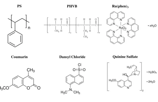
The chemical structures of the polymers and of the probes employed are represented in Figure 2.1.
Figure 2.1: Membrane separation: Chemical structures of polymers and molecular probes
employed in the development of oxygen and temperature sensitive membranes.
Quinine Sulfate
Ru(phen)3
Dansyl Chloride Coumarin