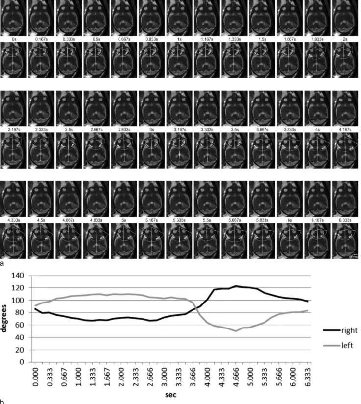
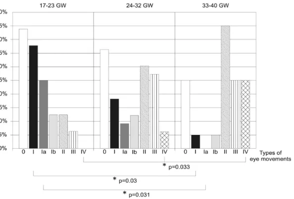
Fetal eye movements on magnetic resonance imaging.
Texto
Imagem
![Table 1. Modified classification of fetal eye movements based on a classification made by Birnholz [3] using ultrasound findings.](https://thumb-eu.123doks.com/thumbv2/123dok_br/18268540.344245/3.918.89.612.93.255/table-modified-classification-movements-classification-birnholz-ultrasound-findings.webp)


Documentos relacionados
Magnetic resonance imaging (MRI) (Figure 1A,B,C) showed sparse hyperintense areas in the white substance, bilaterally on T2-weighted and FLAIR sequences, predominantly in
Evaluation of suspicious nipple discharge by magnetic resonance mammography based on breast imaging reporting and data system magnetic resonance imag- ing descriptors. Manganaro
Spectrum of indings on magnetic resonance imaging of the brain in patients with neurological manifestations of dengue fever..
The image sequence of the right (Figure 1) and left (Figure 2) sides in closed-mouth position showed retroposition of the condyle and partial anterior
Figure 3: Errors in position, given in RNT coordinates, for 2 hours (on the left) and 24 hours (on the right), considering perturbations due to geopotential and direct solar
In the ASIST (Active Surveillance Magnetic Resonance Imaging Study) trial the value of MRI guided confirmatory biopsy in men on surveillance is being studied by the On-
Data on the postural assessment of the child (Table 1) on the right side and left planes are pre- sented in degrees and compared in percentages for assessments with and
(A) Magnetic resonance imaging (MRI) shows an intra-axial right parietal solid lesion, isointense on T2 weighted image; (B) With edema and mass effect seen on FLAIR; (C) MRI T1

