585
Case report doi:10.12980/JCLM.3.2015J5-43 ©2015 by the Journal of Coastal Life Medicine. All rights reserved.
A case of Vogt-Koyanagi-Harada disease mimicking sympathetic ophthalmia
Syed Shoeb Ahmad*
, Faisal Ariff Hassan
Ophthalmology Department, Queen Elizabeth Hospital, Kota Kinabalu, 88586, Malaysia
Journal of Coastal Life Medicine2015; 3(7): 585-587
Journal of Coastal Life Medicine
*Corresponding author: Syed Shoeb Ahmad, Ophthalmology Department, Queen Elizabeth Hospital, Kota Kinabalu, 88586, Malaysia.
Tel: 006-01126868346 Fax: 006-88-252827
E-mail: syedshoebahmad@yahoo.com
1. Introduction
Sympathetic ophthalmia (SO) is a serious ocular disorder which can cause loss of vision in both eyes. Although rare, this bilateral granulomatous inflammation can result from accidental or surgical insult to the uvea. Diagnosis of this condition is imperative in order to save both the eyes from going blind. A number of conditions can mimic SO, including Vogt-Koyanagi-Harada syndrome (VKH). A 34-year-old patient was seen by us complaining of pain, redness and decreased vision in one eye. There was a history of injury to the other eye 20 years ago. A provisional diagnosis of SO was made. However, subsequent investigations showed that the patient was actually a case of VKH.
This case report is being presented to highlight the clinical resemblance between SO and VKH. This can make a definite diagnosis difficult in the early phase of these two conditions.
2. Case report
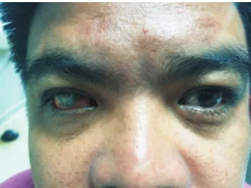
A 34-year-old male patient was involved in a motor-vehicle accident nearly 20 years ago. He sustained severe injury to the right eye and had lost all sight in the affected eye. Subsequently, both eyes had been stable. Recently, he was riding a motorbike behind a lorry when he felt a sudden foreign body sensation in the left eye. He went to a general clinic, where ocular irrigation was done and then the patient was referred to our specialist clinic (Figure 1).
Figure 1. External appearance of the patient showing the inflamed phthisical right eye and inflamed left eye.
According to the patient, he developed redness, discomfort and progressive blurring of vision in the left eye following the recent incident involving a lorry. He had slight pain in the right eye also. He denied any systemic illness, joint pains, aphthous ulcers, neurologic symptoms, contact with tuberculosis, promiscuity, fever, night sweats, rashes, loss of weight or appetite, chronic cough, alopecia, skin changes or ocular accommodative insufficiency. He had no contacts with dogs or pigs, but had 6 cats at home, although he denied any proximity to cat feces.
On examination, his vision was no-perception-to-light in the right eye and 6/60 (with pin-hole 6/48) in the left eye. Slit-lamp biomicroscopic examination showed circumciliary congestion in A RT I C L E I N F O A B S T R AC T
A 34 years old male patient presented to us on 15th February 2015, with complaints of mild pain in right eye and a foreign body sensation to the left eye, associated with redness, discomfort and blurring of vision with 4 days duration. There was history of a penetrating eye injury to his right eye 20 years ago. Examination showed bilaterally inflamed eyes. The right eye was going into phthisis bulbi. Fundus examination of the left eye showed blurred optic disc with hemorrhagic areas, macular edema and exudative retinal detachments. Systemic review did not show any gross skin changes, neurological signs or dysmorphism. He was initially treated as sympathetic ophthalmitis, but the history and examination noted that he had bilateral sensory neural hearing loss. Fundus fluorescein angiography showed that he had hyperfluorescent spots in the fundus. Thus, the diagnosis was changed to Vogt-Koyanagi-Harada disease. The patient was treated with oral steroids (1 mg/kg per day) and subsequent follow up showed a marked improvement in the ocular findings.
Article history:
Received 4 May 2015
Received in revised form 15 May 2015 Accepted 12 Jun 2015
Available online 15 Jun 2015
Keywords:
Uveitis
Uveomeningoencephalitic syndrome Sympathetic ophthalmia
Syed Shoeb Ahmad and Faisal Ariff Hassan/Journal of Coastal Life Medicine 2015; 3(7): 585-587
586
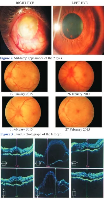
both eyes. The right eye had a vascularised corneal opacity covering the entire cornea. The eyeball was soft and going into phthisis bulbi. The left eye had scattered fine keratic precipitates (KPs), the anterior chamber was deep with +2 cells and a few posterior synechiae were present. Fundus examination showed a swollen optic disc with peripapillary hemorrhages, edematous macula, tortuous vessels and multiple exudative retinal detachments (RDs). A 3-mirror contact lens biomicroscopy did not reveal any retinal breaks (Figures 2 and 3). Optical coherence tomography showed an edematous macula in left eye (Figure 4).
Figure 2. Slit-lamp appearance of the 2 eyes.
RIGHT EYE LEFT EYE
Figure 3. Fundus photograph of the left eye.
19 January 2015 26 January 2015
27 February 2015 3 February 2015
Figure 4. Optical coherence tomography of left eye. N
S S S
I I I
T
256 190 264
65 51
64
N T N T
The patient was suspected to have a SO following trauma to the right eye many years ago. The treatment was started on tablet prednisolone 1 mg/kg per day, which was tapered by 5 mg every 2 weeks. A topical steroid and cycloplegic eyedrops were also prescribed in both eyes.
The patient was referred for a systemic examination. His blood pressure was 128/78 mmHg. Neurologic examination revealed normal cranial nerves except a slight hearing deficiency in both ears and optic nerve involvement in the left eye. A full blood count was normal. Erythrocyte sedimentation ratewas 14 mm/h. Liver, renal function tests and peripheral blood smear were normal. Venereal
disease research laboratory, double stranded DNA, anti-nuclear antibody, serum toxoplasmosis, serum Bartonella and HIV tests were negative. Chest X-ray showed bilateral clear lung fields. The result of mantoux test was 10 mm. Pure tone audiometry showed a mild-moderate sensorineural hearing loss (SNHL) in right ear and moderate to severe SNHL in left ear. A tympanogram of right ear was Type AS, and in left ear was Type A.
Fundus fluorescein angiography (FFA) showed diffuse pinpoint hyperfluorescence and an area of early hyperfluorescence temporal to the macula, which did not increase in size. However, no vasculitis was noted (Figure 5). CT scan noted focal thickening of the sclera in the left eye, while the right eye was going into phthisis bulbi.
Figure 5. FFA of left eye. 03-02-2015, OS
FA 3:10.16 55° ART (45)
03-02-2015, OS FA 9:22.54 55° ART (16)
22 µm
22 µm
On his last visit, 1 month following the episode, his vision was 6/120 in left eye. The anterior segment did not show any signs of inflammation. Fundus examination showed decreasing disc oedema, resolving macular oedema and decreasing exudative detachments. Based on the audiometry results, hearing defect and pinpoint hyperfluorescence on FFA, the diagnosis was changed to VKH. This case report was being presented to highlight the clinical similarities between SO and VKH. This can make a definite diagnosis difficult in the early phase of these conditions.
3. Discussion
Uveitis is proved to be a sight-threatening ocular inflammatory disorder. This condition can affect any or all of the uveal tissues, namely, the iris, ciliary body or choroid. Thus, it could be anterior or posterior and depend upon the duration: acute or chronic. It is commonly associated with a systemic inflammatory disease, a complication of infection or an isolated, idiopathic disorder[1]. One of the rare uveitic conditions seen in clinical practice is SO. This condition was ably defined by Sir Stewart Duke Elder
as “a specific bilateral inflammation of the entire uveal tract of
unknown etiology. It is characterized clinically by an insidious onset and progressive course with exacerbations and pathologically by nodular or diffuse infiltrations of the uveal tract by lymphocytes and epitheloid cells. It almost invariably follows a perforating wound
involving the uveal tissue”. Penetrating injuries are the most common
Syed Shoeb Ahmad and Faisal Ariff Hassan/Journal of Coastal Life Medicine 2015; 3(7): 585-587
587
The condition is characterized by bilateral anterior uveitis with mutton-fat KPs and moderate to severe vitritis, choroiditis and papillitis. The retina is characterized by sub-retinal pigment epithelium yellow-white nodular lesions (corresponding to the histopathologic Dalen-Fuchs nodules). Dalen-Fuchs nodules are not pathognomonic of SO and were seen in only 1/3 of enucleations in one study[3]. At times, the anterior chamber reaction is relatively
mild and non-granulomatous. This is also known as “sympathetic irritation”. Our patient had fine KPs and +2 cells in the anterior
chamber on initial presentation. The differential diagnosis of SO includes conditions like VKH, sarcoidosis, tuberculosis and intraocular lymphoma. In our patient, the tests for the last 3 conditions were negative.
The patients who develop SO may actually have better final vision in the exciting eye. Thus, preservation of both eyes should be attempted if possible. The sympathizing eye may develop sequelae of SO such as: secondary glaucoma, cataract, retinal or optic atrophy, retinal detachment, sub-retinal fibrosis, choroidal atrophy, choroidal neovascularisation and phthisis bulbi[3].
Occasionally, it may be difficult to distinguish SO from another uveitic condition called VKH[5]. This condition is also known as uveomeningoencephalitic syndrome. VKH is one of the most common causes of bilateral panuveitis. Both VKH and SO are autoimmune disorders, which target melanin-bearing cells[6]. They are virtually identical in terms of histopathology, FFA and human leukocyte antigens DR4, DRw53 and Bw54. Unlike SO, which shows no racial predilection, VKH is more common in darkly pigmented races. VKH is also common among females, while
SO doesn’t show any sexual predilection. SO is more common
in young age due to trauma and in old age due to increased frequency of surgeries[7]. The inciting event is also different, usually, a penetrating ocular injury in SO. Occasionally, VKH may be preceded by prodromal symptoms and signs suggestive of a viral infection. In VKH, serous RDs and optic disc involvement is also more frequent compared to SO. Unlike SO, it also shows extraocular manifestations such as meningismus, vitiligo, poliosis and dysacusis. Our patient denied any of these symptoms. However, pure tone audiometry showed a mild-moderate SNHL in the right ear and moderate-severe SNHL in left ear. However, the extraocular manifestations may appear in different phases of the disease and may vary in different ethnic groups. Cutaneous manifestations might be absent during the acute phase and appear later during the course of the disease. While, neurologic and auditory symptoms may precede the ocular manifestations[8].
There are 4 clinical stages of VKH, namely, the prodromal, uveitic, chronic and recurrence phases. The diagnosis of VKH is based on the criteria published by the International Committee on Nomenclature in 2001. These include: 1) absence of previous history of ocular trauma or surgery; 2) no evidence of other ocular diseases; 3) early bilateral ocular involvement (with focal areas of sub-retinal fluid or serous RDs) or late bilateral involvement (depigmentation, sunset glow fundus, Dalen-Fuchs nodules and migration or accumulation of the pigmented epithelium of the retina); 4) history or presentation of auditory and/or neurological symptoms; 5) cutaneous symptoms that appear during or after the neurological and ocular manifestations. On the basis of these criteria, VKH may be classified as “complete”, when all criteria are
met; as “incomplete” when criteria 1, 2 and 3 plus 4 or 5 are met or “probable” when only 1, 2 and 3 are met[9].
Dalen-Fuch’s nodules are not pathognomonic of any disease
and may be seen in VKH, SO and sarcoidosis. FFA findings in VKH go on changing as the disease progresses. In acute stage it may show disseminated spotted choroidal hyperfluorescence and hypofluorescence. In the chronic stage, there is spotted
choroidal hyperfluorescence and hypofluorescence, along with optic disc hyperfluorescence. Retinal vasculitis and a reticular hyperfluorescent pattern may also occur. In our patient, there was a
typical “starry night” appearance seen[10].
Tr e a t m e n t o f V K H i n c l u d e s c o r t i c o s t e r o i d s a n d / o r immunosuppressive drugs. The dose of corticosteroids is usually 1-2 mg/kg of oral prednisone. In more severe cases, pulse therapy of methylprednisolone 1 g/day for 3-5 days can be given. The treatment should be given for 6 months to 1 year prior to withdrawing corticosteroid treatment. Immunomodulators are recommended in patients who do not respond to corticosteroid treatment or those
who develop side-effects such as Cushing’s syndrome, systemic
hypertension or diabetes mellitus. These immunomodulators include methotrexate, etanercept, tacrolimus, cyclosporine, mycophenolate mofetil, azathioprine, cyclophosphamide, chlorambucil and adalimumab. In our patient, tablet of prednisolone 1 mg/kg body weight was given and the patient responded significantly to the treatment[9].
This paper is a reminder that occasionally patients can present with bilateral acute uveitis and it may be difficult to differentiate SO and VKH. Although, the treatment for the 2 conditions is similar,
the prognosis is worse for SO. Thus, in the interest of the patient’s
quality of life it is imperative to reach the correct diagnosis. However, it might be difficult to be sure in the early phase of the disease.
Conflict of interest statement
We declare that we have no conflict of interest.
Acknowledgments
We would like to thank the Head of Ophthalmology Department, Queen Elizabeth Hospital, for permission to present this case.
References
[1] Al-Dhibi HA, Al-Mahmood AM, Arevalo JF. A systematic approach to emergencies in uveitis. Middle East Afr J Ophthalmol 2014; 21: 251-8. [2] Chaithanyaa N, Devireddy SK, Kishore Kumar RV, Galli RS, Aneja V.
Sympathetic ophthalmia: a review of literature. Oral Surg Oral Med Oral Pathol Oral Radiol 2012; 113: 172-6.
[3] Arevalo JF, Garcia RA, Al-Dhibi HA, Sanchez JG, Suarez-Tata L. Update on sympathetic ophthalmia. Middle East Afr J Ophthalmol
2012; 19: 13-21.
[4] Chu XK, Chen CC. Sympathetic ophthalmia: to the twenty-first century and beyond. J Ophthalmic Inflamm Infect 2013; 3: 49.
[5] Das D, Bhattacharjee H, Bhattacharyya PK, Jain L, Paniker MJ, Das K, et al. Pattern of uveitis in North East India: a tertiary eye care center study. Indian J Ophthalmol 2009; 57: 144-6.
[6] Chuang CT, Huang PS, Chen SC, Sheu SJ. Reversible alopecia in Vogt-Koyanagi-Harada disease and sympathetic ophthalmia. J Ophthalmic Inflamm Infect 2013; 3: 41.
[7] Wang Y, Chan CC. Gender differences in Vogt-Koyanagi-Harada disease and sympathetic ophthalmia. J Ophthalmol 2014; doi: 10.1155/2014/157803.
[8] Rao NA, Gupta A, Dustin L, Chee SP, Okada AA, Khairallah M, et al. Frequency of distinguishing clinical features in Vogt-Koyanagi-Harada disease. Ophthalmology 2010; doi: 10.1016/j.ophtha.2009.08.030.
[9] Mota LAA, dos Santos AB. Vogt-Koyanagi-Harada syndrome and it’s
multisystemic effects. Res Assoc Med Bras 2010; 56: 590-5.