18 artigo 484
CASE REPORT
1 – Orthopedic Doctor of the Spine Service, Hospital das Clínicas of the Universidade Federal de Goiás do HC/UFG – Goiânia, GO, Brazil. 2 – Neurosurgeon of the Spine Service, HC/UFG – Goiânia, GO, Brazil,
3 – Head of the Spine Service of the HC– UFG and Head of the Orthopedics and Traumatology Department of the HC/UFG – Goiânia, GO, Brazil, 4 – Neurosurgeon, Spine Service of the HC/UFG – Goiânia, GO, Brazil,
5 – Medical Students of the Faculty of Medicine, HC/UFG – Goiânia, GO, Brazil,
Work carried out at the Orthopedics and Traumatology Department of the HC/UFG – Goiânia, GO.
Correspondence: Primeira Avenida, S/N° - Setor Universitário, Hospital das Clínicas – Departamento de Ortopedia. Goiânia – Go, Cep: 74605-050. E-Mail: [email protected] Received for publication: 01/26/2011, accepted for publication: 06/03/2011
PRIMARy LIPOSARCOMA OF
THE LUMbAR SPINE: CASE REPORT
Frederico Barra de Moraes1, André Luiz Passos Cardoso1, Newton Antônio Tristão1, Wilson Eloy Pimenta Júnior2, Sérgio Daher3,
Siderley de Souza Carneiro4, Nathalia Parrode Machado Barbosa5, Nayanne de Lima Malta5, Noara Barros Ribeiro5
The authors declare that there was no conflict of interest in conducting this work
This article is available online in Portuguese and English at the websites: www.rbo.org.br and www.scielo.br/rbort ABSTRACT
We report a rare case of primary bone liposarcoma of the lumbar spine, for which only one case has been re-ported. A female patient, 60 years of age, with lumbar pain and left sciatalgy for six months. In the imaging exams, a destructive tumor was found in the L4 vertebral body, and magnetic resonance imaging (MRI) revealed a tumoral lesion with T1 hiposignal and T2 hypersignal. Histological diagnosis was difficult, and immunohistochemistry con-firmed the diagnosis. Surgical treatment was performed
with wide ressection, spinal cord decompression, and anterior and posterior fusion of L3 to L5 complemented by radiotherapy and chemotherapy. After three years, a computed tomography (CT) scan evidenced an expansive injury in the lung. Despite its rarity, liposarcoma should be considered in the differential diagnosis of sciatica and primary tumors of the spine.
.
Keywords - Primary Liposarcoma; Bone Neoplasms; Lumbar Vertebrae
INTRODUCTION
Liposarcoma is a malignant soft-tissue tumor that origi-nates from primitive mesenchymal cells, rather than from mature adipose tissue. There are five histological types, of which the pleomorphic type is the rarest. Pleomorphic liposarcoma is a high-grade tumor with highest prevalence between the ages of 60 and 70 years, and it affects both sexes equally. The commonest site for this tumor is the thigh, but other anatomical sites such as the lungs, heart, orbit, retroperitoneum, epidural space and bones(1).
The first reports on primary liposarcoma of bone were made by Ewing, Stewart and Barnard in the 1930s, and Ewing included it in his classification of bone tumors in 1939(2-4). However, its bone origin was subsequently
questioned by several authors for some years. Among these was Jaffe, who only in the 1960s became con-vinced that it existed(5). It is a very rare type of tumor,
with less than 100 cases so far reported in the medical
literature(6). Among these, primary pleomorphic
lipo-sarcoma of vertebrae is an even rarer lesion, with only two cases described (one in the thoracic spine and one in the lumbar spine).
The aim of this study was to report a case of pri-mary liposarcoma of bone, in the lumbar spine, with descriptions of its clinical, radiological and treatment characteristics.
CASE REPORT
125
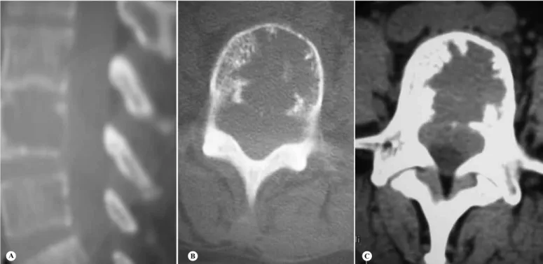
Figure 1 – Radiograph of the lumbar spine without evidence of the tumor at L4: anteroposterior view (A) and lateral view (B).
A
B
PRIMARY LIPOSARCOMA OF THE LUMBAR SPINE: CASE REPORT
The initial radiographs on the lumbar spine did not show bone lesions (Figure 1). Computed tomography showed an extensive lytic lesion in the L4 body, in association with content presenting soft-tissue density. This was seen to be invading the medullary canal and neural foramens and compromising the nerve roots, especially on the left side (Figure 2). Nuclear magnetic resonance showed that the bone structure of the L4 body had been replaced by tissue presenting hyposignal in T1 and hypersignal in T2, with strong highlighting after introducing gadolinium, accompa-nied by invasion and compression of the medullary canal, without changes to the soft tissues (Figure 3).
A tomography-guided biopsy was then performed, from which an initial diagnosis of high-grade sarco-matous pleomorphic neoplasia was made (Figure 4). It was consequently decided to completely resect the L4 vertebra, initially using an anterior route. The body was replaced by a titanium cage filled with autologous bone graft harvested from the iliac, in order to obtain arthrodesis from L3 to L5.
Ten days later, in a second surgical procedure, the resection of L4 was completed by removing its pos-terior bone elements and decompressing the medulla, as well as finishing the arthrodesis from L3 to L5 with pedicled screws and bone graft material harvested from the iliac (Figure 5).
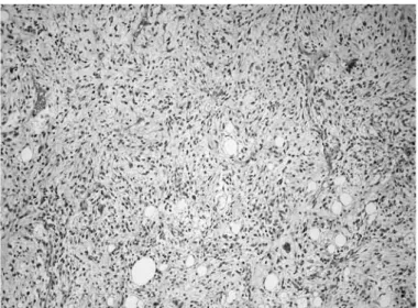
The histological evaluation showed that the bone tissue had been infiltrated by a pleomorphic malignant neoplastic lesion with rounded elongated hyperchro-matic nuclei, mitoses and poorly defined cytoplasm, and with a rich vascular network, areas of hemorrhage and multivacuolated lipoblasts compatible with high-grade liposarcoma. Immunohistochemical analysis was then performed with the following results: vi-mentin +, ENE +, CD31 -, CD34 -, JJF35 - (these last three were CI +), AE1 -, AE3 -, EMA -, S100 - and desmin -, thus confirming the diagnosis of primary liposarcoma of bone (Figure 6).
Figure 2 – Computed tomography of the lumbar spine, showing osteolytic lesion at L4, in sagittal slice (A). Axial slices: showing the destruction in a slice for bone (B) and tumor invasion of the canal in a slice for soft tissues (C).
Figure 3 – Nuclear magnetic resonance imaging on the lumbar column, in a sagittal slice showing hyposignal in T1 (A) and hypersignal in T2 (B).
A B C
127
Figure 4 – Percutaneous biopsy using needle guided by com-puted tomography, in axial slice.
Figure 5 – Postoperative radiographs on lumbar spine showing resec-tion of the L4 vertebral body and anterior arthrodesis with a titanium cage, in anteroposterior view (A), and medullary decompression and posterior arthrodesis with pedicled screws, in lateral view (B).
PRIMARY LIPOSARCOMA OF THE LUMBAR SPINE: CASE REPORT
A
B
DISCUSSION
Lumbalgia is one of the commonest complaints at visits to primary-care physicians, and musculoskeletal complaints are commonest at emergency services(7).
Lumbalgia is a common condition, responsible for significant morbidity, and it has an important eco-nomic and occupational impact within society(8). Most
patients with acute lumbalgia, with or without radicu-lar symptoms, present musculoskeletal disorders or degenerative diseases that do not require specific treatment and are generally self-limited(9). Physicians
Figure 6 – Lesion showing high cellularity and cell atypia, com-patible with fusocellular sarcoma, corresponding to the undif-ferentiated area (hematoxylin-eosin: 100x).
of greater severity may be present, requiring specific treatment, always needs to be ruled out(9).
Most cases of lumbar pain are caused by mechani-cal conditions, including lumbar contraction or relax-ation, degenerative disc and facet processes, disc her-nia, vertebral canal stenosis and fracture compression due to osteoporosis. Non-mechanical conditions such as neoplasia, infections and inflammatory arthritis are important, but rarely cause lumbar pain. Furthermore, around 85% of evaluations on patients with lumbar pain do not reach a definitive diagnosis(9).
Primary tumors of the spine are relatively uncom-mon types of neoplasia, in comparison with metastatic lesions, multiple myeloma and lymphoma. Their pre-sentation is variable and insidious, before signs of spinal cord compression are detected, i.e. the point at which the diagnosis becomes evident(1), as in the
present case.
Primary liposarcoma of bone is preferentially found at the extremities of bones (tibia and femur). Three main morphological characteristics of pleomor-phic liposarcoma have been described: high-grade MFH-like pleomorphic sarcoma, fusiform cell neo-plasia and neoneo-plasia with epithelioid morphology. Histologically, it can be divided into low, medium or high grade. It is formed by areas of normal, multi-vacuolated or even pleomorphic lipoblasts, with large quantities of mitoses. Lesions with low degrees of differentiation are highly vascular(1,10,11).
The difficulty in diagnosing pleomorphic liposar-coma derives from its varied histological appearance. The histological criterion for diagnosing this
neopla-sia consists of recognizing multivacuolated lipoblasts in a high-grade pleomorphic sarcoma. However, al-though the presence of lipoblasts is characteristic of any type of malignant tumor of adipocytes, this is in-sufficient for diagnosing liposarcoma, because lipo-blasts also occur in a series of benign lesions. Lipo-sarcoma can be confounded with malignant fibrous histiocytoma and malignant bone mesenchymoma, mainly because of the pleomorphic cell pattern(12). It
is difficult to differentiate pleomorphic liposarcoma from malignant fibrous histiocytoma. The latter is rarely seen in the spine and only nine cases have been described. Lipoblasts are not histological fin-dings from this entity, and thus, this diagnosis was rejected in the present case. Immunohistochemical or ultrastructural analysis needs to be performed in order to establish the diagnosis(1).
Although pleomorphic liposarcoma has a non-specific immunohistochemical profile, special stains may be necessary in order to identify the presence of intracellular fat, rule out other substances such as mucin or glycogen and rule out other diagnoses. Like other sarcomas, pleomorphic liposarcoma expresses vimentin, while protein S100, smooth-muscle actin, cytokeratins AE1/AE3 and CAM5.2 have variable expression. Desmin is generally absent and the mem-brane antigen or CD34 is negative. The morphological characteristics and positivity for vimentin eliminate the hypothesis of blood diseases such as “signet ring” lymphoma or clear-cell myeloma(1,13).
Radiographically, the lesions are destructive, os-teolytic and poorly defined, and they are observed to expand and destroy the cortex. Nuclear magnetic resonance has great value for anatomical evalua-tions on spinal tumors, showing hyposignal in T1 and hypersignal in T2, mainly due to the non-lipo-matous regions of the tumor. Since the radiologi-cal characteristics of spinal liposarcoma have only rarely been studied, no pathognomonic image has been described through any type of imaging in other parts of the body(1,10,11).
129
PRIMARY LIPOSARCOMA OF THE LUMBAR SPINE: CASE REPORT
avoided by means of vertebral arthrodesis(12).
The radiosensitivity of primary liposarcoma of bone is greater than its radiocurability, and thus, ra-diotherapy is used as adjuvant and/or neoadjuvant treatment to diminish the size of the tumor. Primary liposarcoma of bone does not respond well to chemo-therapy regimens, but these can be tried as adjuvant therapy for reducing the size of the tumor(3,4).
Occurrences of metastases from liposarcoma have been reported in 40.2 to 45.8% of the cases in the literature. These cases often arose many years after apparently successful treatment of the primary
tu-mor, and pulmonary metastases predominate. Thus, the prognosis for such cases would seem to be un-certain, with median survival of two years(1,6,14,15,16).
Primary liposarcoma of the spine is a very rare dis-ease, with only two cases reported in the literature. In cases of suspected primary tumors of the spine, pleo-morphic liposarcoma should be considered in the dif-ferential diagnosis. Nuclear magnetic resonance has great value in investigating suspected spinal tumors, although the conclusive diagnosis of pleomorphic liposarcoma is only obtained through immunohisto-chemical and structural analysis.
REFERENCES
1. Hamlat A, Saikali S, Gueye EM, Le Strat A, Carsin-Nicol B, Brassier G. Primary liposarcoma of the thoracic spine: case report. Eur Spine J. 2005;14(6):613-8. 2. Ewing JA. A review of the classification of bone tumors. Bull Am Coll Surg.
1939;24:290-5.
3. Stewart FW. Primary Liposarcoma of Bone. Am J Pathol. 1931;7(2):87-94.5. 4. Barnard L. Primary liposarcoma of bone. Arch Surg. 1934;29:560-5. 5. Jaffe HL. Tumors and tumorous conditions of the bones and joints. Philadelphia:
Lea & Febiger; 1958.
6. Torok G, Meller Y, Maor E. Primary liposarcoma of bone. Case report and review of the literature. Bull Hosp Jt Dis Orthop Inst. 1983;43(1):28-37.
7. Corwell BN. The emergency department evaluation, management, and treat-ment of back pain. Emerg Med Clin North Am. 2010;28(4):811-39.
8. Duffy RL. Low back pain: an approach to diagnosis and management. Prim Care. 2010;37(4):729-41.
9. Goldman L, Ausiello D. Cecil tratado de medicina interna. Rio de Janeiro: Elsevier; p. 2609-12.
10. Campanacci M. Bone and soft tissue tumors. New York: Springer; 1990.
11. Schajowicz F. Neoplasias ósseas e lesões pseudotumorais. Rio de Janeiro: Revinter; 2000.
12. Simmon M, Springfield D. Surgery for bone and soft tissue tumors. Philadelphia: Lippincott-Raven Publishers; 1997.
13. Oliveira AM, Nascimento AG. Pleomorphic liposarcoma. Semin Diagn Pathol. 2001;18(4):274-85.
14. Addison AK, Payne SR. Primary liposarcoma of bone. Case report. J Bone Joint Surg Am. 1982;64(2):301-4.
15. Schneider HM, Wunderlich T, Puls P. The primary liposarcoma of the bone. Arch Orthop Trauma Surg. 1980;96(3):235-9.
16. Ishii T, Ueda T, Myoui A, Tamai N, Hosono N, Yoshikawa H. Unusual skeletal metastases from myxoid liposarcoma only detectable by MR imaging. Eur