Revista da Sociedade Brasileira de Medicina Tropical 44(2):131-135, mar-abr, 2011
INTRODUCTION
1. Centro de Pesquisa em Virologia, Faculdade de Medicina, Universidade de São Paulo, Ribeirão Preto, SP. 2. Departamento de Ciências Biológicas e da Saúde, Universidade do Oeste de Santa Catarina, São Miguel
Serosurvey of hantavirus infection in humans in the border region
between Brazil and Argentina
Estudo sorológico de infecção por hantavírus em humanos na região de fronteira, entre Brasil
e Argentina
William Marciel de Souza
1, Alex Martins Machado
1, Luiz Tadeu Moraes Figueiredo
1and Everton Bof
2ABSTACT
Introduction: According to reports by the Ministry of Health, in the far western region of the State of Santa Catarina, there have been no reports of hantavirus pulmonary syndrome, a zoonotic disease transmited by feces of infected rodents. A seroepidemiological study of residents of this region, was conducted, with the aim of determining the presence of hantavirus infections. A total of 340 volunteers of both genus, from the towns of Belmonte and Paraíso, were studied. Methods: he serum of these patients was collected and used to detect IgG antibodies against recombinant N protein of Araraquara hantavirus, by ELISA assay. he positive samples were then titrated and conirmed by immunoluorescence assay. Results:
his study demonstrated the presence of IgG antibodies against hantavirus N protein in 3.5% of the population. he most frequent occupation was farm worker, 81% had direct and indirect contact with rodents, 91.7% of positive cases were farm workers, indicating that the probable cause of infection occurred during barn cleaning. hese antibodies are noteworthy, given that the levels of antibodies were veriied in individuals whose contact with hantavirus may have occurred many years ago. Conclusions: his study shows the circulation of hantavirus in the region, a fact that until now, had not reported. All the serum reagents had contact with the pathogen, but did not develop pulmonary and cardiovascular syndrome. It is important to remain alert, because hantavirus is a serious and emerging disease of some relevance.
Keywords: Hantavirus. Seroprevalence in Santa Catarina. Morbidity.
RESUMO
Introdução: De acordo com relatórios do Ministério da Saúde, na região do extremo oeste do Estado de Santa Catarina, não há relatos de síndrome pulmonar por hantavírus, doença zoonótica transmitida por excretas de roedores contaminados. Com a inalidade de demosntrar a infecção por hantavírus, um estudo soroepidemiológico de moradores da região foi realizado. Assim, foi estudado um total de 340 voluntários de ambos os gêneros, dos municípios de Belmonte e Paraíso. Métodos: O soro destes pacientes foi coletado e usado para a detecção de anticorpos IgG contra a proteína N recombinante de hantavírus Araraquara, pelo teste de ELISA. As amostras positivas foram tituladas e conirmadas por imunoluorescência indireta.
Resultados: Este estudo demonstrou a presença de anticorpos IgG contra a proteína N hantavírus em 3,5% da população. A ocupação de lavrador foi a mais frequente, e 81% tiveram contato direto e indireto com os roedores, 91,7% dos casos positivos foram os agricultores, a causa provável da infecção foi através da limpeza de celeiros. Estes anticorpos são notáveis, dado que os níveis de anticorpos são encontrados nos indivíduos cujo contato com o hantavírus pode ter ocorrido há muitos anos. Conclusões: Este estudo mostra a circulação de hantavírus na região, um fato que até então não havia relatado, todos os reagentes soro tiveram contato com o patógeno, mas não desenvolveram a síndrome pulmonar e cardiovascular. É preciso estar alerta, porque é uma doença grave e emergente com grande importância.
Palavras-chaves: Hantavírus. Soroprevalência de hantavirose em Santa Catarina. Morbidade.
Viruses of the genus hantavirus can cause two serious illnesses when transmited from rodents to humans: hemorrhagic fever with renal syndrome (HFSR) or hantavirus pulmonary syndrome (HPS), characterized by respiratory failure, shock and high mortality, making it an important public health problem1.
Hantavirus is an enveloped virus with trisegmented negative sense RNA genome, deined as small, medium and large. Human infection is acquired by inhalation of aerosols containing excreta of rodents infected with hantavirus; i.e., infected by a virus of the family Bunyaviridae2, which includes
than 20 hantaviruses identified throughout the American continent3.
Interpersonal transmission of hantavirus has been reported only in Argentina and Chile, but further investigation indicated that this possibility is very unlikely4-6. he contact with hantavirus in
and of itself does not cause HPS, since infection is dependent on the quantity of viral particles, such that lower concentrations of particles could lead to subclinical infection. However, another possibility is infection by a nonpathogenic strain that induces immune response activation with antibody production3,7.
The pathogenesis of HPS in humans is hypothesized to be mediated by immune activation and excess inflammatory responses, especially macrophages and CD8 + T cells3. Excess cytokines
IL-1-α, IL-1-β, IL-6 and TNF-α interferon-γ, IL-2, IL-4 and TNF-β produced by activated macrophages in hantaviruses, speciic T cells in recognition antigen infected pulmonary endothelial cells, are thought to be critical due to the increased permeability in the pathogenesis of HPS8.
Article/Artigo
METHODS Souza WM et al - Human hantavirus serosurvey on the Brazil Argentina border
Numerous diagnostic methods for these viruses have been produced, among these, the production of recombinant proteins, which are used as antigen in ELISA to enable detection of speciic antibodies against this virus, should be highlighted10,11.
In South America, hantaviruses have been reported in Argentina, Chile, Uruguay, Paraguay, Bolivia and Venezuela12. In Brazil, the irst
evidence that this virus was circulating was observed in the study of three individuals living in a rural area of Juquitiba, São Paulo, in 199313. Today, over 1,200 cases of hantavirus pulmonary syndrome
have occurred in Brazil since 1993, fatalities were reported at a rate of 39%, according to geographic and ethnic diferences14. Five
hantaviruses associated with HPS are currently known in Brazil: the Juquitiba, Araraquara, Laguna Negra-like, Castelo dos Sonhos and Anajatuba viruses15.
he irst conirmed case of hantavirus in Santa Catarina was in 1999 and up to 2009, 205 cases have been conirmed. he irst death from hantavirus in the state was reported in 2001 and 48 deaths have been reported up to the present, making this the state with the second highest death rate16. HPS is an emerging public health problem in Brazil due to
the overlap of urban and agricultural areas, livestock and the disorderly growth of human population in areas where imbalance occurs, which increases human contact with several species of rodents that are reservoirs of hantavirus15. his study was conducted to investigate the
presence of antibodies against hantavirus in the far western region of the State of Santa Catarina and the southern border of Brazil and northern Argentina and correlate this presence through interviews to elucidate the medical history and morbidity of the participants.
The study region has an economic base that comes from agricultural activities and accordingly, the degrading of native forest has occurred throughout the region, which is favorable to colonization by wild rodents. he study revealed the presence of
Necromyslasiurus and Oryzomysnigripes in the region, which are known rodent reservoirs of hantavirus, suggesting there is a chance of viral circulation and, therefore, risk of human infection17.
Although Santa Catarina is the state with the second d highest number of HPS cases, no cases of the disease have been recorded in the far west of the state16 or in municipalities in the study area that
borders northeastern Argentina; however, previous studies have shown that the region has high numbers of HPS18.
hus, it is unclear whether the lack of notiication of hantavirus disease in this region is due, in fact, the absence of local disease or that infections occur and diagnosis is negligent. So far, no accurate survey of the presence of hantavirus has been conducted in the far west of Santa Catarina. Diagnostic tests to detect hantaviruses are only performed following clinical suspicion during hospital atendance, which is not common in the region.
he serological survey aimed to assess, for the irst time, current results and scientific evidence of the absence or circulation of hantavirus in the far west of the state by helping to clarify the real situation of the virus in the region.
Study area
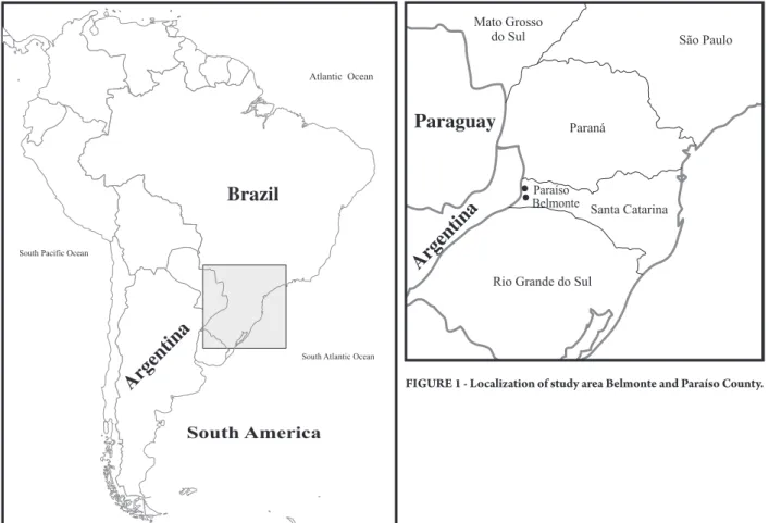
he towns of Belmonte and Paraíso have respective populations of 2,681 and 4,195 inhabitants, who are mostly descended from Italians, Germans and Poles. hey have a mesothermal humid climate, with hot summers and an average temperature of 18.3°C, with the main economic activity based on agriculture, particularly the cultivation of monocultures of corn, wheat, tobacco and, consequently, a large part of the native forest has sufered degradation (Figure 1).
Brazil
Argentina
Argentina
Paraguay
Rev Soc Bras Med Trop 44(2):131-135, mar-abr, 2011
RESULTS
DISCUSSION
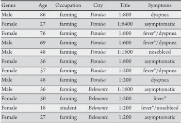
TABLE 1 - Cases of hantavirus serology reagents according to gender, occupation, city, title and symptoms.
Genus Age Occupation City Title Symptoms
Male 86 farming Paraíso 1:800 dyspnea
Female 27 farming Paraíso 1:6400 asymptomatic Female 76 farming Paraíso 1:800 fever*/dyspnea Male 69 farming Paraíso 1:600 fever*/dyspnea
Male 48 farming Paraíso 1:1600 nosebleed
Female 56 farming Paraíso 1:800 asymptomatic Female 57 farming Paraíso 1:200 fever*/dyspnea
Male 48 farming Paraíso 1:200 dyspnea
Male 56 farming Belmonte 1:1600 asymptomatic Female 50 farming Belmonte 1:200 fever* Female 18 student Belmonte 1:200 fever*/nosebleed Female 27 farming Belmonte 1:200 asymptomatic *Fever over 38°C lasting for several days.
Human samples
Prior statistical analysis determined the calculation of a proportional stratiied sample of the population; thus, a total of 340 volunteers of both sexes, aged between 18 and 90 years-old in December (summer) 2009, permited the collection of blood samples.
Immunoassay
he blood samples were subjected to centrifugation to extract the serum, which were subjected to enzyme linked immunosorbent assay (ELISA) using as antigen a recombinant N protein of Araraquara
hantavirus produced in E. coli. he samples were tested in duplicate, using a known sample positive for hantavirus Araraquara as the positive control and as the negative control, a protein extract of
E. coli not containing the plasmid encoding the recombinant protein, according to the protocol developed by Figueiredo 2009 (Evaluation of an ELISA using rNucleoprotein of Araquarara). Following the test, the samples considered positive were then titrated. To achieve this, the sera were subjected to serial dilution of 1:100 to 1:12,800. hese dilutions were retested in ELISA to determine the concentration of IgG anti-hantavirus in each of the serum.
Immunoluorescence
As a conirmatory test, positive sera in ELISA and titrated by the same method were submited to indirect immunoluorescence assay (IFA) using cells infected with Rio Mamoré hantavirus and preixed in spot slides for IFA, as described by Leduc19. he sera were then
added to each of the spots. To determine the presence or absence of IgG hantavirus, the sera were tested in a second antibody anti-human IgG labeled with the luorochrome FITC and then observed under a luorescent microscope. Positive and negative controls were performed using sera with or without anti-hantavirus antibodies.
Ethical considerations
he study was reviewed and approved by the Research Ethics Committee, in accordance with Ministry of Health protocol no. 073/2009. Free informed consent was obtained from all patients,
and the information was kept conidential.
A total of 340 volunteers with no prior history of hantaviruses were studied, residents of the microregion of the far west of the State of Santa Catarina, from the towns of Belmonte and Paraíso, on the Brazilian border with the Province of Misiones of the Republic of Argentina. Of these, 52.7% were men and 47.3%were women. Regarding the occupation of the respondents, 96% were directly related to the agricultural system and the occupation of farm worker was the most frequent with 85%.
Among those interviewed, 80% reported storing food in barns and sheds (grain and tobacco) and at these sites and in the surrounding areas, 81% reported visual or physical contact with rodents. he procedure for cleaning these sites included sweeping and raking in 75.9%. In houses, 64.3% said there was no garbage collection, such that this accumulated and was then incinerated, a
population studied, while quantitative analysis, involving titration of positive sera, showed evidence of variation of up to 1:200 1:6,400. Sera positive for hantavirus by ELISA were conirmed by IFA.
Regarding medical history of morbidity presented by individuals who were positive for hantavirus, these patients reported the following symptoms: fever over 38°C for several days (33.3%), dyspnea (25%) and nasal bleeding (16.6%), though the symptoms evolved to cure in a few days without medical intervention or hospitalization
(Table 1). Although these symptoms are common in HPS, they
are not very speciic and the events cited by respondents cannot be directly linked to any disease or infection, since few responded with certainty regarding the moment of contact and any subsequent production of IgG antibodies against hantavirus.
Evidence of the presence of hantavirus in Brazil have been observed since the 1970's, initially isolated in Brazil in the State of Pará in Ratus norvegicus in the 1980's19. At this time showed,
the presence of antibodies to Hantaan virus was also observed, the prototype of the genus hantavirus in rodent sera in the cities of Belém, São Paulo and Recife19. Seroepidemiology in patients of
northern Brazil identiied Hantaan IgG and IgM in 8.4% to 1.9%. In São Paulo, in 1976, 1.2% of patients admited presented Hantaan
IgM19. In 1993, between November and December, three individuals
living in rural area of Juquitiba, SP7, became ill and from these cases,
the importance of intensive epidemiological studies of hantaviruses in Brazil became apparent. Since then, HPS has been observed in all regions of the country; however, most known cases of HPS occurred in the south and southeast of Brazil, where studies conducted in diferent periods showed volunteer prevalence in these regions.
ACKNOWLEDGMENTS
he authors declare that there is no conlict of interest. CONFLICT OF INTEREST
REFERENCES 0.7% in the City of Jardinópolis, State of São Paulo, veriied that contact
occurred irrespective of genus, profession or history of contact with rodents, indicating that many people had been exposed to hantavirus and had presented severe clinical symptoms7. However, all the positive
samples in this work were rural workers. his inding is similar to a serum prevalence study conducted in Colombia, where all positive samples came from men involved in agricultural activities23.
Other serological surveys in Latin America, show the presence of antibodies against hantavirus in previously healthy populations, with no signiicant diferences between sex or age. Prevalence in Venezuela was 1.7%24; in northeastern Argentina, seroprevalence was
6.5% and was statistically signiicant for rural activity compared to other studies25; while Chile showed a seroprevalence of 1.9% among
family members of patients with HPS25-27. he highest prevalence
occurred in Paraguay, with a mean of 42.7%, and the risk of infection was higher among those who lived in rural environments, similar to the present study21,27.
Two hypotheses are proposed to explain these striking diferences between the epidemiology of countries and regions, as well as diferent areas of a particular country: I) the circulation of distinct hantavirus strains, some of which are less virulent; and II) the existence of two coincident variables, the nature of exposure and the genetic constitution of the host. In the second case, increased strength of the local population could be associated with greater exposure to the virus in some regions, but it is unclear how ethnicity might play a role or interact with environmental factors of exposure27-29.
he probable contact with hantavirus, reported by respondents, may simply involve the procedure of sweeping or raking barns and sheds, causing the suspension of aerosols containing rodent excreta and the aspiration of viral particles in the air7,13; this is the likely form
of infection, especially in women, who are primarily responsible for this task and who presented 58.3% of reactive sera in the present study; in contrast, 41.7% of positive samples were from men, who are primarily responsible for planting and harvesting. he practice of storing food in barns suggests that the infection occurs around the home, since the abundance of food would atract a large population of rodents to the peridomicile area3.
Among seroreactive individuals, 91% were farm workers, who are considered by other studies to be the largest population at risk30. One
serum sample that was reactive was collected from a young student who assisted his parents with the agricultural activities. Similar to that previously reported, all were involved in the practice of storing food in barns and sheds31. he infection probably occurred close to
home or during manual work planting corn in a place abundant with rodents; this is due to both the wider choice of food during planting and the provision of natural forest and Araucaria pines13.
he antibody titers are of some interest, particularly when taking into account that the antibody levels veriied in these individuals could be due to contact with hantavirus that may have occurred many years ago. Despite the severity of HPS, cases of human infection with hantavirus that produce mild disease without respiratory failure do occur; however the causes and factors involved in clinical behavior remain unknown9. hese cases are proven by the presence
of hantavirus antibodies in the general population, detected in serological surveys that include people with no history of HPS: in regions of Paraguay, 40%; Salta Province in Argentina, 17%; and the City of Jardinópolis in São Paulo, 14.3%7. In other similar works, the
highest titers were 1:400 and 1:32007,21.
Among the studies conducted on rodents in the vicinity of the region, reports of the transmission of hantavirus in southern Brazil indicate that the Araraquara virus is associated with N. lasiurus, while the Juquitiba virus is associated with O. nigripes17. he presence of
these rodents in the region suggests that the viruses circulating in this area are Araraquara and Juquitiba.
Each hantavirus is primarily associated with a speciic rodent host in a particular geographic region, but occasional transmissions can occur sporadically among rodents33-36. Conclusive evidence that
the virus studied can establish productive infections in more than one species of rodent host has not been established. We suggest Araraquara and Juquitiba as possible viruses circulating in this study area, based on the geographical distribution of these viruses and the assumption that no other strain is in circulation. However, the possibility of an unknown hantavirus or one not yet reported in Brazil31,32, or even a hantavirus previously reported in Argentina,
given the proximity of this region with the neighboring country, cannot be discarded.
he present indings suggest that in the far west of the State of Santa Catarina, hantavirus is in circulation, even though it is considered harmless by the Ministry of Health. Considering that Argentina is an endemic location for hantavirus, in particular the Province of Misiones, which borders the area under study, further studies should be conducted to assess which virus is circulating in the region and rodents involved in its transmission, since the border in this region is only a geopolitical issue and not a physical barrier.
The authors would like to thank the towns of Paraíso and Belmonte for their contribution to the ieldwork and technician Neusa Salete Fiorini and biomedic Evanio Junior Wronski for their assistance during the sample collection procedures and interviews. he authors are also grateful to the Center for Research in Virology, FMRP-USP, Ribeirão Preto, for their support in the analysis.
1. Jonsson CB, Hooper J, Mertz G. Treatment of hantavirus pulmonary syndrome. Antiviral Res 2008; 78:162-169.
2. Zeier M, Handermann M, Bahr U, Rensch B, Muller S, Kehm R, et al. New ecological aspects of hantavirus infection: a change of a paradigm and a challenge of prevention-a review. Vir Gen 2005; 30:157-180.
3. Easterbrook JD, Klein SL. Immunological Mechanisms Mediating Hantavirus Persistence in Rodent Reservoirs. PLoS Pathog 2008; 4:11.
4. Wells RM, Sosa Stani S, Yadón Z, Enria D, Padula P, Pini N, et al. An unusual hantavirus outbreak in southern Argentina: person-to-person transmission? Emerg Infect Dis 1997; 3:171-174.
5. Padula PJ, Edelstein A, Miguel SD, Lopez NM, Rossi CM, Rabinovich RD. Hantavirus pulmonary syndrome outbreak in Argentina: Molecular evidence for person-to-person transmission of Andes virus. Virology 1998; 241:323-330. 6. Martinez VP, Bellomo C, San Juan J, Pinna D, Forlenza R, Elder M, et al.
Person-to-person transmission of Andes virus. Emerg Infect Dis 2005; 11:1848-1853. 7. Campos GM, Sousa RL, Badra SJ, Pane C, Gomes UA, Figueiredo LT. Serological
survey of hantavirus infection in Jardinopolis County, Brazil. J Med Virol 2003; 71:417-422.
8. Mori M, Rothman AL, Kurane I, Montoya JM, Nolte KB, Norman JE, et al. High levels of cytokine- producing cells in the lung tissues of patients with fatal hantavirus pulmonary syndrome. J Infect Dis 1999; 179:295-302.
9. Mertz GJ. Bunyaviridae: Bunyaviruses, phleboviruses, nairoviruses, and hantaviruses. In: Richmann DD, Whitley RJ, Hayden FG, editors. Clinical virology. 2nd Ed. New York: Churchill-livinsgstone; 1997. p. 943-972. 10. Machado AM, Figueiredo GG, Sabino GSJ, Figueiredo LTM. Laboratory
diagnosis of human hantavirus infection: novel insights and future potential. Future Virol 2009; 4:383-389.
11. Figueiredo LTM, Moreli ML, Borges AA, Figueiredo GG, Bisordi I, Suzuki A, et al. Evaluation of an Enzyme-Linked Immunosorbent Assay Based on Araraquara Virus Recombinant Nucleocapsid Protein. Am J Trop Med Hyg 2009; 81:273-276.
12. Padula PJ, Colavecchia SB, Martinez VP, Gonzalez Della Valle MO, Edelstein A, Miguel SDL, et al. Genetic diversity, distribution, and serological features of hantavirus infection in ive countries in South America. J Clin Microbiol 2000; 38:3029-3035.
13. Figueiredo LTM, Moreli ML, Souza LM, Borges AA, Figueiredo GG, Machado AM, et al. Hantavirus pulmonary syndrome, Central Plateau, Southeastern, and Southern Brazil. Emerg Infect Dis 2009; 15:561-567.
14. Raboni SM, Rubio G, De Borba L, Zeferino A, Skraba SG, Santos CND. Clinical survey of hantavirus in southern Brazil and the development of speciic molecular diagnosis tools. Am J Trop Med Hyg 2005; 72:800-804.
15. Figueiredo LTM, Moreli ML, Borges AA, Figueiredo GG, Souza RLM, Aquino VH. Expression of a hantavirus N protein and its eicacy as antigen in immune assays. Braz J Med Biol Res 2008; 41:596-599.
16. Ministério da Saúde. Ministério da Saúde Web Site [Internet]. [Cited 2010 Feb 8]. Available from: http://portal.saude.gov.br/portal/saude/Gestor/ visualizar_texto.cfm?idtxt=27673/.
17. Suzuki A, Bisordi I, Levis S, Garcia J, Pereira LE, Souza RP, et al. Identifying rodent hantavirus reservoirs. Brazil. Emerg Infect Dis 2004; 10:2127-2134. 18. Padula P, Martinez VP, Bellomo C, Maidana S, San Juan J, Tagliaferri P, et al
Pathogenic Hantaviruses, Northeastern Argentina and Eastern Paraguay. Emerg Infect Dis 2007; 13:1211-1214.
19. Leduc JW, Smith GA, Pinheiro FP, Vasconcelos PFC, Rosa EST, Maiztegui JI. Isolation of a hantaan-.related virus from brazilian rats and serologic evidence of its widespread distribution in south america. Am J Trop Med Hyg1985; 34:810-815.
20. Iversson LB. Doença humana por hantavirus. In: Veronesi R, Focaccia R, editores. Tratado de infectologia. 1st Ed. São Paulo: Atheneu; 1996. p. 219-228. 21. Pereira GW, Teixeira AM,Souza MS, Pereira TSS,Braga AD,Santos Junior GS,
et al. Inquérito sorológico e de morbidade para hantavírus na população rural e periurbana da cidade de Turvo, SC. Jornada Unisul de Iniciação Cientíica 2009; 1-6.
22. Holmes R, Boccanera R, Figueiredo LT, Mançano SR, Pane C. Seroprevalence of human hantavirus infection in the Ribeirao Preto region of Sao Paulo State, Brazil. Emerg Infect Dis 2000; 6:560-561.
23. Máttar S, Parra M. Serologic evidence of hantavirus infection in humans, Colombia. Emerg Infect Dis 2004; 10:2263-2264.
24. Rivas YJ, Moros Z, Moron D, Uzcategui MG, Duran Z, Pujol FH, et al. he seroprevalences of anti-hantavirus IgG antibodies among selected Venezuelan populations. Ann Trop Med Parasitol 2003; 97:61-67.
25. Pini N, Levis S, Calderón G, Ramirez J, Bravo D, Lozano E, et al.. Hantavirus infection in humans and rodents, northwestern Argentina. Emerg Infect Dis 2003; 9:1070-1076.
26. Castillo C, Villagra E, Sanhueza L, Ferres M, Mardones J, Mertz GJ. Prevalence of antibodies to hantavirus among family and health care worker contacts of persons with hantavirus cardiopulmonary syndrome: lack of evidence for nosocomial
29. Täger Frey MT, Vial PC, Castillo CH, Godoy PM, Hjelle B, Ferrés MG. Hantavirus prevalence in the IX Region of Chile. Emerg Infect Dis 2003; 9:827-832. 30. Figueiredo LTM, Moreli ML, Campos GM, Sousa RL. Hantaviruses in São Paulo
State, Brazil. Emerg Infect Dis 2003; 9:891-892.
31. Mendes WS, Aragão NJL, Santos HJ, Raposo L, Vasconcelos PFC, Rosa EST, et al. Hantavirus pulmonary syndrome in Anajatuba, Maranhão, Brazil. Rev Inst Med Trop Sao Paulo 2001; 43:237-240.
32. Johnson AM, Souza LTM, Ferreira IB, Pereira LE, Ksiazek TG, Rollin PE, et al. Genetic investigation of novel hantaviruses causing fatal HPS in Brazil. J Med Virol 1999; 59:527-535.
33. Rosa ES, Mills JN, Padula PJ, Elkhoury MR, Ksiazek TG, Mendes WS, et al. Newly recognized hantaviruses associated with hantavirus pulmonary syndrome in northern Brazil: partial genetic characterization of viruses and serologic implication of likely reservoirs. Vect Born Zoon Dis 2005; 5: 11-19. 34. Plyusnin A, Morzunov SP. Virus evolution and genetic diversity of hantaviruses
and their rodent hosts. Curr Top Microbiol Immunol 2001; 256:47-75. 35. Ramsden C, Melo FL, Figueiredo LM, Holmes EC, Zanotto PM, VGDN
Consortium. High rates of molecular evolution in hantaviruses. Mol Biol Evol 2008; 25:1488-1492.
36. Chu YK, Milligan B, Owen RD, Goodin DG, Jonsson CB. Phylogenetic and geographical relationships of hantavirus strains in eastern and western Paraguay. Am J Trop Med Hyg 2006; 75:1127-1134.